Age-related changes in
radiation-induced micronuclei
among healthy adults
“Vin…a” Institute of Nuclear Sciences, Belgrade, Yugoslavia
G. Joksiƒ, S. Petroviƒ
and ð. Iliƒ
Abstract
The aim of the present study was to establish the extent of in vitro
radioresponseof lymphocytes among 62 healthy adults of both gen-ders and to estimate the distribution of baseline micronuclei and radiosensitivity among individuals of the study population using the cytochalasin block micronucleus test. A younger study group con-sisted of 10 males (mean age, 22.4 years; range, 21-27) and 12 females (mean age, 24.8 years; range, 20-29), whereas an older study group consisted of 18 males (mean age, 35.1 years; range, 30-44) and 22 females (mean age, 38.5 years; range, 30-48). For evaluation of radiosensitivity blood samples were irradiated in vitro using 60Co
γ-ray
source. The radiation dose employed was 2 Gy, the dose rate 0.45 Gy/ min. The study revealed a significant gender effect on baseline micro-nuclei favoring females (Z = 3.25, P < 0.001), while yields of radia-tion-induced micronuclei did not differ significantly (Z = 0.56, P < 0.56) between genders. The distribution of baseline micronuclei among the individuals tested followed Poisson distribution in both study groups and in both genders, whereas the distribution of radiosensitiv-ity among individuals of the older study group did not fulfill Poisson expectations (Kolmogorov-Smirnof test, P < 0.01). In contrast to a nonsignificant difference in radiosensitivity between males and fe-males of the same age group (Z = 1.97, P < 0.56), a statistically significant difference in radiosensitivity between younger and older group for both genders was found (Z = 3.03, P < 0.03). Since the individuals tested were healthy, the observed variability in radiation response is considered to be an early effect of ageing.
Correspondence
G. Joksiƒ
“Vin…a” Institute of Nuclear Sciences P.O. Box 522 11000 Belgrade Yugoslavia
Fax: +381-1125-3967 E-mail: gjoksic@vin.bg.ac.yu
Research supported by the Ministry of Science and Technology of Serbia, Project 1991.
Received February 4, 2003 Accepted March 22, 2004
Key words
•Human lymphocytes •Baseline micronuclei •Radiosensitivity •Ageing
Introduction
The assay most frequently used in geno-toxicity testing is the induction of chromoso-mal aberrations. However, the in vitro mi-cronucleus test is also suitable for the evalu-ation of genotoxicity (1-7) and is applied to human biomonitoring (8,9). Since
radiation-induced micronuclei show a clear depend-ence on radiation dose and its quality (10-14), the micronucleus assay can be applied as a biological dosimeter.
of whole chromosomes or chromatin frag-ments during cell division. Micronuclei are visualized using different staining techniques (Giemsa or fluorescent) and their frequency is quantified microscopically.
The simplicity of micronucleus scoring makes this test very attractive when large numbers of cells must be analyzed. The as-say has been recently applied to estimate radiosensitivity and to the investigation of individual variability in response to ionizing radiation (16-18).
In a previous study by our group (19), the cytochalasin B-blocking micronucleus test was used to estimate the radiosensitivity of human lymphocytes from males of different ages (18, 45 and 65 years on average). The results showed a slight increase in baseline micronuclei with age, and wide variation in number of radiation-induced micronuclei in the middle-aged group. A subsequent study (20) highlighted a significant gender effect on baseline micronuclei, the lack of statisti-cal differences between genders in the yield of radiation-induced micronuclei, and marked variability in radiation response among individuals of the same age group. The likelihood of expressing sensitivity was correlated with the ability of cells to prolifer-ate in vitro more than with the incidence of radiation-induced micronuclei per micro-nucleated cell.
The objective of the present study was to determine the extent of sensitivity of the in vitro radioresponse of lymphocytes from
healthy adults of both genders aged 22 to 40 years and to estimate the baseline distribu-tion of micronuclei and radiosensitivity among the individuals of tested population using the cytochalasin block micronucleus test.
Material and Methods
Subjects
The study was conducted on 62 healthy
individuals of Red Cross volunteers, 28 men and 34 women not exposed occupationally to known mutagenic agents. Blood samples were obtained during a routine health check-up only from healthy persons selected by a physician. The subjects were divided into two age groups as shown in Table 1. In the male group, 4 of 28 subjects were smokers and in the female group, 6 of 34 subjects were smokers.
Irradiation
Blood samples were obtained by veni-puncture using heparinized syringes and needles. Two hours after blood collection, a 3-ml aliquot of each sample was added to a sterile plastic test-tube placed in a 15 x 15-cm Plexiglas container and irradiated using a 60Co γ-ray source. The radiation dose
em-ployed was 2 Gy, the dose rate 0.45 Gy/min, the dimensions of the radiation field were 20 x 20 cm, and the distance from the source was 74 cm. Blood samples were irradiated at room temperature and cultured 1 h after irradiation and non-irradiated samples were used as controls.
Micronucleus analysis
non-irradi-ated control were analyzed for each subject. For each sample 1000 binucleated cells were scored and micronuclei were recorded using a Zeiss microscope (400 or 1000X magnifi-cation when necessary).
Statistical analysis
Data were analyzed statistically using the software package “Statistic” for Windows 9.8, version 5.5. The Wilcoxon matched-pair test was employed. The distribution of spontaneously occurring and radiation-in-duced micronuclei regarding gender and age was assessed using the Kolmogorov-Smirnof test.
Results
The results of micronucleus analysis are presented in Table 1 and Figures 1 to 4. For evaluation of radiosensitivity, baseline mi-cronuclei were subtracted from those re-corded for irradiated samples, representing the level of radiation-induced micronuclei.
In the male group there was no statisti-cally significant difference in baseline mi-cronuclei between the two age groups (Wil-coxon matched-pair test, Z = 0.05, P < 0.95) but a statistically significant difference in the yields of radiation-induced micronuclei was observed between the two age groups of males (Wilcoxon matched-pair test, Z = 3.05, P < 0.03).
In the female group, no significant differ-ence in baseline micronuclei was observed between the two age groups (Wilcoxon matched-pair test, Z = 0.31, P < 0.75), whereas the yields of radiation-induced mi-cronuclei differed significantly (Wilcoxon matched-pair test, Z = 3.05, P < 0.002).
Considering the male and female group as a whole, a significant difference in base-line micronuclei was observed (Z = 3.25, P < 0.001), while yields of radiation-induced micronuclei did not differ significantly (Z = 0.56, P < 0.56).
Table 1. Frequency of baseline and radiation-induced micronuclei in healthy adults of both genders.
Group I Group II Total
Total individuals per group 22 40 62
Sex
Male 10 18 28
Female 12 22 34
Smokers
Yes 7 3 10
No 15 37 52
Median age of group (years) 23.6 36.8 32.1
Median age (males; females) 22.4; 24.8 35.1; 38.5 30.1; 34.1 Age range (males; females) 21-27; 20-29 30-44; 30-48 21-24; 20-48
Micronucleus count Baseline
Male 6.9 ± 3.03 7.62 ± 2.91 7.35 ± 2.92
Female 9.83 ± 2.36 10.33 ± 3.04 10.02 ± 2.79
Total 8.5 ± 3.01 9.0 ± 3.21 8.83 ± 3.17
Radiation-induced
Male 220.10 ± 13.51 206.78 ± 73.72 211.53 ± 59.37 Female 225.0 ± 22.86 205.54 ± 46.18 209.52 ± 44.52 Total 222.77 ± 18.92 206.10 ± 59.31 213.10 ± 49.82
Micronucleus data are reported as mean number ± SD of micronuclei per 1000 binucleated cells.
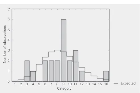
Figure 1. Distribution of baseline micronuclei among individuals aged on average 23.6 years (Kolmogorov-Smirnof test, d = 0.11, nonsignificant).
Number of observations
7
16 Expected
6
5
4
3
2
1
0
1 2 3 4 5 6 7 8 9 10 11 12 13 14 15 Category
Number of observations 10
9
8
7
6
5
4
3
Number of observations
7
288 Expected 6
5
4
3
2
1
0
180 189 198 207 216 225 234 243 252 261 270 279 Category (upper limits)
Number of observations
22
20
18
16
8
Figure 2. Distribution of baseline micronuclei among individuals aged on average 36.8 years (Kolmogorov-Smirnof test, d = 0.15, nonsignificant).
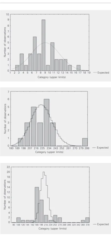
Figure 3. Distribution of radiosensitivity among indi-viduals aged on average 23.6 years (Kolmogorov-Smirnof test, d = 0.68, nonsignificant).
Figure 4. Distribution of radiosensitivity among indi-viduals aged on average 36.8 years (Kolmogorov-Smirnof test, d = 0.36, P < 0.01).
16 Expected
1 2 3 4 5 6 7 8 9 10 11 12 13 14 15 Category (upper limits)
17 18 19 2
1
0
14
12
10
2 6
4
360 Expected
0
90 108 126 144 162 180 198 216 234 252 270 288 306 324 342 Category (upper limits)
0.11 and 0.15; Figures 1 and 2). The distribu-tion of radiosensitivity among individuals of the younger study group did not differ sig-nificantly and also followed Poisson expec-tation (Kolmogorov-Smirnof test, d = 0.68; Figure 3). In contrast to the younger study group, we found a significant difference be-tween the yields of radiation-induced micro-nuclei and the Poisson distributions in the group of older individuals (Kolmogorov-Smirnof test, d = 0.36, P < 0.01; Figure 4).
Discussion
Accumulated mutations seem to be the main cause of accelerated ageing or cancer. Many enzymes and regulatory proteins are devoted to the repair of DNA damage be-cause unrepaired DNA damage can lead to mutations. Recent studies have pointed out that more than 120 repair genes are involved in the repair of DNA damage (21), suggest-ing that it might be more practical to study “repair phenotype” than to try to unravel or predict phenotype from complete genotypic information. Repair phenotype can be easily recognized by provoking cell radioresponse in vitro using a simple test such as the
cy-tochalasin block micronucleus test (22). A significant gender effect on baseline micronuclei favoring females has been pviously reported (23) and confirmed in re-cent large-scale studies (24,25). On this ba-sis, we may hypothesize that X-chromosomes play an important role in the occurrence of micronuclei by interaction of their products with receptors located on the nuclear mem-brane and with proteins of the spindle appa-ratus. Some studies demonstrated that an inactive X-chromosome is preferentially in-cluded in the micronuclei, suggesting that X-chromosome hypodiploidy in aging women is a related phenomenon.
Our earlier study showed a significant gender effect on the baseline level of micro-nuclei favoring females, which was con-firmed in the present study. In addition to
determining the baseline level of micronu-clei, we estimated radiation-induced sensi-tivity in two groups of healthy adults of both genders aged 22 and 40 years. Several im-portant results were obtained: i) the distribu-tion of baseline micronuclei followed Pois-son distributions in both study groups of both genders, ii) there was a nonsignificant difference in baseline micronuclei between the two age groups of both genders, iii) marked variability in radiation-response was observed among individuals near 40 com-pared to those near 23 years old, iv) the distribution of radiosensitivity among older individuals did not follow Poisson expecta-tions, v) in contrast to a nonsignificant dif-ference in baseline micronuclei between the young (23 years) and older (37 years) groups, radiosensitivity differed significantly be-tween older members of both genders and young members.
obser-vation could be explained by ageing, due to cumulative defects in the efficiency of repair genes or of antioxidant defense mechan-isms. Reduced repair efficiency and accu-mulated point mutations due to ageing cer-tainly could increase the levels of radiation-induced micronuclei. A sensitive radiore-sponse was not observed in the younger subjects of both genders, possibly reflecting differences in repair of damaged DNA and the ability to pause while the damage is repaired. This type of “phenotype repair” is seen as a consequence of accelerated ageing, which led to micronucleus distribution di-verging from Poisson expectations. Ageing is usually followed by disturbances in micronutritient status and accumulated point muations of repair genes: folate and vitamin B12 concentrations are particularly
impor-tant determinants of micronucleus frequency (28,29). Recent studies on men aged 50-70 years have confirmed these observations (30,31).
Variability in radioresponse suggests that individual radiosensitivity might play a cru-cial role in radiotherapy. Knowledge of a patient’s individual radiosensitivity before radiotherapy could be of help in planning the most appropriate clinical treatment.
In the near future, new information will be obtained on polymorphisms affecting DNA repair and genome integrity, which are probably of special importance in modulat-ing radiation effects. Fully understandmodulat-ing the mechanisms of radioresponse and the possibility to modulate it is expected to im-prove the applicability of the micronucleus assay to individualized radiotherapy schemes.
References
1. Fenech M (1993). The cytokinesis-block micronucleus technique: a detailed description of the method and its application to genotoxic-ity studies in human populations. Mutation Research, 285: 35-44. 2. Rimoldi R, Miller AC, Freeman SE & Samid D (1991). DNA damage
in cultured human skin fibroblasts exposed to excimer laser radia-tion. Journal of Investigative Dermatology, 96: 898-902.
3. Degen GH (1993). SEMV cell cultures: a model for studies of prosta-glandin-H synthase-mediated metabolism and genotoxicity of xeno-biotics. Toxicology Letters, 67: 187-200.
4. Matsuoka A, Yamazaki N, Suzuki T, Hayashi M & Sofuni T (1992). Evaluation of the micronucleus test using a Chinese hamster cell line as an alternative to the conventional in vitro chromosomal aberration test. Mutation Research, 272: 223-236.
5. Stopper H, Korber C, Schiffmann D & Caspary WJ (1993). Cell-cycle dependent micronucleus formation and mitotic disturbances in-duced by 5-azacytidine in mammalian cells. Mutation Research, 300: 165-177.
6. Miller BM, Pujadas E & Gocke E (1995). Evaluation of the micro-nucleus test in vitro using Chinese hamster cells: results of four chemicals weakly positive in the in vivo micronucleus test. Environ-mental and Molecular Mutagenesis, 26: 240-247.
7. Rosin MP (1992). The use of the micronucleus test on exfoliated cells to identify anti-clastogenic action in humans: a biological marker for the efficacy of chemopreventive agents. Mutation Re-search, 267: 265-276.
8. Natarajan AT, Boei JJ, Darroudi F, Van Diemen PC, Dulout F, Hande MP & Ramalho A (1996). Current cytogenetic methods for detecting exposure and effects of mutagens and carcinogens. Environmental Health Perspectives, 104 (Suppl 3): 445-448.
9. Fenech M, Holland N, Chang WP, Zeiger E & Bonassi S (1999). The
Human Micronucleus Project - An international collaborative study on the use of the micronucleus technique for measuring DNA damage in humans. Mutation Research, 428: 271-283.
10. Ramalho A, Sunjevaric I & Natarajan AT (1988). Use of the frequen-cies of micronuclei as quantitative indicators of X-ray induced chrom-osomal aberrations in human peripheral blood lymphocytes: Com-parison of two methods. Mutation Research, 207: 141-146. 11. Köeteles GJ (1996). Biological dosimetry. Lecture notes for the
IRPA 9 Refresher Course, R-06. International Congress on Radiation Protection, April 14-19, Vienna, Austria, 1-40.
12. Galloway SM, Sofuni T, Shelby MD et al. (1997). Multilaboratory comparison of in vitro tests for chromosome aberrations in CHO and CHL cells tested under the same protocols. Environmental and Molecular Mutagenesis, 29: 189-207.
13. Palitti F (1998). Mechanisms of the origin of chromosomal aberra-tions. Mutation Research, 404: 133-137.
14. Fenech M (1991). Optimisation of micronucleus assays for biologi-cal dosimetry. Progress in Clinical and Biological Research, 372: 373-386.
15. Fenech M & Morely AA (1985). Measurement of micronuclei in lymphocytes. Mutation Research, 147: 29-36.
16. Murnane JP & Kapp LN (1993). A critical look at the association of human genetic syndrome with sensitivity to ionizing radiation. Semi-nars in Cancer Biology, 246: 629-634.
18. Scott D, Barber JB, Levine EL, Burrill W & Roberts SA (1998). Radiation-induced micronucleus induction in lymphocytes identifies a high frequency of radiosensitivity cases among breast cancer patients: a test for predisposition? British Journal of Cancer, 77: 614-620.
19. Joksiƒ G, Nikoliƒ M & Spasojeviƒ-Tišma V (1997). Radiosensitivity of different aged human lymphocytes following electron irradiation
in vitro. Neoplasma, 44: 117-121.
20. Joksiƒ G, Petroviƒ-Novak A, Stankoviƒ M & Kova…eviƒ M (1999). Radiosensitivity of human lymphocytes in vitro correlates more with proliferative ability of cells than with the incidence of radiation-induced damages of the genome. Neoplasma, 46: 40-49.
21. Wood RD, Mitchell M, Sgouros J & Lindahl T (2001). Human DNA repair genes. Science, 29: 1284-1289.
22. Fenech M (2002). Biomarkers of genetic damage for cancer epide-miology. Toxicology, 181-182: 411-416.
23. Fenech M, Neville S & Rinaldi J (1994). Sex is an important variable affecting spontaneous micronucleus frequency in cytokinesis blocked lymphocytes. Mutation Research, 313: 203-207.
24. Hando JC, Nath J & Tucker JD (1994). Sex chromosomes, micronu-clei and aging in women. Chromosoma, 103: 186-192.
25. Tucker JD, Nath J & Hando JC (1996). Activation status of the X-chromosome in human micronucleated lymphocytes. Human
Ge-netics, 97: 471-475.
26. Rothfuss A, Schutz P, Bochum S, Volm T, Eberhardt E, Kreienberg R, Vogel W & Speit G (2000). Induced micronucleus frequencies in peripheral blood lymphocytes as a screening test for carriers of a BRCA1 mutation in breast cancer families. Cancer Research, 60: 390-394.
27. Joksiƒ G, Pajoviƒ SB, Stankoviƒ M, Pejiƒ S, Kasapoviƒ J, Cuttone G, Calonghi N, Masotti L & Kanazir DT (2000). Chromosome aberra-tions, micronuclei and activity of superoxide dismutases in human lymphocytes after irradiation in vitro. Cellular and Molecular Life Sciences, 57: 842-850.
28. Fenech M, Aitken C & Rinaldi J (1998). Folate, vitamin B12, ho-mocysteine status and DNA damage in young Australian adults.
Carcinogenesis, 19: 1163-1171.
29. Fenech M, Dreosti I & Aitken C (1997). Vitamin-E supplements and their effect on vitamin-E status in blood and genetic damage rate in peripheral blood lymphocytes. Carcinogenesis, 18: 359-364. 30. Fenech M, Dreosti IE & Rinaldi JR (1997). Folate, vitamin B12,
homocysteine status and chromosome damage rate in lympho-cytes of older men. Carcinogenesis, 18: 1329-1336.
31. Lenton KJ & Greenstock CL (1999). Ability of human plasma to protect against ionising radiation is inversely correlated with age.