The anti-IRBP IgG1 and IgG2a response
does not correlate with susceptibility to
experimental autoimmune uveitis
1Laboratório de Imunologia Clínica, Departamento de Imunologia,
Instituto de Ciências Biomédicas, Universidade de São Paulo, São Paulo, SP, Brasil
2Laboratório de Imunogenética, 3Laboratório de Imunoquímica, Instituto Butantan,
São Paulo, SP, Brasil
4Instituto de Investigação em Imunologia, Ministério da Ciência e Tecnologia,
Brasília, DF, Brasil
5LIM-60 Disciplina de Imunologia Clínica e Alergia,
Departamento de Clínica Médica e Fundação Zerbini,
Faculdade de Medicina, Universidade de São Paulo, São Paulo, SP, Brasil L. Vieira de Moraes1,
G.A. Martins1,
M. Flangini1,
O.M. Ibañez2,
O.A. Sant’Anna3
and L.V. Rizzo1,4,5
Abstract
Susceptibility to experimental autoimmune uveitis (EAU) in inbred mice has been associated with a dominant Th1 response. Elevated anti-inter-photoreceptor retinoid-binding protein (anti-IRBP) IgG2a/ IgG1 antibody ratios have been implicated as candidate markers to predict disease severity. In the present study, both the anti-IRBP antibody isotype and severity of EAU phenotypes were examined in 4 non-isogenic genetically selected mouse lines to determine if they can be used as general markers of disease. Mice between 8 and 12 weeks old selected for high (HIII)or low (LIII)antibody response and for
maximum (AIRMAX)or minimum (AIRMIN) acute inflammatory
reac-tion (AIR) were immunized with IRBP. Each experiment was per-formed with at least 5 mice per group. EAU was evaluated by histopathology 21 days after immunization and the minimal criterion was inflammatory cell infiltration of the ciliary body, choroid and retina. Serum IgG1- and IgG2a-specific antibodies were determined by ELISA. EAU was graded by histological examination of the enucleated eyes. The incidence of EAU was lower in AIRMIN mice
whereas in the other strains approximately 40% of the animals devel-oped the disease. Low responder animals did not produce anti-IRBP IgG2a antibodies or interferon-γ. No correlation was observed be-tween susceptibility to EAU and anti-IRBP isotype profiles. Suscep-tibility to EAU is related to the intrinsic capacity to mount higher inflammatory reactions and increased production of anti-IRBP IgG2a isotype is not necessarily a marker of this immunologic profile. Correspondence
L.V. Rizzo
Departamento de Imunologia ICB IV, USP
Av. Prof. Lineu Prestes, 1730 05508-900 São Paulo, SP Brasil
Fax: +55-11-309-1739 E-mail: lvrizzo@icb.usp.br Research supported by FAPESP and CNPq. L.V. Rizzo, O.M. Ibañez and O.A. Sant’Anna are recipients of CNPq fellowships for scientific productivity. L.V. de Moraes is the recipient of a post-doctoral fellowship from CNPq.
Received July 18, 2005 Accepted January 10, 2006
Key words •Autoimmunity
•IgG1 and IgG2a isotypes •Inflammation
Introduction
Experimental autoimmune uveitis (EAU) is an organ-specific T cell-mediated disease that targets the neural retina and leads to retinochoroiditis and blindness. The failure of the mechanisms that maintain the state of immune tolerance in the eye can be induced in experimental models by immunization with retinal antigens such as the soluble protein S-antigen and the glycolipoprotein inter-photoreceptor retinoid-binding protein (IRBP) (1-4). EAU is a cell-mediated in-flammatory disease in which the uveitogenic retinal antigen-specific CD4+ T lymphocytes
invade and destroy the retina (5,6). There is evidence implicating Th1-like lymphocytes in the pathogenesis of EAU, whereas induc-tion of a Th2-like response seems to be protective (7-10). Sun et al. (11) demon-strated that susceptibility to the develop-ment of EAU is associated with a predomi-nant Th1 response to IRBP in six different isogenic mouse strains, and that resistance to disease can occur in the presence or ab-sence of a dominant Th2 response.
Several mechanisms have been impli-cated in the susceptibility to the develop-ment of EAU. Mutations in two genes, lpr
and gld, result in defects in apoptosis induc-tion in mice. These deficiencies result in inefficient elimination of autoreactive T cells and consequent accumulation of this lym-phocyte population (12-15). They may have a role in ocular autoimmunity since Fas and FasL play a role in the maintenance of immunoprivilege in the eye (16,17). Abnor-malities during the intra- and/or extrathymic clonal T-cell selection (18) and expansion or even in the capacity of these lymphocytes to recognize ocular antigens and/or proteins that mimic ocular antigens have been associ-ated with certain haplotypes of the major histocompatibility complex (MHC). It has been demonstrated that the MHC haplotypes H-2k, H-2b, or H-2r correlate with
suscepti-bility to EAU in inbred mouse strains,
whereas the presence of other alleles in the I-E sub-region has been related to resistance (19,20). The importance of the MHC is best exemplified by the individual response to the 161-180 peptide of IRBP, which contains a major epitope recognized as pathogenic by the H-2r bearing B10.RIII mice (21).
The investigation of genes controlling acquired or innate immune mechanisms has demonstrated that major immunobiological functions such as inflammatory responses, quantitative antibody production, immuno-logical tolerance, and T cell-mediated reac-tivity, in spite of their functional integration are, at least in part, under the additive effect of independent segregating loci (22-25). The resulting polymorphic regulation of innate and adaptive immunity is the fundamental characteristic ensuring the survival of a ge-netically heterogeneous natural population. This variability is related to the genotypes of the population and the environmental condi-tions to which the individuals are subjected before and after birth.
To better understand the influence of a complex genetic background intervening in the innate and/or adaptive immune functions on EAU development and to establish the possible association of specific anti-IRBP isotype profiles in the susceptibility to de-veloping disease, the present study was car-ried out in genetically selected lineages of mice for high (H) or low (L) antibody re-sponses (26) and for maximal or minimal acute inflammatory reactions (AIRMAX and
AIRMIN, respectively) (23). As pointed out
mice to establish protective mechanisms re-flects a dynamic relationship between gene frequency in natural populations and the nature of the immunogens, pathogens or toxins to which they are exposed. The results of the factors responsible for the complex pleiotropic network of the immune system may be indicative of the main protective mechanisms responsible for survival, includ-ing those responsible for the aspects of au-toimmune pathogenicity.
The advantage of the genetically selected H, L, AIRMAX, and AIRMIN mouse line
mod-els compared to inbred mice is the availabil-ity of individuals genetically homogeneous at the relevant loci controlling a general character but still heterogeneous at other loci, including the MHC antigens, assuring the preservation of the naturally occurring genetic variability in the population study. These features allow the study of the real biological relevance of innate and/or adap-tive immune traits and the correlation amongst distinct immunobiological parameters.
In the present study, we evaluated the responsiveness to IRBP immunization in genetically selected mouse lines by measur-ing both their ability to produce the specific anti-IRBP IgG1 and IgG2a antibody iso-types and their degree of eye pathology dur-ing the course of EAU.
Material and Methods
Animals
Mouse lineages obtained by bidirectional genetic selection for high (HIII) and low (LIII)
immune responsiveness and for maximal (AIRMAX) and minimal (AIRMIN) acute
in-flammatory reaction were used. HIII and LIII
animals were obtained from four indepen-dent outbred Swiss mouse colonies and se-lected on the basis of high or low antibody production to flagellar antigens of Salmo-nellae (26). AIRMAX and AIRMIN mice were
obtained from a Foundation Population
de-riving from intercrosses of eight distinct in-bred mouse strains (A, DBA-2, P, SWR, CBA, SJL, BALB/c, and C57Bl/6) (23). The selective breeding of these animals for maxi-mal or minimaxi-mal acute imflammatory response was based on both cellular infiltrate and protein contents. Animals were obtained and maintained at the animal facilities of the Immunogenetics Laboratory of Instituto Butantan. All experiments were performed following the guidelines for animal use ap-proved by the Ethics Committee in Animal Experimentation from the institutions in-volved in the study and according to the guidelines established by the Association for Research in Vision and Ophthalmology for the care and handling of experimental animals.
Reagents and antibodies
IRBP was isolated from bovine retinas by concanavalin A-Sepharose affinity chro-matography and FPLC (28,29) and stored at -70ºC. Bordetella pertussis toxin, concanava-lin A, bovine serum albumin, and complete Freund adjuvant were purchased from Sigma (St. Louis, MO, USA). Horseradish peroxi-dase-streptavidin goat IgG1 and anti-IgG2a mouse subclasses were purchased from Southern Biotechnologies Associates, Inc. (Birmingham, AL, USA). The hybrido-mas XMG1.2 and AN18 producers of anti-mouse interferon-γ (IFN-γ) were a gift from Dr. I. Abrahamsohn (University of São Paulo). Recombinant murine IFN-γ and streptavidin-horseradish peroxidase were purchased from BD Pharmingen (San Di-ego, CA, USA).
Immunization
HIII, LIII, AIRMAX, and AIRMIN mice of
adjuvant administered subcutaneously and 1 µg Bordetella pertussis toxin by the intra-peritoneal route.
Histology and disease grading
Eyes were collected 20, 28, and 40 days after immunization, fixed for 1 h in 4% sodium phosphate-buffered glutaraldehyde, and transferred to 10% sodium phosphate-buffered formaldehyde until processing. Fixed and dehydrated tissue was embedded in paraffin and cut into 4- to 6-µm sections through the pupillary-optic nerve plane. Six sections cut at different levels were submit-ted to standard hematoxylin-eosin staining and examined for each eye in a masked fashion. The presence and the extent of the lesions were determined and the severity of EAU was scored on a 0 to 4 scale according to a semi-quantitative system described pre-viously (3). Briefly, the minimal criterion to score an animal as positive by histopathol-ogy was inflammatory cell infiltration of the ciliary body, choroid and retina. Progres-sively higher grades were assigned for the presence of discrete lesions in the tissues such as vasculitis, granuloma formation, reti-nal folding, and/or detachment and photore-ceptor damage.
Assay for IRBP-specific antibody IgG isotypes
The individual anti-IRBP IgG1 and IgG2a isotype levels were determined by ELISA as previously described (30). Briefly, 96-well microtiter plates (Costar, Cambridge, MA, USA) were coated with IRBP at 1 µg/mL in carbonate/bicarbonate buffer, blocked with bovine serum albumin and incubated with serially diluted serum samples for 1 h at room temperature. Plates were developed with horseradish peroxidase-conjugated goat anti-mouse IgG1 or with rat anti-mouse IgG2a (diluted 1:2000). The IRBP anti-body titers were expressed as log2 of the
reciprocal serum dilution giving an
absorb-ance value of 20% of the saturation level.
IFN-γγγγγ assay
The draining lymph nodes were collected 20 or 40 days after immunization, teased to a single cell suspension and cultured in Dulbec-co’s modified Eagle’s medium supplemented with 5% fetal bovine serum (Hyclone, Lo-gan, UT, USA), 10 µM 2-mercaptoethanol (Sigma), 2 mM L-glutamine, 0.1 mM vita-mins, 1 mM sodium pyruvate, 0.1 mM non-essential amino acids, and 100 µg/mL gen-tamicin, all purchased from Gibco BRL (Rockville, NY, USA) in a 96-well plate with immobilized anti-CD3 (1 µg/mL; BD/ Pharmingen) for a total of 48 h. IFN-γ was determined in the supernatants by sandwich ELISA employing the rat anti-mouse mono-clonal antibody system prepared in our own laboratory from culturing the respective hy-bridomas, purified and biotinylated when used as the detection antibody. High binding 96-well plates (Corning Corporation, Corn-ing, NY, USA) were coated with 50 µL of the first antibody diluted in carbonate/bicar-bonate buffer, pH 9.6, for 16 h at 4ºC (8 µg/ mL anti-IFN-γ (XMG1.2)). After incuba-tion, wells were blocked for 2 h at room temperature with 150 µL PBS/0.05% Tween 20 (Sigma) and 5% skim milk. Plates were washed three times with PBS/0.05% Tween 20 and received 50 µL of samples or of the recombinant murine IFN-γ in 2-fold serial dilutions. After 4-h incubation at room tem-perature, plates were washed and 50 µL of the biotinylated anti-IFN-γ (AN18 at 8 µg/ mL) was added and incubated for 2 h at room temperature. Plates were washed and 80 µL streptavidin-peroxidase (Sigma) diluted (1:4000) in PBS was added and incubated for 1 h at room temperature. Plates were washed again and 100 µL of substrate solu-tion (3% H2O2, 0.5 µg/mL
min with 0.2 M citric acid. Absorbance was read at λ410 and 490 nm.
Statistical analysis
EAU scores are reported as the average of both eyes for each animal. Disease scores were analyzed using Snedecor and Cochran’s test (31) for linear trend in proportions. Sta-tistical analysis was performed by nonpara-metric analysis of variance (ANOVA) with Dunn’s post-test. Data of the antibody meas-urements were compared using the unpaired
t-test or Mann-Whitney test and were consid-ered to be significantly different at P < 0.05.
Results
Susceptibility to EAU
Susceptibility to EAU was investigated in the genetically selected mouse lineages for high (HIII) or low (LIII) immune response
and for maximal (AIRMAX) or minimal
(AIRMIN) acute inflammation. Mice were
im-munized with IRBP and evaluated for the development of EAU 21 days later. The incidence of uveitis, i.e., the number of ani-mals affected with EAU of the total number of injected mice, was 6/15 in the HIII line and
3/9 in the LIII line; the EAU in the AIRMAX
mice was 6/13 whereas 10/11 of AIRMIN
mice were resistant to the disease - only 9% developed a mild cell infiltration (Figure 1). The estimated EAU mean grade of cellular infiltration and structural alteration scored on a scale from 0 to 4 among the affected mice was 0.73 for HIII, 0.25 for LIII, 0.34 for
AIRMAX, and 0.09 for AIRMIN mice.
Serum IgG1 and IgG2a antibodies to IRBP
Antibody isotype switching to IgG1 and IgG2a is promoted mostly by IL-4 and
IFN-γ, respectively. Since susceptibility to EAU in inbred mice has been associated with Th1 lymphocytes in the pathogenesis of the
dis-Figure 1. EAU scores for inter-photoreceptor retinoid-binding protein (IRBP)-immunized HIII, LIII, AIRMAX, and AIRMIN genetically selected
mouse lines. Mice were immunized with 50 µg IRBP and 1 µg Borde-tella pertussis toxin. Eyes were collected for histopathology 21 days after immunization. Scores were assigned on a scale of 0 to 4 accord-ing to the extent of inflammation and tissue damage. Each symbol represents one animal (average of both eyes). The horizontal lines indicate the mean for each group. EAU = experimental autoimmune uveitis; AIRMAX, AIRMIN = maximal and minimal acute inflammatory
reactions; HIII, LIII = high and low immune responsiveness.
ease, we investigated anti-IRBP IgG1 and IgG2a levels in response to IRBP, 21 days after immunization in an attempt to deter-mine the T-cell subset and the cytokine in-volvement in a more direct fashion than simply measuring IL-4 and IFN-γ in vitro. The comparative analysis of IgG isotypes in both genetic selections showed peculiar fea-tures. For the HIII and LIII lines, as expected,
significant interline differences were ob-tained for both isotypes, mainly for IgG2a antibodies, since the LIII mice did not
pro-duce this subclass in response to IRBP (Fig-ure 2A). Interestingly, AIRMAX animals did
not produce this anti-IRBP isotype whereas AIRMIN mice showed both an IgG1 and IgG2a
response to IRBP (Figure 2B). The lack of anti-IRBP IgG2a production in LIII was
ob-served 40 and 65 days after immunization (data not shown). At the former time AIRMAX
mice showed detectable IgG2a antibody ti-ters (3.33 ± 2.5) in response to IRBP. Of note is the fact that in all lineages the IgG2a/IgG1
EAU score
3
2
1
0
T
iter (log2) of anti-IRBP
antibody isotypes 10
5
*
** **
0
HIII LIII HIII LIII AIRMAX AIRMIN AIRMAX AIRMIN
IgG2a
IgG1
IgG2a
IgG1
T
iter (log2) of anti-IRBP
antibody isotypes
15
10
5
0
A B
15 **
AIRMIN
AIRMAX HIII LIII
IgG2a/IgG1 ratio
3
2
1
0 C
ratio was below the “1” value, which means that the production of anti-IRBP IgG2a was decreased when compared to the production of IgG1 (Figure 2C).
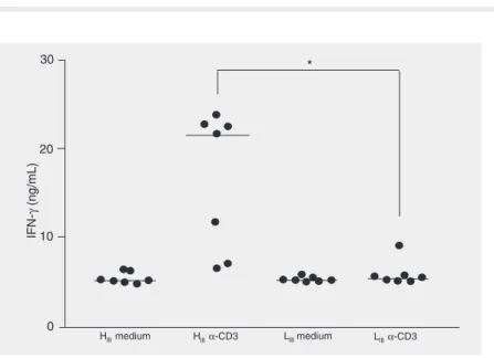
IFN-γγγγγ secretion by lymph node cells
The persistent lack of anti-IRBP IgG2a antibody production in LIII mice following
immunization with IRBP led us to investi-gate IFN-γ production in the supernatants of anti-CD3-stimulated lymph node cell cul-tures. Cytokine secretion was analyzed in HIII and LIII mice 21 days after immunization
(Figure 3). As expected, the production of IFN-γ was significantly lower in LIII when
compared to high responder animals (P < 0.01). Although HIII animals were more
sus-ceptible to developing disease with higher scores of severity, the incidence of EAU was similar in both HIII and LIII lineages,
indicat-ing that the participation of IFN-γ in the susceptibility to developing moderate levels of EAU may not be so important.
Lack of correlation between IgG2a/IgG1 ratios and EAU scores
It has been previously shown in suscep-tible inbred mouse strains that disease sever-ity is directly proportional to their anti-IRBP IgG2a/IgG1 ratios. Here we investigated the relationship between these two phenotypes. The present results in non-inbred mouse strains clearly demonstrate the absence of correlation between IgG2a/IgG1 ratio and
Figure 2. Anti-IRBP IgG1 and IgG2a antibody pro-duction of HIII, LIII (A), AIRMAX, and AIRMIN mice
(B). Antibodies were measured by isotype-specif-ic ELISA in serum 21 days after IRBP immuniza-tion. C, IgG2a/IgG1 antibody ratios for each group. The unbroken horizontal lines indicate the mean (A and B) or median of each group (C). The ani-mals above the broken horizontal line in panel C presented anti-IRBP IgG2a antibody titers greater than IgG1. IRBP = inter-photoreceptor retinoid-binding protein; AIRMAX, AIRMIN = maximal and
minimal acute inflammatory reactions; HIII, LIII =
susceptibility to EAU as well as the severity of disease, as evidenced by the HIII, LIII,
AIRMAX, and AIRMIN profiles (Figure 4). Discussion
The design of the present study was based on an alternative concept for the investiga-tion of autoimmune uveitis in mice. Human uveitis is a complex disease in which the genetic background plays a significant role even when the same entity is under study (32). The goal of an animal model of human disease is to be as closely similar to the human entity as possible, and for this reason we set out to establish EAU in a non-inbred mouse population. Four genetically selected lines of mice resulted in the convergent fixa-tion of alleles affecting the high or low anti-body or inflammatory responses regarding
Figure 4. Absence of correlation between individual EAU scores and IRBP IgG2a/IgG1 anti-body ratios. Anti-IRBP IgG2a/ IgG1 ratios were plotted indi-vidually against the EAU scores. The correlation line was plotted by the linear curve fitting. EAU = experimental autoimmune uvei-tis; AIRMAX, AIRMIN = maximal
and minimal acute inflammatory reactions; HIII, LIII = high and
low immune responsiveness. Figure 3. IFN-γ synthesis in HIII and LIII mice. The cytokine was measured in the
superna-tants of activated cell cultures from individual mice. Draining lymph node cells were collected 21 days after immunization and stimulated or not with anti-CD3 antibody for 48 h. The horizontal lines indicate the means. IFN-γ = interferon-γ; AIRMAX, AIRMIN = maximal
and minimal acute inflammatory reactions; HIII, LIII = high and low immune
responsive-ness. *P < 0.01 (Mann-Whitney test).
IFN-(ng/mL)
γ
30
H mediumIII HIIIα-CD3 L mediumIII LIIIα-CD3
*
20
10
0
EAU score
4
3
2
1
0
0.0 0.1 0.2 0.3 0.4 0.5 0.6 0.7 0.8 IgG2a/IgG1 ratio
EAU score
4
3
2
1
0
0.0 0.5 1.0 1.5 2.0 2.5 3.0 IgG2a/IgG1 ratio
0.0 0.5 1.0 1.5 2.0 2.5
IgG2a/IgG1 ratio
0.0 0.25 0.50 0.75 1.25
IgG2a/IgG1 ratio 1.00
EAU score
4
3
2
1
0
EAU score
4
3
2
1
0
r = 0.0082 r = 0.00052
AIRMAX
r = 0.0082
AIRMIN
r = 0.102
HIII
r = 0.0072
the innate and/or acquired immune compart-ments. This approach allows the study of the disease in populations that have been se-lected for definite immunobiological traits but still, at least in part, heterogeneous with regards to the rest of their genome, thus better resembling the human population. Here, we investigated EAU development and the anti-IRBP IgG1 and IgG2a antibody production in HIII, LIII, AIRMAX, and AIRMIN
mice. It was demonstrated that in these mice, unlike in inbred strains, susceptibility to ex-perimental autoimmune uveitis does not cor-relate with the production of anti-IRBP IgG2a isotype.
The data indicate that the amount of anti-IRBP IgG1 antibody was similar among the lines during the period evaluated and titer-wise higher than the anti-IRBP IgG2a in all selected mouse strains, with the exception of AIRMIN mice that expressed the same titers
for both IgG1 and IgG2a isotypes 21 days after IRBP administration. The levels of anti-IRBP IgG2a subclass were nearly undetect-able in LIII mice. In other experiments serum
was tested for the presence of IgG1 and IgG2a antibodies in response to IRBP 40 and 65 days after immunization. It was intri-guing to notice that LIII mice did not produce
IRBP-specific IgG2a antibody whereas the other mouse lines responded with high anti-body titers although always lower when com-pared to IgG1. This peculiar pattern seems to be dependent on the isotype-restricted regu-latory effect related to the immunization procedure used during the selective breed-ing process to which the parental animals were submitted to develop the lineages. In fact, a similar picture in the IgG2a profile of the HIII and LIII line was observed after
im-munization with Salmonella flagellar and somatic antigens, both present during the selective process (26). Interestingly, all lines exhibited IgG2a/IgG1 ratios below “1”, irre-spective of the susceptibility to the disease, suggesting that under any circumstances anti-IRBP IgG2a levels were higher than IgG1
antibody. Histopathologic examination of the eyes after antigen administration showed disorganization of the retinal architecture in 40 to 50% of the animals of the AIRMAX, HIII,
and LIII strains. AIRMIN mice were resistant
to the development of EAU with only 1 affected individual of 11. In a similar study performed with high responder mice selected for heterologous erythrocytes (19), the so-called Biozzi ABH mice, a higher percent-age of individuals susceptible to uveoretinitis and low levels of antigen-specific IgG2a sub-class were observed (33). Our data support previously published results on the subject.
The balance between Th1 and Th2 cells and its role in determining the outcome of the immune response to the uveitogenic pro-teins has been an issue for some time. It has been shown that mouse strains susceptible to EAU polarize their response against the reti-nal antigen towards a type 1 response, con-sequently enhancing the production of anti-uveitogen IgG2a antibody, an IFN-γ-induced isotype, even after EAU onset. In contrast, a type 2 response and a dominant IgG1 profile have been associated with a resistant pheno-type (11). This paradigm does not hold true in these genetically selected non-inbred lines of mice, suggesting it may be a consequence of the inbreeding process by carrying to-gether with the quantitative trait loci that code for susceptibility, those that control isotype switching to IgG1 and IgG2a. The differential result obtained in outbred mice may explain why only a percentage of pa-tients with autoimmune uveitis develop a cytokine profile similar to the one described in inbred mice, whereas all patients present a similar inflammatory reaction (34). Contin-ued studies on these strains will help associ-ate specific clinical features such as retinal detachment with sustained patterns of im-mune response to ocular antigens.
The data presented here show a predomi-nant anti-IRBP IgG1 antibody response in the serum of AIRMAX and LIII mice 21 days
response did not correlate with a resistant phenotype, since 27% of LIII mice and 40%
of AIRMAX mice were sick after
immuniza-tion with IRBP. On the other hand, AIRMIN
animals showed enhanced anti-IRBP IgG2a antibody titers compared to AIRMAX mice
but were resistant to the disease. Another interesting feature was the observation that LIII mice did not produce IFN-γ even after
anti-CD3 stimulation. This event was com-patible with the lack of production of anti-IRBP IgG2a isotype after immunization. Based on these data, one could expect a resistant phenotype for LIII regarding the
development of uveitis since the disease has been correlated with a dominant type-1 re-sponse (11). In fact, in high responder ani-mals the production of IFN-γ seems to corre-late with susceptibility since these animals develop a milder disease with an incidence of EAU in almost 50% of the population. Although AIRMAX mice do not produce
IRBP-specific IgG2a 21 days after immuni-zation, over longer periods of time these animals were able to secrete this isotype, differently from LIII mice. Still, this
unbal-anced situation between the two isotypes in favor of anti-IRBP IgG1 antibody did not protect AIRMAX from developing EAU. The
development of EAU in the context of lack of secretion of IFN-γ has already been shown. Mice treated with anti-IFN-γ antibody de-velop EAU and this feature was also ob-served in some resistant strains of mice after systemic depletion of this cytokine (35). Other studies have shown that IFN-γ -defi-cient mice are highly susceptible to develop-ing EAU after immunization with whole IRBP (36) or with the major uveitogenic human peptide 1-20 (37) and in the latter case the resulting disease is significantly more severe when compared to wild-type controls. The cytokine profile in the super-natants of these animals lacking IFN-γ was skewed toward a Th2-like response, show-ing an up-regulation of IL-5. Cellular infil-trates contained significantly more
eosino-phils compared to their wild-type littermates. In our experiments, although we did not look at the mechanisms involved in the de-velopment of EAU in low IFN-γ-producing animals such as AIRMAX and LIII, we cannot
rule out the possibility of a deviant immune response in these mice.
For both antigen-specific IgG1 and IgG2a antibodies, analysis of variance supported the importance of multigenic factors regu-lating the adaptive response to IRBP. More-over, based on the distinct H-2 genes of the four mouse lines and the similarities of the EAU scores, especially between the AIRMAX
and LIII strains, no specific MHC allele seems
to be crucial for the development of the disease, as it is in inbred strains of mice. However, we must point out that the LIII
mice are H-2z, H
III are H-2º3 (3,38), AIRMAX
are predominantly H-2b, and in AIR
MIN mice
the H-2d and H-2k haplotypes are more
preva-lent. Therefore, we suggest that the genetic control of the adaptive immune characteris-tics during the autoimmune process in EAU is polygenic, since the interline variances were always higher than the intraline ones and there were continuous distributions among individuals.
The evaluation of the development of the disease in AIRMAX and AIRMIN mice can also
be considered from the viewpoint of the inflammatory potential that these lines pres-ent in response to distinct pathogens, immu-nogens and chemical agents. In uveitis, dam-age to the eye is a consequence of the re-cruitment of lymphocytes, macrophages and polymorphonuclear cells. The lymphocyte population that migrates to the eye is consti-tuted of both retinal antigen-specific lym-phocytes and “non-specific cells” (39,40). The fact that AIRMIN mice are resistant to
References
1. Gery I, Mochizuki M & Nussenblatt RB (1986). Retinal specific antigens and immunopathogenic processes they provoke. Progress in Retinal and Eye Research, 5: 75-109.
2. Caspi RR, Roberge FG, Chan CC et al. (1988). A new model of autoimmune disease: experimental autoimmune uveoretinitis in-duced in mice with two different retinal antigens. Journal of Immu-nology, 140: 1490-1495.
3. Wacker WB, Donoso LA, Kalsow CM et al. (1977). Experimental allergic uveitis. Isolation, characterization, and localization of a soluble uveitopathogenic antigen from bovine retina. Journal of Immunology, 119: 1949-1958.
4. Hankey DJ, Lightman SL & Baker D (2001). Interphotoreceptor retinoid binding protein peptide-induced uveitis in B10.RIII mice: characterization of disease parameters and immunomodulation. Ex-perimental Eye Research, 72: 341-350.
5. Caspi RR, Silver PB, Chan CC et al. (1996). Genetic susceptibility to experimental autoimmune uveoretinitis (EAU) in the rat is associ-ated with an elevassoci-ated Th1 response. Journal of Immunology, 157: 2668-2675.
6. Liblau RS, Singer SM & McDevitt HO (1995). Th1 and Th2 CD4+ T cells in the pathogenesis of organ-specific autoimmune disease.
Immunology Today, 16: 34-38.
7. Rizzo LV, Silver P, Wiggert B et al. (1996). Establishment and characterization of a murine CD4+ T cell line and clone that induce
experimental autoimmune uveoretinitis in B10.A mice. Journal of Immunology, 156: 1654-1660.
8. Caspi RR (1994). Th1 and Th2 lymphocytes in experimental autoim-mune uveoretinitis. In: Whitcup SM, Nussenblatt RB, Caspi RR et al. (Editors), Advances in Ocular Immunology. International Congress Series, Elsevier Science Publishers, Amsterdam, The Netherlands, 55.
9. Saoudi A, Kuhn J, Huygen K et al. (1993). Th2 activated cells prevent experimental autoimmune uveoretinitis, a Th1-dependent autoimmune disease. European Journal of Immunology, 23: 3096-3103.
10. Chan CC, Mochizuki M, Nussenblatt RB et al. (1985). T-lymphocyte subsets in experimental autoimmune uveitis. Clinical Immunology and Immunopathology, 35: 103-110.
11. Sun B, Rizzo LV, Sun SH et al. (1997). Genetic susceptibility to experimental autoimmune uveitis involves more than a predisposi-tion to generate a T helper-1-like or a T helper-2-like response.
Journal of Immunology, 158: 1004-1011.
12. Bossu P, Singer GG, Andres P et al. (1993). Mature CD4+ T lym-phocytes from MRL/lpr mice are resistant to receptor-mediated tol-erance and apoptosis. Journal of Immunology,151: 7233-7239. 13. Chu JL, Drappa J, Parnassa A et al. (1993). The defect in Fas
mRNA expression in MRL/lpr mice is associated with insertion of the retrotransposon, ETn. Journal of Experimental Medicine, 178: 723-730.
14. Drappa J, Brot N & Elkon KB (1993). The Fas protein is expressed at
high levels on CD4+CD8+ thymocytes and activated mature lym-phocytes in normal mice but not in the lupus-prone strain, MRL lpr/ lpr. Proceedings of the National Academy of Sciences, USA,90: 10340-10344.
15. Broekhuyse RM, Kuhlmann ED & Winkens HJ (1993). Experimental autoimmune anterior uveitis (EAAU): induction by melanin antigen and suppression by various treatments. Pigment Cell Research, 6: 1-6.
16. Griffith TS, Yu X, Herndon JM et al. (1996). CD95-induced apoptosis of lymphocytes in an immune privileged site induces immunological tolerance. Immunity, 5: 7-16.
17. Griffith TS, Brunner T, Fletcher SM et al. (1995). Fas ligand-induced apoptosis as a mechanism of immune privilege. Science, 270: 1189-1192.
18. Egwuagu CE, Charukamnoetkanok P & Gery I (1997). Thymic ex-pression of autoantigens correlates with resistance to autoimmune disease. Journal of Immunology, 159: 3109-3112.
19. Silver PB, Rizzo LV, Chan CC et al. (1995). Identification of a major pathogenic epitope in the human IRBP molecule recognized by mice of the H-2r haplotype. Investigative Ophthalmology and Visual Science, 36: 946-954.
20. Caspi RR, Grubbs BG, Chan C-C et al. (1992). Genetic control of susceptibility to experimental autoimmune uveoretinitis in the mouse model. Concomitant regulation by MHC and non-MHC genes. Jour-nal of Immunology, 148: 2384-2389.
21. Caspi RR (1992). Immunogenetic aspects of clinical and experi-mental uveitis. Regional Immunology, 4: 321-330.
22. Biozzi G, Mouton D, Sant’Anna OA et al. (1979). Genetics of immu-noresponsiveness to natural antigens in the mouse. Current Topics in Microbiology and Immunology, 85: 31-98.
23. Ibañez OM, Stiffel C, Ribeiro OG et al. (1992). Genetics of non-specific immunity: I. Bidirectional selective breeding of lines of mice endowed with maximal and minimal inflammatory responsiveness.
European Journal of Immunology, 22: 2555-2563.
24. da Silva AC, Souza KW, Machado RC et al. (1998). Genetics of immunological tolerance: I. Bidirectional selective breeding of mice for oral tolerance. Research in Immunology, 49: 151-161. 25. da Silva MF, da Costa SC, Ribeiro RC et al. (2001). Independent
genetic control of B- and T-cell tolerance in strains of mouse se-lected for extreme phenotypes of oral tolerance. Scandinavian Jour-nal of Immunology,53: 148-154.
26. Siqueira M, Bandieri A, Reis MS et al. (1976). Selective breeding of mice for antibody responsiveness to flagellar and somatic antigens of salmonellae. European Journal of Immunology,4: 241-249. 27. Boyartchuk V & Dietrich W (2002). Genetic dissection of host
im-mune response. Genes and Immunity, 3: 119-122.
28. Pepperberg DR, Okajima TL, Ripps H et al. (1991). Functional properties of interphotoreceptor retinoid-binding protein. Photochem-istry and Photobiology, 54: 1057-1060.
29. Pepperberg DR, Okajima TL, Wiggert B et al. (1993).
Interphotore-In conclusion, our data suggest that the mechanisms involved in the resistance or susceptibility to EAU development involve polymorphic intervention of independent
ceptor retinoid-binding protein (IRBP): molecular biology and physi-ological role in the visual cycle of rhodopsin. Molecular Neurobiol-ogy, 7: 61-85.
30. Rizzo LV, DeKruyff RH, Umetsu DT et al. (1995). Regulation of the interaction between Th1 and Th2 cell clones to provide help for antibody production in vivo. European Journal of Immunology, 25: 708-716.
31. Snedecor GW & Cochran WG (1967). Statistical Methods. Iowa State University Press, Ames, IA, USA, 248.
32. Tugal-Tutkun I, Onal S, Altan-Yaycioglu R et al. (2004). Uveitis in Behcet disease: an analysis of 880 patients. American Journal of Ophthalmology, 138: 373-380.
33. Hankey JR, Nickerson JM, Donoso LA et al. (2001). Experimental autoimmune uveoretinitis in mice (Biozzi ABH and NOD) expressing the autoimmune-associated H-2Ag7 molecule: identification of a
uveitogenic epitope. Journal of Neuroimmunology,118: 212-222. 34. Singh VK & Rai G (2001). Cytokines in posterior uveitis: an update.
Immunologic Research, 23: 59-74.
35. Caspi RR, Chan CC, Grubbs BG et al. (1994). Endogenous
sys-temic IFN-gamma has a protective role against ocular auto-immuni-ty in mice. Journal of Immunology,152: 890-899.
36. Jones SL, Rizzo LV, Agarwal RK et al. (1997). IFN-γ-deficient mice develop experimental autoimmune uveitis in the context of a deviant effector response. Journal of Immunology, 158: 5997-6005. 37. Avichezer D, Chan C-C, Silver PB et al. (2000). Residues 1-20 of
IRBP and whole IRBP elicit different uveitogenic and immunological responses in interferon gamma deficient mice. Experimental Eye Research, 71: 111-118.
38. Frangoulis B, Mouton D, Sant’Anna OA et al. (1990). H-2 typing of mice genetically selected for high or low antibody production. Immu-nogenetics, 31: 389-392.
39. Gritz DC, Montes C, Atalla LR et al. (1991). Histochemical localiza-tion of superoxide produclocaliza-tion in experimental autoimmune uveitis.
Current Eye Research, 10: 927-931.