Selegiline increases heme oxygenase-1
expression and the cytotoxicity
produced by dopamine treatment
of
neuroblastoma SK-N-SH cells
1Departamento de Neurologia, Hospital de Clínicas de Porto Alegre, Porto Alegre, RS, Brasil
Departments of 2Clinical Neuroscience and 3Medicine,
The University of Birmingham, Queen Elizabeth Hospital, Birmingham, UK C.R.M. Rieder1,
A.C. Williams2 and D.B. Ramsden3
Abstract
Increased dopamine catabolism may be associated with oxidative stress and neuronal cell death in Parkinson’s disease. The present study was carried out to examine the effect of dopamine on the expression of heme oxygenase-1 and -2 (HO-1 and HO-2) in human neuroblastomas (SK-N-SH cell line) and the effects of selegiline and antioxidants on this expression. Cells were kept with close control of pH and were incubated with varying concentrations of dopamine (0.1-100 µM) for 24 h. HO-1 and HO-2 cDNA probes were prepared by reverse transcription-polymerase chain reaction am-plification. The mRNA expression of HO-1 and HO-2 was measured by Northern blot analysis. The levels of HO-1 mRNA increased after dopamine treatment, in a dose-dependent manner, in all cell lines studied, whereas levels of the two HO-2 transcripts did not. The HO-1 and HO-2 protein expression was analyzed by Western blotting. HO-1 protein was undetectable in untreated SK-N-SH cells and increased after treatment with dopamine. In contrast, the HO-2 protein (36 kDa) was detected in untreated cells and the levels did not change as a result of treatment. α-Tocopherol (10-100 µM) and ascorbic acid ((10-100 µM) did not attenuate the effects of dopamine. Selegiline (10 µM) produced significant increase (P < 0.01) in the induction of HO-1 by dopamine (more than six times the control values). The increased expression of HO-1 following dopa-mine treatment indicates that dopadopa-mine produces oxidative stress in this cell line.
Correspondence
C.R.M. Rieder
R. Ten. Cel. Fabrício Pilar, 945/402 90450-040 Porto Alegre, RS Brasil
E-mail: carlosrieder@terra.com.br
Received September 1, 2003 Accepted April 19, 2004
Key words
•Oxidative stress •Parkinson’s disease •Dopamine •Selegiline
Introduction
Idiopathic Parkinson’s disease (IPD) is characterized by a loss of dopamine in the striatum caused by degeneration of dopamin-ergic neurons in the zona compacta of the
enzy-matic (monoamine oxidase B; MAO-B) and nonenzymatic pathways (2). Thus, dopa-mine itself may be the primary agent at the heart of the degenerative process. Further-more, selegiline, a specific inhibitor of MAO-B, is commonly used in conjunction with L-3,4-dihydroxyphenylalanine (levodopa) in the treatment of IPD.
Heme oxygenase (HO) is the rate-limiting enzyme in heme catabolism. It catalyzes the degradation of heme to biliverdin, with the concurrent release of iron and carbon mon-oxide (5,6). In humans two HO isoenzymes, the products of distinct genes, have been characterized to date (7,8). HO-1, an induc-ible enzyme, is a stress response protein (6). HO-1, but not HO-2, is strongly induced by heme, metals, sulfhydryl compounds, hor-mones, and certain adverse conditions such as oxidative stress (9-11). The chemical diversity of HO-1 inducers has led to the speculation that HO-1, in addition to its role in heme degradation, may also have a vital function in maintaining cellular homeostasis (12). Thus, regulation of HO-1 may have a role in conferring cytoprotection to vulner-able neurons in the presence of sustained oxidative stress. Increased HO-1 expression has been reported in neurodegenerative dis-eases, such as Alzheimer disease and IPD (13-16). HO-1 immunoreactivity has been recently demonstrated in the substantia nigra Lewy bodies of IPD (16,17). However, the cause and significance of the altered expres-sion are unclear. It has been reported that dopamine induces the expression of HO-1 in glial-derived cell lines and human endothelial cells (18,19).
In this study we investigated the effects of dopamine on the levels of mRNAs encod-ing HO-1 and HO-2 and protein levels in SK-N-SH neuroblastoma, a catecholaminergic human cell line, to determine whether these cellsexpress HO-1 and whether expression is related to cell survival. The effect of selegiline and antioxidants on HO regulation was also investigated.
Material and Methods
Treatment with dopamine
The human neuroblastoma (SK-N-SH) cell line was obtained from the European Collection of Animal Cell Cultures, Porton Down, Salisbury, UK. The cells were culti-vated in Dulbecco’s modified Eagle’s medi-um supplemented with 10% fetal calf sermedi-um, L-glutamine (2 mM), penicillin (100 U/ml), and streptomycin (100 µg/ml) (Sigma, Poole, UK). Cells were kept in a humidified cell culture incubator at 37ºC under 5% CO2 with
close control of pH.
On day 2 after plating (about 70% of cell confluence) the cells were treated with 0.1-100 µM dopamine (Sigma, St. Louis, MO, USA) for 24 h. Flasks maintained under the same conditions but without exposure to the drugs served as control.
Treatment with dopamine and antioxidants
On day 2 after plating, SK-N-SH cells were treated with either 10 or 100 µM dopamine plus either selegiline (10 µM) or α -tocopherol (10, 50, 100 µM) or ascorbic acid (100 µM), or the appropriate control solu-tion. Dopamine, ascorbic acid and α -toco-pherol were purchased from Sigma, and selegiline was obtained from Aldrich Chem-ical Company Inc. (Milwaukee, WI, USA).
mRNA estimation by Northern blotting
X06985); HO-2 forward primer, 5' ATG TCA GCG GAA GTG GAA ACC 3' (position 85) and reverse primer 5' GCC AAG AGT CCA GCA GCT AGG 3' (position 990) were predicted to produce a 926-bp product (Genbank accession code file: S34389). Be-tween the upper and lower primers there is at least one intron to differentiate the RT-PCR product from that produced by genomic DNA amplification. PCR, using a hot start technique, was performed in a final volume of 50 µl consisting of 1 µl of the reverse transcribed product, 5 µl of Taq 10 x NH4 buffer, 5 µl of 2.5 mM dNTP’s, 5 µl of 10 µM forward primer, 5 µl of 10 µM reverse primer, 1.5 µl of 50 mM MgCl2, 27 µl of
Milli-Q water, and 0.5 µl of Taq polymerase enzyme (Promega, Southampton, UK). The thermocycler conditions used were the fol-lowing: 93ºC for 30 s (denaturation), 60ºC for 30 s (annealing) and 72ºC for 30 s (extension) performed for 30 cycles. To exclude the chances of cross-hybridization with other sequences, the sequence was compared with Genebank entries using the Genetics Computer Group FASTA program. The PCR-amplified cDNA of human HO-1 and HO-2 yielded amplification products of the expected size, as confirmed by DNA sequencing.
Total RNA (2 µg/lane), isolated from the cell lines using RNAzol™ B reagent (AMS Biotechnology, Benelux, UK) was electro-phoresed on 1.0% agarose gel containing 2.2 M formaldehyde, transferred to a Hybond-N nylon membrane (Amersham, Little Chalfont, UK) and fixed by UV irradiation. The blots were probed with 32P-labeled cDNAs
(HO-1, HO-2 and ß-actin) by the method of Sambrook et al. (20). The relative amounts of radiolabeled cDNA that hybridized to the blots were quantified by densitometric anal-ysis of autoradiograms (LKB 2202 laser densitometer, Pharmacia Biotech, St. Albans, UK)and normalized to ß-actin levels to con-trol for loading errors. HO/ß-actin signal ratio was set at an arbitrary value of 1 in
control experiments (untreated control). Experiments were repeated at least three times.
HO-1 protein estimation by Western blotting
For the Western blot analysis, cells were lysed with 10% Triton X-100 and vortexing. Soluble lysate samples (20 µg protein/lane determined by BioRad DC protein assay; BioRad Laboratories, Watford, UK) were fractionated by SDS/polyacrylamide gel elec-trophoresis, and transferred to a nitrocellu-lose membrane using a Trans-Blot transfer cell (Pharmacia Biotech). Membranes were exposed to rabbit anti-HO-1 or anti-HO-2 (Affinity Research Products Limited, Exeter, UK) diluted 1:1000 and then to alkaline phos-phatase conjugated sheep anti-rabbit IgG (1:2000) (Binding Site Limited, Birmingham, UK) and the color was developed using Sigma Fast™ BCIP/NBT tablets prepared according to manufacturer instructions.
Cell viability
Cell viability was assessed by Trypan blue exclusion and by lactate dehydrogenase (LDH) release assays (21). The viability of treated cells was expressed as percentage in relation to the untreated cells. Cytotoxicity was measured using the CytoTox 96® Assay
from Promega. This method indirectly meas-ures the release of LDH. For this assay the cells were plated and treated in 96-well cell culture plates and the assay was performed according to manufacturer instructions.
Lipid peroxidation assay
treated cells.
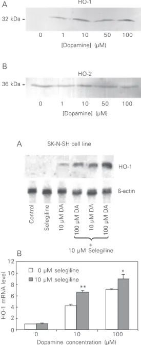
HO-1 (32 kDa) protein was not detected in the control (not treated with dopamine) homogenates from SK-N-SH but was pres-ent at detectable levels after treatmpres-ent with dopamine (1-100 µM). HO-2 protein was observed in untreated cells and the levels did not change as a result of dopamine treatment (Figure 2).
Up-regulation of HO-1 was inversely correlated with survival. Exposure to in-creasing dopamine concentrations was cy-totoxic to the SK-N-SH cell line analyzed. Increasing exposure to dopamine caused a progressive decrease in cell viability assessed by LDH release and Trypan blue exclusion. The survival of treated cells expressed as percentage of the viable untreated cells was as follows: 100% with 0.1 µM dopamine, 100% with 1 µM dopamine, 62% with 10 µM dopamine, 54% with 50 µM dopamine, and 42% with 100 µM dopamine. The experi-ments were repeated six times. The percent-ages of cytotoxicity, estimated by LDH re-lease for SK-N-SH (repeated five times) were as follows: 0% with 0.1 dopamine, 0% with 1 µM dopamine, 14% with 10 µM dopamine, 21% with 50 µM dopamine, and 29% with 100 µM dopamine. HO-1 mRNA levels were inversely correlated with cell survival in each case. Dopamine toxicity positively correlated with HO-1 induction.
The decrease in cell survival as a result of dopamine exposure was accompanied by an increase in lipid peroxidation. Malonaldehyde levels in untreated cells and in cells treated with 100 µM dopamine ranged respectively from undetectable to 0.071 µM and from 0.056 to 0.37 µM malonaldehyde/mg extracted protein (N = 6 extractions per group; P < 0.01, Mann-Whitney U-test).
Effect of antioxidants on dopamine-induced HO-1 expression in SK-N-SH cells
Dopamine-induced reduction in cell vi-ability was not attenuated by antioxidants (α
-Figure 1. Effects of exposure to dopamine on the levels of heme oxygenase 1 and 2 (HO-1 and HO-2) mRNA in SK-N-SH cells. A, Northern blot analysis. Cells were treated for 24 h with increasing concentrations of dopamine (0.1-100 µM). B, Relative expression levels of HO-1 mRNA. Data are reported as means ± SEM (N = 3). 2.4 kb
1.4 kb HO-1
HO-2
ß-actin
0 0.1 1.0 2.5 5.0 10 25 50 100 [Dopamine] (µM)
R2 = 0.9536
HO-1
HO-2 (1.4)
HO-2 (2.4) 10
8
6
4
2
0
0.1 1.0 10 100
[Dopamine] (µM)
HO mRNA level (ratio to control)
The amount of malonaldehyde determined was corrected for protein extracted, assayed by the method of Bradford (22).
Results
Effects of dopamine on steady-state levels of HO-1 and HO-2 mRNA and intracellular protein and on cell viability
Dopamine induced HO-1 mRNA expres-sion in the SK-N-SH cell line in a dose-dependent manner, as shown in Figure 1. HO-2 mRNA was detected as two bands of ~1.4 and ~2.4 kb. Expression of HO-2 mRNA did not change significantly in either the ~1.4- or ~2.4-kb transcript in
dopamine-A
tocopherol and ascorbic acid). The dopa-mine induction of HO-1 mRNA in the SK-N-SH cell line (10 and 100 µM) was not affected by L-ascorbic acid or by α -toco-pherol treatment at concentrations of 100 µM and 50 µM, respectively. In contrast, combined selegiline at 10 µM and dopamine treatment at 10 and 100 µM increased the expression of HO-1 mRNA and reduced significantly the level of cell viability com-pared with the results obtained with dopa-mine alone (Figures 3 and 4).
Discussion
Exposure to dopamine induced steady-state HO-1 mRNA levels in the human neu-roblastoma SK-N-SH cell line. Neuroblas-toma derives from embryonic sympathetic tissue and these cells have been widely used as an in vitro model system to investigate catecholamine metabolism in Parkinson’s disease. In contrast, the two HO-2 tran-scripts were evident in all cell lines tested and were not induced by dopamine treatment. Levels of intracellular HO-1 protein mirrored the changes seen with the mRNA. The pat-tern of regulation observed here was similar to that seen in other situations where oxida-tive stress was induced, i.e., HO-1 is highly sensitive, whereas HO-2 is refractory (23). Given that dopamine metabolism generates oxidative free radicals, as evidenced again here by the increase in lipid peroxidation products, the induction of HO-1 seen here would seem to be a consequence of the formation of these free radicals. In another study, pre-treatment of cultured C6 glioma cells with a dopamine antagonist failed to attenuate HO-1 induction by dopamine. This suggested that dopamine induction of HO-1 is not mediated by conventional dopamine receptors. This conclusion was supported by the finding that neither D1 nor D2 agonists activated HO-1 expression in C6 cells (18). Co-exposure to classical and therapeutic antioxidants failed to attenuate the
dopamine-Figure 2. Effect of dopamine treatment on HO protein ex-pression in SK-N-SH cell line. A, Immunoblot of the homoge-nate incubated with anti-HO-1 (1:2000). B, Immunoblot of the homogenate incubated with anti-HO-2 (1:2000). The molec-ular weight is shown on the left. Twenty micrograms of protein was loaded per lane.
32 kDa
36 kDa
0 1 10 50 100
[Dopamine] (µM)
0 1 10 50 100
[Dopamine] (µM) HO-1
HO-2
A
B
0 µg selegiline
% Cell viability (Trypan blue)
120 100 80 60 40 20 0
10 µg selegiline SK-N-SH
0 10 100
Dopamine concentration (µM)
Figure 4. Effect of 10 µM selegi-line on the survival of SK-N-SH cells exposed to dopamine. Data are reported as means ± SEM (N = 5). Statistical analyses were by two-way ANOVA with pairwise comparisons. +P <
0.05 and ++P < 0.001
com-pared with 0 µM selegiline at 0 µM dopamine; *P < 0.05 com-pared with 0 µM selegiline at 10 µM or 100 µM dopamine. ß-actin
HO-1
Control
100 µM DA
10 µM DA
100 µM DA
10 µM DA
Selegiline
+ 10 µM Selegiline
HO-1 mRNA level
12 10 8 6 4 2 0
0 µM selegiline
**
*
10 µM selegiline
0 10 100
Dopamine concentration (µM)
Figure 3. Effect of selegiline on the levels of HO-1 mRNA in the SK-N-SH cell line exposed to dopamine for 24 h. A: Northern blot analysis. B: Relative expres-sion levels of HO-1 mRNA. Data are reported as means ± SEM (N = 5). DA = dopamine *P < 0.05 and **P < 0.01 compared with 0 µM selegiline at 10 µM or 100 µM dopamine (two-way ANOVA pairwise comparisons). SK-N-SH cell line
induced increase in HO-1 expression and reduction in cell viability. Rather, selegiline, a specific MAO-B inhibitor, enhanced dopa-mine-induced HO-1 expression and reduced cell viability further compared with treat-ment with dopamine alone. This would sug-gest that the antioxidants were rapidly me-tabolized or were in a separate intracellular compartment compared with that where the free radicals were generated. Nevertheless, the antioxidant results agree with the assess-ment of their therapeutic effectiveness in vivo.
A similar observation of an in vitro cyto-toxic effect of selegiline has been shown in SH-SY5Y cells treated with dopamine (24). The results suggest that the toxic effect of dopamine is enhanced by selegiline. Dopa-mine levels would of course be increased due to the blocking of oxidative deamination by selegiline. The mechanism of selegiline-en-hanced toxicity, however, is still unclear. The lack of effect of MAO-B inhibitors suggests an involvement of enzymatic mechanisms in dopamine toxicity. Overall, it appears that the toxicity of dopamine in-volves more auto-oxidation than enzymatic metabolism to produce reactive oxygen spe-cies. If dopamine auto-oxidation results in toxicity, this may occur via reactive semi-quinone or semi-quinone formation, leading to generation of free radicals. Although selegiline by itself did not produce any change in HO-1 expression, cells exposed to dopamine plus selegiline showed higher levels of HO-1 mRNA compared to cells exposed to dopa-mine only.
Selegiline rather than reducing the dopa-mine-induced toxicity in vitro actually en-hances it. This finding seems to be related to a controversial issue, namely, that selegiline can cause an increase of the mortality of Parkinson’s disease patients following chronic treatment with the drug (25).
Selegiline alters the gene expression of HO-1 in neuroblastoma cell lines treated with dopamine. This is perhaps an indication that
the interaction between selegiline and dopa-mine exacerbates the cytotoxicity to SK-N-SH cells. Interestingly, research has shown that selegiline can induce altered expression of a number of genes in neurons including the genes for superoxide dismutase 1 and 2 and catalase (26,27). It has been suggested that the above changes in gene expression appear to reduce oxidative radical damage (28). Nevertheless, our findings suggest that selegiline in fact may be toxic to dopaminer-gic cells treated with dopamine, and that HO induction may possibly be induced by an increase in the oxidative stress condition. Because the enzymatic route is inhibited, more toxic radicals could be formed by a non-enzymatic pathway.
Our findings suggest that the apparent toxic effect by selegiline observed in some in vivo (25) and in vitro (24) studies may be due to potentiation of dopamine toxicity. The mechanism of selegiline-enhanced toxicity, however, is still unclear.
stimulating cyclic guanosine monophosphate production (35,36). Generated in excess in disease states, however, carbon monoxide possesses cellular effects that may impair cell vitality (31).
The present study showed that dopamine induces HO-1 in the neuroblastoma cell line
and induction is inversely correlated with cell viability. The increased HO-1 expression is strongly indicative that dopamine produces oxidative stress in these cell lines, but co-exposure to dopamine and antioxidants did not attenuate the induction or the effects of dopamine on cell viability.
References
1. Hirsch EC (1994). Biochemistry of Parkinson’s disease with special reference to the dopaminergic systems. Molecular Neurobiology, 9: 135-142.
2. Olanow CW (1992). An introduction to the free-radical hypothesis in Parkinson’s disease. Annals of Neurology, 32: S2-S9.
3. Jenner P (1994). Oxidative damage in neurodegenerative disease.
Lancet, 344: 796-798.
4. Simonian NA & Coyle JT (1996). Oxidative stress in neurodegen-erative diseases. Annual Review of Pharmacology and Toxicology, 36: 83-106.
5. Maines M (1988). Heme oxygenase: function, multiplicity, regula-tory mechanisms, and clinical applications. FASEB Journal, 2: 2557-2568.
6. Abraham NG, Drummond GS, Lutton JD & Kappas A (1996). The biological significance and physiological-role of heme oxygenase.
Cellular Physiology and Biochemistry, 6: 129-168.
7. Shibahara S, Sato M, Muller R & Yoshida T (1989). Structural organization of the human heme oxygenase gene and the function of its promoter. European Journal of Biochemistry, 179: 557-563. 8. McCoubrey WK, Ewing JF & Maines MD (1992). Human heme oxygenase-2 - characterization and expression of a full-length cDNA and evidence suggesting that the 2 HO-2 transcripts may differ by choice of polyadenylation signal. Archives of Biochemistry and Biophysics, 295: 13-20.
9. Keyse SM & Tyrrell RM (1989). Heme oxygenase is the major 32-kDa stress protein induced in human-skin fibroblasts by UVA radiation, hydrogen-peroxide, and sodium arsenite. Proceedings of the National Academy of Sciences, USA, 86: 99-103. 10. Applegate LA, Luscher P & Tyrrell RM (1991). Induction of heme
oxygenase: a general response to oxidant stress in cultured-mammalian-cells. Cancer Research, 51: 974-978.
11. Ewing J & Maines M (1991). Rapid induction of heme oxygenase 1 mRNA and protein by hyperthermia in rat brain: heme oxygenase 2 is not a heat shock protein. Proceedings of the National Academy of Sciences, USA, 88: 5364-5368.
12. Choi AMK & Alam J (1996). Heme oxygenase-1: function, regula-tion, and implication of a novel stress-inducible protein in oxidant-induced lung injury. American Journal of Respiratory Cell and Molecular Biology, 15: 9-19.
13. Castellani R, Smith MA, Richey PL, Kalaria R, Gambetti P & Perry G (1995). Evidence for oxidative stress in Pick disease and corticoba-sal degeneration. Brain Research, 696: 268-271.
14. Premkumar DRD, Smith MA, Richey PL, Petersen RB, Castellani R, Kutty RK, Wiggert B, Perry G & Kalaria RN (1995). Induction of heme oxygenase-1 messenger-RNA and protein in neocortex and cerebral vessels in Alzheimer’s disease. Journal of
Neurochemis-try, 65: 1399-1402.
15. Schipper HM & Stopa EG (1995). Expression of heme oxygenase-1 in the senescent and Alzheimer-diseased brain. Annals of Neu-rology, 37: 758-768.
16. Castellani R, Smith MA, Richey PL & Perry G (1996). Glycoxidation and oxidative stress in Parkinson disease and diffuse Lewy body disease. Brain Research, 737: 195-200.
17. Schipper HM, Liberman A & Stopa EG (1998). Neural heme oxyge-nase-1 expression in idiopathic Parkinson’s disease. Experimental Neurology, 150: 60-68.
18. Schmidt J, Mertz K & Morgan JI (1999). Regulation of heme oxygenase-1 expression by dopamine in cultured C6 glioma and primary astrocytes. Brain Research: Molecular Brain Research, 73: 50-59.
19. Berger SP, Hunger M, Yard BA, Schnuelle P & Van Der Woude FJ (2000). Dopamine induces the expression of heme oxygenase-1 by human endothelial cells in vitro. Kidney International, 58: 2314-2319.
20. Sambrook J, Fritsch EF & Maniatis T (1989). Molecular Cloning: A Laboratory Manual. 2nd edn. Cold Spring Harbor Laboratory Press, New York.
21. Moldeus P, Hogbord J & Orrenius S (1978). Isolation and use of liver cells. Methods in Enzymology, 52: 60-71.
22. Bradford MM (1976). A rapid and sensitive method for the quanti-tation of microgram quantities of protein utilizing the principle of protein-dye binding. Analytical Biochemistry,72: 248-254. 23. Kutty RK, Kutty G, Nagineni CN, Hooks JJ, Chader GJ & Wiggert B
(1994). RT-PCR assay for heme oxygenase-1 and heme oxyge-nase-2: a sensitive method to estimate cellular oxidative damage.
Annals of the New York Academy of Sciences, 738: 427-430. 24. Lai CT & Yu PH (1997). Dopamine- and
L-beta-3,4-dihydroxyphen-ylalanine hydrochloride (L-DOPA)-induced cytotoxicity towards cat-echolaminergic neuroblastoma SH-SY5Y cells - Effects of oxidative stress and antioxidative factors. Biochemical Pharmacology, 53: 363-372.
25. Parkinson’s Disease Research Group of the United Kingdom (1995). Comparison of therapeutic effects and mortality data of levodopa and levodopa combined with selegiline in Parkinson’s disease. British Medical Journal, 16: 1602-1607.
26. Carrillo MC, Kitani K, Kanai S, Sato Y, Miyasaka K & Ivy GO (1994). Deprenyl increases activities of superoxide dismutase and catalase in certain brain regions in old male mice. Life Sciences, 54: 975-981.
in Fischer rats. Annals of the New York Academy of Sciences, 717: 60-71.
28. Tatton WG & Chalmersredman RME (1996). Modulation of gene-expression rather than monoamine-oxidase inhibition (-)deprenyl-related compounds in controlling neurodegeneration. Neurology, 47: S171-S183.
29. Sian J, Dexter DT, Lees AJ, Daniel S, Agid Y, Javoyagid F, Jenner P & Marsden CD (1994). Alterations in glutathione levels in Park-inson’s disease and other neurodegenerative disorders affecting basal ganglia. Annals of Neurology,36: 348-355.
30. Nutter LM, Sierra EE & Ngo EO (1994). Heme oxygenase does not protect human cells against oxidant stress. Journal of Laboratory and Clinical Medicine, 123: 506-514.
31. Nath KA (1994). The functional-significance of induction of home oxygenase by oxidant stress. Journal of Laboratory and Clinical Medicine, 123: 461-463.
32. Mann VM, Cooper JM, Daniel SE, Srai K, Jenner P, Marsden CD & Schapira AHV (1994). Complex-I, iron, and ferritin in Parkinson’s disease substantia nigra. Annals of Neurology, 36: 876-881. 33. Dexter DT, Carayon A, Javoy-Agid F, Agid Y, Wells F, Daniel S,
Lees A, Jenner P & Marsden C (1991). Alterations in the levels of iron, ferritin and other trace metals in Parkinson’s disease and other neurodegenerative diseases affecting the basal ganglia. Brain, 114: 1953-1975.
34. Connor JR & Menzies SL (1995). Cellular management of iron in the brain. Journal of the NeurologicalSciences, 134: 33-44. 35. Marks GS, Brien JF, Nakatsu K & McLaughlin BE (1991). Does
carbon-monoxide have a physiological-function. Trends in Pharma-cological Sciences, 12: 185-188.