ABSTRACT: Postharvest diseases are a major problem in guava crops as the symptoms normally appear during fruit ripening. This study aimed to detect and characterize the temporal dynamics and spatial patterns of the most important guava diseases in orchards with and without removal of crop residues as a sanitation practice. The experiment was conducted in an orchard of ´Pedro Sato´ guavas, over two consecutive seasons, and data were collected from the flowering to the fruit ripening stage. In immature guavas treated with paraquat and ethrel, Colletotrichum spp. was detected from the 5th day of incubation. Anthracnose was detected in flowers at incidences higher than 50 % and black spot in fruit larger than 5.5 cm in length. The monomolecular and the exponential models provided the best fit to anthracnose and black spot incidence progress curve data, respectively. Both diseases showed a predominantly random spatial pattern in the orchard. The removal of crop residues reduced the rate of disease progress in at least one season, and was effective in reducing the areas under the quiescent disease progress curves (AUDPC) of anthracnose. Anthracnose incidence increased from 57 to 96 % and black spot from 1 to 48 %, respectively, at fruit maturation levels 1 and 3. A negative correlation was found between disease incidence and the color of the fruit skin (°h). Fruit harvested during the later maturation stages showed higher incidence of the diseases. Due to the wide distribution and early infection of quiescent diseases, starting at flowering, preventive management should consider disease monitoring and removal of crop residues.
Keywords: Colletotrichum, Guignardia, Psidium guajava,postharvest fruit rot 1Agency Paulista Agribusiness Technology Regional Midwest
Pole, Av. Rodrigues Alves, 40-40 − 17030-000 − Bauru, SP − Brazil.
2University of São Paulo/ESALQ − Dept. of Plant Pathology and Nematology, Av. Pádua Dias, 11, C.P. 09 − 13418-900 − Piracicaba, SP − Brazil.
3Agency Paulista Agribusiness Technology Regional Center South Pole, Rod. SP 127, km 30, C.P. 28 − 13400-970 − Piracicaba, SP − Brazil.
*Corresponding author <[email protected]>
Edited by: Emerson Medeiros Del Ponte
Temporal progress and spatial patterns of quiescent diseases in guava influenced by
Ivan Herman Fischer1*, Ana Raquel Soares-Colletti2, Maria Cecília de Arruda Palharini1, Marise Cagnin Martins Parisi3, Lilian Amorim2
Received November 05, 2015 Accepted April 15, 2016
Introduction
Guava is a highly perishable fruit and is, therefore, more susceptible to postharvest diseases. Once the skin of guava becomes edible, the presence of pathogenic in-juries, even a small spot, can compromise fruit quality and make the product unfit for marketing. Such damage can occur not only during harvesting but subsequently, such as in packing houses, and at the grocery whole-saler, retailer and consumer stages. The major posthar-vest diseases affecting guava are anthracnose, caused by
Colletotrichum spp., and black spot caused by Guignardia psidii Ullasa and Rawal (Fischer et al., 2011).
Anthracnose and black spot are defined as quies-cent diseases because the pathogens infect the fruit be-fore harvest and remain latent until maturation when physical and physiological changes in the fruit favor pathogen development (Prusky and Lichter, 2007). Meth-ods for detecting quiescent infection of Botryosphaeria dothidea (Moug.) Ces. & De Not. in apples (Valdebenito-Sanhueza et al., 2005) and Colletotrichum sp. in mangos (Paramasivan et al., 2009) already exist. For example, im-mature fruits are immersed in a paraquat solution that generates free radicals which induce lipid peroxidation and loss of membrane integrity (Dodge, 1971). Thus, se-nescence is induced in herbicide-treated fruit and the symptoms are expressed.
Colletotrichum spp. survives in mummified fruit on trees or on the ground and in infected twigs and
branch-es. The spores are dispersed by rain splashing combined with wind exposure (Singh, 2000). Spore germination and appressorium formation of G. psidii increase as the fruit ages (Escanferla et al., 2009). Nevertheless, knowl-edge of temporal and spatial analysis of guava diseases is limited but has the potential to provide important information for disease management. The analysis of these patterns may allow inferences about the influence of wind or rain in the dissemination of pathogens, and can provide information on sources of inocula to predict disease levels (Benson et al., 2006).
Little is known about the field epidemiology of guava diseases and the importance of early disease de-tection in disease management. This study aimed to (i) establish a method for detecting quiescent infections in guavas; (ii) quantify the incidence of postharvest dis-eases and (iii) characterize their temporal dynamics and spatial patterns in managed and unmanaged orchards by removal of crop residues.
Materials and Methods
Disease detection in immature fruit
for 5 min and washed in running water. To express the symptoms, fruit received different treatments by immer-sion for 1 min in: (i) paraquat (3 g L−1) (Northover and
Cerkauskas, 1994); (ii) ethrel (0.1 g L−1)
(Valdebenito-Sanhueza et al., 2005); (iii) urea (50 g L−1)
(Valdebenito-Sanhueza et al., 2005); or water. Afterwards, the fruits were washed for 3 min under running water. The fruits were placed in plastic pocket trays individually and stored under light at 25 °C and 80-85 % relative hu-midity (RH). The experimental design was completely randomized with five replicates per treatment and each replicate was represented by ten fruits. The experiment was repeated once. The best treatment allowing for the quickest detection was used in the field experiments.
Visible symptoms on fruit were assessed daily for 10 days. The Anova assumptions of normality and homoscedasticity were verified and data from two ex-periments were combined for analysis. A linear mixed effect model was fitted to the logit-transformed disease incidence progress data. The R software (R Core Team, 2015) and the function “lmer” from “lme4” package (Bates et al., 2015) were used for analysis.
Fifty isolates of Colletotrichum spp. representing 50 plants in a plot as well as four isolates of L. theobro-mae were obtained from immature fruit by direct iso-lation on potato-dextrose-agar (PDA). Pathogenicity was confirmed by inoculation of fruit at harvest.
Temporal progress and spatial distribution of qui-escent diseases
Fifteen temporal evaluations of guava quiescent diseases were performed on 50 plants from two plots of 'Pedro Sato' guava orchard located in Itajú, for two harvesting seasons as follows: the first from 29 Sept 2009 to 18 Feb 2010 and the second from 19 July 2010 to 12 Nov 2010. Weather information was recorded in a sta-tion located 40 km from the orchard. For 143 days in the first season, accumulated precipitation was 1,069.0 mm, the average maximum temperature was 31.0 °C and the average minimum temperature was 17.9 °C. In the 117 days of the second crop season, the accumulated precipi-tation was 233.6 mm, the average maximum tempera-ture was 28.8 °C and the average minimum temperatempera-ture 15.6 °C.
Crop residues (stems, leaves and fruit) lying on the ground were removed in one of the plots one month before the start of sampling to evaluate its importance as a source of inoculum of quiescent diseases. Dur-ing the fruitDur-ing period, fallen fruit was removed on a monthly basis to reduce the inoculum amount. Once a week, two flowers/fruits per guava tree were collected from 50 plants in each plot. Sampling started during the flowering period and continued until harvesting, totaling 15 samples in each plot. Three commercial maturation stages were recorded according to skin color (1 = dark green, 2 = light green and 3 = yellow-green).
During the first season, the flowering and sam-pling period differed by one month between plots
be-cause pruning was conducted at different times in the plots. Flowers and immature fruit were disinfected with NaOCl (5 g L−1) for 5 min, washed in running water,
immersed for 1 min in paraquat (3 g L−1) and washed
for 3 min under running water. The flowers and imma-ture fruit were subsequently incubated under light at 25 °C and 80-85 % RH. Disease incidence was assessed 10 days later, except for mature fruit that were assessed every 2 days for 10 days. Different population dynamic models including monomolecular (y = 1- (1- yo)exp(-rt)), logistic y = 1/[1+exp{-lnyo/(1-yo)+rt}], exponential (y =
yoexp(rt)) and Gompertz (y = exp(lnyoexp(-rt)), where y
is the rate of disease incidence, yo the initial inoculum,
r the disease progress rate and t the time (Campbell and Madden, 1990), were fitted to incidence progress data for anthracnose and black spot recorded using non-linear regression models in the Statistica 6.0 software (Statsoft, Tulsa, OK, EUA). The best-fitted model was chosen based on the coefficient of determination (R2)
and distribution of residuals. The parameters (yo - initial inoculum and r – disease progress rate) of the models fit-ted to data from each treatment (with and without crop residues) were compared for both years based on the
t-test (Campbell and Madden, 1990). The areas under the quiescent disease progress curves (AUDPC) were cal-culated by trapezoidal integration (Berger, 1988) and the data for each disease were analyzed in a 2 × 2 factorial design, comparing the two harvesting seasons and with and without crop residues, using the Tukey’s test at a 5 % probability level, considering each row of ten plants as a replication.
The spatial pattern of diseased trees (based on fruit symptoms of black spot and anthracnose) was evaluated weekly in 40 plants in the plot during the same period of the temporal progress evaluation. The plots were divid-ed into quadrats of 2 × 2 trees. The spatial pattern was determined based on the dispersion index D = Vobs/Vbin, where Vobs = [(Xi-np)2/n2(N-1)] and V
bin = p(1-p)/n, and
where Vobs is the observed variance, Vbin is the binomial variance, Xi represents the number of diseased plants per quadrat, n the number of plants per quadrat, N the total number of quadrats and p is the probability of a plant having diseased fruit (Hughes et al., 1996). The value of D was tested against the χ2 using p ≤ 0.05 as the
significance level. The binary Taylor’s Power Law model was fitted to all weekly data of the two crop seasons, but separately for plots with and without crop residues, as follows: log(Vobs) = A+b*log(Vbin), where A and b are pa-rameters (Madden and Hughes, 1995) calculated by lin-ear regression. According to this equation, where b = 1 and a = 0, the distribution is random and where a > 0 and b > 1, the distribution is aggregated. The hypoth-eses were tested at a 5 % probability.
Incidence of quiescent diseases at postharvest
each row of ten plants as a replication. Different models [y = 1-b2 exp(-rt); y = b1(1-b2 exp(-rt)], where y is the inci-dence of disease in proportion, b1 the maximum asymp-tote, b2 the parameter related to the initial inoculum of disease, r the rate of disease progression and t the time in days after the beginning of storage, were fitted to the anthracnose and black spot progress curves.
Detection of Colletotrichum spp. in tree bark
In a separate plot without crop residues,
Colle-totrichum spp. occurrence was evaluated on guava tree
trunks 1 month after the end of harvesting (03/2010) and during the flowering of the following season (07/2010). The pathogen was detected by the direct transfer of 36 small fragments of bark (trunk) per plant on potato dex-trose agar (PDA) for five plants randomly chosen in the field. The effect of surface disinfestation on bark with NaOCl (5 g L−1) for 1 min was not significantly
differ-ent (p ≤ 0.05) from that with absence of disinfestation in isolation of Colletotrichum spp. in the preliminary test. After 5 days of plate incubation at 25 °C, Colletotrichum
spp. isolates were transferred and purified in PDA. Dif-ferences in mean isolation frequency between the five sampled plants were compared by the Tukey’s test, at the 5 % probability level. Pathogenicity was confirmed by inoculation with mycelial discs of fungus isolates in guava fruit (Fischer et al., 2011).
Physicochemical fruit traits and diseases
In addition to disease detection, fruits were sam-pled at three maturation stages (1, 2 and 3) to charac-terize the following physicochemical parameters: a) skin color, determined by a colorimeter with readings in the equatorial region of the fruit and the results were ex-pressed as hue angles (ºh); b) flesh firmness, determined by a digital penetrometer with an 8 mm tip with read-ings in the equatorial region of the fruit and the results were expressed in Newtons (N); c) soluble solids con-tent, determined in a digital refractometer and the re-sults were expressed in ºBrix; d) acidity, determined by titration with NaOH and the results expressed as g L−1
citric acid; and e) ascorbic acid content, determined by titration with DCFI (2,6 dichlorophenol indophenol so-dium salt) and the results were expressed in mg 100 g−1
(AOAC, 2005). The experimental design was completely randomized with five replicates of six fruits per treat-ment. The physicochemical parameters in each fruit maturation stage for the plots with and without crop res-idues were compared by the Tukey’s test (p≤ 0.05). The physicochemical parameters were correlated with dis-ease incidence using the Spearman correlation (p≤ 0.05).
Results and Discussion
Detection of latent pathogen infections in imma-ture guavas
Colletotrichum conidia were observed at the 5th day
of incubation of guava fruit immersed in paraquat and
ethrel solution, at higher incidence compared with urea and water on the 6th and 7th day of incubation onward.
Significant interaction between treatment and day of in-cubation was observed (p≤ 0.05). Colletotrichum was de-tected in 96 % of fruit treated with paraquat and ethrel on the 7th day. In the urea and water treatment,
inci-dence was 93 % only at the 9th day (Figure 1).
Lasiodiplo-diatheobromae Pat. Griffon & Maubl was also detected, but at a low incidence (2 %).
Based on these results, paraquat or ethrel could be recommended for the quick and reliable detection of latent Colletotrichum spp. infections in immature guava fruit. The effect of ethrel may be associated with the stimulation of ethylene production in fruit and thus the activation of quiescent C. gloeosporioides (Penz.) Penz & Sacc.infections, as previously reported for avocados (Prusky et al., 1996). When treating mango fruit with paraquat, anthracnose symptoms were anticipated in 4-5 days, which assisted in decisions regarding storage time and whether to export or send the fruit to the domestic market in India (Paramasivan et al., 2009).
Temporal progress and spatial distribution of qui-escent diseases
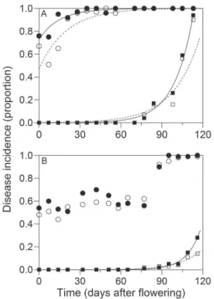
Anthracnose was the main disease in 'Pedro Sato' guavas (Table 1) with virtually 100 % incidence in im-mature small fruit (3 cm) during the first season (Figure 2A). According to Moraes et al. (2015), there is no differ-ence in conidial germination and appressorium forma-tion of C. gloeosporioides in guavas at different ages. Fifty percent of Colletotrichum spp. infection was observed during the flower opening stage. The incidence of major postharvest diseases was lower during the second crop season (Figure 2B), possibly because of less favorable weather conditions with lower temperatures and
pre-Figure 1 − Incidence (%) of anthracnose in immature fruit (2 cm long) of 'Pedro Sato' guavas for different postharvest treatments. The disease assessment was performed for 10 days of storage at
cipitation. The optimum temperature for infection by C.
gloeosporioides lies between 22 and 25 °C (Piccinin et al., 2005). The optimal temperature for germination and formation of G. psidii appressoria ranges between 25 and 30 °C (Escanferla et al., 2009).
Black spot in guava, found in fruit at 5.5 cm in length, was observed at high intensity in both plots when fruit reached maturation (Figure 2A). The high in-cidence of black spot and anthracnose is related to the ability of the pathogens to infect unripe and uninjured fruit (Picinnin et al., 2005; Martins et al., 2007). Other postharvest diseases detected in both plots were pestalo-tia, phomopsis, fusicocum and lasiodiplodia rots (Table 1). Mucor and dothiorella rots were found at low inci-dences, with AUDPC values below 5.4. Since no interac-tion was observed (p > 0.05) between harvesting seasons and crop residues, the average of harvesting seasons is presented in Table 1. Except for black spot and lasiodi-plodia rot, crop residue removal was effective in reduc-ing the incidence of diseases.
The monomolecular model provided the best fit to the anthracnose progress curve in the plots without crop residues (b2 = 1.84; r = 0.05, R2 = 0.78) and with
crop residues (b2 = 0.27; r = 0.07, R2 = 0.87) for the
first sampled crop season (Figure 2A). The monomo-lecular model most frequently describes the progress of monocyclic diseases. However, it also describes the progress of quiescent diseases, regardless of their desig-nation as monocyclic or polycyclic (Bergamin Filho and Amorim, 2002). The monomolecular model showed a good fit to the temporal progress of apple leaf spot inci-dence caused by C. gloeosporioides (Guerra et al., 2012) and of witches’ broom observed in cocoa fruit (Alves et al., 2006). In the second crop season, none of the models tested was fitted to the anthracnose progress curves (Figure 2B).
The exponential model presented the best fit to the progress curve for black spot in the crops with (1st
year: yo = 0.0003; r = 0.072; and R2 = 0.97 and 2nd year:
yo = 0.000003; r = 0.098; and R2 = 0.98) and without
Figure 2 − Progress incidence (proportion) of anthracnose (circles) and black spot (squares) for different developmental stages of 'Pedro Sato' guavas (flower to commercial fruit) from the plot without crop residues (empty) and with crop residues (full) after 10 days of storage at 25 °C and 80-85 % relative humidity. Points represent disease incidence in proportion and lines fit with the monomolecular model (y = 1- y0 exp(- r t)) (anthracnose) and exponential (y = y0 exp (r t)) (black spot), where y is the disease incidence in proportion, y0 is the disease initial inoculum, r is the rate of disease progression and t is the time in days. Data from the first year of sampling (29/09/2009 to 18/02/2010) (A) and the second year of sampling (19/07/2010 to 12/11/2010) (B). Table 1 − Area under the disease progress curve (AUDPC) calculated
from 15 weekly evaluations (flowering to harvest) of postharvest diseases in 'Pedro Sato' guavas grown in plots with and without crop residues after 10 days of storage at 25 °C and 80-85 % relative humidity. Averages for two crop seasons (2009/10 and 2010).
Diseases AUDPC
Plot without crop residues Plot with crop residues
Anthracnose 9,143.8 a* 9,534.3 b
Black spot 666.5 a 772.8 a
Pestalotia rot 297.5 a 714.8 b
Phomopsis rot 238.8 a 323.8 b
Fusicocum rot 208.5 a 329.8 b
Lasiodiplodia rot 25.0 a 40.0 a
*Values followed by different letters in each row are significantly different by Tukey’s test (p≤ 0.05).
residues (1st year: y
o = 0.0007; r = 0.050; and R
2 = 0.93
and 2nd year: y
o = 0.00008; r = 0.065; and R
2 = 0.93)
(Figure 2A and B). In citrus, the monomolecular model best fitted the progress curves of black spot caused by
The progress rates of anthracnose and black spot were higher when crop residues were present (p ≤ 0.05), which suggests that both pathogens can survive in these residues that perpetuate within-field inoculum in the ab-sence of flowers or fruit. The mucilage around the conid-ia of Colletotrichum sp. allows for pathogen survival for long periods in plant debris. In addition, Colletotrichum
spp. is known to survive in several species of wild and cultivated plants (Silva and Michereff, 2013). The high disease incidence at the beginning of the season is prob-ably related to the high survival capacity of this patho-gen. Moreover, as a perennial crop, guava trees remain in the field for many years enabling a continued increase in the incidence of pathogens that survive in crop resi-dues. Therefore, crop residues may be sources of local inocula besides other planting areas. Further studies are recommended, preferably with evaluations performed over a longer period. According to Piccinin et al. (2005), among the measures to control postharvest diseases of guava, especially quiescent diseases, pruning diseased branches, cleaning the orchard and immediately burn-ing all crop residues are of paramount importance to re-duce the amount of inocula in the field.
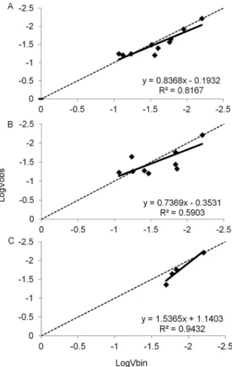
A modified Taylor’s Power Law provided a good fit for the relationship between the log of the two variances for black spot in both plots without (R2 = 0.82, Figure
3A) and with (R2 = 0.59, Figure 3B) crop residues. For
anthracnose, this Taylor’s Power Law fitted well the data from plots with crop residues only (R2 = 0.94, Figure
3C). The hypothesis that b = 1 was not rejected (p > 0.05) based on the t-test, suggesting a random distribu-tion of trees with black spot and anthracnose. In most assessments, the dispersion index (D) did not differ from 1 (p≤ 0.05), suggesting a random distribution of the dis-ease. An exception was the assessment of 22 Dec 2009 for black spot in the plot with crop residues, where D = 2.59, indicating aggregation (Table 2).
The causal agent of black spot is known to pro-duce ascospores and conidia. Spore traps in the field detect only G. psidii ascospores. Phyllosticta psidiicola
(Petrak) van der Aa conidia are not found, suggesting that ascospores might act as the main inoculum (Lin et al., 2003). In the G. citricarpa-citrus pathosystem, asco-spores that form in decaying leaves on the ground are spread over short and long distances by the wind (Spósi-to et al., 2007), which could explain the random tion pattern of black spot in guava or extensive distribu-tion of the inoculum in the field with all trees acting as a source of pathogen survival. Unlike our findings for guava black spot, diseased citrus plant exhibited a clus-tered pattern, regardless of disease incidence, indicating the high importance of inocula dispersal over short dis-tances and suggesting the involvement of conidia in the disease increase under field conditions in the state of São Paulo (Spósito et al., 2007).
The most likely explanation for the high initial in-cidence of anthracnose at flowering is that the inoculum was already present and homogeneously distributed in
Figure 3 − Relationship between the logarithm of the expected binomial variance and logarithm of the variance observed in incidence evaluations of 'Pedro Sato' guava plants with black spot symptoms in without (A) and with crop residues (B) plots and anthracnose in a with crop residues plot (C) for a quadrat size of 2 × 2 in Itajú-SP, with harvests in 2009 and 2010.
the area. Further studies are necessary preferably in younger trees with lower incidences of anthracnose to verify a possible clustering of diseased trees. According to Timmer et al. (1994), mechanisms of C. acutatum J.H. Simmonds dispersal over long distances are not clear. Thus, it is not possible to explain how a disease caused by Colletotrichum occurs suddenly and causes losses of up to 100 % in citrus. In São Paulo, the postbloom fruit drop of citrus had a variable spatial pattern, which was initially random and then, it became moderately aggre-gated, suggesting the existence of other dispersal mecha-nisms besides rain with winds (Silva Júnior et al., 2014).
Disease incidence at postharvest
and D). A similar result was observed for anthracnose during fruit development in the first crop season (Figure 2A) and for citrus black spot (Spósito et al., 2004). The monomolecular model presented a good fit also for the guava anthracnose progress curve established in fruit marketed wholesale in Brazil. However, none of the tested models had a good fit for the black spot incidence progress curves (Fischer et al., 2011).
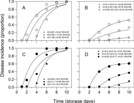
The overall incidence of postharvest diseases, con-sidering the average of the two plots (with and without crop residues) was higher than 50 % in the three ripen-ing stages. On average, anthracnose incidence was 78 % followed by black spot with 22 % of diseased fruit. Fusicocum, phomopsis, pestalotia, mucor, lasiodiplodia and dothiorella rot incidences were below 3 % and did not differ among themselves (p > 0.05) (data not shown). Differences in anthracnose and black spot incidences were observed among the three maturation stages. An-thracnose incidences increased from 57 to 96 % of dis-eased fruit and black spot incidences incrdis-eased from 1 to 48 % of diseased fruit, respectively, in fruit at matura-tion levels 1 and 3 (Figure 5 A and B). 'Pedro Sato' gua-vas harvested at maturation stage 1 should be commer-cialized within 6 days. This time is reduced to four and two days if fruit are harvested at stages 2 and 3, mainly because of the occurrence of rots (Azzolini et al., 2004).
Postharvest diseases downgrade fruit for sale and must not exceed 20 % during the fruit shelf life (Paull,
Figure 4 − Progress incidence (proportion) of anthracnose (A and C) and black spot (B and D) in 'Pedro Sato' guavas harvested at three maturation stages, according to skin color [1 = dark green (triangles), 2 = light green (squares) and 3 = yellow-green (circles)] in plots (area) with (C and D) and without crop residues (A and B) for 10 days of storage at 25 °C and 80 to 85 % relative humidity. Averages for 2009 and 2010. Parameters and determination coefficients of regression (R2) for the models y = 1-b
2 exp(-rt) (anthracnose) and y = b1(1-b2 exp(-rt) (black
spot), where y is the incidence of disease in proportion, b1 the maximum asymptote, b2 the parameter related to the initial inoculum of disease,
r the rate of disease progression and t the time in days after the beginning of storage, which set the progress curves of diseases. Table 2 − Dispersion index (D) of the incidence of black spot
symptoms during weekly evaluations in ‘Pedro Sato’ guava plants in without and with crop residues plots and of anthracnose in with crop residues plot. The quadrat size was 2 × 2 in Itajú-SP, with harvests in 2009 and 2010.
Dispersion index (D)
Date Black spot Anthracnose
with crop residues
without crop residues
with crop residues
29 Sept 2009 -* - 0.74
22 Dec 2009 2.59** -
-05 Jan 2010 1.48 -
-12 Jan 2010 1.03 -
-20 Jan -2010 1.03 0.63
-26 Jan 2010 - 1.51
-02 Feb 2010 - 1.21
-18 Feb 2010 - 1.03
-19 July 2010 - - 0.46
26 July 2010 - - 0.84
02 Aug 2010 - - 1.03
04 Oct 2010 1.03 0.94
-15 Oct 2010 0.84 0.74
-22 Oct 2010 0.40 0.96
-29 Oct 2010 0.30 0.62
-05 Nov 2010 0.55 1.02
-12 Nov 2010 0.74 0.44
1999). However, incidences of postharvest diseases ex-ceeding 40 % in guava have been reported in the state of São Paulo (Fischer et al., 2011). In this study, the market-ing period was even lower than that found by Azzolini et al. (2004) because fruit maturation at stage 2 had levels of anthracnose symptoms exceeding 20 % by the 4th day
of storage (Figure 4A and C). According to Martins et al. (2007), the high incidence of fruit rot during the summer of each year explains why the fruit is harvested before the maturation point with negative consequences for the final quality of the product, which suggests the necessity to adopt measures for more effective control during the production and postharvest phases.
The results indicate that anthracnose was the most common postharvest disease, confirming findings in the literature (Piccinin et al., 2005; Fischer et al., 2011). Af-ter 10 days of storage at 25 °C, the average incidence of black spot and anthracnose reached values above 30 % in guavas marketed in the São Paulo wholesale market (Soares-Colletti et al., 2014). In Taiwan, where black spot
causes large losses, the disease reached 80 and 95 % in-cidences in the Pearl and Crystal cultivars, respectively, after 5 days of fruit storage (Lin et al., 2003).
Detection of Colletotrichum spp. in tree bark
One month after the end of thefirst season’s har-vesting, Colletotrichum spp. was present in 25 % of guava tree trunks and in 14 % of tree trunks during the flower-ing of the followflower-ing season. Although there was a de-crease in 45 % incidence after the offseason, tree trunks are a possible source of pathogen inoculum for the next crop season. No differences (p > 0.05) were observed in isolation frequency among the five plants sampled and pathogenicity of isolates was confirmed in guava fruit.
Relationships between physicochemical character-istics and postharvest diseases for guavas
The physicochemical variables of fruit were similar in both plots (with and without crop residues) of 'Pedro Sato' guavas. Differences in skin color, flesh firmness, acidity and solids-acidity ratio were observed between the three maturation stages (Table 3). Similar results were observed by Azzolini et al. (2004) for 'Pedro Sato' guavas at harvesting.
Flesh firmness of fruit at maturation stage 2 was between 66.72 and 78.56 N in both plots, averaging 45 % less than that of stage 1 and 49 % greater than that of stage 3 (Table 3). Titratable acidity of fruit decreased as the fruit ripened with average values 7; 6 and 5 g L−1
citric acid for maturation stages 1, 2 and 3, respectively, in both plots. According to Wills et al. (2007), during rip-ening, a pronounced decrease in organic acid content is found in most fruits because these compounds are used as substrates during the respiration process. The ratio (solids:acidity) in fruit from plots 1 and 2 increased with advancing maturation stages because of acidity reduc-tion (Table 3).
The soluble solids content of the fruit did not dif-fer (p > 0.05) over the three maturation stages (Table 3), which was inconsistent with the results of Azzolini et al. (2004). The total soluble solids (SS), as expressed in °Brix, is used as an indirect measurement of sugar con-tent. Generally, sweetness increases with the ripening process because of the accumulation of sugars when the fruit stays on the tree.
Ascorbic acid levels were similar for fruit from the plot with crop residues at stages 2 and 3 and higher (p≤ 0.05) than those in stage 1. Azzolini et al. (2004) only found differences between stages 1 and 3 with ascorbic acid values of approximately 37 %, which were lower than those obtained in this work. For the fruit from the plot without crop residues, ascorbic acid found during stages 1 and 3 did not differ and a lower ascorbic acid content was observed during stage 2 (Table 3).
A negative correlation (p ≤ 0.05) was observed between the incidence of anthracnose and skin color (r = -0.90), flesh firmness (r = -0.85) and titratable acidity (r = -0.71) parameters, that is, as these physi-Figure 5 − Incidence of anthracnose (A) and black spot (B) on ‘Pedro
cochemical parameters decreased with fruit ripening, there was a proportional increase in disease incidence. Several physiological changes that occur during fruit rip-ening, such as the activation of ethylene biosynthesis, cuticular changes and loosening of cell walls, which is accompanied by a decline in antifungal compounds, led to the occurrence of fruit diseases (Prusky et al., 2013). Associations between the increased disease occurrence with decreasing acidity or increasing soluble solids con-centrations have been reported in apples (Sharma and Kaul, 1988) and cherry fruit (Northover and Biggs, 1990) making these parameters useful for identifying periods of fruit susceptibility in the field. Similarly, a negative correlation between the incidence of black spot and skin color (r = -0.69) and a positive correlation between dis-ease incidence and ascorbic acid content (r = 0.60) were observed.
Thus, skin color, flesh firmness and titratable acidity parameters for three maturation stages contrib-uted to an understanding of the causes of resistance or susceptibility of guavas to the primary postharvest guava disease and may be considered when planning for a harvesting point aimed to achieve a greater shelf life for this fruit.
Acknowledgements
The authors wish to the São Paulo State Founda-tion for Research Support for the financial support to the research project (FAPESP 09/09337-2) and Juan Pablo Edwards Molina for collaboration in the statistical analy-sis.
References
Alves, S.A.M.; Pomella, A.W.V.; Aitken, W.M.; Bergamin Filho, A. 2006. Disease progress curves and gradients of witches' broom disease in a cloned cocoa area, in Uruçuca, Bahia. Fitopatologia Brasileira31: 483-491 (in Portuguese, with abstract in English). Association of Official Analytical Chemists [AOAC]. 2005. Official
Methods of Analysis. 18ed. AOAC, Gaithersburg, MD, USA. Azzolini, M.; Jacomino, A.P.; Bron, I.U. 2004. Indices to evaluate
postharvest quality of guavas under different maturation stages. Pesquisa Agropecuária Brasileira 39: 139-145 (in Portuguese, with abstract in English).
Table 3 – Physical-chemical characteristics of 'Pedro Sato' guava harvested at three maturation stages (1 = dark green, 2 = light green and 3 = yellow-green) in plots with and without crop residues. Averages for two seasons (2009/10 and 2010).
Physical-chemical characteristics Plot with crop residues Plot without crop residues
Stage 1 Stage 2 Stage 3 Stage 1 Stage 2 Stage 3
Skin color (°h) 118.13 a* 114.38 b 109.40 c 117.43 a 114.72 b 109.80 c
Flesh firmness (N) 106.31 a 78.56 b 33.48 c 104.55 a 66.72 b 40.43 c
Soluble solids (°Brix) 8.99 ns** 9.05 9.05 9.34 ns 8.94 9.33
Acidity (g L−1 citric acid) 6.8 a 5.4 b 4.6 c 7.0 a 5.7 b 5.2 c
Ratio 13.33 c 16.92 b 19.59 a 13.42 c 15.64 b 17.82 a
Ascorbic acid (mg 100 g−1) 56.39 b 70.85 a 66.85 a 70.41 a 61.91 b 71.78 a
*Means followed by the same letter in the row do not differ in each plot (Tukey's test, p≤ 0.05). **ns = not significant (p≤ 0.05).
Bates, D.; Maechler, M.; Bolker, B.; Walker, S. 2015. Fitting linear mixed-effects models using lme4. Journal of Statistical Software 67: 1-48.
Benson, D.M.; Grand, L.F.; Vernia, C.S.; Gottwald, T.R. 2006. Temporal and spatial epidemiology of Phytophthora root rot in Fraser fir plantations. Plant Disease 90: 1171-1180.
Bergamin Filho, A.; Amorim, L. 2002. Diseases with a variable incubation period as a function of the host phenology. Fitopatologia Brasileira 27: 561-565 (in Portuguese, with abstract in English).
Berger, R.D. 1988. The analysis of the effects of control measures on the development of epidemics. p. 137-151. In: Kranz, J.; Rotem, J., eds. Experimental techniques in plant disease epidemiology. Springer, Heidelberg, Germany.
Campbell, C.L.; Madden, L.V. 1990. Introduction to Plant Disease Epidemiology. John Wiley, New York, NY, USA.
Dodge, A.D. 1971. The mode of action of bipyridylium herbicides, paraquat and diquat. Endeavour 30: 130-135.
Escanferla, M.E.; Moraes, S.R.G.; Salaroli, R.B.; Massola Júnior, N.S. 2009. Pre-penetration stages of Guignardia psidii in guava: effect of temperature, wetness duration and fruit age. Journal of Phytopathology 157: 618-624.
Fischer, I.H.; Almeida, A.M.; Arruda, M.C.; Bertani, M.A.R.; Garcia, M.J.M.; Amorim, L. 2011. Postharvest damages in guavas from the Midwest region of the State of São Paulo. Bragantia 70: 570-576 (in Portuguese, with abstract in English).
Guerra, D.S.; Nickel, O.; Del Ponte, E.M.; Sanhueza, R.M.V.; Fajardo, T.V.M.; Marodin, G.A.B. 2012. Development of Glomerella leaf spot is enhanced in virus-infected maxi gala apples. Journal of Plant Pathology 94: 237-241.
Hughes, G.; Madden, L.V.; Munkvold, G.P. 1996. Cluster sampling for disease incidence data. Phytopathology 86: 132-137. Lin, C.C.; Lai, C.S.; Tsai, S.F. 2003. Ecological survey of guava
new fruit rot: Phyllosticta rot (black spot) and other fruit rots. Plant Protection Bulletin 45: 263-270.
Madden, L.V.; Hughes, G. 1995. Plant disease incidence: distributions, heterogeneity, and temporal analysis. Annual Review of Phytopathology 33: 529-564.
Moraes, S.R.G.; Escanferla, M.E.; Massola Júnior, N.S. 2015. Pre-penetration and Pre-penetration of Colletotrichum gloeosporioides into guava fruit (Psidium guajava L.): effects of temperature, wetness period and fruit age. Journal of Phytopathology 163: 149-159.
Northover, J.; Biggs, A.R. 1990. Susceptibility of immature and mature sweet and sour cherries to Monilinia fructicola. Plant Disease 74: 280-284.
Northover, J.; Cerkauskas, R.F. 1994. Detection and significance of symptomless latent infections of Monilia fruticola in plums. Canadian Journal of Plant Pathology 16: 30-36.
Paramasivan, M.; Ohan, S.; Li, G.S.; Mathiyazhagan, S.; Uthukrishanan, N. 2009. Detection of latent infections in mango fruit with herbicides. Archives of Phytopathology and Plant Protection 42: 318-326.
Paull, R.E. 1999. Effects of temperature and relative humidity on fresh commodity quality. PostharvestBiology and Technology 15: 263-277.
Piccinin, E.; Pascholati, S.F.; Di Piero, R.M. 2005. Guava diseases (Psidium guajava L. = Doenças da goiabeira (Psidium guajava L.). p. 401-405. In: Kimati, H.; Amorim, L.; Rezende, J.A.M.; Bergamin Filho, A.; Camargo, L.E.A., eds. Manual de fitopatologia: doenças das plantas cultivadas. 4ed. Ceres, São Paulo, SP, Brazil (in Portuguese).
Prusky, D.; Wattad, C.; Kobiler, I. 1996. Effect of ethylene on activation of lesion development from quiescent infections of Colletotrichum gloeosporioides in avocado fruits. Molecular Plant Microbe Interactions 9: 864-868.
Prusky, D.; Lichter, A. 2007. Activation of quiescent infections by postharvest pathogens during transition from the biotrophic to the necrotrophic stage. FEMS Microbiology Letters 268: 1-8. Prusky, D.; Alkan, N.; Mengiste, T.; Fluhr, R. 2013. Quiescent
and necrotrophic lifestyle choice during postharvest disease development. Annual Review of Phytopathology 51: 155-176.
Sharma, R.L.; Kaul, J.L. 1988. Susceptibility of apples to brown rot in relation to quantitative characters. Indian Phytopathology 43: 113-115.
Silva, C.F.B.; Michereff, S.J. 2013. Biology of Colletotrichum spp. and epidemiology of the anthracnose in tropical fruit trees. Revista Caatinga 26: 130-138.
Silva-Junior, G.J.; Spósito, M.B.; Marin, D.R.; Ribeiro-Junior, P.J.; Amorim, L. 2014. Spatiotemporal characterization of citrus postbloom fruit drop in Brazil and its relationship to pathogen dispersal. Plant Pathology 63: 519-529.
Singh, R.S. 2000. Diseases of Fruit Crops. Science Publishers, Enfield, NH, USA.
Soares-Colletti, A.R.; Fischer, I.H.; Lourenço, S.A. 2014. The incidence of postharvest diseases on ‘Kumagai’ and ‘Pedro Sato’ guavas at wholesale markets in Brazil. Tropical Plant Pathology 39: 478-482.
Spósito, M.B.; Bassanezi, R.B.; Amorim, L. 2004. Resistance to citrus black spot by the analyses of disease progress curves. Fitopatologia Brasileira 29: 532-537 (in Portuguese, with abstract in English).
Spósito, M.B.; Amorim, L.; Ribeiro, P.J.; Bassanezi, R.B.; Krainski, E.T. 2007. Spatial pattern of trees affected by black spot in citrus groves in Brazil. Plant Disease 91: 36-40.
Timmer, L.W.; Agostini, J.P.; Zitko, S.E.; Zulfiqar, M. 1994. Postbloom fruit drop of citrus, an increasingly prevalent disease of citrus in the Americas. Plant Disease 78: 329-334. Valdebenito-Sanhueza, R.M.; Duarte, V.; Amorim, L.; Porto,
M.D.M. 2005. Detection and epidemiology of white rot on apples. Fitopatologia Brasileira 30: 217-223 (in Portuguese, with abstract in English).