ISSN 0100-879X
BIOMEDICAL SCIENCES
AND
CLINICAL INVESTIGATION
www.bjournal.com.br
www.bjournal.com.br
Volume 45 (2) 93-178 February 2012
Braz J Med Biol Res, February 2012, Volume 45(2) 139-146
doi: 10.1590/S0100-879X2012007500012
Effect of lipoarabinomannan from Mycobacterium avium subsp
avium in Freund's incomplete adjuvant on the immune response of
cattle
S.B. Colavecchia, A. Jolly, B. Fernández, A.M. Fontanals, E. Fernández and S.L. Mundo
Institutional Sponsors
The Brazilian Journal of Medical and Biological Research is partially financed by
Faculdade de Medicina de Ribeirão Preto Campus
Ribeirão Preto
Ex plor e H igh - Pe r for m a n ce M S Or bit r a p Te ch n ology I n Pr ot e om ics & M e t a bolom ics
Effect of lipoarabinomannan from
Mycobacterium avium
subsp
avium
in
Freund’s incomplete adjuvant on the
immune response of cattle
S.B. Colavecchia
1, A. Jolly
1, B. Fernández
1, A.M. Fontanals
1,
E. Fernández
2and S.L. Mundo
11Inmunología, Facultad de Ciencias Veterinarias, Universidad de Buenos Aires,
Ciudad Autónoma de Buenos Aires, Argentina
2Clínica de Rumiantes, Facultad de Ciencias Veterinarias, Universidad de Buenos Aires,
Ciudad Autónoma de Buenos Aires, Argentina
Abstract
The aim of the present study was to determine whether lipoarabinomannan (LAM), in combination with Freund’s incomplete adjuvant (FIA), was able to improve cell-mediated and antibody-mediated immune responses against ovalbumin (OVA) in cattle. Twenty-three calves were assigned to four treatment groups, which were subcutaneously immunized with either OVA plus FIA, OVA plus FIA and LAM from Mycobacterium avium subsp avium, FIA plus LAM, or FIA alone. Lymphoproliferation, IFN-γ
production and cell subpopulations on peripheral blood mononuclear cells before and 15 days after treatment were evaluated.
Delayed hypersensitivity was evaluated on day 57. Specific humoral immune response was measured by ELISA. Inoculation with LAM induced higher levels of lymphoproliferation and IFN-γ production in response to ConA and OVA (P < 0.05). Specific
antibody titers were similar in both OVA-immunized groups. Interestingly, our results showed that the use of LAM in vaccine
preparations improved specific cell immune response evaluated by lymphoproliferation and IFN-γ production by at least 50 and
25%, respectively, in cattle without interfering with tuberculosis and paratuberculosis diagnosis.
Key words: Lipoarabinomannan; Cattle; Immunomodulation; Mycobacterium avium subsp avium
Introduction
Correspondence: S.L. Mundo, Inmunología, Facultad de Ciencias Veterinarias, Universidad de Buenos Aires, Chorroarín 280, (1427) Ciudad Autónoma de Buenos Aires, Argentina. Fax: +54-11-4524-8480. E-mail: [email protected]
Received April 24, 2011. Accepted January 16, 2012. Available online February 3, 2012. Published February 17, 2012.
Protection against infectious diseases may require a
cell-mediated immune response (CMIR) and/or an antibody-mediated immune response (AMIR), depending on the agent. Adjuvants have been used since the early 1920’s
(reviewed in Ref. 1) to improve vaccine efficacy through the enhancement of a specific CMIR and/or AMIR. Since adjuvants may cause a number of adverse consequences,
either locally at the injection site or systemically, these reactions must be minimized for the development of new safer vaccines (2). The mechanisms of action by which adjuvants promote an increased immune response may be: the depot effect, antigen presentation, antigen distribution or targeting and immune activation/modulation, and CD8 cytotoxic T lymphocyte induction (1,2).
Microbial cells, their components, or chemically modified
microbial products have historically been used as adjuvants
in vaccines. Freund’s complete adjuvant (FCA) is a
water-in-mineral-oil adjuvant containing heat-killed Mycobacterium
tuberculosis that stimulates both a CMIR and an AMIR and was considered to be the gold standard adjuvant for many years (2). However, FCA causes severe lesions at the site of injection (3) and cannot be used in commercial herds since mycobacterial antigens may interfere with the
in vitro and in vivo diagnosis of tuberculosis. The same
effect is detected with the use of Mycobacterium sp walls
or protein antigens in experimental immunology and vac-cination procedures (4,5). On the other hand, Freund’s incomplete adjuvant (FIA), which uses mannide monooleate
into which the antigen is emulsified, has been shown to
140 S.B. Colavecchia et al.
use of high-grade oils and purified surfactants. Besides,
several studies using Marcol 52, Arlacel C, and Tween 80
as oil adjuvants in a Tritrichomonas vaccine have detected
an increase in resistance to infection in immunized cattle (8). One disadvantage of FIA is that it does not potentiate the CMIR, which is critical for the control of many infections
(7,9). The inclusion of purified components of mycobacteria
could be an alternative to improve these responses. Lipoarabinomannan (LAM) is an important component of the cell wall of mycobacteria. It is a conserved
mannosyl-phosphatidyl-myo-inositol that possesses a mannan core
and a branched arabinan polymer, which in some cases is decorated with a terminal cap motif (10-12). LAM can
be classified into three major structural families: ManLAM,
present in pathogenic strains (including Mycobacterium
avium subsp avium; Maa) with a short capping of mannoses;
PILAM, present in fast-growing non-pathogenic strains
with inositol phosphate caps, and AraLAM, present in M.
chelonae, which does not have capping (12,13). Modulation
studies with LAM have been carried out in vitro with mouse
(14) and human cells (15), and in vivo in mouse models
(16,17), using different doses and immunization protocols, indicating that LAM and different mycobacteria induce a Th1 biased response in allergic and parasitic diseases.
The aim of the present study was to determine whether LAM, in combination with FIA, is able to improve CMIR and AMIR against ovalbumin (OVA) in cattle. To our knowledge,
this is the first study about the immunomodulatory effects
of LAM on the immune response in a bovine model. The results could be useful for future applications, such as the development of new vaccines in cattle.
Material and Methods
Bacterial strain
Maa (R4 ER strain, kindly provided by Dr. A. Bernardelli, Servicio Nacional de Sanidad Animal, Argentina) was grown in Dorset Herley medium for 8 weeks, heat-inactivated and lyophilized for LAM extraction and vaccine preparation.
Preparation and characterization of LAM extract
LAM was extracted from 91.8 g Maa as previously
described (18). Briefly, crude LAM was purified on a 100 x 25 cm Sephadex G-200 column equilibrated with PBS at a flow rate of 25 mL/h. Fractions of 3.5 mL were collected and
examined for carbohydrate content by the phenol-sulfuric acid method using glucose as standard (19) and for protein content by the Bradford method using bovine serum albumin
as standard (20). LAM-containing fractions were identified by ELISA using a monoclonal antibody (mAb) specific for
LAM of M. tuberculosis (mAb CS-35, kindly provided by Dr.
J. Belisle, Colorado State University, Fort Collins, CO, USA). Fractions that strongly reacted with mAb CS-35 were pooled,
concentrated by ultrafiltration and characterized by SDS-PAGE and immunoblot as previously described (18).
Animals, groups and immunization protocols
Twenty-three 6-month-old Holstein calves from tuberculosis-free accredited herds were kept on a natural
farm inthe Pampas region of Argentina throughout the
experiment. Calves were randomly assigned to one of the
following experimental groups: OF(N = 7), which received
1 mg OVA (Sigma Chemical Co., USA) dissolved in 1 mL
PBS, pH 7.4, and emulsified in 1 mL FIA (Sigma-Aldrich
Co., USA); OFL (N = 8), which received 1 mg OVA and 1
mg LAM, both dissolved in 1 mL PBS and emulsified in 1
mL FIA; FL (N = 3), which received 1 mg LAM dissolved
in 1 mL PBS and emulsified in 1 mL FIA, and F (N = 5), which received 1 mL PBS emulsified in 1 mL FIA. Calves
were inoculated subcutaneously on days 0, 21, and 42 on the left scapular region. The experiment was performed with the approval and under the supervision of the Insti-tutional Committee for the care and use of experimental animals of Facultad de Ciencias Veterinarias, Universidad de Buenos Aires.
Proliferation assays
Proliferation assays were performed on days 0
(pre-immunization) and 57 (15 days after the third
immuniza-tion). PBMC were isolated from heparinized blood by density gradient centrifugation using Histopaque 1077 (Sigma-Aldrich Co.) according to standard techniques (21).
Lymphoproliferation assays were performed in U-shaped 96-well plates (BD Biosciences, USA) containing 100 µL/
well PBMC (0.5 x 10⁶ viable cells/well) in RPMI 1640 (Invit
-rogen Corporation, USA) with 10% fetal calf serum (FCS,
Invitrogen Corporation). Cells were cultured in 5% CO2 at
37°C and stimulated for 4 days with 2.5 µg/mL concanavalin A (ConA; Sigma-Aldrich Co.), 250 µg/mL OVA or 250 µg/mL
LAM. Non-stimulated wells were incubated only with RPMI
as control, and 0.5 µCi methyl-[3H]-thymidine (New England
Nuclear Radiochemicals, USA) was added to each well. Twenty hours later, cells were harvested onto Whatman GF/A paper and the incorporated radioactivity (counts per
minute, cpm) was measured by liquid scintillation counting (beta counter 1214 Rackbeta, Vallac; Pharmacia, Finland).
Experiments were run in triplicate and the stimulation index (SI) was calculated as mean cpm of stimulated wells/mean cpm of non-stimulated wells. A control of LAM toxicity was made by comparing the proliferation response to ConA
and ConA plus LAM and no significant differences were
detected between these treatments.
IFN-γ production
IFN-γ ELISA was performed according to manufacturer specifications (duoset ELISA development system, R&D Systems Inc., USA). Briefly, 96-well plates (Immulon 2HB,
Dynex Technologies, USA) were coated with 100 µL
anti-IFN-γ mAb (R&D), 2 µg/mL in PBS overnight at room tem -perature. The plates were washed three times and blocked
sodium azide for 2 h at room temperature. After washing, 100 µL of the culture supernatants of the proliferation assays
or recombinant IFN-γ (R&D, to construct a reference curve)
was incubated for 2 h at room temperature. Then, 100 µL
bio-tin labeled-anti-IFN-γ (R&D), 0.4 µg/mL, was added to each
well and the plates were further incubated for 2 h at room
temperature. One hundred microliters of HRP-conjugated streptavidin (R&D) 1:200 was added and incubated for 2 h at room temperature. After subsequent washes, plates were
developed using ortho-phenylenediamine dihydrochloride (Sigma-Aldrich Co.) in citrate buffer (Sigma-Aldrich Co.). Absorbance at 490 nm was measured with an OpsysMR spectrophotometer (Dynex Technologies).
Flow cytometry
Blood leukocytes were labeled for flow cytometry using
a single staining procedure. A total of 1 x 10⁶ viable cells
in 100 µL PBS were labeled in round-bottom microplates
(GBO, Greiner Bio One Inc., USA) with FITC-conjugated goat anti-bovine IgG (Sigma-Aldrich Co.), mouse IgG1 anti-bovine CD4 (CACT138A, VMRD, USA), mouse IgG1 anti-bovine CD8 (BAT82A, VMRD), mouse IgG1 anti-bovine CD25 (CACT116A, VMRD), and FITC-conjugated goat
anti-mouse IgG (KPL, Kierkegaard & Perry Laboratories,
Inc., USA) was used as a secondary antibody. Cells were also incubated with the mouse IgG1 isotype control (e-Bioscience Inc., USA). Stained cells were analyzed with
a FacsCalibur flow cytometer (Becton Dickinson, USA) with standard optical equipment using an argon ion laser
tuned at 488 nm. A total of 10,000 events were collected for each sample.
Cutaneous delayed-type hypersensitivity
On day 57 (15 days after the third immunization), all vaccinated calves were injected intradermally with 250 µg
OVA, 50 µg bovine purified protein derivative (PPDb, Centro Panamericano de Zoonosis) according to the guidelines of SENASA, Argentina (22) and 0.1 mL PBS as negative con -trol, using 28-gauge needles. Immediately before injection, the sites were shaved, cleaned and disinfected. Delayed
type hypersensitivity (DTH) responses were determined
as the difference in skin thickness between the time of
inoculation and 72 h later. The results are reportedas the
mean increase in skin thickness in mm ± standard deviation
(SD) for each group. A 3-mm increase in skin thicknesswas
considered to be a positive reaction.
Serum antibodies against OVA
Blood from cattle was collected on days 0, 21, 42, and 57 to obtain serum for determining antibodies against OVA. Flat-bottomed 96-well polystyrene plates were coated with 50 µL 2.5 mg/mL OVA in 0.05 M sodium carbonate buffer, pH 9.6 (coating buffer), overnight at 4°C. The plates were
washed three times with rinsing buffer (PBS containing 0.05% Tween 20) and then blocked with blocking buffer (PBS
containing 0.05% Tween 20 and 10% dry skim milk) for 1 h at 37°C. After washing, 50 µL serum samples at different dilutions in blocking buffer was added and the plates were
incubated for 1 h at 37°C. Then, 50 µL HRP-conjugated goat anti-bovine IgG (KPL) diluted 1:1000 was added to each
well and the plates were further incubated for 1 h at 37°C.
Plates were washed three times after each step, substrate
was added and plates were read at 490 nm.
Anti-OVA Ig isotypes
Anti-OVA IgM, IgG1, IgG2, and IgG3 in sera of all im-munized calves were detected by ELISA. The coating and blocking steps were performed as above. Sample and
control sera (1:500) were incubated for 1 h at 37°C.
HRP-conjugated sheep anti-bovine IgM, IgG1 and IgG2 (1:300) (Bethyl Labs Inc., USA) and unlabeled rabbit anti-bovine IgG3 diluted 1:500 (23) were added and incubated for 1 h
at 37°C. Then, HRP-conjugated goat anti-rabbit IgG (KPL) diluted 1:1000 was added to anti-IgG3 wells. Plates were
washed three times after each incubation, substrate was added and plates were read at 490 nm.
Serological diagnosis of paratuberculosis
Specific antibodies against paratuberculosis protoplas
-mic antigen (PPA; Allied Monitor Inc., USA) were detected by ELISA. Briefly, flat-bottomed 96-well polystyrene plates were coated with 2 µg PPA in coating buffer. For adsorption
of sera to avoid cross-reactions, 25 µL sera was added to
100 µL heat-inactivated M. phlei at an absorbance value of
1.0 at 600 nm. The blocking step was performed as above. Sample and control sera (1:100) were incubated for 1 h
at 37°C. Then, 50 µL HRP-conjugated goat anti-bovine IgG (KPL) diluted 1:1000 was added to each well and the plates were further incubated for 1 h at 37°C. Plates were
washed three times after each step, substrate was added and plates were read at 490 nm. Internal ELISA controls were 5 negative sera with a mean absorbance value of 0.35 and infected sera with a mean absorbance value of 1.9.
Statistical analysis
Data were analyzed by either the Student t-test or
one-way analysis of variance (ANOVA) followed by the Tukey multiple comparison test using STATISTIX 8.0 (Analytical
Software, USA). Significance was determined as a P value
<0.05.
Results
Characterization of the LAM extract
The total carbohydrate yield obtained after LAM extrac-tion was 15.7% of starting weight. Fracextrac-tions that reacted strongly by ELISA with mAb CS-35 were pooled and
con-centrated by ultrafiltration, yielding 201.2 mg/mL (total 13.3
142 S.B. Colavecchia et al.
at 490 nm by ELISA with mAb CS-35, corresponding to
87% of positive control M. tuberculosis LAM (0.94 ± 0.06
absorbance at 490 nm). The LAM extract contained protein
of similar apparentmolecular mass(between 50 and 25
kDa) and reactivity as M. tuberculosis LAM used as
refer-ence (Figure 1).
Proliferation assays
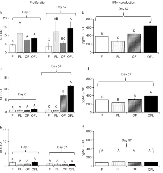
As regards the effect of LAM on the proliferation of peripheral cells, no differences between groups were
de-tected on day 0. PBMC from the four groups responded to
ConA on both day 0 and day 57. The OFL group showed a higher proliferation against ConA than the OF and F groups
(P = 0.009) on day 57 (Figure 2a). Responses against OVA were significantly higher in the OFL than in the OF group (P = 0.001). As expected, an increase in specific OVA SI
was detected in OFL and OF when that observed before immunization was compared to that observed 15 days
af-Figure 1. Characterization of the lipoarabinomannan (LAM) ex-tract. A, Coomassie staining 12% SDS-PAGE electrophoresis
gel. B, Immunoblot of the same gel using monoclonal antibody CS-35. Lane 1 = LAM extract; lane 2 = LAM of Mycobacterium tuberculosis.
Figure 2. Effect of LAM on the cellular response. a, c
and e, PBMC proliferation. b, d and f, IFN-γ production.
Data are reported as stimu-lation index (SI = mean cpm stimulated wells/mean cpm non-stimulated wells) or pg/ mL. a and b, PBMC stimu -lated with ConA (2.5 µg/ mL); c and d, PBMC stimu -lated with OVA (250 µg/mL);
e and f, PBMC stimulated
with LAM (250 µg/mL). Er-ror bars indicate standard deviation (SD) and different
letters indicate significant
differences between groups
(P < 0.05, ANOVA). Group F
(N = 5) wasimmunized with
PBS and FIA; group FL (N =
3)with PBS, FIA and LAM; group OF (N = 7)with OVA and FIA; group OFL (N = 8) with FIA and LAM. LAM =
li-poarabinomannan; PBMC =
peripheral blood mononuclear
ter the last immunization (Figure 2c). When
cells were stimulated with LAM, no significant
differences were detected between groups (Figure 2e).
IFN-γ detection in culture supernatants
PBMC from OFL stimulated with ConA showed a significantly higher IFN-γ production than those from the OF group (P = 0.01; Fig
-ure 2b). Similarly, PBMC stimulated with OVA showed significantly higher IFN-γ production in the OFL than in the OF group (P = 0.03;
Figure 2d). When cells were stimulated with
LAM, no significant differences were detected
between groups (Figure 2f). These results suggest that LAM is an immunomodulator
capable of increasing IFN-γ production.
Cell subpopulations
IgG+, CD4+, CD8+, and CD25+ cell subpopulations from
PBMC were evaluated before and 15 days after treatment.
After immunization, IgG+, CD4+, and CD8+ cell counts
showed similar levels of expression in all groups. Remark-ably, expression of CD25 in the OF and OFL groups was
higher after immunization than before (P = 0.003 and P =
0.01, respectively). Comparison between groups after the
third immunization showed a higher CD25 expression (P
= 0.01) in OFL than in OF, FL, and F (Table 1).
Delayed type hypersensitivity
DTH reactions against OVA were measured in order
to determine whether LAM was able to modify the in vivo
response to this protein antigen. All calves gave negative re-actions to the intradermal skin test using OVA (Table 2).
Specific humoral immune response
Specific anti-OVA titers and IgG isotypes were signifi
-cantly higher (P < 0.05) after the immunizations than on
day 0 for both the OFL and OF groups. This indicates that
the immunization protocol used to induce specific antibody
responses was successful. Log titer antibody responses
to OVA did not show significant differences between OVA
immunized groups on days 21, 42, and 57 (Figure 3). The
specific anti-IgM response did not differ between groups and was lower than IgG isotype responses. No significant differences were observed in the specific isotype responses
between the OF and OFL groups (Figure 4).
Tuberculosis and paratuberculosis diagnosis
DTH reactions against PPDb were evaluated to identify
a possible interference with the diagnosis of this tubercu-losis and negative reactions were detected in all calves (Table 2).
Serum-specific anti-PPA responses showed nega -tive results on days 0 and 57 for all the groups analyzed
Figure 3. Logarithms of anti-OVA titers measured by ELISA on days 21, 42, and 57. Error bars indicate the standard deviation
(SD) using ANOVA. Group F (N = 5) was immunized with PBS
and FIA; group FL (N = 3) with PBS, FIA and LAM; group OF (N = 7)with OVA and FIA; group OFL (N = 8)with FIA and LAM. PBS = phosphate-buffered saline; FIA = Freund’s incomplete adjuvant; LAM = lipoarabinomannan; OVA = ovalbumin.
Table 1. Expression of IgG+, CD4+, CD8+, and CD25+ in bovine peripheral blood
mononuclear cells before and 15 days after the third immunization.
Group IgG+ CD4+ CD8+ CD25+
Pre-immunization 12.64 ± 1.58 23.73 ± 13.06 11.3 ± 2.01 2.00 ± 0.90 F 9.25 ± 6.51 18.38 ± 0.32 6.96 ± 2.91 3.20 ± 0.40 FL 8.10 ± 4.90 11.10 ± 5.00 9.10 ± 5.20 2.60 ± 0.03 OF 13.08 ± 5.75 19.50 ± 9.23 9.61 ± 1.06 6.25 ± 0.96 OFL 17.69 ± 3.60 20.34 ± 4.22 10.73 ± 4.92 11.15 ± 3.20*
Data are reported as percent ± SD. Pre-immunization values correspond to all
animals before being assigned to each group. Group F (N = 5) wasimmunized
with PBS and FIA; group FL (N = 3)with PBS, FIA and LAM; group OF (N = 7)
with OVA and FIA; group OFL (N = 8)with OVA, FIA and LAM. FIA = Freund’s
incomplete adjuvant; PBS = phosphate-buffered saline; LAM = lipoarabinoman
-nan; OVA = ovalbumin. *P < 0.05 compared to othergroups (ANOVA).
Table 2. Delayed-type hypersensitivity immune response in cattle.
Group PBS OVA PPDb
F 0 0 0.4 ± 0.5
FL 0 ND 0.9 ± 0.7
OF 0.3 ± 0.6 0.5 ± 0.5 0.5 ± 0.5 OFL 1.0 ± 0.9 0.3 ± 0.6 0.5 ± 0.7
Data are reported as mean increase in skin thickness (mm) ± SD
on day 60 (P < 0.05, ANOVA). ND = not determined. Group F (N =
5) was immunized with PBS and FIA; group FL (N = 3) with PBS, FIA and LAM; group OF (N = 7)with OVA and FIA; group OFL (N = 8) with OVA, FIA and LAM. FIA = Freund’s incomplete adjuvant;
144 S.B. Colavecchia et al.
(Table 3). This indicates that LAM inoculation did not
induce cross-reactions with PPA used for the diagnosis of
paratuberculosis.
Discussion
The present study was conducted in order to evaluate the effect of LAM addition to FIA inoculated with a non-infectious antigen, to improve the immune response in calves. Several studies have shown that LAM is a highly immunogenic molecule (24,25), which has been related to mycobacterial pathogenesis (12,26). Then, LAM has been proposed as immunomodulator (12,16,27,28).
Investigations in humans and animals have used FIA in vaccine formulation to induce persistent, high-titer protective humoral immune responses (4,7). In fact, the use of an oil adjuvant in foot- and mouth-disease vaccine has allowed an increase in the interval between immunizations. Actually, the use of FIA in our immunization protocol is supported by the fact that it is included in commercial formulations assigned to bovine (29) and human vaccines (30).
We chose OVA as a T-dependent antigen because it has been previously reported to elicit cellular and humoral immune responses in calves and mice (4,31). The OVA doses and the immunization protocol used in our study were based on experiments carried out in cattle by Hernández et al. (4). As expected, in our model, OVA antigen induced high antibody titers with a major IgG response after only one dose.
With our extraction methodology, characterization of
crude LAM was similar to that obtained from M. tuberculosis
by Hamasur et al. (18).
The stimulation of a specific cell immune response is
important to increase protection against intracellular
patho-gens (28). Our study showed that LAM increases the specific
cell immune response, evaluated by lymphoproliferation, compared to vaccination with antigen with incomplete Freund’s adjuvant alone. Likewise, immunization with LAM as an immunomodulator induced a seven times higher proliferation than the preimmunization value and than that reported by Sun et al. (32) using OVA with other adjuvants. Similar results have been reported with other oil adjuvants compared to cationic liposomes in a bovine model (33). We showed here an increase in proliferation to ConA in the OFL group, indicating that stimulation with LAM affects
both antigen-specific and -non-specific lymphocytes. In the same way, we detected an increase in IFN-γ production in
LAM-treated animals. These results are in agreement with
other experiments in which LAM from M. bovis induced
an increase in IFN-γ production in a mouse model (34). IFN-γ production involves macrophage and lymphocyte
activation and is associated with the Th1 response in cattle
(3,33). As demonstrated by flow cytometry analysis, our
experiments showed an increase in CD25+ cells. Some
investigators have demonstrated a proliferative immune
response by bovine CD4+ T cells to soluble mycobacterial
antigens linked with a markedly increased expression of
CD25 (35). The functional relevance and correlation with in
vitro findings remain unclear. In our model, the increase in
CD25 could be related to the immunomodulation induced by glycolipids. The CD4/CD8 ratio detected in our experiments remained within the normal values previously reported for cattle (36). This seems to indicate that LAM addition does not modify the proportion of these subpopulations. We
could not detect OVA-specific DTH responses. Different
results have been previously reported by Hernández et al.
(4) in cattle inoculated with cell walls of M. phlei and OVA.
Since, in our model, we used purified LAM, we can sug -gest that the use of LAM does not cause the same effect
Figure 4. Anti-OVA IgM, IgG1, IgG2, and IgG3 were measured by ELISA on day 57 after the initial immunization. Absorbance values of IgM, IgG1, IgG2, and IgG3 responses to OVA are
re-ported as means ± SD. Different letters indicate significant differ
-ences among groups (P < 0.05, ANOVA). Group F (N = 5) was immunized with PBS and FIA; group FL (N = 3)with PBS, FIA
and LAM; group OF (N = 7)with OVA and FIA; group OFL (N = 8)with FIA and LAM. PBS = phosphate-buffered saline; FIA = Freund’s incomplete adjuvant; LAM = lipoarabinomannan; OVA = ovalbumin.
Table 3. Specific antibodies against paratuberculosis protoplas -mic antigen.
Group Day 0 (A at 490 nm ± SD)
Day 57 (A at 490 nm ± SD)
F 0.19 ± 0.03 0.18 ± 0.04 FL 0.16 ± 0.08 0.13 ± 0.03 OF 0.18 ± 0.08 0.14 ± 0.02 OFL 0.20 ± 0.14 0.17 ± 0.12
Data are reported as absorbance (A) at 490 nm ± SD (ANOVA). Internal ELISA controls were 5 negative sera (A = 0.35) and
in-fected sera (A = 1.90). Group F (N = 5) was immunized with PBS and FIA; group FL (N = 3) with PBS, FIA and LAM; group OF (N =
7) with OVA and FIA; group OFL (N = 8) with OVA, FIA and LAM.
as mycobacterial cell walls.
The study of serum immunoglobulins by the evaluation of relative concentration and isotypic composition is useful for testing immunomodulators (37). Our results show that
modulation of LAM induced high titers of OVA-specific an
-tibodies after the first immunization in cattle. These levels
were increased in the second dose and remained after the third immunization, in agreement with results published by Heriazon et al. (3) using OVA with saponins. A relationship between the isotypes produced and the immune response
profile in cattle has been previously established (38). Similarly, in sheep, an increase in the specific production of IgG2 isotype and IFN-γ has been detected when using
a recombinant protein of Taenia ovis and FIA as adjuvant
(39). Our results indicate that the serum response against OVA predominantly involved IgG1 and IgG2 and, to a lesser extent, IgG3 but no differences were detected, probably for the high dispersion found between animals belong-ing to the OFL group. The measurements of cutaneous
hypersensitivity produced against PPD and ELISA-PPA are the methods most frequently used in Argentina for the
diagnosis of tuberculosis and paratuberculosis in cattle. Even if cross-reactions have been described with other mycobacteria (40), we did not detect positive reactions in LAM-treated groups.
Our study provides original data on the effect of LAM
on the lymphoproliferative response, IFN-γ production, the
humoral immune response, and isotypes involved against OVA in cattle.
We demonstrated that LAM, at the dose, route and immu-nization protocol used in the present study, in combination
with FIA, is able to improve specific cell immune response evaluated by lymphoproliferation and IFN-γ production by
at least 50 and 25%, respectively, in cattle.
Acknowledgments
We thank Dr. Ana M. Jar for her kind help and valuable suggestions, VMD Liliana Ramayo and student María Laura Fortuny for their technical assistance. Research supported
by UBACyT, Project VE023 (#2008-2011) and BID PICT (#2010-2672).
References
1. Cox JC, Coulter AR. Adjuvants - a classification and review
of their modes of action. Vaccine 1997; 15: 248-256. 2. Stills HF Jr. Adjuvants and antibody production: dispelling
the myths associated with Freund’s complete and other adjuvants. ILAR J 2005; 46: 280-293.
3. Heriazon A, Thompson KA, Wilkie BN, Mathes-Sears W, Quinton M, Mallard BA. Antibody to ovalbumin and delayed-type hypersensitivity to Candida albicans and mycobacteria in lactating Holstein cows using Quil A or Freund’s complete adjuvant. Vet Immunol Immunopathol 2009; 127: 220-227. 4. Hernández A, Yager JA, Wilkie BN, Leslie KE, Mallard BA.
Evaluation of bovine cutaneous delayed-type hypersensi-tivity (DTH) to various test antigens and a mitogen using several adjuvants. Vet Immunol Immunopathol 2005; 104: 45-58.
5. Wedlock DN, Denis M, Painter GF, Ainge GD, Vordermeier
HM, Hewinson RG, et al. Enhanced protection against bo-vine tuberculosis after coadministration of Mycobacterium bovis BCG with a Mycobacterial protein vaccine-adjuvant combination but not after coadministration of adjuvant alone.
Clin Vaccine Immunol 2008; 15: 765-772.
6. Aucouturier J, Dupuis L, Deville S, Ascarateil S, Ganne V. Montanide ISA 720 and 51: a new generation of water in oil emulsions as adjuvants for human vaccines. Expert Rev Vaccines 2002; 1: 111-118.
7. Miller LH, Saul A, Mahanty S. Revisiting Freund’s incomplete adjuvant for vaccines in the developing world. Trends Para-sitol 2005; 21: 412-414.
8. Cobo ER, Morsella C, Cano D, Cipolla A, Campero CM. Immunization in heifers with dual vaccines containing Tri-trichomonas foetus and Campylobacter fetus antigens using systemic and mucosal routes. Theriogenology 2004; 62: 1367-1382.
9. Jensen FC, Savary JR, Diveley JP, Chang JC. Adjuvant
activity of incomplete Freund’s adjuvant. Adv Drug Deliv Rev
1998; 32: 173-186.
10. Hunter SW, Brennan PJ. Evidence for the presence of a
phosphatidylinositol anchor on the lipoarabinomannan and lipomannan of Mycobacterium tuberculosis. J Biol Chem
1990; 265: 9272-9279.
11. Nigou J, Gilleron M, Puzo G. Lipoarabinomannans: from
structure to biosynthesis. Biochimie 2003; 85: 153-166.
12. Briken V, Porcelli SA, Besra GS, Kremer L. Mycobacterial
lipoarabinomannan and related lipoglycans: from biogenesis to modulation of the immune response. Mol Microbiol 2004; 53: 391-403.
13. Guerardel Y, Maes E, Elass E, Leroy Y, Timmerman P,
Besra GS, et al. Structural study of lipomannan and lipoara-binomannan from Mycobacterium chelonae. Presence of
unusual components with alpha 1,3-mannopyranose side chains. J Biol Chem 2002; 277: 30635-30648.
14. Bhattacharjee S, Majumder N, Bhattacharyya P, Bhattacha -ryya S, Majumdar S. Immunomodulatory role of arabinosy-lated lipoarabinomannan on Leishmania donovani infected murine macrophages. Indian J Biochem Biophys 2007; 44: 366-372.
15. Barrow WW, de Sousa JP, Davis TL, Wright EL, Bachelet M,
Rastogi N. Immunomodulation of human peripheral blood
mononuclear cell functions by defined lipid fractions of My-cobacterium avium. Infect Immun 1993; 61: 5286-5293. 16. Smit JJ, Van Loveren H, Hoekstra MO, Schijf MA, Folkerts
G, Nijkamp FP. Mycobacterium vaccae administration during allergen sensitization or challenge suppresses asthmatic features. Clin Exp Allergy 2003; 33: 1083-1089.
146 S.B. Colavecchia et al.
mycobacterial lipoglycans. J Allergy Clin Immunol 2004; 114: 302-309.
18. Hamasur B, Kallenius G, Svenson SB. A new rapid and
simple method for large-scale purification of mycobacterial
lipoarabinomannan. FEMS Immunol Med Microbiol 1999; 24: 11-17.
19. Dubois M, Gilles K, Hamilton JK, Rebers PA, Smith F. A
colorimetric method for the determination of sugars. Nature
1951; 168: 167.
20. Bradford MM. A rapid and sensitive method for the quantita
-tion of microgram quantities of protein utilizing the principle
of protein-dye binding. Anal Biochem 1976; 72: 248-254. 21. Deringer JR, Ely RJ, Monday SR, Stauffacher CV, Bohach
GA. Vbeta-dependent stimulation of bovine and human T
cells by host-specific staphylococcal enterotoxins. Infect Immun 1997; 65: 4048-4054.
22. SENASA Argentina. Programa nacional de control y erradi -cación de la tuberculosis bovina. http://www.senasa.gov.ar/
contenido.php?to=n&iN = 858&io=15899.
23. Mundo SL, Fontanals AM, Garcia M, Durrieu M, Alvarez E, Gentilini ER, et al. Bovine IgG1 antibodies against Myco-bacterium avium subsp. paratuberculosis protein p34-cx improve association of bacteria and macrophages. Vet Res
2008; 39: 6.
24. Koets AP, Rutten VP, de Boer M, Bakker D, Valentin-Weigand P, van Eden W. Differential changes in heat shock protein-, lipoarabinomannan-, and purified protein derivative-specific
immunoglobulin G1 and G2 isotype responses during bovine
Mycobacterium avium subsp. paratuberculosis infection.
Infect Immun 2001; 69: 1492-1498.
25. Watanabe Y, Watari E, Matsunaga I, Hiromatsu K, Dascher CC, Kawashima T, et al. BCG vaccine elicits both T-cell mediated and humoral immune responses directed against mycobacterial lipid components. Vaccine 2006; 24: 5700-5707.
26. Flynn JL, Chan J. Immunology of tuberculosis. Annu Rev Immunol 2001; 19: 93-129.
27. Ito T, Hasegawa A, Hosokawa H, Yamashita M, Motohashi S, Naka T, et al. Human Th1 differentiation induced by lipoarabinomannan/lipomannan from Mycobacterium bovis
BCG Tokyo-172. Int Immunol 2008; 20: 849-860.
28. Guenin-Mace L, Simeone R, Demangel C. Lipids of patho-genic Mycobacteria: contributions to virulence and host immune suppression. Transbound Emerg Dis 2009; 56: 255-268.
29. Robiolo B, La Torre J, Maradei E, Beascoechea CP, Perez A, Seki C, et al. Confidence in indirect assessment of
foot-and-mouth disease vaccine potency and vaccine matching
carried out by liquid phase ELISA and virus neutralization
tests. Vaccine 2010; 28: 6235-6241.
30. Roman F, Vaman T, Gerlach B, Markendorf A, Gillard P,
Devaster JM. Immunogenicity and safety in adults of one
dose of influenza A H1N1v 2009 vaccine formulated with and
without AS03A-adjuvant: preliminary report of an observer-blind, randomised trial. Vaccine 2010; 28: 1740-1745. 31. Habjanec L, Halassy B, Tomasic J. Comparative study of
structurally related peptidoglycan monomer and muramyl dipeptide on humoral IgG immune response to ovalbumin in mouse. Int Immunopharmacol 2010; 10: 751-759.
32. Sun HX, Ye YP, Pan HJ, Pan YJ. Adjuvant effect of Panax notoginseng saponins on the immune responses to ovalbu-min in mice. Vaccine 2004; 22: 3882-3889.
33. Vordermeier HM, Dean GS, Rosenkrands I, Agger EM,
Andersen P, Kaveh DA, et al. Adjuvants induce distinct im -munological phenotypes in a bovine tuberculosis vaccine model. Clin Vaccine Immunol 2009; 16: 1443-1448. 34. Rosenkrands I, Agger EM, Olsen AW, Korsholm KS,
Ander-sen CS, JenAnder-sen KT, et al. Cationic liposomes containing mycobacterial lipids: a new powerful Th1 adjuvant system.
Infect Immun 2005; 73: 5817-5826.
35. Waters WR, Rahner TE, Palmer MV, Cheng D, Nonnecke
BJ, Whipple DL. Expression of L-Selectin (CD62L), CD44, and CD25 on activated bovine T cells. Infect Immun 2003; 71: 317-326.
36. Seo KS, Lee SU, Park YH, Davis WC, Fox LK, Bohach GA.
Long-term staphylococcal enterotoxin C1 exposure induces soluble factor-mediated immunosuppression by bovine CD4+ and CD8+ T cells. Infect Immun 2007; 75: 260-269. 37. Berghaus LJ, Corbeil LB, Berghaus RD, Kalina WV, Kimball
RA, Gershwin LJ. Effects of dual vaccination for bovine respiratory syncytial virus and Haemophilus somnus on im-mune responses. Vaccine 2006; 24: 6018-6027.
38. Estes DM, Brown WC. Type 1 and type 2 responses in regulation of Ig isotype expression in cattle. Vet Immunol Immunopathol 2002; 90: 1-10.
39. Rothel JS, Corner LA, Lightowlers MW, Seow HF, McWaters
P, Entrican G, et al. Antibody and cytokine responses in ef -ferent lymph following vaccination with dif-ferent adjuvants.
Vet Immunol Immunopathol 1998; 63: 167-183.
40. Waters WR, Palmer MV, Thacker TC, Payeur JB, Harris NB, Minion FC, et al. Immune responses to defined antigens of Mycobacterium bovis in cattle experimentally infected with