Integrative Approach to Analyze Biodiversity
and Anti-Inflammatory Bioactivity of
Wedelia
Medicinal Plants
Wen-Ching Lin1,2☯, Chih-Chun Wen1☯, Yung-Hsiang Chen1, Pei-Wen Hsiao1, Jiunn-Wang Liao3, Ching-I Peng4‡, Ning-Sun Yang1‡*
1Agricultural Biotechnology Research Center, Academia Sinica, Taipei, Taiwan,2Institute of Plant Biology, National Taiwan University, Taipei, Taiwan,3Graduate Institute of Veterinary Pathology, National Chung Hsing University, Taichung, Taiwan,4Biodiversity Research Center, Academia Sinica, Taipei, Taiwan
☯These authors contributed equally to this work. ‡These authors also contributed equally to this work. *[email protected]
Abstract
For the development of“medical foods”and/or botanical drugs as defined USA FDA, clear and systemic characterizations of the taxonomy, index phytochemical components, and the functional or medicinal bioactivities of the reputed or candidate medicinal plant are needed. In this study, we used an integrative approach, including macroscopic and microscopic ex-amination, marker gene analysis, and chemical fingerprinting, to authenticate and validate various species/varieties ofWedelia, a reputed medicinal plant that grows naturally and commonly used in Asian countries. The anti-inflammatory bioactivities ofWedeliaextracts were then evaluated in a DSS-induced murine colitis model. Different species/varieties of
Wedeliaexhibited distinguishable morphology and histological structures. Analysis of the ri-bosomal DNA internal transcribed spacer (ITS) region revealed significant differences among these plants. Chemical profiling of testWedeliaspecies demonstrated candidate index compounds and distinguishable secondary metabolites, such as caffeic acid deriva-tives, which may serve as phytochemical markers or index for quality control and identifica-tion of specificWedeliaspecies. In assessing their effect on treating DSS induced-murine colitis, we observed that only the phytoextract fromW.chinensisspecies exhibited signifi-cant anti-inflammatory bioactivity on DSS-induced murine colitis among the variousWedelia
species commonly found in Taiwan. Our results provide a translational research approach that may serve as a useful reference platform for biotechnological applications of traditional phytomedicines. Our findings indicate that specificWedeliaspecies warrant further investi-gation for potential treatment of human inflammatory bowel disease.
a11111
OPEN ACCESS
Citation:Lin W-C, Wen C-C, Chen Y-H, Hsiao P-W, Liao J-W, Peng C-I, et al. (2015) Integrative Approach to Analyze Biodiversity and Anti-Inflammatory Bioactivity ofWedeliaMedicinal Plants. PLoS ONE 10(6): e0129067. doi:10.1371/journal.pone.0129067
Academic Editor:Francesca Borrelli, University of Naples Federico II, ITALY
Received:October 3, 2014
Accepted:May 4, 2015
Published:June 4, 2015
Copyright:© 2015 Lin et al. This is an open access article distributed under the terms of theCreative Commons Attribution License, which permits unrestricted use, distribution, and reproduction in any medium, provided the original author and source are credited.
Data Availability Statement:All relevant data are within the paper and its Supporting Information files.
Funding:This study was supported by a National Science Council grant (NSC-103-2325-B-001-003) and by a grant from the Academia Sinica, Taiwan (AS-103-TP2-B04). The funders had no role in study design, data collection and analysis, decision to publish, or preparation of the manuscript.
Introduction
As complementary and alternative medicine (CAM) is increasingly recognized for its potential in evidence-based public healthcare applications, research into the safety and efficacy of medic-inal plants, has become vital for public health. A number of reputed medicmedic-inal herbs that are likely to be clinically useful have been proven to be highly toxic if used incorrectly, and medica-tion errors, misuse of plant species with similar appearances, and adulterants or consumpmedica-tion of unknown and complex herbs in various clinical situations may also hinder toxicology and effectiveness analyses [1]. The key to herbal medicine safety is positive verification and guaran-teed purity of medicinal plants and the derived extracts. To date, various macroscopic and microscopic techniques, molecular marker authentication, metabolite profiling and metabolo-mics analyses have been employed, often individually or non-systematically, for verification and validation of putative medicinal plants in an effort to authenticate medicinal herbs [2–4]. Chemical chromatographic fingerprinting by high performance liquid chromatography (HPLC) [5] and ultra performance liquid chromatography coupled with electrospray ioniza-tion and quadrupole time of flight mass spectrometry (UPLC-ESI-Q-TOF-MS) [6,7], in combi-nation with principal component analysis (PCA) [8] have been proven to be a powerful platform for metabolomics studies, as they not only are useful for comparing the chemical pro-files of herbal medicines, but also for identifying chemical markers or indices. In this study, we intentionally combined all of these various technology systems mentioned above for and inte-grated investigation for phytomedicine use of a specific medicinal herb,Wedelia chinensis, that we recently demonstrate to confer strong anti-colitis activity in experimental mouse sys-tem [9].
Inflammatory bowel disease (IBD), with a high incidence worldwide, is a result of chronical-ly relapsing idiopathic inflammation of the gastrointestinal tract [10,11]. Patients with IBD typ-ically also run a high risk of the disease developing into colorectal cancer due to chronic damage and inflammation in the colon and tissues of the rectum [12]. The DSS-induced mouse colitis model is well accepted for studying IBD due to its resemblance to human ulcera-tive colitis [13,14]. Drugs for treating IBD including 5-aminosalicyclic acid, sulfasalazine, and others [15,16]; however, IBD patients usually need long-term and repeated treatment, involv-ing potentially toxic drug doses [16,17]. Because of such side-effects or/and lack of effectiveness of standard western medicine therapies, many IBD patients turn to complementary and alter-native medicine (CAM). Herbal therapy is the most common type of CAM used for gastroin-testinal disorders [18–21], but so far there have been limited reports of efficacious CAM therapeutic strategies or safe phytomedicines to remedy IBD.
Wedelia chinensisbelonging to family Asteraceae (sunflower family) is commonly con-sumed orally. It is often infused with hot water to make an herbal tea. Such herbal tea drinks, as cold beverages, are readily available commercially in Taiwan, China Mainland and other Asian countries. We revealed in our previous study [9] that the crude plant extract ofW. chi-nensiscan confer a specific anti-inflammatory effect on dextran sulphate sodium (DSS)-in-duced murine colitis.In vivotreatment with the hot water extract ofWedelia chinensiscan effectively suppress the DSS-induced increase in expression of macrophage-derived/Th1 (TNF-α, IFN-γ) and Th17 (IL-17) cytokines, but has no effect on the expression of Th2 (IL-4) cytokines. According to theFlora of Taiwan(2nd edition), fourWedeliaspecies and two addi-tional varieties grow in Taiwan [22]. Since taxonomists can make use of different parameters and resolutions for the definition of a species or in defining speciation, we were in fact able to find a range of different classifications of these sixWedeliaspecies in the literature. For exam-ple, a number of different reports reducedW.trilobatato the genusSphagneticola, andW.
Sphagneticola trilobataandWollastonia bifloraare the academically accepted taxonomic names, whileWedelia bifloraandWedelia trilobataare synonyms.W.biflora,W.prostrataand their varieties were also reduced to the genusMalanthera[26]. These sixWedeliaspecies have similar appearances, particularlyW.chinensisandW.trilobata. These plants often bewilder the general public and herbal medicine users because of the lack of distinction in their common trivial names and their similar morphology. Different species are sold in herbal markets in Asia, not only as an ingredient in herbal tea, but also as an anti-inflammatory tonic or medici-nal food. The Zhongyao Da Cidian (Encyclopedia of Chinese Materia Medica), the most com-prehensive work on Chinese Materia Medica since the Bencao Gangmu (Compendium of Materia Medica), records thatWedeliaplants confer benefit against dysentery, an inflamma-tion of the intestines especially the colon [27]. There is, however, little or no information about which species or cultivar strains ofWedeliahave the potency or specificity to serve as desirable medicinal food, especially for control of various inflammatory activities related to human health. Therefore we used a DSS-induced mouse colitis model, which is well-established in our laboratory and has many similarities to human ulcerative colitis, as a biotechnological platform for systematic analysis of the anti-inflammatory activities of the six differentWedeliaspecies/ varieties found in Taiwan.
In this study, our primary goal was to establish an effective biotechnological approach and platform through which morphological, histological and phytochemical profiles and biodiver-sities are comparatively studied among differentWedeliaspecies. Effects were also made to evaluate the safety and potential medicinal efficacy of these traditional medicinal herbs. To this end, we integrated several authentication methodologies, including macroscopic and micro-scopic examination, molecular identification, and metabolomics analysis to identify and char-acterize the six medicinal plants from theWedeliagenus commonly found in Taiwan. To evaluate the anti-inflammatory bioactivity and therapeutic efficacy of theseWedeliaspecies, we used a DSS-induced acute murine colitis system as an animal model. We then explored the possible correlation between our metabolite fingerprinting and phytochemical analyses data (obtained with principle component analysis [PCA]), and the bioactivity results obtained from
in vivo, anti-colitis animal experiments. Our findings provide useful baseline and regional spe-ciality information on the biodiversity of theWedeliagenus plants. Specific plant secondary metabolites that may confer potent bioactivities in both mammalian and plant systems are discussed. The possible application of our findings to systematic exploration of plant secondary metabolites as phytomedicines present in reputed, traditional medicinal plants is contemplated.
Methods and Materials
Materials
Aerial plant parts of four different species and two additional varieties ofWedeliawere collect-ed from the northern counties of Taiwan. SomeWedeliaplant samples were kindly provided by the Miaoli District Agricultural Research and Extension Station (MDARES), National Mu-seum of Natural Science (NMNS) and I-Shun herbal shop in Taiwan (see details described in
Table 1). Each of these species was initially authenticated by taxonomy expert Professor Ching-I Peng (Research Center for Biodiversity, Academia Sinica). Voucher specimens were deposited in the HAST Herbarium, Biodiversity Research Museum, Academia Sinica, Taiwan.
Extraction
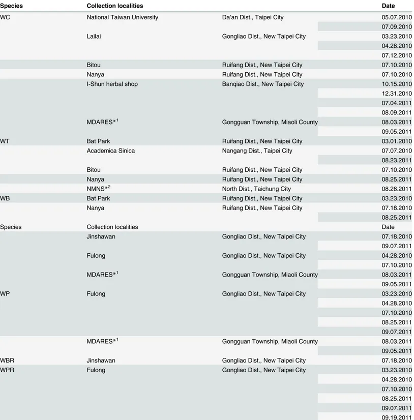
Table 1. Taiwan local collection sites ofWedeliaspecies.
Species Collection localities Date
WC National Taiwan University Da’an Dist., Taipei City 05.07.2010
07.09.2010
Lailai Gongliao Dist., New Taipei City 03.23.2010
04.28.2010 07.12.2010
Bitou Ruifang Dist., New Taipei City 07.10.2010
Nanya Ruifang Dist., New Taipei City 07.10.2010
I-Shun herbal shop Banqiao Dist., New Taipei City 10.15.2010
12.31.2010 07.04.2011 08.09.2011
MDARES*1 Gongguan Township, Miaoli County 08.03.2011
09.05.2011
WT Bat Park Ruifang Dist., New Taipei City 03.01.2010
Academica Sinica Nangang Dist., Taipei City 07.07.2010
08.23.2011
Bitou Ruifang Dist., New Taipei City 07.10.2010
Nanya Ruifang Dist., New Taipei City 08.25.2011
NMNS*2 North Dist., Taichung City 08.26.2011
WB Bat Park Ruifang Dist., New Taipei City 03.23.2010
Nanya Ruifang Dist., New Taipei City 07.18.2010
08.25.2011
Species Collection localities Date
Jinshawan Gongliao Dist., New Taipei City 07.18.2010
09.07.2011
Fulong Gongliao Dist., New Taipei City 04.28.2010
07.10.2010
MDARES*1 Gongguan Township, Miaoli County 08.03.2011
09.05.2011
WP Fulong Gongliao Dist., New Taipei City 03.23.2010
04.28.2010 07.10.2010 08.25.2011 09.07.2011
MDARES*1 Gongguan Township, Miaoli County 08.03.2011
09.05.2011
WBR Jinshawan Gongliao Dist., New Taipei City 07.18.2010
WPR Fulong Gongliao Dist., New Taipei City 03.23.2010
04.28.2010 07.10.2010 08.25.2011 09.07.2011 09.19.2011
*1MDARES: Miaoli District Agricultural Research and Extension Station. *2NMNS: National Museum of Natural Science.
filtered using a glass funnel (Buchner GF16-25G3, Iwaki TE-32 Pyrex) and filter paper No. 2, 90 mm (Toyo Roshi Kaisha). Finally, the extracted samples were concentrated using a rotary evaporator (Büchi, Rotavapor R-200), and subsequently lyophilized using a freeze-drying sys-tem (Freezone 4.5, Labconco).
Tissue sectioning and histological analysis
Transverse sections of the plant stem and murine colon tissues were prepared using a Leica RM2125RT microtome (Leica Instruments, Nussloch, Germany). Photomicrographs were taken using an AxioCam HRc videocamera (Zeiss) connected to an optical microscope (Zeiss Imager Z1) using AxioVision Rel. 4.8 software. A stereomicroscope (SteREO Lumar V12, Zeiss) equipped with AxioCam MRc and AxioVision Rel. 4.8 software was also used to photo-graphically record test specimens.
Safranin-fast green staining of plant tissue sections
The fresh stems of six species/variants ofWedeliawere cut into appropriate segment sizes and fixed in FAA (formalin, acetic acid, and 70% ethanol mixed at a ratio of 1:1:18, respectively). Tissue samples were processed through a gradual ethanol series (from 30% to 100%) and dimethylbenzene for specimen dehydration [28], then buried in paraffin blocks according to the technique described by Ruzin (1999). Subsequently, tissues were sectioned with a micro-tome to a suitable thickness (10μM) and stained with safranin-fast green (1% Safranin O in methylcellosolve, 95% ethanol and water at a ratio of 2:1:1, respectively), then in 1% NaOAc and 2% formalin solution, followed by 0.5% Fast Green FCF in methyl cellosolve, 100% ethanol and clove oil at a ratio of 1:1:1, respectively. Finally, samples were mounted in Histokitt mount-ing medium (Assistant) for observation.
DNA extraction
The CTAB method [29], with some modification, was employed for DNA extraction. Small quantities (50–100 mg) of fresh leaves were frozen in liquid nitrogen and ground to fine pow-ders. The powder derived from 50–100 mg fresh leaves was added to 0.7 mL of extraction buff-er [2% (w/v) cetyltrimethylammonium bromide (CTAB), 0.1M tris-HCl (pH 8.0), 20 mM EDTA (pH 8.0), 0.2% (w/v) PVP-40, 1.42 M NaCl, 5 mM ascorbic acid, 0.02% 2-mercaptoetha-nol]. Subsequently, 1μL RNase A (10 mg/mL) was added to the test sample and incubated at 65°C for 30 min. The tissue homogenate was then centrifuged at 13,000 rpm for 10 min. DNA was extracted using 0.8 volume of phenol/chloroform/isoamyl alcohol (IAA) at a ratio of 25:24:1, respectively, and 0.8 volume chloroform/IAA (24:1). After centrifugation at 13,000 rpm, the supernatant was purified using a DNeasy Plant Mini Kit (Qiagen), according to the manufacturer’s instructions.
Amplification and analysis of ITS sequences
Plant genomic DNA of sixWedeliaspecies were used as templates to amplify the ITS se-quences, by employing 20 pmol each of the primers TCM-1 (50-CACAAAGCCCGTCGCTCCT
ACCGA-30) and TCM-2 (50-ACTCGCCGTTACTAGGGGAA-30) [4] in a 20μL solution
Purified DNA plasmids were sequenced using an ABI Prism 3730 DNA Analyzer. Sample sizes: (1)Wedelia chinensis: n = 39; (2)Wedelia trilobata: n = 18; (3)Wedelia biflora: n = 27; (4)Wedelia prostrate: n = 21; (5)Wedelia prostrata var.robusta: n = 3; (6)Wedelia prostrata var.robusta: n = 18.
Sequence alignment and data analysis
Multiple alignment and analysis of ITS DNA sequences were carried out using Clustal X2 [30]. Genetic distances were computationally analyzed using MEGA, version 4.0, according to the Kimura Two Parameter (K2P) model [31]. The neighbor joining (NJ) consensus tree was also constructed using MEGA 4.0.
Mice
Seven to eight-week-old male C57BL/6 mice (17–20 g) were purchased from the National Lab-oratory Animal Center (Tainan, Taiwan). Every five mice were fostered in one cage in a special pathogen free (SPF) animal room kept at 22°C with 55% relative humidity on a 14-h light/10-h dark cycle. The mice were fed with a sterilized diet (Laboratory Autoclavable Rodent Diet 5010, USA) during their one-week acclimatization.
Dextran sodium sulfate-induced acute murine colitis model
After acclimatization, test mice were randomized into four groups: vehicle control, dextran so-dium sulfate (DSS, colitis control), sulfasalazine (positive control, Sigma-Aldrich, Lot No. 460842) and the treatment group (n = 10). All mice received normal sterilized drinking water from day -7 to 0. Subsequently, acute colitis was induced by adding 2% (w/v) DSS (molecular weight: 36,000–50,000 Da, MP Biochemicals, Solon) solution to the drinking water for each group (except the vehicle control) from day 0 to day 8 as described previously [10]. The DSS solution was filtered and changed every three days and mean DSS consumption was recorded. Throughout the whole experimental period (i.e., from 7 days before DSS treatment to 8 days post DSS treatment, with a total of 15 day treatment duration), mice in the control and DSS groups were orally administrated with vehicle control solution (sterilized water as the vehicle). Mice in the treatment group were fed by oral gavage with water extracts derived from various
Wedeliaspecies dissolved in sterilized Milli-Q water at a dose of 50 mg/kg body weight. The dose of 50 mg/kg as the optimal dosage is based on our preliminary test and on our results pre-viously published in PLOS ONE [9]. Sulfasalazine was orally administered as a positive control at a dose of 200 mg/kg after the sterilized drinking water was changed to a 2% DSS. Sulfasala-zine solution was dissolved in 0.5% cellulose, the 0.5% cellulose was first dissolved in saline and autoclaved, and then Tween 80 was added to 1%. At the end of the experiment, all test mice were weighed and sacrificed by cervical dislocation.
Evaluation of colitis severity
Histological evaluation of colitis severity and hematoxylin-eosin staining
Excised fresh colons were cut into defined 0.5 cm long segments and fixed in 10% formalin overnight. The samples were then dehydrated by passing through a gradual ethanol series (from 70% to 100%) and dimethylbenzene, and then embedded in paraffin. Tissues were sec-tioned to 4μM thicknesses using a microtome, and slides were prepared. The paraffin was re-moved by passing samples through dimethylbenzene and a gradual ethanol series (from 100% to 95%). Tissue slides were then stained with hematoxylin and eosin (Muto pure chemicals, Lot. No 3008–1). Finally, tissue sections were mounted in Histokitt mounting medium (Assis-tant) for microscopic examinations.
High performance liquid chromatography
For analysis by high performance liquid chromatography (HPLC), powders of plant tissue ex-tracts were dissolved in deionized water at a concentration of 4 mg/ml, and centrifuged at 14,000 rpm for 10 minutes to remove debris. Test extracts (4 mg/ml, 20μl) were analyzed by HPLC (Agilent 1100 series with a G131A Quat pump) using a C18-AR-II 5μm column
(250 × 4.6 mm Cosmosil) at a flow rate of 1 ml/min with a elution gradient program of 15% methanol with 0.05% trifluoroacetic acid (TFA) to 70% methanol with 0.05% TFA over 30 minutes, and then held at 100% methanol with 0.05% TFA for 20 min. A photodiode array de-tector (DAD, Agilent 1100) was used to identify various plant metabolites at 254 nm, 280 nm, 300 nm and 350 nm.
LC-ESI-Q-TOF MS analysis
For LC-MS analysis, acetonitrile (ACN) with 0.1% formic acid (FA) and water with 0.1% for-mic acid (LC-MS grade, J. T. Baker, Phillipsburg, NJ) were used in the mobile phase. Sulfadi-methoxine and formic acid were purchased from Fluka (Germany). LC-MS was performed with a LC system, (Acquity UPLC, Waters, Millford, MA), coupled to a hybrid Q-TOF mass spectrometer (Synapt HDMS, Waters, Manchester, UK). Test samples were separated online with a reverse-phase column, (HSS T3 C18, 1.8μm, 2.1 mm × 150 mm, Waters, Milford, MA), which was kept in a column oven at 40°C. The mobile phases for positive ion mode consisted of 0.1% formic acid in 2% ACN (buffer A) and 0.1% formic acid in 100% ACN (buffer B). The mobile phase in the negative-ion mode consisted of 2% ACN (buffer A) and pure 100% AcCN (buffer B). The injection volume was 10μL and the mobile phase flow rate was 400μl/min using a 4 min gradient of 5–95% acetonitrile/water. The mass spectrometer equipped with a lock electrospray ionization probe was operated in both positive and negative mode. The elec-trospray voltage was set to 3 kV for the positive ion mode and -2.5kV for the negative ion mode, and the cone voltage was 40 V. The cone and desolvation gas flow were 50 and 700μL/ h, respectively. A lock mass calibration of sulfadimethoxine (0.5 mg/L) in water/MeOH (50:50 v/v) was introduced by the HPLC pump (LC-10ATVP, Shimadzu, Japan) and split to the lock-spray probe at 5μL/min. The acquisition method was set to one full MS scan (50–990 m/z) with a 0.2 sec scan time in centroid data mode.
Processing of data files
the LC-MS data was generated using retention time (RT) and m/z data pairs as the identifier of each peak. The processed data for each of the samples were combined and aligned for each of the RT-m/z pair to generate the final data table. The ion intensities for each peak were then normalized within each sample and the 3-dimensional data, peak identifier (RT-m/z pair), sample name, and ion intensity were analyzed by principle component analysis (PCA).
Statistical analysis
Results (including body weight, disease activity index, colon length, histological score) are pre-sented as mean ± standard deviation (SD). Overall differences between test groups were deter-mined by ANOVA, followed by Dunnett’s test. A P value of less than 0.05 was
considered significant.
Ethics statement
All animal studies for this research work were performed in strict accordance with the recom-mendations in the Guide for the Institutional Animal Care and Use Committee (IACUC) of Academia Sinica. The protocol was approved by the IACUC of Academia Sinica (Protocol ID: 11-10-232) (S1 File). This research complies with the Animal Research: Reporting ofIn Vivo
Experiments (ARRIVE) guidelines (S1 ARRIVE Checklist). AllWedeliaplant species are not the endangered or protected species, and were not collected from national park or protected area. No specific permissions were required for collection of plants from these locations/ activities.
Results
Macroscopic characterization and identification of
Wedelia
species
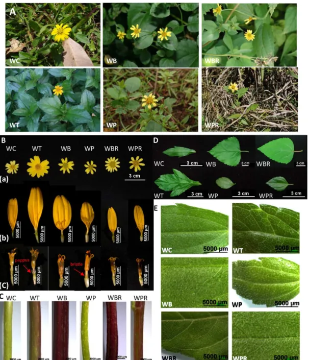
Four species and two additional varieties ofWedeliagrow naturally in Taiwan (Table 2) [22]. TheseWedeliaspecies or cultivar strains were collected from areas in the northern and central counties of Taiwan (details inTable 1). Some of theseWedeliaspecies are similar in morpholog-ical appearance (Fig 1A); and all bear yellow flowers (Fig 1B). The number of ray florets varies:
W.trilobatahas 8 to 14;W.chinensisandW.biflorahave 8 to 12;W.prostrataandW.prostrata
var.robustahave 8; andW.bifloravar.ryukyuensishaving 13 to 15 (Table 3). Ovaries ofW. chi-nensisandW.trilobataare crowned with a cup-shaped pappus on the summit.W.biflora,W.
prostrata,W.prostratavar.robustaandW.bifloravar.ryukyuensis, on the other hand, do not have a pappus or it has degenerated into one or two bristles (Fig1B(b) and1B(c)). The shape of the ovaries ofW.chinensisis similar to those ofW.trilobata. AndW.biflora,W.prostrata,W.
bifloravar.ryukyuensisandW.prostratavar.robustahave remarkably similarly shaped ovaries (Fig1B(b) and1B(c)) that differ from those ofW.chinensisandW.trilobata.
Stems ofW.biflora,W.prostrata,W.bifloravar.ryukyuensisandW.prostratavar.robusta
exhibit deep angular structures with strigose hairs (Fig 1C). In contrast, the stem ofW.
Table 2. Wedeliaspecies in Taiwan.
Species Acronym Additional Varieties Acronym
Wedelia chinensis WC
Wedelia trilobata WT
Wedelia biflora WB Wedelia bifloravar.ryukyuensis WBR
Wedelia prostrate WP Wedelia prostratavar.robusta WPR
trilobatais polygonal or circular in shape with pubescent hairs, and that ofW.chinensisis cir-cular with appressed-pilose hairs (Fig 1C). TheW.chinensisleaf is linear-oblong with ap-pressed-pilose hairs; theW.trilobataleaf is always three-lobate with pubescent hairs;W.
bifloraandW.bifloravar.ryukyuensisleaves are ovate-lanceolate with appressed-strigose hairs;W.prostratais oblong with densely strigose hairs; andW.prostratavar.robustais ovate with appressed-strigose hairs (Fig1Dand1E,Table 3).
Fig 1. Macroscopic characteristics ofWedeliaspecies. A,habitat;B, flower structures: (a) flowers, (b) ray florets, (c) disc florets;C,stems;D&E,leaves.
Anatomical analysis of plant tissues
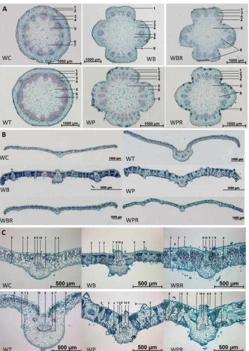
The transverse tissue section of the stem ofW.chinensisis circular in outline and that ofW. tri-lobatais virtually polygonal with the vascular bundles cross-connected; those ofW.biflora,W.
prostrata,W.prostratavar.robustaandW.bifloravar.ryukyuensisare cross-shaped (Fig 2A). According to observations of free-hand cross sections (data not shown),W.chinensishas 12 to 17 vascular bundles in the stems;W.trilobatahas 17 to 22;W.biflorahas 20 to 29;W.prostrata
has 12 to 26;W.prostratavar.robustahas 18 to 28; andW.bifloravar.ryukyuensishas 20 to 29 (Table 3). With the exception ofW.prostrate, all of theWedeliaspecies examined had resin ducts which are located in the cortex and pith of stems, (S1 FigandTable 3). The resin ducts of
W.prostrataare only located in the cortex (S1 Fig). In addition to resin ducts, secretory cells can be found in the stems ofW.biflora,W.prostrata,W.prostratavar.robustaandW.biflora
var.ryukyuensis. The arrangements of the vascular bundles in theseWedeliastems can vary among the species (S1 Fig).
The transverse sections of leaves ofW.chinensisandW.trilobataare quite similar and their leaves are generally thinner than those of the other species (Fig2Band2C).Wedelia trilobata
usually has multiple vascular bundles in the midrib whereas the otherWedeliaspecies have a single vascular bundle (Fig 2BandS2 Fig). This feature may be related to the fact thatW. trila-batahas three-lobate leaves. The arrangements of spongy and palisade tissues inW.chinensis
are similar to those inW.trilobata; inW.biflorathey are similar to those inW.bifloravar. ryu-kyuensis; and inW.prostratathey are similar to those inW.prostratavar.robusta(Fig2Band
2C). All the species and varieties have trichomes on both the upper and lower epidermis (Fig 2CandS2 Fig). They also have resin ducts, usually located near the vascular bundles in the midrib and veins (S2 FigandTable 3).
Comparative molecular and genotyping analyses of internal transcribed
spacer sequences of
Wedelia
species
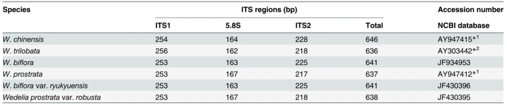
The internal transcribed spacer (ITS) regions of analyzedWedeliaspecies were measured to be between 637 and 646 bp (Table 4) in length. The ITS sequences ofW.biflora(accession number JF934953),W.bifloravar.ryukyuensis(accession number JF430396) andW. pros-trata var.robusta(accession number JF430395) have been submitted to GenBank. And the ITS sequences ofW.chinensis,W.trilobataandW.prostrateare the same as the sequences AY303442 (W.trilobata), AY947415 (W.chinensis) and AY947412 (W.prostrate) submitted
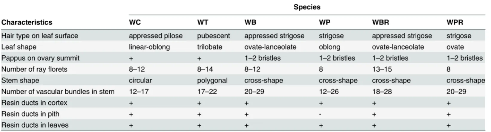
Table 3. Summary of macroscopic and microscopic characteristics observed for stem and leaf tissues ofWedeliaspecies.
Species
Characteristics WC WT WB WP WBR WPR
Hair type on leaf surface appressed pilose pubescent appressed strigose strigose appressed strigose strigose
Leaf shape linear-oblong trilobate ovate-lanceolate oblong ovate-lanceolate ovate
Pappus on ovary summit + + 1–2 bristles 1–2 bristles 1–2 bristles 1–2 bristles
Number of rayflorets 8–12 8–14 8–12 8 13–15 8
Stem shape circular polygonal cross-shape cross-shape cross-shape cross-shape
Number of vascular bundles in stem 12–17 17–22 20–29 12–26 18–28 20–29
Resin ducts in cortex + + + + + +
Resin ducts in pith + + + - + +
Resin ducts in leaves + + + + + +
Abbreviations, including WC, WT, WB, WP, WBR and WPR, are described as shown inTable 2.
previously. They are composed of ITS1, 253 to 256 bp; 5.8S, 162 to 167 bp; and ITS2, 217 to 228 bp (Table 4). The GC content of the ITS in all species was determined to be approximate-ly 53.2% in ITS1, 53.4% in 5.8S, and 57.5% in ITS2. We detected 124 variable sites in the ITS region, and among them 76 variable sites were detected in ITS1, five in 5.8S and 43 in ITS2 (Fig 3A). The smallest genetic distance was found at 0.002 betweenW.bifloraandW.biflora
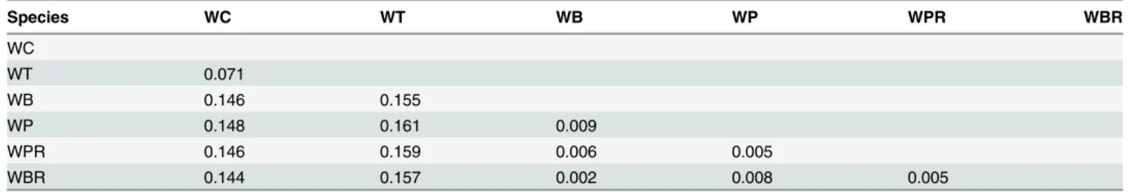
var.ryukyuensisand the largest genetic distance was 0.161 betweenW.prostrataandW. trilo-bata(Table 5). According to the neighbor-joining tree analysis, the testedWedeliacan be
Fig 2. Transverse sections of stems and leaves ofWedeliaspecies. A,Tissue sections of stems: 1. Epidermis; 2. Cortex; 3. Phloem; 4. Xylem; 5. Pith; 6. Resin duct; 7. Pro-resin duct; and 8. Secretory cells.B & C,Tissue sections of leaves: 1. Upper epidermis; 2. Palisade tissue; 3. Lower epidermis; 4. Spongy tissue; 5. Bundle sheath; 6. Xylem; 7. Phloem; 8. Stoma; 9. Trichome; 10. Resin duct.
divided into two clades: one clade composed ofW.prostrata,W.biflora,W.prostratavar. ro-bustaandW.bifloravar.ryukyuensis, and a second clade composed ofW.chinensisandW.
trilobata(Fig 3B). This neighbor-joining tree was found to appropriately correspond not only with the histological analysis of stem and leaf tissues of theseWedeliaspecies but also with the HPLC chemical profiles of the water-extracted metabolites present in theseWedelia
species (see below).
Analyses of chemical fingerprints of
Wedelia
species by HPLC
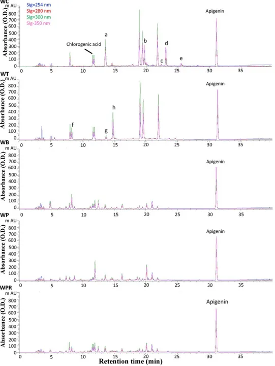
HPLC analysis revealed five distinguishable metabolite profiles from tissue extracts of test
Wedeliaspecies (Fig 4). The population ofW.bifloravar.ryukyuensisplants in plantations or fields in Taiwan was found to be limited, impeding harvesting of sufficient materials for sys-tematic studies of this species. Chromatogram peaks exhibiting specific metabolite profiles were complemented using a photo-diode array detector (S3 Fig). Thus, the metabolite peaks were characterized by both retention time and UV spectra. Some peaks exhibiting the same re-tention time for bothW.chinensisandW.trilobataplants also have similar UV spectra (Fig 4
andS3 Fig). We observed that peaks f, g and h were present only inW.trilobata, whereas peaks a, b, c, d and e were present only inW.chinensis(Fig 4, WC and WT). Although the retention time of peak a inW.chinensisis similar to peak g inW.trilobata, their UV spectra are quite dif-ferent (S3 Fig). The UV spectra for several peaks shown inS3B FiginW.chinensisandW. trilo-bataare similar to that of caffeic acid, indicating they are likely caffeic acid derivatives (S3A
andS3BFig). Comparison of these two HPLC metabolite profiles with those from the other threeWedeliaspecies/variants revealed thatW.biflora,W.prostrataandW.prostratavar. ro-bustahave relatively similar metabolite profiles as a group (Fig 4), that are dramatically differ-ent from those ofW.chinensisandW.trilobata.
We further identified a number of phytochemical peaks as candidate“index compounds”, which may identify the metabolite profiles of theWedeliaspecies. We observed that the re-tention time and UV spectra for peaks 3 and 4 were quite similar to that exhibited by chloro-genic acid, while peak 5 was similar to ferulic acid. These results were further verified by LC-MS using authentic pure compounds for comparison (data not shown). We hence suggest that the various secondary metabolites extracted from the five testedWedeliaspecies may all contain chlorogenic acid or its analogue(s). Another compound, ferulic acid, may also be an index compound for bothW.chinensisandW.trilobataextracts. We and others [36–38] have reported a spectrum of bio-activities of chlorogenic acid, caffeic acid and ferilic acid on
Table 4. Nucleotide sequence length of ITS1, 5.8S rRNA and ITS2 genomic DNA fragments of four species and two additional varieties ofWedelia.
Species ITS regions (bp) Accession number
ITS1 5.8S ITS2 Total NCBI database
W.chinensis 254 164 228 646 AY947415*1
W.trilobata 256 162 218 636 AY303442*2
W.biflora 253 163 225 641 JF934953
W.prostrata 253 167 217 637 AY947412*1
W.bifloravar.ryukyuensis 253 163 225 641 JF430396
Wedelia prostratavar.robusta 253 167 218 638 JF430395
*1The sequences AY947415 and AY947412 were submitted to GenBank by Yuan, C.I., Hsieh, Y.C. and Chiang, M.Y. *2The sequence AY303442 was submitted to GenBank by Dias de Moraes, M., Panero, J.L. and Semir, J.
various mammalian systems. On the other hand, these phytochemicals, as secondary metabo-lites, were also shown to confer important stress or pathogen-responsive activities in plants. Possible correlation and significance of these cross-kingdom bio-activities are addressed in Discussion.
Fig 3. Multiple alignment of the ITS regions ofWedeliaspecies in Taiwan. A,ITS1 and ITS2 genes are located between 1 and 256 bp, and between 422 and 657 bp of the ribosomal DNA (rDNA) gene,
respectively. The 5.8S rDNA is located between 257 and 421 bp in the rDNA gene.B,The neighbor-joining consensus tree ofWedeliaspecies in Taiwan established based on the ITS sequence analogy (Bootstrap: 500 replicates, seed: 954).
UPLC-ESI-Q-TOF MS analyses of chromatograph fingerprints of
Wedelia
species
To further analyze the chemical constituents of plant metabolites from testWedeliaspecies, we investigated the metabolite profiles using the UPLC-ESI-Q-TOF MS system. Various chemical components were detected by this MS system in both ES+and ES-modes (S4 Fig) suggesting that the sensitivity of MS we set to optimize the assay conditions was adequate for the present study. Using the LC-MS dataset, we obtained 3164 peaks and 1849 peaks from the fiveWedelia
species in the ES+and ES-mode, respectively. Subsequently, principal component analysis (PCA) was performed to analyze these peaks. PCA for data from both ES-and ES+modes re-vealed a clear variation in the metabolite content, effectively separating the fiveWedeliaspecies into the following three groups: (1)W.chinensis; (2)W.trilobata; and (3)W.biflora,W. pros-trataandW.prostratavar.rubosta(Fig 5).
Functional analysis of
in vivo
anti-inflammatory bioactivities in
DSS-induced acute colitis mice
To determine the possible specific anti-inflammatory functions ofWedeliaspecies, we used a DSS-induced acute murine colitis model to evaluate the bioactivities of variousWedeliaherbal extracts. Throughout the experiment, we monitored the oral intake of drinking water spiked with 2.0% DSS: the consumption level of DSS-spiked water was found to be statistically indis-tinguishable among all test groups (data not shown). Sulfasalazine, a commercialized drug for treating and preventing relapse of IBD was used as the positive control for this experiment [39,40]. Under normal conditions, i.e., DSS was not added to drinking water,Wedeliaherbal extracts did not promote mouse body weight loss (data not shown). Mice treated with 2% DSS in drinking water developed typical symptoms of acute colitis including diarrhea, rectal bleed-ing and loss of body weight. Comparison of the DSS group with the vehicle group showed that the relative body weight (%) of the DSS-treated group decreased significantly on days 6, 7 and 8 (Fig 6A). After treatment with 2% DSS, mouse body weights were found to be significantly lower in the DSS, sulfasalazine (Sul), WB, WP and WPR-treated groups than in the WC and WT groups on days 7 and 8 (Fig 6A). The disease activity index (DAI) was calculated accord-ing to the severity of clinical colitis symptoms (S1 Table). Statistical analysis revealed that the DAI scores for variousWedeliaspecies groups were not significantly different from that for the DSS group (Fig 6B). Although we can not co-relate clearly the bioactivities of different
Wedeliaplant extracts to the DAI score, we consider that the specific difference between the WC and DSS only group is in agreement with other data shown inFig 6and as we previously reported [9]. Decrease in colon length is widely accepted as a critical symptomatic parameter in DSS-induced colitis.Fig 6Cshows that the colon length of test mice was significantly de-creased after eight days of DSS administration. A comparison of theWedeliaextract-treated
Table 5. Kimura’s two-parameter’s genetic distances as determined based on total ITS sequence.
Species WC WT WB WP WPR WBR
WC
WT 0.071
WB 0.146 0.155
WP 0.148 0.161 0.009
WPR 0.146 0.159 0.006 0.005
WBR 0.144 0.157 0.002 0.008 0.005
experimental groups and the untreated, DSS only group showed that WC, WT, WB, WP and WPR treatments effectively protected the colon tissues of test mice from physically contract-ing or shortencontract-ing (Fig 6C). These treatments, except for WT and WP, also attenuated the his-topathological manifestations in test colons (Fig6Dand6E). Microscopic examination of haematoxylin and eosin (H&E)-stained middle colon tissue sections from groups adminis-tered 2% DSS alone for eight days showed a substantial decrease in colon length, regeneration
Fig 4. HPLC chromatograms of hot water extracts of fiveWedeliaspecies.Apigenin was supplemented only as an internal standard to calibrate the HPLC data. The colors of signals, blue, red, green and pink, denote different absorbance levels of 254 nm, 280 nm, 300 nm and 350 nm, respectively. The peaks of a, b, c, d, e, f and g represent the different peaks detected between WC and WT extracts.
or losing of the large area crypts, disruption of epithelial lining, massive infiltration of the lam-ina propria and submucosa tissues by inflammatory cells, and submucosal erosion with edema (Fig 6E). Comparison of colon tissue sections from variousWedeliaextract-treated and DSS only-treated mice showed a notably lower degree of edema, crypt abscesses, and ero-sion with infiltration of mononuclear cells or other lymphocytes into the mucosal tissues in the WC-treated groups as compared to the DSS only-treated group. Interestingly, the WP-treated group showed little or no reduction in colitis symptoms on histopathological examina-tion, whereas the WC group showed histological features, tissue size and morphology that closely resembled those of vehicle (control) mice (Fig 6E). Sulfasalazine-treatment had a mod-est effect on colon length (Fig 6C), histological score (Fig 6D) and histopathology microscopy (Fig 6E). The histological scores, as quantitatively determined according to standard histo-pathological examinations (S2 Table), were also drastically decreased in the WC-treated mice, with varying degrees of reduction in the WPR and WB groups (Fig 6D). These results corre-sponded reasonably well with the histopathological examinations. Together, the multiple
Fig 5. PCA analysis of plant metabolites from aqueous tissue extracts ofWedeliaspecies using UPLC-ESI-MS. A,Electrospray ionization positive (ES+) LC-MS data.B,Electrospray ionization negative (ES-) LC-MS data.
assay results obtained from the DSS-induced acute colitis murine model (Fig 6) suggest that dietary treatment with WC can markedly ameliorate both the colitis-like symptoms and the tissue damage caused by induced inflammation and tissue-wounding in colons of DSS-induced acute colitis mice.
Fig 6. Effect of different extracts ofWedeliaspecies in mice with DSS-induced acute colitis.Mice were simultaneously administered orally with vehicle (sterilized water), Sul: sulfasalazine (positive control, 50 mg/ kg) or different extracts ofWedeliaspecies (50 mg/kg), respectively.A,Percentage of murine body weight.B, Disease activity index.C,Colon length.D,Histological score.E,Histopathology of mice with DSS-induced acute colitis. Data are expressed as mean±SD (n = 10). WC and WT, respectively*P<0.05,**P<0.01, ***P<0.001, significant difference compared with the DSS group.
Discussion
In this study, we used a combined technological approach to authenticate, validate or profile the various species/varieties and biodiversities ofWedeliaplants that grow naturally in Taiwan, and further investigated their reputed bioactivitiesin vivoin a clinically-relevant animal/dis-ease model as an example of a systematic exploration of plant secondary metabolites found in traditional medicinal plants. Among various medicinal plants from theWedeliagenus com-monly found in Taiwan, as compared to the untreated (DSS-treated only) mice, only the phy-toextract from theW.chinensisspecies exhibited statistically significant anti-inflammatory bioactivity on DSS-induced murine colitis. This result may be connected to two groups of sec-ondary plant metabolites, caffeic acid derivatives and chlorogenic acid, present inW.chinensis
extracts. Further study is needed however to verify whether these index compound-like phyto-chemicals are indeed the active ingredient(s) of the detected bioactivities.
Caffeic acid and derivatives are efficacious phytochemicals that may help defend plants against abiotic and/or biotic stresses. They were reported to help protect plants against plant pathogens, including fungi, bacteria and insects [41–43]. They have also been shown to con-tribute or be associated with the control of physical or chemical stresses including drought, sa-linity and heat [44–46]. On the other hand, the oxidant, carcinogenic,
anti-inflammatory, immunomodulatory and even anti-colitis activities of caffeic acid and some of its derivatives have also been previously reported by our own group and others [36,47–52]. Chlorogenic acid has also been reported to exhibit various bioactivities in mammalian systems, including anti-inflammatory and cytokine-modulatory activities [53–59]. Caffeic acid has been recently shown to increase the expression of CYP4B1 mRNA in DSS-treated mice and effec-tively inhibited DSS-induced murine colitis [60]. In light of these findings, on chlorogenic acid and caffeic acid, we suggest that these two plant secondary metabolites or/and their derivatives may act target in plant extract mixtures on effective inhibition of colon inflammation in DSS-treated mice with colitis. Dietary uptake ofW.chinensisplant extracts may thus confer phyto-medicinal or nutraceutical effects on IBD in humans. Obviously, such claims would need to be evaluated and verified by human clinical studies or trials.
The other five species/variants other thanW.chinensistested in this study had a moderate effect on ameliorating body weight loss, colon length shortening, histological scores, DAI and histopathological activities in colitis mice. Among the various parameters evaluated (Fig6Ato
6E),W.chinensisplant extract was found to exhibit coordinated, correspondent, and routine efficacy in test bioactivities. Some plants, e.g.,W.prostrate, showed little or no anti-inflamma-tory effects in DSS colitis model mice. Our studies hence effectively demonstrated the need for proper clarification and verification of reputed medicinal herbs for use and future development of medicinal food or botanical drugs.
Assessment of whole, fragmented, and even dried-botanical materials through microscopic identification and comparative analysis improves our capability to identify or verify medicinal plants [61]. Effective authentication of medicinal plants for botanical drug development is in-deed a key requirement for future advancement of phytomedicines. In this study, in terms of biodiversity studies, we also revealed interesting and important results on the taxonomy of
W.prostratavar.robustaandW.prostrata, and 0.006 betweenW.prostratavar.robustaand
W.biflora, strongly support this conclusion (Table 5).
For another comparison in grouping,W.bifloravar.ryukyuensisis notably similar toW.
biflora, not only in terms of general morphology, but also in the anatomical structure of the stems and leaves. Using macroscopic identification, we found that there was only one way to distinguish them:W.bifloravar.ryukyuensishas a higher number of ray florets and disc florets thanW.biflora, the former has 14 to15 ray florets and 45 to 70 disc florets, whereasW.biflora
has 8 to 12 and 20 to 35, ray florels and disc florets respectively. It was previously reported that
W.bifloravar.ryukyuensisis a triploid plant with 2n = 45, whereasW.biflorais a diploid plant with 2n = 30 [22]. In addition to the morphological and anatomical characteristics, the genetic distance betweenW.bifloraandW.bifloravar.ryukyuensiswas determined as 0.002 (Table 5). These results hence support the notion thatW.bifloravar.ryukyuensisis indeed likely a variety ofW.biflora.
Based on the results of the neighbor-joining tree that we established using comparative se-quence analysis of the ITS regions, we suggest here that theseWedeliaspecies can be effec-tively classified into two clades (Fig 3), a result that correlates well with the similarity in the microscopic characteristics. This result on confirmation of previous speciation study also re-vealed the usefulness and importance of our current crosstalk (see below too) with different biotechnology study systems. Anatomical studies showed that numerous resin ducts are pres-ent in the stems and leaves ofWedeliaspecies (S1 FigandS2 Fig). The resin duct, a secretory structure, usually contains terpenoids [62] and essential oils [63]. It has been suggested that the structure of secretory cell plastids is related to their role in monoterpene synthesis [64]. Secretory structures are present in a wide spectrum of vascular plants and play an important mechanistic and ecological role in the defense against herbivores and pathogens [65]. These lipid substances, including terpenoids and essential oils, are widespread internally in secreto-ry structures in the Asteraceae [65]. A number of studies have indicated that the chemical constituents fromWedeliaspecies extracted by organic solvents may contain various terpe-noids [66–69]. Our present study may suggest that specific terpenoids may be usefully em-ployed chemical markers ofWedeliaspecies. It may be also important to investigate in the future whether specific anti-pathogen and/or anti-biotic stress plant terpenoids, can also reg-ulate inflammatory or/and immunomodulatory activities in mammalian systems. The possi-bility of cross-kingdom bioactivities of phytochemicals may warrant future systematic studies.
Based on the metabolite profiles of differentWedeliaspecies analyzed by HPLC,W. chinen-sisandW.trilobatamay be categorized into a group with relatively similar chemical finger-prints, whereasW.biflora,W.prostrataand their varieties can be assigned to another
fingerprint group. Interestingly, althoughW.chinensisandW.trilobataexhibit a similar HPLC metabolite profile, we were still able to identify a number of phytochemical peaks distinctively deleted in these two different plant extracts. UPLC-MS profiling accompanied by PCA statisti-cal analyses [2,5,70] were employed to investigate the relatively minor but still specific differ-ences in secondary metabolites, as categorized by the mass peaks revealed from the six test
Conclusions
This study set out to establish a cross-disciplinary, integrated technology platform for system-atic evaluation of the safety and efficacy of traditional medicinal plants to aid the development of new botanical drugs. As now publically defined and recognized, crude or partially purified (fractionated) medicinal plant extracts or phytochemical mixtures can be employed to develop botanical drugs in USA, Europe and Asian countries. Despite this new development, the scien-tific and pharmaceutical communities have made very limited progress, likely due to the lack of experimental/technical know-how and useful technology platforms for systematic research and development of new phytomedicines [71]. The approach outlined here shows that a com-bination of taxonomy, molecular biology and chemical fingerprinting methodologies are re-quired to effectively and accurately authenticate and characterize medicinal plants and reveal their associated bioactive compounds. Macroscopic and microscopic identification alone may not be sufficient to distinguish a spectrum of reputed or claimed traditional medicinal plants. In addition, specific genome sequencing, secondary plant metabolite profiling and index com-pound analysis, and specific and representative bioactivity assaysin vivoandin vitroare need-ed to make up a sequential and integratneed-ed system to evaluate the efficacy of traditional herbal medicines and the derived phytochemicals. Here, a“real case”plant system, in contrast to a
“model plant system”, was explored using the above approaches, andW.chinensiswas identi-fied and veriidenti-fied as candidate medicinal plant that conferred specific anti-inflammatory effects on DSS-induced murine colitis. These results provide possible leads for future use and develop-ment of evidence-based phytomedicines or botanical drugs. Further, in terms of plant science, specific plant secondary metabolites are expected to be revealed as candidate active compounds for anti-inflammatory, anti-colitis therapies. We hypothesize that these“medicinal”secondary metabolites may also confer useful anti-stress and pro-innate immunity bioactivities in host plants, due to the orthologous molecular and cellular mechanisms seen in plants. These mecha-nisms may well be mirrored in mammalian systems. The possible effects of this specific class of secondary plant metabolites fromWedeliamay hence warrant further systematic investigation in the future.
Supporting Information
S1 ARRIVE Checklist. (PDF)
S1 Fig. Transverse sections of stem tissues ofWedeliaspecies.The arrows show resin ducts. (TIF)
S2 Fig. Transverse sections of leaf tissues ofWedeliaspecies.Ph: phloem Rd: resin duct Tc: trichome X: xylem.
(TIF)
S3 Fig. A,UV spectra detected for caffeic acid.B,UV spectra detected for some putative caffeic acid derivatives in the HPLC chromatograms ofW.chinensisandW.trilobata. Apigenin is spiked in as the internal standard.
(TIF)
S4 Fig. ES-BPI chromatograms of plant metabolites from aqueous tissue extracts of differ-entWedeliaspecies. A,Electrospray ionization positive LC-MS data.B,Electrospray ioniza-tion negative LC-MS data.
S1 File. Approval letter.This is to certify that the animal protocol by the following applicant has been evaluated and approved by the Institutional Animal Care and Use Committee of Aca-demia Sinica (AS IACUC).
(PDF)
S1 Table. Disease activity index. (DOCX)
S2 Table. Scoring system for histological pathology study. (DOCX)
Acknowledgments
We thank Ms. Miranda Loney for professional editing of the manuscript. In addition, the au-thors would like to thank the Laboratory Animal Core Facility which is funded by Agricultural Biotechnology Research Center at Academia Sinica for their services.
Author Contributions
Conceived and designed the experiments: WCL CCW NSY. Performed the experiments: WCL CCW. Analyzed the data: WCL CCW JWL CIP. Contributed reagents/materials/analysis tools: PWH CIP. Wrote the paper: WCL CCW YHC NSY.
References
1. Berrin Y, Ali O, Umut S, Meltem E, Murat B, Barut Y. Multi-organ toxicity following ingestion of mixed herbal preparations: an unusual but dangerous adverse effect of phytotherapy. Eur J Intern Med. 2006; 17: 130–132. PMID:16490692
2. Li SL, Song JZ, Qiao CF, Zhou Y, Qian KD, Lee KH, et al. A novel strategy to rapidly explore potential chemical markers for the discrimination between raw and processed Radix Rehmanniae by UHPLC-TOFMS with multivariate statistical analysis. J Pharm Biomed Anal. 2010; 51: 812–823. doi:10.1016/j. jpba.2009.10.002PMID:19879709
3. Xue D, Yin HX, Li J, Liu XB, Zhang H, Peng C. Application of Microscopy in Authentication and Distin-guishing of 11 Paris Species in West Sichuan. Microscopy Research and Technique. 2009; 72: 744–754. doi:10.1002/jemt.20726PMID:19455685
4. Lin WY, Chen LR, Lin TY. Rapid authentication of Blupleurum species using an array of immobilized se-quence-specific oligonucleotide probes. Planta Med. 2008; 74: 464–469. doi: 10.1055/s-2008-1034325PMID:18484544
5. Zhong XK, Li DC, Jiang JG. Identification and Quality Control of Chinese Medicine Based on the Fin-gerprint Techniques. Current Medicinal Chemistry. 2009; 16: 3064–3075. PMID:19689283
6. Guy PA, Tavazzi I, Bruce SJ, Ramadan Z, Kochhar S. Global metabolic profiling analysis on human urine by UPLC-TOFMS: issues and method validation in nutritional metabolomics. J Chromatogr B Analyt Technol Biomed Life Sci. 2008; 871: 253–260. doi:10.1016/j.jchromb.2008.04.034PMID:
18490203
7. Brown M, Dunn WB, Dobson P, Patel Y, Winder CL, Francis-McIntyre S, et al. Mass spectrometry tools and metabolite-specific databases for molecular identification in metabolomics. Analyst. 2009; 134: 1322–1332. doi:10.1039/b901179jPMID:19562197
8. Xie PS, Yan YZ, Guo BL, Lam CW, Chui SH, Yu QX. Chemical pattern-aided classification to simplify the intricacy of morphological taxonomy of Epimedium species using chromatographic fingerprinting. J Pharm Biomed Anal. 2010; 52: 452–460. doi:10.1016/j.jpba.2010.01.025PMID:20144519
9. Huang YT, Wen CC, Chen YH, Huang WC, Huang LT, Lin WC, et al. Dietary Uptake of Wedelia chinen-sis Extract Attenuates Dextran Sulfate Sodium-Induced Colitis in Mice. PLoS One. 2013; 8: e64152. doi:10.1371/journal.pone.0064152PMID:23734189
10. Wirtz S, Neufert C, Weigmann B, Neurath MF. Chemically induced mouse models of intestinal inflam-mation. Nature Protocols. 2007; 2: 541–546. PMID:17406617
12. Danese S, Mantovani A. Inflammatory bowel disease and intestinal cancer: a paradigm of the Yin-Yang interplay between inflammation and cancer. Oncogene. 2010; 29: 3313–3323. doi:10.1038/onc.2010. 109PMID:20400974
13. Melgar S, Karlsson A, Michaelsson E. Acute colitis induced by dextran sulfate sodium progresses to chronicity in C57BL/6 but not in BALB/c mice: correlation between symptoms and inflammation. Am J Physiol Gastrointest Liver Physiol. 2005; 288: G1328–1338. PMID:15637179
14. Shteingart S, Rapoport M, Grodzovski I, Sabag O, Lichtenstein M, Eavri R, et al. Therapeutic potency of IL2-caspase 3 targeted treatment in a murine experimental model of inflammatory bowel disease. Gut. 2009; 58: 790–798. doi:10.1136/gut.2008.153981PMID:18978179
15. Bouma G, Strober W. The immunological and genetic basis of inflammatory bowel disease. Nat Rev Immunol. 2003; 3: 521–533. PMID:12876555
16. Deguchi Y, Andoh A, Inatomi O, Yagi Y, Bamba S, Araki Y, et al. Curcumin prevents the development of dextran sulfate Sodium (DSS)-induced experimental colitis. Dig Dis Sci. 2007; 52: 2993–2998. PMID:17429738
17. Cullen G, Donnellan F, Long S, Forry M, Murray FE. Perceptions of medication safety among patients with inflammatory bowel disease. Scandinavian Journal of Gastroenterology. 2010; 45: 1076–1083. doi:10.3109/00365521.2010.490595PMID:20459367
18. Moody GA, Eaden JA, Bhakta P, Sher K, Mayberry JF. The role of complementary medicine in Europe-an Europe-and AsiEurope-an patients with inflammatory bowel disease. Public Health. 1998; 112: 269–271. PMID:
9724953
19. Rawsthorne P, Shanahan F, Cronin NC, Anton PA, Lofberg R, Bohman L, et al. An international survey of the use and attitudes regarding alternative medicine by patients with inflammatory bowel disease. American Journal of Gastroenterology. 1999; 94: 1298–1303. PMID:10235210
20. Ke F, Yadav PK, Ju LZ. Herbal medicine in the treatment of ulcerative colitis. Saudi J Gastroenterol. 2012; 18: 3–10. doi:10.4103/1319-3767.91726PMID:22249085
21. Rahimi R, Mozaffari S, Abdollahi M. On the use of herbal medicines in management of inflammatory bowel diseases: a systematic review of animal and human studies. Dig Dis Sci. 2009; 54: 471–480. doi:10.1007/s10620-008-0368-xPMID:18618255
22. Peng CI, Chung KF, Li HL. Flora of Taiwan. Taipei, Taiwan: Editorial Committee of the Flora of Taiwan. v. p. 1998
23. Fosberg FR, Sachet M-H. Systematic studies of Micronesian plants. Washington: Smithsonian Institu-tion Press. iii, 40 p. p. 1980
24. Panero JL, Jansen RK, Clevinger JA. Phylogenetic relationships of subtribe Ecliptinae (Asteraceae: Heliantheae) based on chloroplast DNA restriction site data. Am J Bot. 1999; 86: 413–427. PMID:
10077503
25. Decaisne J. Herbarii timorensis descripto. Nouvelles Annales du Museum d'Histoire Naturelle. 1834; 3: 333–501.
26. Wagner WL, Robinson H. Lipochaeta and Melanthera (Asteraceae: Heliantheae subtribe Ecliptinae): establishing their natural limits and a synopsis. Brittonia. 2001; 53: 539–561.
27. (Jiangsu) JioMM. Zhongyao Da Cidian (Encyclopedia of Chinese Materia Medica). Shanghai: Shang-hai Scientific and Technical Press. 1977
28. McLean RC, Ivimey-Cook WR. Plant science formulæ; a reference book for plant science laboratories (including bacteriology). London,: Macmillan and co., limited. vii, 203, 201 p. p. 1941
29. Rogers SO, Bendich AJ. Extraction of DNA from plant tissues. Plant Molecular Biology Manual. 1988; 1–10 p.
30. Thompson JD, Higgins DG, Gibson TJ. CLUSTAL W: improving the sensitivity of progressive multiple sequence alignment through sequence weighting, position-specific gap penalties and weight matrix choice. Nucleic Acids Res. 1994; 22: 4673–4680. PMID:7984417
31. Tamura K, Dudley J, Nei M, Kumar S. MEGA4: Molecular Evolutionary Genetics Analysis (MEGA) soft-ware version 4.0. Mol Biol Evol. 2007; 24: 1596–1599. PMID:17488738
32. Cooper HS, Murthy SN, Shah RS, Sedergran DJ. Clinicopathologic study of dextran sulfate sodium ex-perimental murine colitis. Lab Invest. 1993; 69: 238–249. PMID:8350599
33. Jiang GL, Im WB, Donde Y, Wheeler LA. Comparison of prostaglandin E2 receptor subtype 4 agonist and sulfasalazine in mouse colitis prevention and treatment. J Pharmacol Exp Ther. 2010; 335: 546– 552. doi:10.1124/jpet.110.173252PMID:20833794
35. Kabashima K, Saji T, Murata T, Nagamachi M, Matsuoka T, Segi E, et al. The prostaglandin receptor EP4 suppresses colitis, mucosal damage and CD4 cell activation in the gut. J Clin Invest. 2002; 109: 883–893. PMID:11927615
36. Staniforth V, Huang WC, Aravindaram K, Yang NS. Ferulic acid, a phenolic phytochemical, inhibits UVB-induced matrix metalloproteinases in mouse skin via posttranslational mechanisms. J Nutr Bio-chem. 2012; 23: 443–451. doi:10.1016/j.jnutbio.2011.01.009PMID:21543204
37. Cho AS, Jeon SM, Kim MJ, Yeo J, Seo KI, Choi MS, et al. Chlorogenic acid exhibits anti-obesity proper-ty and improves lipid metabolism in high-fat diet-induced-obese mice. Food Chem Toxicol. 2010; 48: 937–943. doi:10.1016/j.fct.2010.01.003PMID:20064576
38. Abdel-Latif MM, Windle HJ, Homasany BS, Sabra K, Kelleher D. Caffeic acid phenethyl ester modu-lates Helicobacter pylori-induced nuclear factor-kappa B and activator protein-1 expression in gastric epithelial cells. Br J Pharmacol. 2005; 146: 1139–1147. PMID:16247412
39. Zhao W, Song L, Hongzhu D. Amelioration of dextran sulfate sodium-induced chronic colitis by sulfasa-lazine salicylazosulfapyridine via reducing NF-kappaB transcription factor p65 recruitment to ICAM-1 gene promoters. Yakugaku Zasshi. 2010; 130: 1239–1249. PMID:20823682
40. Ford AC, Kane SV, Khan KJ, Achkar JP, Talley NJ, Marshall JK, et al. Efficacy of 5-Aminosalicylates in Crohn's Disease: Systematic Review and Meta-Analysis. American Journal of Gastroenterology. 2011; 106: 617–629. doi:10.1038/ajg.2011.71PMID:21407190
41. Harrison HF, Peterson JK, Snook ME, Bohac JR, Jackson DM. Quantity and potential biological activity of caffeic acid in sweet potato [Ipomoea batatas (L.) Lam.] storage root periderm. J Agric Food Chem. 2003; 51: 2943–2948. PMID:12720375
42. Summers CB, Felton GW. Prooxidant effects of phenolic acids on the generalist herbivore HelicoVerpa zea (Lepidoptera: Noctuidae): potential mode of action for phenolic compounds in plant anti-herbivore chemistry. Insect Biochem Mol Biol. 1994; 24: 943–953.
43. Cvikrová M, Nedělník J, Eder J, Binarová P. Changes in pattern of phenolic acids induced by culture
fil-trate of Fusarium oxysporum in alfalfa plants differing in susceptibility to the pathogen. J Plant Physiol. 1993; 142: 1–5.
44. Vasquez-Robinet C, Mane SP, Ulanov AV, Watkinson JI, Stromberg VK, De Koeyer D, et al. Physiolog-ical and molecular adaptations to drought in Andean potato genotypes. J Exp Bot. 2008; 59: 2109– 2123. doi:10.1093/jxb/ern073PMID:18535297
45. Kovacik J, Klejdus B, Hedbavny J, Backor M. Salicylic acid alleviates NaCl-induced changes in the me-tabolism of Matricaria chamomilla plants. Ecotoxicology. 2009; 18: 544–554. doi: 10.1007/s10646-009-0312-7PMID:19381803
46. Rivero RM, Ruiz JM, Romero L. Can grafting in tomato plants strengthen resistance to thermal stress? J Sci Food Agric. 2003; 83: 1315–1319.
47. da Cunha FM, Duma D, Assreuy J, Buzzi FC, Niero R, Campos MM, et al. Caffeic acid derivatives: in vitro and in vivo anti-inflammatory properties. Free Radic Res. 2004; 38: 1241–1253. PMID:15621702
48. Ek RO, Serter M, Ergin K, Yildiz Y, Cecen S, Kavak T, et al. The effects of caffeic acid phenethyl ester (CAPE) on TNBS-induced colitis in ovariectomized rats. Dig Dis Sci. 2008; 53: 1609–1617. PMID:
17957471
49. Fitzpatrick LR, Wang J, Le T. Caffeic acid phenethyl ester, an inhibitor of nuclear factor-kappaB, attenu-ates bacterial peptidoglycan polysaccharide-induced colitis in rats. J Pharmacol Exp Ther. 2001; 299: 915–920. PMID:11714876
50. Marquez N, Sancho R, Macho A, Calzado MA, Fiebich BL, Muñoz E. Caffeic acid phenethyl ester inhib-its T-cell activation by targeting both nuclear factor of activated T-cells and NF-kappaB transcription fac-tors. J Pharmacol Exp Ther. 2004; 308: 993–1001. PMID:14617683
51. Russo A, Longo R, Vanella A. Antioxidant activity of propolis: role of caffeic acid phenethyl ester and galangin. Fitoterapia. 2002; 73 Suppl 1: S21–29. PMID:12495706
52. Toyoda T, Tsukamoto T, Takasu S, Shi L, Hirano N, Ban H, et al. Anti-inflammatory effects of caffeic acid phenethyl ester (CAPE), a nuclear factor-kappaB inhibitor, on Helicobacter pylori-induced gastritis in Mongolian gerbils. Int J Cancer. 2009; 125: 1786–1795. doi:10.1002/ijc.24586PMID:19610061
53. Krakauer T. The polyphenol chlorogenic acid inhibits staphylococcal exotoxin-induced inflammatory cy-tokines and chemokines. Immunopharmacol Immunotoxicol. 2002; 24: 113–119. PMID:12022439
54. Shan J, Fu J, Zhao Z, Kong X, Huang H, Luo L, et al. Chlorogenic acid inhibits lipopolysaccharide-in-duced cyclooxygenase-2 expression in RAW264.7 cells through suppressing NF-kappaB and JNK/AP-1 activation. Int Immunopharmacol. 2009; 9: JNK/AP-1042–JNK/AP-1048. doi:10.1016/j.intimp.2009.04.011PMID:
19393773
56. Ding X, Kaminsky LS. Human extrahepatic cytochromes P450: function in xenobiotic metabolism and tissue-selective chemical toxicity in the respiratory and gastrointestinal tracts. Annu Rev Pharmacol Toxicol. 2003; 43: 149–173. PMID:12171978
57. Gu X, Ke S, Liu D, Sheng T, Thomas PE, Rabson AB, et al. Role of NF-kappaB in regulation of PXR-mediated gene expression: a mechanism for the suppression of cytochrome P-450 3A4 by proinflam-matory agents. J Biol Chem. 2006; 281: 17882–17889. PMID:16608838
58. Kwon KH, Murakami A, Tanaka T, Ohigashi H. Dietary rutin, but not its aglycone quercetin, ameliorates dextran sulfate sodium-induced experimental colitis in mice: attenuation of pro-inflammatory gene ex-pression. Biochem Pharmacol. 2005; 69: 395–406. PMID:15652231
59. Reed KL, Fruin AB, Gower AC, Gonzales KD, Stucchi AF, Andry CD, et al. NF-kappaB activation pre-cedes increases in mRNA encoding neurokinin-1 receptor, proinflammatory cytokines, and adhesion molecules in dextran sulfate sodium-induced colitis in rats. Dig Dis Sci. 2005; 50: 2366–2378. PMID:
16416193
60. Ye Z, Liu ZP, Henderson A, Lee K, Hostetter J, Wannemuehler M, et al. Increased CYP4B1 mRNA Is Associated with the Inhibition of Dextran Sulfate Sodium-Induced Colitis by Caffeic Acid in Mice. Exper-imental Biology and Medicine. 2009; 234: 605–616. doi:10.3181/0901-RM-1PMID:19307459
61. Techen N, Crockett SL, Khan IA, Scheffler BE. Authentication of medicinal plants using molecular biology techniques to compliment conventional methods. Current Medicinal Chemistry. 2004; 11: 1391–1401. PMID:15180573
62. Fahn A. Structure and function of secretory cells. Advances in Botanical Research Incorporating Ad-vances in Plant Pathology 2000; 31: 37–75.
63. Dell B, McComb A. Plant resins-their formation, secretion and possible functions. Advances in botanical research. 1979; 6: 277–316.
64. Cheniclet C, Carde JP. Presence of leucoplasts in secretory cells and of monoterpenes in the essential oil: a correlative study. Israel J Bot. 1985; 34: 219–238.
65. Cury G, Appezzato-da-Glória B. Internal secretory spaces in thickened underground systems of Astera-ceae species. Australian Journal of Botany. 2009; 57: 229–239.
66. Li X, Dong M, Liu Y, Shi QW, Kiyota H. Structures and biological properties of the chemical constituents from the genus Wedelia. Chemistry & Biodiversity. 2007; 4: 823–836. PMID:17510998
67. That QT, Jossang J, Jossang A, Kim PPN, Jaureguiberry G. Wedelolides A and B: Novel sesquiterpene delta-Lactones, (9R)-Eudesman-9,12-olides, from Wedelia trilobata. Journal of Organic Chemistry. 2007; 72: 7102–7105. PMID:17705534
68. Chen WL, Tang WD, Zhang RJ, Lou LG, Zhao WM. Cytotoxic germacrane-type sesquiterpenes, pimar-ane-type diterpenes, and a naphthalene derivative from Wollastonia biflora. Journal of Natural Prod-ucts. 2007; 70: 567–570. PMID:17291042
69. Kumar RM. pharmacognostical studies of the plant Wedelia chinensis (Osbeck) MERR. International Journal of Pharmaceutical Research and Development. 2011; 2: 53–57.
70. Dan M, Su MM, Gao XF, Zhao T, Zhao AH, Xie G, et al. Metabolite profiling of Panax notoginseng using UPLC-ESI-MS. Phytochemistry. 2008; 69: 2237–2244. doi:10.1016/j.phytochem.2008.04.015PMID:
18550132