Fluorescence Activated Cell Sorting of
Rickettsia prowazekii
-Infected Host Cells
Based on Bacterial Burden and Early
Detection of Fluorescent Rickettsial
Transformants
Lonnie O. Driskell, Aimee M. Tucker, Andrew Woodard, Raphael R. Wood, David O. Wood*
Department of Microbiology and Immunology, College of Medicine, University of South Alabama, Mobile, Alabama, United States of America
*dowood@southalabama.edu
Abstract
Rickettsia prowazekii, the causative agent of epidemic typhus, is an obligate intracellular bacterium that replicates only within the cytosol of a eukaryotic host cell. Despite the barri-ers to genetic manipulation that such a life style creates, rickettsial mutants have been gen-erated by transposon insertion as well as by homologous recombination mechanisms. However, progress is hampered by the length of time required to identify and isolateR. pro-wazekiitransformants. To reduce the time required and variability associated with propaga-tion and harvesting of rickettsiae for each transformapropaga-tion experiment, characterized frozen stocks were used to generate electrocompetent rickettsiae. Transformation experiments employing these rickettsiae established that fluorescent rickettsial populations could be identified using a fluorescence activated cell sorter within one week following electropora-tion. Early detection was improved with increasing amounts of transforming DNA. In addi-tion, we demonstrate that heterogeneous populations of rickettsiae-infected cells can be sorted into distinct sub-populations based on the number of rickettsiae per cell. Together our data suggest the combination of fluorescent reporters and cell sorting represent an important technical advance that will facilitate isolation of distinctR.prowazekiimutants and allow for closer examination of the effects of infection on host cells at various infectious burdens.
Introduction
Rickettsia prowazekiicauses the serious and historically significant human disease epidemic typhus. This malady is transmitted by the human body louse and is associated with crowded populations living in unhygienic environments [1–3]. In addition, a zoonotic reservoir, the southeastern flying squirrel, has been associated with sporadic cases ofR.prowazekiiinfection OPEN ACCESS
Citation:Driskell LO, Tucker AM, Woodard A, Wood RR, Wood DO (2016) Fluorescence Activated Cell Sorting ofRickettsia prowazekii-Infected Host Cells Based on Bacterial Burden and Early Detection of Fluorescent Rickettsial Transformants. PLoS ONE 11 (3): e0152365. doi:10.1371/journal.pone.0152365
Editor:James E Samuel, Texas A&M Health Science Center, UNITED STATES
Received:November 20, 2015
Accepted:March 14, 2016
Published:March 24, 2016
Copyright:© 2016 Driskell et al. This is an open access article distributed under the terms of the Creative Commons Attribution License, which permits unrestricted use, distribution, and reproduction in any medium, provided the original author and source are credited.
Data Availability Statement:All relevant data are within the paper and its Supporting Information file.
Funding:This work was funded by National Institutes of Health, National Institute of Allergy and Infectious Diseases (https://www.niaid.nih.gov/Pages/ default.aspx) grants 1R21AI103272 and
5R01AI020384 to DOW. The funders had no role in study design, data collection and analysis, decision to publish, or preparation of the manuscript.
in the United States as recently as 2009 [4–7]. Due to a low infectious dose and the fact thatR.
prowazekiiis stable for months in louse feces, there is the potential for aerosol spread andR.
prowazekiiwas previously weaponized for use as a biological warfare agent [8,9]. Thus, it is currently classified as a Category B Select Agent.
Rickettsial species are classified into four phylogenetic groups (ancestral, typhus, transi-tional, spotted fever) with the typhus and spotted fever groups containing some of the most notorious rickettsial pathogens [10,11].R.prowazekiiis a member of the typhus group and dif-fers from spotted fever group rickettsiae in several significant ways.R.prowazekiidoes not polymerize actin and is unable to spread by this active mechanism from cell to adjacent cell [12,13]. Also, in contrast to spotted fever group rickettsiae, which induce early damage to the host cell,R.prowazekiireplicates to high rickettsial numbers per cell with little apparent dam-age until the cell lyses [14–17]. The lack of directional spread to adjacent cells preventsR. pro-wazekiifrom forming distinct, isolatable plaques as proficiently as spotted fever group rickettsiae [18–21]. Similarities in intracellular growth between the different groups are also visible. For example, in cell culture models, rickettsial infections are not uniform and growth within individual host cells, as well as between cells, is non-synchronous. This results in cell populations exhibiting a wide range of rickettsiae per cell. Characterizing the changes in gene expression as a few rickettsiae grow within a cell replete with nutrients to a later stage when there are hundreds of rickettsiae per cell, is hampered by the lack of homogeneous populations of infected cells. Here we describe a protocol to separate cells infected with fluorescent rickett-siae into distinct populations based on bacterial burden.
Despite the challenges an obligate intracellular lifestyle presents to genetic analysis, rickett-sial mutants have been generated via transformation using both plasmid and linear DNA [21– 28]. Characterization of these mutants has increased our understanding of rickettsial virulence mechanisms[21,27] and generated an attenuated strain that could serve as a live vaccine based on its ability to grow in culture but not exhibit a virulence phenotype in an animal model [24]. However, in contrast to bacteria that can form colonies on the surface of an agar medium, the identification ofR.prowazekiimutants and the isolation of pure clones is currently a lengthy process. The protocol involves weeks of growth followed by limiting dilution to separate, for example, a transposon insertion mutant from a background composed of other insertions and spontaneously resistant bacteria. As noted above, mutant isolation by the formation of plaques on monolayers, used successfully to purify spotted fever group rickettsial mutants, is also prob-lematic forR.prowazekii. To circumvent these issues we have taken advantage of the fact that rickettsial species can express fluorescent proteins [23,28–30]. In combination with antibiotic selection, fluorescent reporters offer a complementary method for the early identification and isolation of rickettsial transformants and for the examination of experimental parameters, such as DNA concentration, on transformant detection. In this report, we describe the utility of this approach in the genetic analysis ofR.prowazekii.
Materials and Methods
Bacterial strains, host cell lines, and culture conditions
AnR.prowazekiicloned, transposon insertion mutant, designated Madrid E-RP880::arr2-Rp/ gfp[23], was used for fluorescence gating experiments. The transposon is inserted into theR.
(0.218 M sucrose, 3.76 mM KH2PO4, 7.1 mM K2HPO4, 4.9 mM potassium glutamate, and 10 mM MgCl2), designated SPGMg, and stored frozen at -80°C. Murine fibroblast L929 cells (American Type Culture Collection, Manassas, VA, ATCC Number CCL-1) were cultured at 34°C with 5% CO2in modified Eagle’s medium (Mediatech, Inc., Herndon, VA), supplemented with 10% heat-inactivated newborn calf serum (HyClone Laboratories, Logan, UT), and 2 mM glutamine (Mediatech, Inc.), designated SMEM. When indicated for the selection of rickettsial mutants, rifampin (Sigma-Aldrich, St. Louis, MO) dissolved in 100% ethanol at 2 mg/ml was added to SMEM to a final concentration of 200 ng/ml.Escherichia colistrain XL1-Blue (Strata-gene, La Jolla, CA) was used as a recipient for construction and maintenance of shuttle vector pMW1710 and for preparation of plasmid DNA used in rickettsial transformations. XL1-Blue was cultured in Luria-Bertani (LB Lennox) medium at 37°C. For selection ofE.coli transfor-mants, rifampin was added to a final concentration of 50μg/ml.
Plasmid construction
A derivative of the rickettsial shuttle vector pRAM18dRGA [32] was generated by replacing the gene encoding GFPUVwith a rickettsial codon-adapted gene encoding mCherry (desig-nated RpCherry). This gene was synthesized based on the sequence of mCherry (Clontech, Mountain View, CA) using codons optimized for expression inR.prowazekiiby GenScript (Piscataway, NJ). RpCherry gene expression was placed under the control of the rickettsial
ompApromoter fromR.rickettsii[28] in a plasmid containing the rickettsial rifampin resis-tance gene. A cassette (rpsLP- Rparr-2/ompAP-RpCherry)was amplified using this intermediate plasmid as template and primer pair DW316/DW1378 (AACATACTTGCTTTTATAGG and GTCGACGGGCCCGGGATCC). The amplified fragment was inserted into pRAM18dRGA digested with BstBI and EagI to remove the GFP cassette (rpsLP- Rparr-2/ompAP-GFPUV) and
End-It™(Epicentre, Madison, WI) repaired to generate blunt ends. The sequence of the result-ing plasmid, pMW1710, was verified (Iowa State DNA Facility, Ames, IA) (S1 Sequence). Plas-mid pMW1710 was purified using a MP Biomedicals Maxiprep kit (MP Biomedicals, Solon, OH), precipitated with ethanol, and suspended in sterile PCR-quality water at a concentration of ~2.0μg/μl and stored at 4°C.
FACS analysis of fluorescent populations of
R
.
prowazekii
infected L929
cells
Flow cytometric analysis of rickettsiae-infected cells was conducted using a Beckman Coulter Moflo XDP contained in a biosafety cabinet located within a dedicated biosafety level 3 (BSL-3) suite. The instrument is equipped with a 100μm tip. GFPUVwas excited using the 488 nm
laser and detected using a 529/28 band-pass filter. For sorting, a population of L929 cells infected with anR.prowazekiitransposon insertion mutant, Madrid E-RP880::arr2-Rp/gfp [23], that exhibited a range of rickettsiae per cell, was harvested from 175 cm2flasks using tryp-sin as previously described [25]. Cells were collected by centrifugation at 700 X g, washed once in 10 ml of sorting buffer (1X Dulbecco’s PBS, 1 mM EDTA, 25 mM HEPES [pH 7.0], 1% heat-inactivated newborn calf serum) collected by centrifugation as above and suspended in sorting buffer to a density of ~1 X 10⁷cells/ml. Cells were placed in a filtered (35μm) flow
cytometry tube (BD Falcon, Franklin Lakes, NJ) for introduction into the MoFlo XDP. Cells from individual flasks were analyzed independently using the same gating parameters and a histogram of cell counts and GFPUVfluorescence was generated.
To generate a population of RpCherry-expressingR.prowazekiifor use in gating experi-ments,R.prowazekiiBreinl was transformed with 13μg of pMW1710 and cells infected with
experimental samples, background fluorescence was determined using rickettsiae-infected L929 cells electroporated in the absence of DNA. For experimental samples, the gate was set to exclude any background cells. The sorted cells, positive for RpCherry expression, were then planted into a 25 cm2flask containing 10 ml of SMEM with the following antibiotics: 50μg/ml
gentamicin, 200 ng/ml rifampin, and 1X penicillin-streptomycin-neomycin antibiotic mixture with final concentrations of 50 units, 50, and 100μg/ml, respectively. Uninfected L929 cells
(1x10⁶) were added to provide host cells for expansion of the infection. Medium was changed at 24 hours after sorting, and replaced with SMEM containing 200 ng/ml of rifampin. The 24 hour treatment with the antibiotic cocktail was used to kill extracellular contaminants that may have been acquired during sorting. This treatment did not significantly inhibit rickettsial growth. The infection was amplified and rickettsial growth monitored microscopically by Gimenez staining [33]. Transformed rickettsiae were isolated by ballistic shearing as previously described [22] and the purified rickettsiae stored at -80°C in SPGMg prior to use in infections.
To better represent the heterogeneous populations that result from long-term selection of transformants, two 175 cm2flasks were infected at different (low and high) multiplicities of infection (MOI) with purified pMW1710 rickettsial transformants (see above). After 48 hours of growth, infections were pooled prior to analysis. A sample of each pooled infection was col-lected prior to sorting to provide a baseline average of the number of rickettsiae per host cell (Presort) compared to the populations collected from two gates representing low and high numbers of rickettsiae per cell. RpCherry was excited with the 561 nm laser and detected using a 625/26 bandpass filter. Three independent infections were analyzed. Parameters for sorting, including selection of gates were identical for each independent infection tested.
Fluorescence microscopy
For microscopic examination, cells were fixed using 2.5% formaldehyde and cytofuged onto glass slides. Slides were washed with PBS, mounted with Vectashield anti-fade mounting medium (Vector Laboratories, Burlingame, CA), and examined on a Nikon Eclipse TE2000-U microscope using MetaMorph software
Quantitative PCR (qPCR)
qPCR was used to determine the number of rickettsiae per L929 cell as previously described [26]. Briefly, for rickettsial quantitation, primers DW664/DW665 (CCTGCAAGTAGACAT GTGC and AGTGCATTAGCATCAACACC) were used to amplify theR.prowazekii rho
DNA concentration and the detection of
R
.
prowazekii
transformants
Electroporation ofR.prowazekiiwas conducted as previously described [22,25] with one exception. Rather than harvest rickettsiae from infected L929 cell monolayers for each experi-ment, a large pool of rickettsiae was generated from egg yolk sacs and stored as aliquots at -80°C in SPGMg prior to use. The approximate number of purified, viable rickettsiae in 1 ml aliquots was determined by infecting specific numbers of L929 cells with varying amounts of the pool. The infected cells were planted on cover slips, incubated for 24 hours at 34°C and 5% CO2, and visualized by Gimenez staining. The percent of L929 cells infected and the number of rickettsiae per cell were determined microscopically by randomly counting 100 cells and these numbers were used to estimate the number of viable rickettsiae within the aliquots (~3.5 X 10⁹ rickettsiae/ml). To render these bacteria competent for electroporation, rickettsiae were thawed on ice, collected by centrifugation (11,000 x g for 10 minutes at 4°C) and suspended in 20 ml of ice-cold 0.25 M sucrose. Following centrifugation as described above, the supernatant was removed, and the rickettsiae suspended in 400μl of ice-cold 0.25 M sucrose. A 50μl aliquot of
the competent rickettsiae, along with varied amounts of pMW1710 DNA, was used for each transformation. Rickettsiae were electroporated in 1 mm gap cuvettes using a BTX ECM600 electroporator (BTX Harvard, Holliston, MA) as previously described [22,25]. Following elec-troporation, rickettsiae were transferred to a 50 ml conical tube containing 2x10⁷L929 cells [multiplicity of infection (MOI) ~10–20 rickettsiae/cell] in 3 ml of Hank’s balanced salt solu-tion (Corning, Manassas, VA) supplemented with 5 mM glutamic acid and 0.1% gelatin. Cells were incubated at 34°C at 400 rpm for 1 hour in a Thermomixer R (Eppendorf, Hauppauge, NY). After incubation, rickettsiae-infected L929 cells were planted to three 175 cm2tissue cul-ture flasks containing 25 ml of SMEM and placed in an incubator at 34°C with 5% CO2. SMEM medium was changed at 24 hours, and rifampin added to a final concentration of 200 ng/ml for selection of rickettsial transformants. Cells were harvested using trypsin, pooled, and expanded to 6 flasks on day 2 post-transformation and incubated. Fluorescence analysis was performed on days 5–13 as indicated for specific experiments. Medium with rifampin was changed every 2–3 days during the course of the experiment. For analysis of RpCherry-express-ing rickettsial transformants, rickettsiae-infected L929 cells were harvested on selected days from one 175 cm2tissue culture flask for each DNA concentration tested. Cells were collected and prepared for analysis as described above. A histogram of forward scatter area (FSC-A) and RpCherry fluorescence area was generated for each DNA concentration tested and the percent of the total population reported.
Results
FACS analysis of L929 cells infected with GFP
UV-expressing fluorescent
rickettsiae
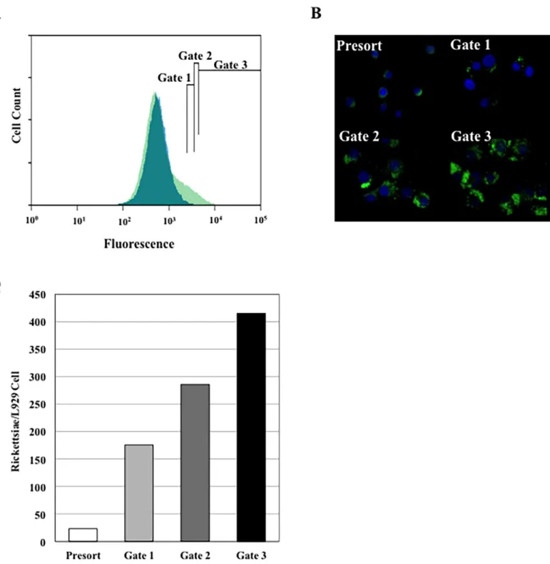
exhibited considerable auto-fluorescence, cells containing rickettsiae expressing GFPUVcould be readily detected above background (Fig 1, Panel A). To establish that fluorescence intensity correlated with increasing rickettsial numbers and to obtain data on gate selection for
Fig 1. FACS analysis of L929 mouse fibroblasts infected withR.prowazekiiexpressing GFPUV.A) Fluorescence plot of the cell populations.
Uninfected L929 control cells (Blue) and a population of infected cells (Green). Three sorting gates are shown and labeled Gate 1, Gate 2, and Gate 3. B) Fluorescence microscopy of presort and sorted populations. Four panels presenting the presorted cell population, Gate 1, Gate 2; and Gate 3. C) Rickettsiae per host cell for sorted populations. Rickettsial and L929 genome equivalents were derived from a single infection sorted three times with each qPCR sample run in duplicate.
subsequent experiments, an initial fluorescence profile of uninfected L929 cells was established. Gate 1 was set at the boundary of uninfected L929 cell auto-fluorescence. Subsequent gates, Gate 2 and Gate 3 were set to collect infected cells with increasing fluorescence intensity. The number of rickettsiae per host cell found in each peak was examined both microscopically (Fig 1, Panel B) and by qPCR (Fig 1, Panel C). These data show that host cells containing fluores-cent rickettsiae can be separated from uninfected cells and sorted into populations where spe-cific gates harbor infected cells with distinct rickettsial loads.
FACS analysis and gating of L929 cells infected with
RpCherry-expressing fluorescent rickettsiae
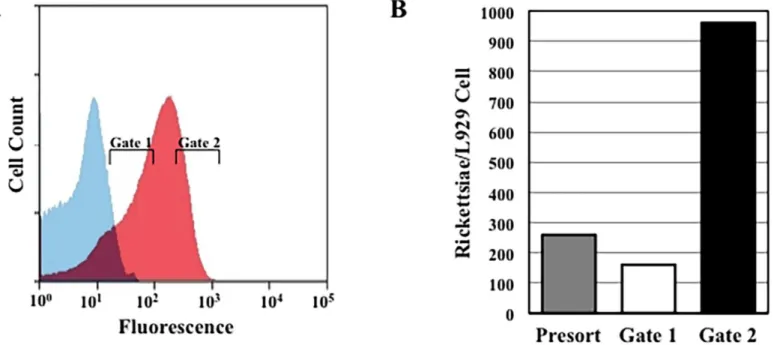
To expand the repertoire of fluorescent proteins available for rickettsial studies and to gener-ate a marker that would have a better signal to noise ratio, crucial for identifying rare trans-formants, we generated a rickettsial codon-adapted mCherry gene, designated RpCherry, to substitute for the GFPUVgene in genetic experiments. In plasmid pMW1710, the RpCherry gene replaces the GFPUVgene of the rickettsial shuttle vector pRAM18dRGA. A population of cells infected with rickettsiae transformed with pMW1710 was analyzed using the 561 nm yellow laser. As seen inFig 2, Panel A, under these conditions the auto-fluorescence of unin-fected L929 cells is low and there is minimal overlap with the fluorescence curve of cells infected with rickettsiae expressing RpCherry. Gating experiments were also performed using this pMW1710 transformed population. As shown by the qPCR results from three independent experiments (Fig 2, Panel B), the RpCherry fluorescent protein can also be used in gating experiments to reproducibly obtain populations of cells with low and high rickett-sial loads.
Fig 2. FACS analysis using RpCherry.A) FACS analysis of L929 mouse fibroblasts infected withR.prowazekiiexpressing RpCherry. Fluorescence plots of uninfected (Blue) and infected (Red) cell populations are shown. Gates used for sorting lightly infected (Gate 1) and heavily infected (Gate 2) host cells are indicated. B) Data represents the ratio of the average number of genome equivalents of rickettsiae per L929 cells from three independent infections and five independent qPCR reactions for each condition.
Effect of DNA concentration on the detection of
R
.
prowazekii
RpCherry-expressing transformants
The identification and isolation ofR.prowazekiitransformants is a lengthy process, involving long-term growth in tissue culture and, if a pure clone is desired, limiting dilution procedures [22,23]. However, the expression of fluorescent proteins byR.prowazekiiprovides a tool for optimizing this process. To demonstrate the potential utility of FACS analysis in rickettsial genetics, we followed the appearance of rickettsiae transformed with plasmid pMW1710. L929 cells infected with control rickettsiae that were mock transformed (no DNA) or transformed with 26μg of pMW1710 DNA were analyzed 13 days post–electroporation (Fig 3, Panel A,
Inset). The no DNA control was used to establish the upper limit of background fluorescence (horizontal line). A fluorescent population above background was readily detected and quantified.
FACS analysis provides a rapid method to quantify and evaluate transformation protocols. One important parameter is the amount of DNA needed to provide the earliest detection and isolation of rickettsial transformants. The effect of plasmid pMW1710 DNA concentration on the appearance of rickettsial transformants is presented inFig 3, A and B. Two independent experiments are shown in order to demonstrate the variability associated with rickettsial exper-iments (see y-axis scale differences). Although each of these independent experexper-iments used ali-quots from the same egg yolk sac purified rickettsial pool, the manipulations required for rickettsial competence and transformation led to two different levels of initial infection. Experi-ment 1 (Fig 3, Panel A) exhibited 88–97% infected cells with an average of 7–14 rickettsiae per cell at 24 hours post-infection. In Experiment 2 (Fig 3, Panel B) the initial infection was higher with 96–100% of the cells infected and an average of 23–34 rickettsiae per cell. In the experi-ment with lower percentages, transformants could be detected by day 8 with the higher concen-tration of pMW1710 DNA. In contrast, previously published isolations ofR.prowazekii
transformants required at least 27 days [22–24,34]. The number of transformants and the rate of detection increased with DNA concentration, although even the lowest amount of plasmid DNA tested generated transformants. Most importantly, transformants could be detected as early as 6 days following electroporation when using the highest concentration of DNA (Fig 3, Panel B). When normalized to the days after the first appearance of transformants, the percent positive per microgram of DNA for both experiments show similar DNA concentration depen-dent numbers of transformants (Fig 3, Panel C).
Discussion
In this paper our goal was to establish fluorescence parameters for sortingR.prowazekii
infected cells into distinct populations based on the number of rickettsiae per cell and to dem-onstrate the early detection ofR.prowazekiifluorescent transformants. Expression byR. pro-wazekiiof both GFPUVand a mCherry derivative, RpCherry, were used successfully to isolate, by FACS gating, populations of infected host cells containing distinct bacterial loads. These populations can now be examined for gene expression changes that are hypothesized to occur asR.prowazekiigrows within a cell replete with nutrients to one where the host cell contains hundreds of rickettsiae and is nearing lysis.
separation between the background fluorescence of uninfected cells and cells infected with RpCherry-expressing rickettsiae will provide an even more efficient tool for mutant identifica-tion and isolaidentifica-tion.
Refining transformation protocols and accelerating mutant isolation using the FACS were also addressed. Extracellular rickettsiae lose viability over time when isolated from their host cell. Thus, early transformation experiments used rickettsiae isolated from freshly inoculated and propagated infections [25]. In the experiments described here, one major modification to our standard transformation protocol was the use of rickettsiae propagated in hen egg yolk sacs, purified, dispensed into multiple aliquots, and stored frozen at -80°C until needed. This eliminated the additional days needed to repeatedly propagate rickettsiae in L929 cells and to perform a separate rickettsial purification for each transformation. In addition, because yolk sac preparations yield a large quantity of pure rickettsiae, rickettsial viability and infectivity could be determined and the pool used for multiple experiments thereby increasing repro-ducibility. While the variability inherent in competence induction, electroporation, and infection remains, the use of frozen aliquots removes a significant variable of previous proto-cols. This also eliminates the two days required to produce competent rickettsiae from L929 cells. This was coupled with the early detection afforded by FACS analysis and the positive effect of increasing transforming DNA concentration. While amounts as low as 1μg have
been used successfully with some rickettsial species [32] our experiments demonstrated that the number of transformants continued to increase from 6μg up to 26μg (the highest
amount tested). This is in contrast to a previous study that showed no significant increase in
R.rickettsiitransposon transformants recovered with increasing DNA concentration (5– 20μg) [35]. This may be due to species differences or transposon versus plasmid
transform-ing DNAs. Most importantly, for ourR.prowazekiiexperiments, transformants could be detected as early as 6 days after electroporation, a significant improvement over our 23 day standard.
In conclusion, we have confirmed the efficacy of two fluorescent proteins, GFPUVand RpCherry, for identifying and isolatingR.prowazekii- infected cells using laser excitation that is standard for most cell sorters. In addition, we demonstrated that rickettsial populations could be separated based on rickettsiae per host cell, an important advancement for studies examining the effect of rickettsial burden on intracellular growth. In regard to the transforma-tion experiments, since these experiments used plasmid DNA, we have not yet applied these techniques to isolate a pure chromosomal transposon insertion or knockout mutant clone. However the early detection ofR.prowazekiitransformants and the ability to sort transfor-mants away from the large population of uninfected cells or those harboring spontaneous resis-tant muresis-tants will greatly decrease the time, effort and cost associated with the isolation and cloning of a variety of rickettsial mutants by limiting dilution. Eventually, we anticipate the sorting of individual cells containing only one or a few rickettsiae and the propagation of these rickettsiae as cloned populations.
Fig 3. Appearance of pMW1710 transformants based on the concentration of transforming DNA.
Competent rickettsiae were electroporated in the presence of increasing amounts of pMW1710 DNA (♦0μg,
●6μg,▲13μg,■26μg). Cells were analyzed on days 5–13 and the percent of RpCherry positive cells was determined. Two independent experiments (A and B) were performed. A) Experiment 1; Inset—Analysis was performed at 13 days following electroporation in the presence of 0μg (Left) or 26μg (Right) of pMW1710. B) Experiment 2; Inset—Area from days 5–10 on an expanded scale. C) RpCherry positive population perμg of DNA normalized to the first appearance of transformants. Bars represent the average of the two experiments with the range indicated.
Supporting Information
S1 Sequence. Plasmid pMW1710 Sequence. (DOCX)
Acknowledgments
We thank Andria Hines for excellent technical expertise and Jonathon Audia, Robert Barring-ton, and John Foster for critical review of the manuscript.
Author Contributions
Conceived and designed the experiments: DOW AMT. Performed the experiments: LOD AMT RRW AW. Analyzed the data: DOW AMT LOD RRW. Contributed reagents/materials/ analysis tools: DOW. Wrote the paper: DOW AMT LOD RRW.
References
1. Azad AF, Beard CB. Rickettsial pathogens and their arthropod vectors. Emerg Infect Dis. 1998; 4 (2):179–86. doi:10.3201/eid0402.980205PMID:9621188; PubMed Central PMCID: PMC2640117.
2. Raoult D, Roux V, Ndihokubwayo JB, Bise G, Baudon D, Marte G, et al. Jail fever (epidemic typhus) outbreak in Burundi. Emerg Infect Dis. 1997; 3(3):357–60. doi:10.3201/eid0303.970313PMID: 9284381; PubMed Central PMCID: PMC2627627.
3. Bechah Y, Capo C, Mege JL, Raoult D. Epidemic typhus. Lancet Infect Dis. 2008; 8(7):417–26. doi:10. 1016/S1473-3099(08)70150-6PMID:18582834.
4. Chapman AS, Swerdlow DL, Dato VM, Anderson AD, Moodie CE, Marriott C, et al. Cluster of sylvatic epidemic typhus cases associated with flying squirrels, 2004–2006. Emerg Infect Dis. 2009; 15 (7):1005–11. doi:10.3201/eid1507.081305PMID:19624912; PubMed Central PMCID: PMC2744229.
5. Reynolds MG, Krebs JS, Comer JA, Sumner JW, Rushton TC, Lopez CE, et al. Flying squirrel-associ-ated typhus, United States. Emerg Infect Dis. 2003; 9(10):1341–3. doi:10.3201/eid0910.030278PMID: 14609478; PubMed Central PMCID: PMC3033063.
6. Bozeman FM, Masiello SA, Williams MS, Elisberg BL. Epidemic typhus rickettsiae isolated from flying squirrels. Nature. 1975; 255(5509):545–7. PMID:806809.
7. Prusinski MA, White JL, Wong SJ, Conlon MA, Egan C, Kelly-Cirino CD, et al. Sylvatic typhus associ-ated with flying squirrels (Glaucomys volans) in New York State, United States. Vector Borne Zoonotic Dis. 2014; 14(4):240–4. doi:10.1089/vbz.2013.1392PMID:24689928.
8. Azad AF, Radulovic S. Pathogenic rickettsiae as bioterrorism agents. Ann N Y Acad Sci. 2003; 990:734–8. PMID:12860715.
9. Walker DH. The realities of biodefense vaccines againstRickettsia. Vaccine. 2009; 27 Suppl 4:D52–5. doi:10.1016/j.vaccine.2009.07.045PMID:19837287; PubMed Central PMCID: PMC2909128.
10. Gillespie JJ, Beier MS, Rahman MS, Ammerman NC, Shallom JM, Purkayastha A, et al. Plasmids and rickettsial evolution: insight fromRickettsia felis. PLoS One. 2007; 2(3):e266. doi:10.1371/journal. pone.0000266PMID:17342200; PubMed Central PMCID: PMC1800911.
11. Gillespie JJ, Williams K, Shukla M, Snyder EE, Nordberg EK, Ceraul SM, et al.Rickettsia phyloge-nomics: unwinding the intricacies of obligate intracellular life. PLoS One. 2008; 3(4):e2018. doi:10. 1371/journal.pone.0002018PMID:19194535; PubMed Central PMCID: PMC2635572.
12. Heinzen RA, Hayes SF, Peacock MG, Hackstadt T. Directional actin polymerization associated with spotted fever groupRickettsiainfection of Vero cells. Infect Immun. 1993; 61(5):1926–35. PMID: 8478082; PubMed Central PMCID: PMC280785.
13. Teysseire N, Chiche-Portiche C, Raoult D. Intracellular movements ofRickettsia conoriiandR.typhi based on actin polymerization. Res Microbiol. 1992; 143(9):821–9. PMID:1299836.
14. Silverman DJ.Rickettsia rickettsii-induced cellular injury of human vascular endothelium in vitro. Infect Immun. 1984; 44(3):545–53. PMID:6724689; PubMed Central PMCID: PMC263617.
15. Silverman DJ. Infection and injury of human endothelial cells byRickettsia rickettsii. Ann Inst Pasteur Microbiol. 1986; 137A(3):336–41. PMID:3122642.
17. Hackstadt T. The biology of rickettsiae. Infect Agents Dis. 1996; 5(3):127–43. PMID:8805076.
18. Cory J, Yunker CE, Ormsbee RA, Peacock M, Meibos H, Tallent G. Plaque assay of rickettsiae in a mammalian cell line. Appl Microbiol. 1974; 27(6):1157–61. PMID:4208640; PubMed Central PMCID: PMC380226.
19. McDade JE, Stakebake JR, Gerone PJ. Plaque assay system for several species ofRickettsia. J Bac-teriol. 1969; 99(3):910–2. PMID:4984178; PubMed Central PMCID: PMC250118.
20. Policastro PF, Peacock MG, Hackstadt T. Improved plaque assays forRickettsia prowazekiiin Vero 76 cells. J Clin Microbiol. 1996; 34(8):1944–8. PMID:8818887; PubMed Central PMCID: PMC229159.
21. Kleba B, Clark TR, Lutter EI, Ellison DW, Hackstadt T. Disruption of theRickettsia rickettsiiSca2 auto-transporter inhibits actin-based motility. Infect Immun. 2010; 78(5):2240–7. doi:10.1128/IAI.00100-10 PMID:20194597; PubMed Central PMCID: PMC2863521.
22. Qin A, Tucker AM, Hines A, Wood DO. Transposon mutagenesis of the obligate intracellular pathogen Rickettsia prowazekii. Appl Environ Microbiol. 2004; 70(5):2816–22. PMID:15128537; PubMed Central PMCID: PMC404435.
23. Liu ZM, Tucker AM, Driskell LO, Wood DO. Mariner-based transposon mutagenesis ofRickettsia pro-wazekii. Appl Environ Microbiol. 2007; 73(20):6644–9. doi:10.1128/AEM.01727-07PMID:17720821; PubMed Central PMCID: PMC2075046.
24. Driskell LO, Yu XJ, Zhang L, Liu Y, Popov VL, Walker DH, et al. Directed mutagenesis of theRickettsia prowazekii pldgene encoding phospholipase D. Infect Immun. 2009; 77(8):3244–8. doi:10.1128/IAI. 00395-09PMID:19506016; PubMed Central PMCID: PMC2715659.
25. Rachek LI, Tucker AM, Winkler HH, Wood DO. Transformation ofRickettsia prowazekiito rifampin resistance. J Bacteriol. 1998; 180(8):2118–24. PMID:9555894; PubMed Central PMCID: PMC107138.
26. Wood DO, Hines A, Tucker AM, Woodard A, Driskell LO, Burkhardt NY, et al. Establishment of a repli-cating plasmid inRickettsia prowazekii. PLoS One. 2012; 7(4):e34715. doi:10.1371/journal.pone. 0034715PMID:22529927; PubMed Central PMCID: PMC3328469.
27. Noriea NF, Clark TR, Hackstadt T. Targeted knockout of theRickettsia rickettsiiOmpA surface antigen does not diminish virulence in a mammalian model system. MBio. 2015; 6(2). doi:10.1128/mBio. 00323-15PMID:25827414; PubMed Central PMCID: PMC4453529.
28. Baldridge GD, Burkhardt N, Herron MJ, Kurtti TJ, Munderloh UG. Analysis of fluorescent protein expression in transformants ofRickettsia monacensis, an obligate intracellular tick symbiont. Appl Envi-ron Microbiol. 2005; 71(4):2095–105. doi:10.1128/AEM.71.4.2095–2105.2005PMID:15812043;
PubMed Central PMCID: PMC1082560.
29. Renesto P, Gouin E, Raoult D. Expression of green fluorescent protein inRickettsia conorii. Microb Pathog. 2002; 33(1):17–21. PMID:12127796.
30. Troyer JM, Radulovic S, Azad AF. Green fluorescent protein as a marker inRickettsia typhi transforma-tion. Infect Immun. 1999; 67(7):3308–11. PMID:10377106; PubMed Central PMCID: PMC116511.
31. Winkler HH. Rickettsial permeability. An ADP-ATP transport system. J Biol Chem. 1976; 251(2):389– 96. PMID:1389.
32. Burkhardt NY, Baldridge GD, Williamson PC, Billingsley PM, Heu CC, Felsheim RF, et al. Development of shuttle vectors for transformation of diverseRickettsiaspecies. PLoS One. 2011; 6(12):e29511. doi: 10.1371/journal.pone.0029511PMID:22216299; PubMed Central PMCID: PMC3244465.
33. Gimenez DF. Staining Rickettsiae in Yolk-Sac Cultures. Stain Technol. 1964; 39:135–40. PMID: 14157454.
34. Rachek LI, Hines A, Tucker AM, Winkler HH, Wood DO. Transformation ofRickettsia prowazekiito erythromycin resistance encoded by theEscherichia coli ereBgene. J Bacteriol. 2000; 182(11):3289– 91. PMID:10809714; PubMed Central PMCID: PMC94521.

