of Polycystic Kidneys and Renal Neoplasia
Jindong Chen1*, Kunihiko Futami1,2, David Petillo1, Jun Peng3, Pengfei Wang4, Jared Knol5, Yan Li1, Sok-Kean Khoo1,6, Dan Huang1, Chao-Nan Qian1,7, Ping Zhao8, Karl Dykyma9, Racheal Zhang1, Brian Cao8, Ximing J. Yang10, Kyle Furge9, Bart O. Williams5, Bin Tean Teh1,11*
1Laboratory of Cancer Genetics, Van Andel Research Institute, Grand Rapids, Michigan, United States of America,2Course of Applied Marine Biosciences, Graduate School of Marine Science and Technology, Tokyo University of Marine Science and Technology, Konan, Minato-ku, Tokyo, Japan,3Genomic Medicine Institute, Cleveland Clinic, Cleveland, Ohio, United States of America,4Stowers Institute for Medical Research, Kansas City, Missouri, United States of America,5Laboratory of Cell Signaling and Cancinogenesis, Van Andel Research Institute, Grand Rapids, Michigan, United States of America,6Laboratory of Germline Modification and Cytogenetics, Van Andel Research Institute, Grand Rapids, Michigan, United States of America,7Sun Yat-Sen University Cancer Center, Guangzhou, Guangdong, China,8Laboratory of Antibody Technology, Van Andel Research Institute, Grand Rapids, Michigan, United States of America,9Laboratory of Computational Biology, Van Andel Research Institute, Grand Rapids, Michigan, United States of America,10Surgical Pathology, Northwestern University Feinberg School of Medicine, Feinberg, Chicago, Illinois, United States of America,11NCCS-VARI Translational Cancer Research Laboratory, National Cancer Centre, Singapore
Abstract
The Birt–Hogg–Dube´ (BHD) disease is a genetic cancer syndrome. The responsible gene, BHD, has been identified by positional cloning and thought to be a novel tumor suppressor gene. BHD mutations cause many types of diseases including renal cell carcinomas, fibrofolliculomas, spontaneous pneumothorax, lung cysts, and colonic polyps/cancers. By combining Gateway Technology with the Ksp-Cre gene knockout system, we have developed a kidney-specific BHD knockout mouse model. BHDflox/flox/Ksp-Cre mice developed enlarged kidneys characterized by polycystic kidneys, hyperplasia, and cystic renal cell carcinoma. The affectedBHDflox/flox/Ksp-Cremice died of renal failure at approximate three
weeks of age, having blood urea nitrogen levels over tenfold higher than those ofBHDflox/+/Ksp-Creand wild-type littermate controls. We further demonstrated that these phenotypes were caused by inactivation ofBHDand subsequent activation of the mTOR pathway. Application of rapamycin, which inhibits mTOR activity, to the affected mice led to extended survival and inhibited further progression of cystogenesis. These results provide a correlation of kidney-targeted gene inactivation with renal carcinoma, and they suggest that theBHDproduct FLCN, functioning as a cyst and tumor suppressor, like other hamartoma syndrome–related proteins such as PTEN, LKB1, and TSC1/2, is a component of the mTOR pathway, constituting a novel FLCN-mTOR signaling branch that regulates cell growth/proliferation.
Citation:Chen J, Futami K, Petillo D, Peng J, Wang P, et al. (2008) Deficiency of FLCN in Mouse Kidney Led to Development of Polycystic Kidneys and Renal Neoplasia. PLoS ONE 3(10): e3581. doi:10.1371/journal.pone.0003581
Editor:Iris Schrijver, Stanford University, United States of America
ReceivedAugust 11, 2008;AcceptedOctober 9, 2008;PublishedOctober 30, 2008
Copyright:ß2008 Chen et al. This is an open-access article distributed under the terms of the Creative Commons Attribution License, which permits unrestricted use, distribution, and reproduction in any medium, provided the original author and source are credited.
Funding:K.F. and S.K.K. were funded by the Japan Amway and the National Cancer Center postdoctoral fellowship grant, respectively. J.K. was supported by the Frederik and Lena Meijer student internship. This work was also supported by the Van Andel Foundation.
Competing Interests:The authors have declared that no competing interests exist.
* E-mail: [email protected] (BTT); [email protected] (J-DC)
Introduction
Birt–Hogg–Dube´ (BHD) syndrome is an autosomal dominant genetic disease characterized by fibrofolliculomas (follicular ham-artomas), renal cell carcinomas, spontaneous pneumothorax, and lung cysts[1]. Renal cysts were also observed in some patients[2,3].
The BHD gene (accession number, BC015687), located on
chromosome 17p11.2, contains 14 exons spanning approximately 20 kb of genomic DNA and encodes a protein of 579 amino acids, folliculin (FLCN) that has no known functional domains [4,5,6].
Germ-line mutations, somatic alterations, and loss ofBHDmRNA
have been observed in patients with BHD, colorectal cancer, and in
some cases of gastric cancer; thus, BHD may be viewed as a
candidate tumor-suppressor gene[7,8,9,10]. Germ-line mutations of
the counterpart BHD have also been identified in dogs and rats
having renal cell carcinomas and renal multiple cysts [11,12,13]. As one of the hamartoma syndromes, BHD shares many clinical features (such as follicular hamartomas, mucosal fibromas, and
internal malignancy) with Cowden syndrome (CD, affected gene
PTEN), Peutz-Jeghers syndrome (PJS, affected gene LKB1), and
tuberous sclerosis complex (affected genesTSC1/TSC2) [14,15,16].
Of these, Cowden syndrome shares the most clinical features with BHD. While PTEN, LKB1, and TSC1/2 are critical members of
the mTOR pathway [17], theBHDprotein FLCN has also been
suggested to be involved [18,19]. These findings imply that FLCN, like PTEN, may also be a pivotal tumor suppressor gene and a potential player in mTOR pathway. Over the last few years, interest in FLCN has grown significantly. A few model organisms have been used to explore the physiological role of FLCN. However, these studies presented discrepant results, which leave the function of
FLCN elusive. InDrosophila, the Bhd homologue was linked to
JAK-STAT and Dpp pathway[20]. Anin vitroexperiment revealed that
Since no in vitro experiments or nonmammalian model can replicate the complex processes of tumorigenesis in humans, the
development ofBHD-deficient animal models will shed light on the
role ofBHD in vivoand on theBHD-related biochemical pathways
responsible for neoplasia, which eventually could lead to the
development of therapeutic agents against BHD-related diseases.
Although natural mutants could be used for experimental models, the possibilities of homozygous embryonic lethality and additional unknown genetic changes often impede further analysis of the phenotypes and the physiological role of the gene. The genetically engineered conditional knockout mouse model can bypass this barrier and provide a ‘‘cleaner’’ and more versatile system for
functional studies ofBHDgene protein FLCN. While it might be a
suppressor of mouse cystogenesis demonstrated by a recent
study[21], BHD is expected to be a potential tumor suppressor
gene whose mutations have led to renal tumors and other diseases in BHD patients. Therefore, it is essential to further elucidate whether
kidney-specific knockout ofBHDin the mouse is also implicated in
kidney tumorigenesis, and what mechanism is involved.
Results
Generation ofBHDconditional knockout construct and mice
To generate a conditional knockout construct, we adopted the
MultiSite GatewayHThree-Fragment Vector Construction system
(Invitrogen, Carlsbad, CA) to inactivate theBHDgene by deleting
exons 3 and 4 (Figure 1A). The construct was electroporated into
129/Sv strain embryonic stem (ES) cells. Correctly targeted ES cell clones were obtained after being selected with G418, screened by long-range PCR, and confirmed using PCR and Southern blot
analysis (Figure 1B–E). For the generation of chimeras, ES cells
heterozygous for theBHD-floxed allele were injected into C57BL/
6 blastocysts by standard procedures. Chimeras were bred to
C57BL/6 mice to produceBHDflox/+
heterozygotes, and germ-line
offspring were identified by PCR genotyping (Figure 1C).
BHDnull mice are embryonic lethal
To determine whether ablation ofBHDaffected the viability of
mice, we first generated a conventional BHD-deficient mouse
model by intercrossingBHDflox/floxmice withCMV-Cre transgenic
strains that express Cre recombinase in all tissues. While most
heterozygousBHD+/2/CMV-Cremice showed no obvious
abnor-malities at age of 18 months, the homozygous mutation was
embryonic lethal andBHD2/2mutants died between 3.5 dpc and
8.5 dpc, underscoring the importance of BHD in development.
Indeed, genes that are important in embryonic development are frequently found to be the culprits in human cancers.
Kidney-specific inactivation ofbhdresults in renal cysts BHD patients have a strong predisposition to develop bilateral and multifocal renal tumors with a variety of histologies [22],
implying an effect of BHD on kidney tumorigenesis. We thus
generated a kidney-specific knockout by breedingBHDflox/floxmice
to Ksp-Cre transgenic mice with expression of Cre-recombinase under the control of the kidney-specific cadherin promoter [23]. While theBHDflox/+
/Ksp-Creheterozygous mice showed a normal
phenotype at the age of 18 months, the homozygousBHDflox/flox/
Ksp-Cremice developed bilateral polycystic kidneys that were over
tenfold heavier than those of BHDflox/+
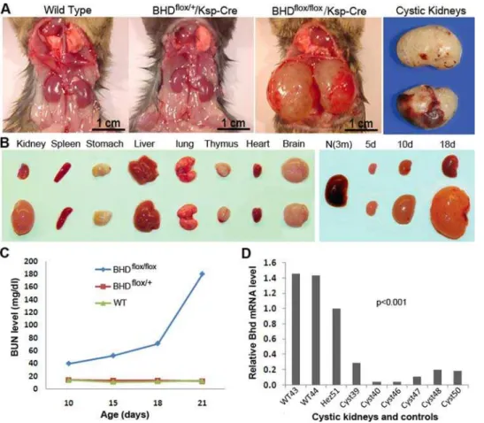
/Ksp-Cre and wild-type littermate controls (Figure 2A,B). TheBHDflox/flox/Ksp-Cremice died of kidney failure at the age of 3 weeks, having over 10 times higher levels of blood urea nitrogen (BUN) than normal littermate
controls (Figure 2C). The considerably low levels of BHD mRNA
detected by real time RT-PCR demonstrated inactivation ofBHD
in most of the kidney cells (Figure 2D). The appearance of the
cysts here is similar to that found in polycystic kidney disease
caused by mutatedPKDgenes (Figure 3A,B). Histopathological
examination of theBHDflox/flox
/Ksp-Crekidneys revealed extremely dilated renal tubules that predominantly originated from collecting ducts due to high expression of Ksp-Cre recombinase. While some proximal tubules were highly or moderately dilated, most of the other proximal tubules remained relatively normal as a result of
extremely low expression levels of Ksp-cre recombinase
(Figure 3C–F). Atrophic, compressed glomeruli were also observed, and degeneration, necrosis, and haemorrhage were frequently observed in the late stages. These morphological
changes suggest that homozygousBHDinactivation in the kidney
may cause loss of growth control in tubular epithelial cells.
Kidney-specific inactivation ofbhd produced renal cell carcinoma (RCC)
We further examined whetherBHDflox/flox/ Ksp-cre mice develop
renal carcinomas along with the cysts. We observed that kidneys from mice less than two weeks old predominately presented dilated tubules and cysts, whereas mice more than 18 days old also developed hyperplasia and renal cell carcinoma in their polycystic kidneys (Figure 3G–J). Hyperplastic areas frequently exhibited as multiple layers of epithelial cells along the inner surface of the tubules (Figure 3G, H). Renal cell carcinoma, which presents as cystic RCC, was frequently observed in the extremely enlarged kidneys. Cystic RCC was first described in 1986 and more cases have been reported since then [24,25,26,27,28,29,30,31,32]. Images of human cystic RCC are also available in the webpathology website (http:// www.webpathology.com/image.asp?case = 66&n = 8; http://www. webpathology.com/image.asp?case = 66&n = 9). The occurrence of cystic RCC in the general population is 4 to 10%, or 1 to 2% of all renal tumors. The cystic RCC does not present as a solid mass, but rather as a unilocular or multilocular cystic mass that is composed of cancer cells growing in the form of cysts that are distinct from regular cysts (Figures 3I,J and S1). While some of the tumor cells lined the septa, the others protruded into the cystic lumen. Most of the tumor cells were larger than the regular cystic cells. Binucleated cystic RCC cells were also observed. Many cystic spaces are filled with hemorrhage or proteinaceous fluid. No solid tumors were observed in any of the affected mice, which may be attributed to their short lifespan; three weeks might not be sufficient for solid tumor development.
Deficiency of FLCN and subsequent activation of mTOR contributed to renal cysts and RCCs
To elucidate the biochemical mechanisms of the cystogenesis and
carcinogenesis related to inactivation of the BHD gene, we
investigated the possible relevance ofBHDto the mTOR signaling
pathway for the following reasons: 1) our microarray analysis revealed
that ectopic expression of the BHDgene product, FLCN, led to
down-regulation of the AKT- related mTOR pathway signature (Figure S2); 2)BHD,PTEN,LKB1, andTSC1/2are all hamartoma syndrome–related genes, and the roles of PTEN, LKB1, and TSC1/
2 in the mTOR pathway have been well-established; and 3)in vitro
experiments indicated that FLCN interacted with AMPK, a member
of the mTOR pathway [18]. All these clues implied thatBHDgene
may play an important role in suppression of cystogenesis and tumorigenesis and that its inactivation could lead to the formation of renal cysts and RCC through the mTOR pathway.
mouse kidney and polycystic kidney. To do this, we designed and developed a human BHD monoclonal antibody that is compatible with immunohistochemical analysis in the mouse. While FLCN was predominantly expressed in the normal proximal tubules and collecting ducts in the cortex, obvious expression was rarely observed in the kidney distal tubules of mice at age of 3 weeks (Figure 3K–N). In the polycystic kidney, FLCN was only detected
in relatively normal tubules (Figure 3C–F), which are mainly
proximal tubules. A small number of proximal tubules were also enlarged due to moderate expression of Ksp-Cre recombinase (Figure 3F, arrow), which is different from the previous report
where the proximal tubules are not involved. All the enlarged
tubules were FLCN-negative (Figure 3C,D), indicating a
corre-lation of the formation of cysts with inactivation of theBHDgene.
We then explored whether the inactivation ofBHDresulted in
the activation of mTOR in affected cysts and RCCs. Immuno-histochemical analysis showed that mTOR was activated through
phosphorylation in cysts and cystic RCCs (Figure 4A–C), which
stained FLCN-negative (Figure 4B). We further examined the
phosphorylation status of the downstream target S6 (Figure 4D).
Phosphorylated S6 has been observed in some cysts and in cystic RCC. Although FLCN was reported to be a possible downstream
Figure 1. Targeting strategy and generation ofBHDconditional knockout mice. (A)Construction of theBHDgene targeting vector using a combination of the Gateway andloxpsystems. A 3.5-kb 59homology arm containing exon 2 and a 3.0-kb 39arm carrying exons 5 and 6 were integrated into the pDONR P4-P1R and pDONR P2R-P through a BP (attB and attP) reaction to generate theBHD-59andBHD-39homology entry clones, respectively. A 1.3-kb fragment of genomic DNA bearing exons 3 and 4 of theBHDgene was inserted to the modified pDONR vector pENTR3C-loxPMCS-loxP-FRT-neo-FRTbetween theSalI andNotI sites to generate aBHD-exon3-4-pENTR3Centry clone. The three entry clones, in combination with the modified destination vector, were incubated to create aBHD-pDESTR4R3 targeting construct through BP recombination reaction.(B)
Positive-targeting ES clones were selected by long-range PCR and confirmed by Southern blot analysis.(C)PCR genotyping of mouse offspring using tail DNA.(D, E)Knockout mice and normal controls were validated by Southern blot analysis.
effector of mTOR in anin vitroexperiment[18], our data revealed
that deficiency of FLCN activated mTOR pathway in vivo,
suggesting mTOR might a downstream target of FLCN. To further elucidate the correlation of FLCN and mTOR, we applied the mTOR inhibitor rapamycin to affected mice to see whether we could inhibit or reverse the development of cysts. Rapamycin treatment significantly extended the survival period ofBHDflox/flox/
Ksp-Cre mice and inhibited the development of cysts relative to control mice; some mice survived more than 50 days. However, once the rapamycin treatment was stopped, cysts redeveloped rapidly and the mice died within 10 days. This result indicated that rapamycin can inhibit cystic cell growth, but cannot reverse the cystic kidney phenotype. We also tested a few other members of the mTOR pathway (e.g. AKT) through IHC; no significant changes were observed or inconsistent results were obtained
following inactivation of BHD, implying a novel FLCN-mTOR
pathway branch may exists. In addition, FLCN might be related to
other signaling pathways. Obviously, the precisein vivo mode of
action of FLCN merits more investigation.
Discussion
In this study, we provide the first evidence that theBHDprotein FLCN predominantly expresses in the proximal tubules and
collecting ducts of the renal cortex (Figure 3K–N). By developing
and subsequently analyzing the conditional BHD knockout mouse
model, we demonstrate that the deletion ofBHD in the mouse
kidney leads to cystic renal cell carcinoma (cystic RCC) in addition to polycystic kidney and hyperplasia. The cystic RCC was only
observed in the older affected mice ($20 days old). This implies
that most of the polycystic kidneys would only present regular cysts and various extents of hyperplasia if the affected mice are sacrificed earlier. Thus, although some kidney-specific knockout
animal models of RCC-related genes failed to develop
RCC[21,33,34], our data provide a connection between
kidney-specific BHD gene inactivation and renal carcinogenesis. This
finding suggests that BHD may act as a suppressor for both cystogenesis and tumorigenesis.
No solid kidney tumors were observed in any of the affected mice, which may be attributed to their short lifespan and mouse distinct genetic background. It is entirely possible that if the cysts had not caused kidney failure at age of three weeks, progression of these cystic RCC to solid tumors would have occurred. In
addition, inactivation ofBHD gene in the kidney causes a large
proportion of tubules to form cysts. Once cystogenesis starts, fast-growing cysts become dominant and lead to highly cystic kidneys, kidney failure, and early death. Thus, lack of appropriate microenvironment might be another reason that the malignant/
Figure 2. Phenotypes ofBHDflox/flox/Ksp-Cremice. (A)BHDflox/flox/Ksp-Cremice developed polycystic kidney and died at age of three weeks.The kidneys from heterozygotes were phenotypically normal, similar to the ones from wild-type mice.(B)The organs from the affected mice were normal except for the kidneys (10 days old). There was not much size difference between the normal control kidneys and the affected kidneys at birth. However, the difference became apparent after the age of 5 days. At age of 10 days, the polycystic kidneys were approximate 15 times larger than those from normal controls. (C) Biochemical analysis revealed that the conditional knockout mice died of kidney failure due to high level of blood urea nitrogen (BUN). The BUN level dramatically elevated 15 days after birth. Most of the mice died at the age of 21 days. (D) The mRNA level of
BHD in the kidneys ofBHDflox/flox/Ksp-Cremice is significantly lower than that in wild-type kidneys. The heterozygote also shows lower mRNA expression relative to the wild type. m = month; d = day. f/f = flox/flox; f/+= flox/+.
pre-malignant cells failed to form solid renal tumors, which is a more complicated and slower process.
Our results further demonstrated that deficiency of BHD
product FLCN led to activation of mTOR pathway in cystic cells, supporting the recent report and consolidating that FLCN is involved in mTOR pathway and mTOR may be downstream target of FLCN[21]. Interestingly, BHD is a member of the hamartoma syndrome family that includes Cowden syndrome
(CD, affected genePTEN), Peutz-Jeghers syndrome (PJS, affected
geneLKB1), and tuberous sclerosis complex (affected genesTSC1/
TSC2) [14,15,16]. While PTEN, LKB1, and TSC1/2 have played
pivotal roles in the mTOR pathway, our findings suggest that
BHD protein FLCN, like other hamartoma syndrome–related
proteins such as PTEN, LKB1, and TSC1/2, is an important component of the mTOR pathway, constituting a novel FLCN-mTOR signaling branch that regulates cell growth/proliferation, though FLCN may involve in other pathways.
Materials and Methods
Design and generation ofBHDconditional knockout construct
The MultiSite GatewayHThree-Fragment Vector Construction
system (Invitrogen, Carlsbad, CA) was modified for the purpose of fabricating recombination vectors [35]. Of the four vectors supplied in the system, the pDONR vectors, pDONR P4-P1R, and pDONR
P2R-P3 were used to generate the 59and 39homology arm entry
clones. Another vector, pENTR3C, was used to carry a targeted gene sequence of interest. To meet the gene targeting purpose, a
1.8-kb loxP-FRT-neo-FRT fragment excised from
p-loxp-2FRTPGKneo (a gift of D. Gordon) was added to generate pENTR3CloxP-FRT-neo-FRT, which allowed later excision of
BHD exons 3 and 4 and the neomycin-resistance gene by
cre-mediated recombination in vivo. Synthetic oligonucleotides were
used to insert an additional loxP site into the DraI site of the
pENTR3C-loxPFRT- neo-FRT vector. Oligonucleotides loxPF (59-ATAACTTCGTATAGCATACATTATACGAAGTTATTT-39) and loxPR1
(59-AAATAACTTCGTATAATGTATGCTATAC-GAAGTTAT-39) (IDT, Coralville, IA) were phosphorylated with
T4 polynucleotide kinase (Invitrogen), annealed, and inserted into
theDraI-digested pENTR3C-loxP-FRTneo-FRT vector to
gener-ate pENTR3C-loxPMCS-loxP-FRT-neo-FRT.
To enrich targeted ES cell clones, we inserted a TK-negative selection cassette downstream of the attR3 site in the destination
vector. The attR4-ccdB-attR3 domain was amplified from the
pDEST/R4-R3 vector using the following primers: 59
-GCCT-CGAGCAGGAAACAGCTATGAC-39 and 59
-GCCTCGAG-TAAAACGACGGCCAGTG-39. After digestion by XhoI, this
domain was inserted into theXhoI site of the pPGKneo/TK vector
(a gift from T. Gridley).
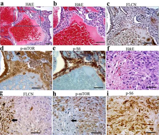
Figure 3. Inactivation of FLCN inBHDflox/flox/Ksp-Cremice led to
polycystic disease, hyperplasia, cystic renal cell RCC. (A,B) Deficiency of FLCN resulted in polycystic disease. (C, D) No FLCN expression was detected in cysts (enlarged tubules). However, FLCN was still expressed in relatively normal tubules where BHD was not deleted or completely eliminated by Ksp-Cre due to no or low Ksp-Cre
expression in some proximal tubules.(E, F)Most of the relative normal tubules were proximal tubules stained by proximal tubule-specific marker lotus tetragonolobus lectin (LTL). Many of the proximal tubules remained relatively normal, though some proximal tubules were also enlarged (indicated by arrow.(G, H)Hyperplasia (indicated by arrow) was frequently observed in the cysts.(I, J)Cystic renal cell carcinomas were also one of the important consequences of kidney-targetedBHD
gene inactivation inBHDflox/flox/Ksp-Cre
mice, which is morphologically distinct from regular cysts showed in A and B. (K, L) FLCN is predominately expressed in proximal tubules, which was demonstrated by the proximal-specific marker LTL(M). FLCN expression is quite weak in distal tubules, which was marked by the distal-tubule-specific marker, Na-Cl-cotransporter (TSC)(N). Scale bar = 50mm.
To generate a BHD gene targeting construct, a 3.5-kb 59
homology arm containing exon 2 and a 3.0-kb 39 arm carrying
exons 5 and 6, PCR-amplified usingPfxpolymerase (Invitrogen),
were integrated into the pDONR P4-P1R and pDONR P2R-P
through BP reaction (attB and attP sites) to generate the BHD-59
and BHD-39 homology entry clones, respectively. A 1.3-kb
fragment of genomic DNA bearing exons 3 and 4 of the BHD
gene was inserted into the modified pDONR vector
pENTR3C-loxPMCS-loxP-FRT-neo-FRT between theSalI andNotI sites to
generate a BHD-exon3-4-pENTR3C entry clone. Finally, the three entry clones, in combination with the modified destination vector, were incubated to create a BHD-pDESTR4R3 targeting construct through BP recombination reaction.
Identification of homologous recombinant ES cells and generation of kidney-specific knockout mice
The generated BHD-pDESTR4R3 targeting construct carries an ampicillin-resistant gene and a neomycin-resistant gene flanked
by FRT sites. The construct was linearized with ScaI for
electroporation into 129/sj strain ES cells. After selection with
500mg/ml G418 (Invitrogen), 1,039 ES cell clones were isolated.
The G418-positive ES clones were first screened by long-range PCR and then confirmed by Southern blot analysis. For the
generation of chimeras, ES cells heterozygous for the BHDflox/+
allele were injected into C57BL/6 blastocysts by standard procedures. Chimeras were bred to C57BL/6 mice, and germline offspring were identified by PCR genotyping. To remove the
neomycin gene flanked by two FTR sites, BHDflox/+
mice were crossed to FlpeR transgenic mice that express the site-specific
recombinase FLP (FLPe). Then,BHDflox/+
heterozygous mice were intercrossed to give rise to mice homozygous for theBHDfloxallele, i.e.,BHDflox/floxmice.
To obtain mice with kidney-specific inactivation of BHD,
BHDflox/flox mice were first bred to Ksp-Cre transgenic mice to
generateBHD heterozygous mice (BHDflox/+
/Ksp-Cre in kidneys).
BHDflox/+
/Ksp-Cremice then were backcrossed toBHDflox/floxmice to
generateBHDhomozygous mice (BHDflox/flox/Ksp-Cre
in kidneys). All mice were manipulated and housed according to protocols approved by the Institutional Animal Care and Use Committee (IACUC) of Van Andel Institute and conducted in an ethical, humane, and scientifically justified manner, and in full compliance with applicable regulations.
Genotyping, RNA and protein analysis
ES cell DNA and tail DNA was extracted by using automated DNA isolation system (Kurabo Industries) and subjected to regular PCR and long-range PCR genotyping analysis (see supplementary Table S1 for PCR primers). For genotyping by Southern blot analysis, DNA from ES cells or tissues was extracted using standard DNA extraction procedure. Purified DNA was digested byXmnI orHind III, isolated by 0.8% agarose gel, and transferred onto nylon membrane. UV-linked or dried membranes were
subjected to DNA hybridization with 59or 39probes.
Total RNA was isolated from various mouse tissues and cystic cell lines with Trizol reagent (Invitrogen) according to the manufacturer’s instructions. Purified RNA was used for quantita-tive analysis (real-time RT-PCR) through ABI Prism 7700 Sequences Detector (Applied Biosystems).
Figure 4. mTOR signaling pathway was activated in the cystic cells, cystic RCC cells.(A) Cystic RCC was stained by hematoxylin and eosin (H&E).(B)No FLCN expression was detected in cystic RCC, indicating deletion of theBHDgene. Phosphorylated mTOR (C) and phosphorylated S6 (D) staining was observed in the corresponding FLCN-deficient cells. Scale bar = 50mm.
For protein detection by Western blot, cultured cells and kidney whole-cell extracts prepared by homogenization were lysed in 1% Nonidet P-40, 50 mM Tris (pH 7.4), 150 mM NaCl, 1 mM EDTA, and 15% glycerol, plus standard protease inhibitors (protease inhibitor cocktail tablets, Roche Diagnostics). Equal amounts of mutant and control kidney cell protein extracts were size-separated by 10% SDS-PAGE and transferred to PVDF membranes (Invitrogen). FLCN was detected with a mouse monoclonal anti-FLCN antibody (developed by Laboratory of Antibody Technology) at a dilution of 1:750 using the enhanced chemiluminescence detection system.
Immunohistochemistry and tubular marker staining Immunohistochemical analysis was performed following the manufactory’s protocols. The antibodies used include anti-FLCN mAb, Phospho-mTOR Rabbit mAb (Cell Signaling), anti-Phospho-S6 Ribosomal Protein (Cell Signaling). Proximal tubules were stained by biotinylated Lotus Tetragonolobus Lectin (LTL, Vector Laboratories), and distal tubules were detected by using rabbit anti-thiazide-sensitive NaCl contransporter affinity purified polyclonal antibody (TSC, Chemicon) Tubular markers. Marker biotinylated Peanut Agglutinin (PNA, Vector Laboratories) was used to stain collecting ducts.
Phenotyping and histopathology
Newborn mice were monitored daily. Sick mice were distinguished from healthy ones by enlarged abdomen at age of
10 days. Totally 73 BHDflox/flox/Ksp-Cre mice and 55 normal
control littermates were collected for phenotyping and histopath-ological analysis. Mouse body weight, kidney weight were
measured upon euthanasia by CO2inhalation. Tissues including
kidneys, lung, liver, spleen, heart, stomach, intestine, brain, testes were collected and fixed in 4% paraformaldehyde for 24 hours and kept in 70% ethanol for 12 hours before paraffin block
preparation. Paraffin blocks were sectioned at 3mm thick and
stained with hematoxylin and eosin (H&E). Stained slides were evaluated by a board-certified veterinary pathologist B. Sigler and pathologists X. Yang and J. Peng.
Blood biochemical analysis
Mouse blood was collected by cardiac puncture at age of 5 days, 10 days, and 15 days, and 20 days. Serum was collected after centrifugation and stored at280uC for further biochemical analysis.
Rapamycin treatment ofBHDflox/flox/Ksp-Cremice and BHDflox/+/Ksp-Cre and wild-type control littermates
Totally 29 mice from three litters were used for rapamycin treatment (n = 15) and control (n = 14). Mice from each litter at
postnatal 7 days were randomly divided into two groups of rapamycin treatment and control. Rapamycin (LC Laboratories, Woburn, MA) was dissolved in ethanol at a concentration of
20 mg/mL and stored at 220uC. The rapamycin solution was
freshly prepared by diluting the rapamycin stock to 250mg/ml in
buffer (1:1 10% PEG-400, 8% ethanol:10% Tween 80) and were injected by intraperitoneally daily at a dose of 2.5 mg/kg body weight for the duration of the treatment. Control animals received i.p. injection of equal amount of vehicle (5% PEG-400, 4% ethanol, and 5% Tween 80). Mice were treated for at least two weeks starting at postnatal day 7. Moribund mice were subjected
to CO2euthanasia, dissection, and analysis in the duration of the
treatment.
Supporting Information
Table S1 Primer used for BHD knockout genotyping
Found at: doi:10.1371/journal.pone.0003581.s001 (0.02 MB XLS)
Figure S1 Additional cystic RCC samples stained by hematox-ylin and eosin. Cystic spaces are filled with proteinaceous fluid (A– J) or hemorrhage (K,L) in cystic RCC. The tumor cells have clear cytoplasm and hyperchromatic nuclei lining the septa or growing into the cystic lumina. Scale bar = 50mm.
Found at: doi:10.1371/journal.pone.0003581.s002 (5.38 MB TIF)
Figure S2 Microarray analysis revealed that ectopic expression of FLCN led to down-regulation of the AKT- related mTOR pathway signature.
Found at: doi:10.1371/journal.pone.0003581.s003 (0.29 MB TIF)
Acknowledgments
We thank the following Van Andel Research Institute core facilities for their services: gene targeting core for generating the knockout; the vivarium for mouse husbandry; the sequencing core for nucleotide sequencing; the analytical, cellular, and molecular microscopy core for immunohistochemical analysis; and the flow cytometry core for blood analysis. In addition, we thank Dr. Bob Sigler for technical assistance in mouse pathology. We also thank David Nadziejka for technical editing and Sabrina Noyes for her administrative support.
Author Contributions
Conceived and designed the experiments: JC BOW BTT. Performed the experiments: JC KF DP PW JK YL DH. Analyzed the data: JC JP CNQ KD RZ XJY KAF BTT. Contributed reagents/materials/analysis tools: SKK PZ BC. Wrote the paper: JC SKK BTT.
References
1. Birt AR, Hogg GR, Dube WJ (1977) Hereditary multiple fibrofolliculomas with trichodiscomas and acrochordons. Arch Dermatol 113: 1674–1677. 2. Welsch MJ, Krunic A, Medenica MM (2005) Birt-Hogg-Dube Syndrome.
Int J Dermatol 44: 668–673.
3. Lindor NM, Hand J, Burch PA, Gibson LE (2001) Birt-Hogg-Dube syndrome: an autosomal dominant disorder with predisposition to cancers of the kidney, fibrofolliculomas, and focal cutaneous mucinosis. Int J Dermatol 40: 653– 656.
4. Khoo SK, Bradley M, Wong FK, Hedblad MA, Nordenskjold M, et al. (2001) Birt-Hogg-Dube syndrome: mapping of a novel hereditary neoplasia gene to chromosome 17p12–q11.2. Oncogene 20: 5239–5242.
5. Nickerson ML, Warren MB, Toro JR, Matrosova V, Glenn G, et al. (2002) Mutations in a novel gene lead to kidney tumors, lung wall defects, and benign tumors of the hair follicle in patients with the Birt-Hogg-Dube syndrome. Cancer Cell 2: 157–164.
6. Schmidt LS, Warren MB, Nickerson ML, Weirich G, Matrosova V, et al. (2001) Birt-Hogg-Dube syndrome, a genodermatosis associated with spontaneous
pneumothorax and kidney neoplasia, maps to chromosome 17p11.2. Am J Hum Genet 69: 876–882.
7. Schmidt LS, Nickerson ML, Warren MB, Glenn GM, Toro JR, et al. (2005) Germline BHD-mutation spectrum and phenotype analysis of a large cohort of families with Birt-Hogg-Dube syndrome. Am J Hum Genet 76: 1023–1033. 8. Vocke CD, Yang Y, Pavlovich CP, Schmidt LS, Nickerson ML, et al. (2005)
High frequency of somatic frameshift BHD gene mutations in Birt-Hogg-Dube-associated renal tumors. J Natl Cancer Inst 97: 931–935.
9. Warren MB, Torres-Cabala CA, Turner ML, Merino MJ, Matrosova VY, et al. (2004) Expression of Birt-Hogg-Dube gene mRNA in normal and neoplastic human tissues. Mod Pathol 17: 998–1011.
10. Khoo SK, Kahnoski K, Sugimura J, Petillo D, Chen J, et al. (2003) Inactivation of BHD in sporadic renal tumors. Cancer Res 63: 4583–4587.
12. Okimoto K, Kouchi M, Matsumoto I, Sakurai J, Kobayashi T, et al. (2004) Natural history of the Nihon rat model of BHD. Curr Mol Med 4: 887–893. 13. Okimoto K, Sakurai J, Kobayashi T, Mitani H, Hirayama Y, et al. (2004) A
germ-line insertion in the Birt-Hogg-Dube (BHD) gene gives rise to the Nihon rat model of inherited renal cancer. Proc Natl Acad Sci U S A 101: 2023–2027. 14. Liaw D, Marsh DJ, Li J, Dahia PL, Wang SI, et al. (1997) Germline mutations of the PTEN gene in Cowden disease, an inherited breast and thyroid cancer syndrome. Nat Genet 16: 64–67.
15. Marsh DJ, Kum JB, Lunetta KL, Bennett MJ, Gorlin RJ, et al. (1999) PTEN mutation spectrum and genotype-phenotype correlations in Bannayan-Riley-Ruvalcaba syndrome suggest a single entity with Cowden syndrome. Hum Mol Genet 8: 1461–1472.
16. Toro JR, Shevchenko YO, Compton JG, Bale SJ (2002) Exclusion of PTEN, CTNNB1, and PTCH as candidate genes for Birt-Hogg-Dube syndrome. J Med Genet 39: E10.
17. Inoki K, Corradetti MN, Guan KL (2005) Dysregulation of the TSC-mTOR pathway in human disease. Nat Genet 37: 19–24.
18. Baba M, Hong SB, Sharma N, Warren MB, Nickerson ML, et al. (2006) Folliculin encoded by the BHD gene interacts with a binding protein, FNIP1, and AMPK, and is involved in AMPK and mTOR signaling. Proc Natl Acad Sci U S A 103: 15552–15557.
19. van Slegtenhorst M, Khabibullin D, Hartman TR, Nicolas E, Kruger WD, et al. (2007) The Birt-Hogg-Dube and tuberous sclerosis complex homologs have opposing roles in amino acid homeostasis in Schizosaccharomyces pombe. J Biol Chem 282: 24583–24590.
20. Singh SR, Zhen W, Zheng Z, Wang H, Oh SW, et al. (2006) The Drosophila homolog of the human tumor suppressor gene BHD interacts with the JAK-STAT and Dpp signaling pathways in regulating male germline stem cell maintenance. Oncogene 25: 5933–5941.
21. Baba M, Furihata M, Hong SB, Tessarollo L, Haines DC, et al. (2008) Kidney-targeted Birt-Hogg-Dube gene inactivation in a mouse model: Erk1/2 and Akt-mTOR activation, cell hyperproliferation, and polycystic kidneys. J Natl Cancer Inst 100: 140–154.
22. Pavlovich CP, Walther MM, Eyler RA, Hewitt SM, Zbar B, et al. (2002) Renal tumors in the Birt-Hogg-Dube syndrome. Am J Surg Pathol 26: 1542–1552.
23. Shao X, Somlo S, Igarashi P (2002) Epithelial-specific Cre/lox recombination in the developing kidney and genitourinary tract. J Am Soc Nephrol 13: 1837–1846.
24. Feldberg MA, van Waes PF (1982) Multilocular cystic renal cell carcinoma. AJR Am J Roentgenol 138: 953–955.
25. Deshpande RB, Pandit AA, Vora IM, Ravi R, Dalvi AN, et al. (1986) Multilocular, predominantly cystic, renal cell carcinoma: an unusual gross appearance (a case report). J Postgrad Med 32: 239–240.
26. Yousef GM, Ejeckam GC, Best LM, Diamandis EP (2005) Collecting duct carcinoma associated with oncocytoma. Int Braz J Urol 31: 465–467. discussion 467–469..
27. Hartman DS, Weatherby E 3rd, Laskin WB, Brody JM, Corse W, et al. (1992) Cystic renal cell carcinoma: CT findings simulating a benign hyperdense cyst. AJR Am J Roentgenol 159: 1235–1237.
28. Hartman DS, Davis CJ Jr., Johns T, Goldman SM (1986) Cystic renal cell carcinoma. Urology 28: 145–153.
29. Azoulay S, Vieillefond A, Paraf F, Pasquier D, Cussenot O, et al. (2007) Tubulocystic carcinoma of the kidney: a new entity among renal tumors. Virchows Arch 451: 905–909.
30. Han KR, Janzen NK, McWhorter VC, Kim HL, Pantuck AJ, et al. (2004) Cystic renal cell carcinoma: biology and clinical behavior. Urol Oncol 22: 410–414.
31. Truong LD, Choi YJ, Shen SS, Ayala G, Amato R, et al. (2003) Renal cystic neoplasms and renal neoplasms associated with cystic renal diseases: pathoge-netic and molecular links. Adv Anat Pathol 10: 135–159.
32. Nassir A, Jollimore J, Gupta R, Bell D, Norman R (2002) Multilocular cystic renal cell carcinoma: a series of 12 cases and review of the literature. Urology 60: 421–427.
33. Gnarra JR, Ward JM, Porter FD, Wagner JR, Devor DE, et al. (1997) Defective placental vasculogenesis causes embryonic lethality in VHL-deficient mice. Proc Natl Acad Sci U S A 94: 9102–9107.
34. Pollard PJ, Spencer-Dene B, Shukla D, Howarth K, Nye E, et al. (2007) Targeted inactivation of fh1 causes proliferative renal cyst development and activation of the hypoxia pathway. Cancer Cell 11: 311–319.