AUTOSOMAL DOMINANT POLYCYSTIC KIDNEY DISEASE:
REVIEW AND MANAGEMENT UPDATE
*Víctor Martínez
Nephrology Department, Hospital Reína Sofía, Murcia, Spain *Correspondence to victormj80@gmail.com
Disclosure: The author declares no potential conflict of interest.
Received: 21.02.14 Accepted: 08.05.14
Citation: EMJ Neph. 2014;1:61-66.
ABSTRACT
Autosomal dominant polycystic kidney disease (ADPKD) is the most common inherited nephropathy. Initially, it is characterised by the growth of renal cysts. Later, progressive deterioration of renal function determines the prognosis of ADPKD, depending on the main factors of progression (genetic, renal volume, and hypertension). Ultrasonography is the diagnostic technique of choice in the screening for relatives of ADPKD patients. Due to the absence of specific treatment it is necessary, in many cases, to start with renal replacement therapy. ADPKD can also be associated with extrarenal manifestations, mainly polycystic liver disease and cerebral aneurysms, which contribute to increased morbidity and mortality of these patients.
Keywords: Autosomal dominant polycystic kidney disease (ADPKD), diagnosis, cysts complications, renal disease progression, extrarenal manifestations.
INTRODUCTION
Autosomal dominant polycystic kidney disease (ADPKD) is the most frequent inherited renal disease with an estimated prevalence of approximately 1 in 800 live births. It is caused by mutations in two genes: PKD1 in 85% and PKD2 in 15% of cases.1 Ofspring
have a 50% risk of inheriting the mutated gene; however, there are sporadic mutations in 10-15% of patients. ADPKD is the cause of 6-10% of patients undergoing renal replacement therapy (RRT).2
The PKD1 and PKD2 genes encode for proteins polycystin 1 and polycystin 2, respectively, which are an associated complex in the cell membrane, acting as a receptor and non-selective cation channel.3 The polycystin complex is localised mainly
in the primary cilium of the tubular epithelium, but also in focal adhesions between cells and cell-matrix. Its functions are receptor-sensor of the urinary low, orientation of the cell, and regulation of the cell cycle.
In ADPKD, these alterations in cilia function cause a cascade of biochemical signals with the increase of intracellular cyclic adenosine monophosphate (cAMP)4 that stimulate cell proliferation and
transverse growth instead of longitudinal tubule. The cell loses its orientation,5 initiating the formation
of renal cysts. The Na-K-ATPase pumps translocate from the basolateral membrane to the luminal membrane, whereby promoting sodium and water secretion into the cyst, stimulating its growth.6
With reference to the two-hit hypothesis, the second hit could explain the variable phenotypic expression of the disease in families with the same mutation and all renal cysts are forming only from 1% of tubular cells. Although a mutated allele is inherited, cyst genesis begins when a somatic mutation in the normal allele (second hit) appears.7
The severity of ADPKD in the individual depends on the precocity of this mutation occurring, as has been observed in experiments with mice.8 Environmental
factors (chronic inlammation, cytokines, renal ischaemia) may accelerate this process.9
The progressive growth of renal cysts can cause tubular obstruction and renal ischaemia, which increase activity of the renin-angiotensin system (RAS) with arterial hypertension.10 Deterioration of
vascular sclerosis, and interstitial ibrosis. The kidneys increase their volume greatly.
DIAGNOSIS
Screening diagnosis should be performed in all irst-degree adult relatives (>18 years) of patients with ADPKD.11 Ultrasonography is the technique of
choice to diagnose and follow-up ADPKD patients, detecting cysts >1 cm and abdominal extrarenal features such as hepatic cysts. Ultrasonography is cheap and safe, and its sensitivity for PKD1 is signiicantly higher than for PKD2.12 Computed
tomography (CT) and magnetic resonance imaging (MRI) can detect cysts as small as 0.5 cm but are reserved for limited cases (suspicion of kidney stones or renal tumours) as they are more expensive and expose patients to radiation. MRI is the best imaging modality to monitor renal volume as used in research studies but not in clinical practice.13
Ultrasonographic diagnostic criteria14 are applied
to patients with unknown genotype and a family history of ADPKD (Table 1). In the case of a negative family history, the presence of renal cysts with increased kidney size, deterioration of renal function, and liver cysts may suggest the diagnosis of ADPKD;15 however, it must be diferentiated from
other cystic acquired disorders (multiple simple cysts, acquired renal cystic disease, medullary sponge kidney) or genetic disorders (autosomal recessive PKD, tuberous sclerosis, orofaciodigital syndrome Type 1, medullary cystic disease).16
Genetic testing should not be used as a screening tool when imaging diagnosis is clear. It is only indicated in speciic situations: diagnosis of exclusion in younger at-risk individuals with a family history of ADPKD (such as a potential living-related kidney donor), patients with cystic disease but without afected relatives, individuals with early onset of PKD in families with ADPKD, and couples who wish a prenatal or preimplantation genetic diagnosis.17
There are two methods to perform genetic testing: genetic linkage analysis (indirect study that requires several afected family members) or mutation analysis by Sanger sequencing (direct study, useful in sporadic mutations).18
Urinary Findings
The urinary concentration defect is one of the earliest indings and may be associated with symptoms of thirst and polyuria.19 It has been
demonstrated that urinary osmolality is less in
ADPKD after water restriction test.20 Proteinuria
is usually minimal and is associated with more advanced renal dysfunciton.21 Urolithiasis occurs
in 20-36% of cases, mainly because of the cystic collector compression system.22 In addition, the
stones are favoured by other alterations in the urine which may be present: low urine pH, hypocitraturia, hyperoxaluria, or hypercalciuria.
Cyst Compilations
Cystic infection causes sudden acute pain, localised in the lank, and fever with elevated markers of inlammation. Imaging tests should be performed for diagnosis since blood or urine cultures are often negative.23 The treatment of choice is soluble
antibiotics (quinolones) that penetrate the walls of the cyst for several weeks. Depending on the severity of infection, other treatment options to consider are percutaneous or surgical drainage, or even nephrectomy in recurrent infections.
In symptomatic cystic bleeding, the management is typically conservative with rest, hydration, and analgesia. In persistent bleeding, the following may be necessary: transfusion, desmopressin, percutaneous embolisation, or nephrectomy.24 When
haematuria persists for more than 1 week or when the irst episode occurs in over 50 years, screening for kidney cancer with MRI or CT with contrast is recommended. The prevalence of kidney cancer is not increased in patients with ADPKD.25
RENAL DISEASE PROGRESSION
Although during the irst decades of life the renal function remains normal, the growth and development of renal cysts continues. After starting the deterioration, the annual reduction of glomerular iltration rate (GFR) is 4.4-5.9 ml/ minute.26 Depending on the GFR, the patients
Years Number Of Cysts
15-39 ≥3 cysts (unilateral or bilateral)
40-59 ≥2 cysts in each kidney
>60 ≥4 cysts in each kidney
should be regularly monitored by the nephrologist, although there are no established protocols. ADPKD is considered by some authors the prototype of cardiorenal syndrome Type 4,27 associated with
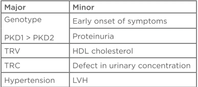
lower prevalence of cardiovascular disease. The main factors that determine progression of chronic kidney disease (CKD) are: genetic, renal volume, and hypertension (Table 2).
The mutation in PKD1 has a worse prognosis, beginning the RRT several years before PKD2 (53 years versus 69 years). A recent study in patients with PKD1 observed that individuals with truncating mutations progress faster to RRT than patients without this mutation (55 years versus 67 years).28
Total renal volume is the best predictor of progression of CKD.29 The increase of renal volume
rate is 1-10% per year. Data from CRISP (Consortium for Radiologic Imaging Studies of polycystic kidney disease) of kidney volume, measured by MRI, demonstrated that the growth in renal volume is associated with deterioration of renal function,30
reduction of blood low renal,31 and hypertension.32
The control of blood pressure slows the progression of CKD and usually precedes the decline of GFR.33 Initial treatment option is the use of RAS
inhibitors to achieve blood pressure of <130/80 mmHg;34 however, these have not demonstrated
superiority in the progression of CKD versus beta-blockers35 or calcium channel blockers,36 only
with diuretics.37 45% of ADPKD patients have a
non-dipping circadian rhythm on ambulatory blood pressure monitoring.38
Others relevant factors include: early age of onset of symptoms,39 low high-density lipoprotein
cholesterol levels, proteinuria, haematuria, left ventricular hypertrophy, and defect in urinary concentration (Table 2).40 Although traditionally
the male sex has been considered a risk factor for progression, in many articles there are no diferences.41 Normotensive pregnant women with
normal renal function usually have uncomplicated pregnancies without reduction of GFR; however, several pregnancies may accelerate progression to CKD.42
TREATMENT
Experimental studies with vasopressin V2 receptor antagonist (tolvaptan) in mice with PKD found lower renal cAMP levels and inhibition of cystogenesis.43
The TEMPO study included 1,445 ADPKD patients with GFR >60 ml/minute and renal volume >750 ml, who received treatment with tolvaptan or placebo (double-blind) for 3 years. In the results of the tolvaptan group, a slowing of the deterioration in renal function was observed and there was a smaller increase in renal volume, although adverse efects were higher (hypernatraemia, polyuria, or hyperuricaemia).44 Other treatments, such as
somatostatin analogues45 and the mammalian target
of rapamycin (mTOR) inhibitor46 have shown no
beneicial efect in these patients.
RRT is required in 50% of patients older than 60 years. In recent years, it has been observed that the age at initiation of RRT has increased, attributable to therapeutic advances. However, one study found no diference in the last 25 years in ADPKD patients who started RRT, despite a delay of 9 years in the age of onset of RRT in patients without ADPKD.47
Kidney transplantation is the best therapeutic choice for ADPKD patients in RRT.48 Nephrectomy is
indicated previously in presence of repeated cystic infections or bleeding and when an enormous renal size may complicate a correct placement of the graft. Peritoneal dialysis is a technique of irst choice49
which gives a better prognosis than haemodialysis for some authors;50 however, in patients with
massive polycystic liver disease, very large kidneys, abdominal hernias, or recurrent diverticulitis it is not recommended.
In most articles, the survival of the ADPKD patients in RRT was higher than non-ADPKD patients, may be in relation to: starting RRT at younger age, lower prevalence of cardiovascular risk factors,
Major Minor
Genotype
PKD1 > PKD2
Early onset of symptoms
Proteinuria
TRV HDL cholesterol
TRC Defect in urinary concentration
Hypertension LVH
Table 2: Factors that determine progression of CKD in ADPKD patients.
and higher haemoglobin levels.51 In another study,
ADPKD patients showed a higher survival rate than diabetes-free non-ADPKD patiens.52 The primary
cause of death in these patients was cardiovascular disease, followed by infections.53,54
EXTRARENAL MANIFESTATIONS
Polycystic liver disease is the most common extrarenal manifestation and is associated with increased renal volume, older age, and female sex55 (several pregnancies or oestrogen intake),56
as oestrogen can stimulate cyst hepatic growth. Liver cysts are usually asymptomatic and the liver function is normal, so follow-up of these patients is not necessary. The diagnosis is performed by ultrasonography; however, MRI is more sensitive for the detection of small cysts.57
The increased liver volume may cause symptoms of extrinsic compression: abdominal pain, early satiety, and obstruction of the hepatic veins or biliar duct.57 Partial hepatic resection or even liver
transplantation may be necessary in the case of massive polycystic liver.58 Treatment with
somatostatin analogues for 2 years has shown decreased liver volume but more studies are needed. The mTOR inhibitors have not obtained any results.59 Liver cyst infection causes fever, right
upper abdominal pain, and possible elevated CA19.9 and alkaline phosphatase levels. Treatment consists of antibiotics that penetrate the cyst wall, and the response to percutaneous drainage is better than in cysts kidney.60
The prevalence of cerebral aneurysms (CA) is 5-times higher in the general population and is increased with a positive family history of CA.61 A rupture of
a CA, resulting in an intracerebral or subarachnoid haemorrhage, is the most severe complication of ADPKD,62 and depends on size of the CA and other
factors such as age, uncontrolled hypertension, tobacco, cocaine, or anticoagulants.63 Patients with
CA are usually asymptomatic.
Magnetic resonance angiography (MRA) or CT angiography are the procedures of choice for diagnosis of CA.64 Screening is indicated in
ADPKD patients with previous family history of aneurysm or cerebral bleeding, previous rupture, or neurological symptoms.65 In high-risk
patients, when the diagnostic test detects no aneurysms, it should be repeated every 5 years. When an aneurysm is diagnosed, the therapeutic options are:66 conservative management with
radiological monitoring in CA <7 mm, and surgical intervention or endovascular repair67 in AC >7-11 mm
or rapidly growing.
With regards to cysts in other organs, heart or abdominal manifestations are less common and usually asymptomatic so screening is not recommended. The cysts in the epididymis and seminal vesicles are a rare cause of infertility,68
arachnoid cysts are usually asymptomatic, although they may increase the risk of subdural haematoma, and there does not appear to be an increased risk of ovarian cysts.69
Left ventricular hypertrophy is the most frequent cardiac manifestation, usually associated with hypertension, but it has also been observed in normotensive patients.70 The mitral valve prolapse
is the most common valve disease;71 others that
are less common are tricuspid valve prolapse and aortic regurgitation due to aortic root dilatation, which increases the risk of aortic dissection. Studies suggest an increased incidence of coronary and aortic abdominal aneurysms.72 The increased risk of
colonic diverticula and abdominal hernias in ADPKD patients may contraindicate the peritoneal dialysis.73
CONCLUSIONS AND
RECOMMENDATIONS
ADPKD is a hereditary systemic disorder characterised by the growth of renal cysts. The irst symptoms appear from adulthood, such as hypertension, haematuria, nephrolithiasis, or cyst complications (infection and bleeding) that usually are due to the number and size of renal cysts. Total renal volume is the best predictor of prognostics for ADPKD, and factors of progression of CKD should be controlled.
with MRA should be performed in high-risk ADPKD patients.
Despite the absence of a speciic treatment for ADPKD as yet, the initial results of treatment with tolvaptan showed a slowing reduction in
deterioration of renal function and a smaller increase in renal volume; however, comparisons with more long-term studies are necessary in the search for a deinitive treatment. Therefore, we should follow the recommendations in this review to slow the progression of CKD in ADPKD patients.
REFERENCES
1. Hateboer N et al. Comparison of phenotypes of polycystic kidney disease types 1 and 2. Lancet. 1999;353(9147): 103-7.
2. Bleyer AJ, Hart TC. Polycystic kidney disease. N Engl J Med. 2004;350(25):2622. 3. Sharif-Naeini R et al. Polycystin-1 and -2 dosage regulates pressure sensing. Cell. 2009;139(3):587-96.
4. Ortiz A. [Cilia and cystogenesis]. Nefrología. 2004;24(4):307-11.
5. Chapin HC, Caplan MJ. The cell biology of polycystic kidney disease. J Cell Biol. 2010;191(4):701-10.
6. Davidow CJ et al. The cystic ibrosis transmembrane conductance regulator mediates transepithelial luid secretion by human autosomal dominant polycystic kidney disease epithelium in vitro. Kidney Int. 1996;50(1):208-18.
7. Pei Y et al. Somatic PKD2 mutations in individual kidney and liver cysts support a “two-hit” model of cystogenesis in type 2 autosomal dominant polycystic kidney disease. J Am Soc Nephrol. 1999;10(7):1524-9.
8. Joly D et al. Ciliary function of polycystins: a new model for cystogenesis. Nephrol Dial Transplant. 2003;18(9): 1689-92.
9. Karihaloo A et al. Macrophages promote cyst growth in polycystic kidney disease. J Am Soc Nephrol. 2011;22(10):1809-14. 10. Jafar TH et al. The efect of a n g i o t e n s i n - c o n v e r t i n g - e n z y m e inhibitors on progression of advanced polycystic kidney disease. Kidney Int. 2005;67(1):265-71.
11. O’Neill WC et al. Sonographic assessment of the severity and progression of autosomal dominant polycystic kidney disease: the Consortium of Renal Imaging Studies in Polycystic Kidney Disease (CRISP). Am J Kidney Dis. 2005;46(6):1058-64.
12. Nicolau C et al. Autosomal dominant polycystic kidney disease types 1 and 2: assessment of US sensitivity for diagnosis. Radiology. 1999;213(1):273-6.
13. Kistler AD et al. Increases in kidney volume in autosomal dominant polycystic kidney disease can be detected within 6 months. Kidney Int. 2009;75(2):235-41.
14. Pei Y et al. Uniied criteria for ultrasonographic diagnosis of ADPKD. J Am Soc Nephrol. 2009;20(1):205-12. 15. Gabow PA. Autosomal dominant polycystic kidney disease. N Engl J Med. 1993;329(5):332-42.
16. Thauvin-Robinet C et al. Clinical, molecular, and genotype–phenotype correlation studies from 25 cases of oral-facial-digital syndrome type 1: a French and Belgian collaborative study. J Med Genet. 2006;43(1):54-61.
17. Pei Y. Diagnostic approach in autosomal dominant polycystic kidney disease. Clin J Am Soc Nephrol. 2006;1(5):1108-14. 18. Rossetti S et al. Identiication of gene mutations in autosomal dominant polycystic kidney disease through targeted resequencing. J Am Soc Nephrol. 2012;23(5):915-33.
19. Seeman T et al. Renal concentrating capacity is linked to blood pressure in children with autosomal dominant polycystic kidney disease. Physiol Res. 2004;53(6):629-34.
20. Zittema D et al. Vasopressin, copeptin, and renal concentrating capacity in patients with autosomal dominant polycystic kidney disease without renal impairment. Clin J Am Soc Nephrol. 2012;7(6):906-13.
21. Chapman AB et al. Overt proteinuria and microalbuminuria in autosomal dominant polycystic kidney disease. J Am Soc Nephrol. 1994;5(6):1349-54.
22. Torres VE, et al. Renal stone disease in autosomal dominant polycystic kidney disease: contribution of extrarenal complications to mortality. Am J Kidney Dis. 1993;22(4):513–9.
23. Jouret F et al. Diagnosis of cyst infection in patients with autosomal dominant polycystic kidney disease: attributes and limitations of the current modalities. Nephrol Dial Transplant. 2012;27(10):3746-51.
24. Gabow PA et al. Clinical proiles of gross hematuria in autosomal dominant polycystic kidney disease. Am J Kidney Dis. 1992;20(2):140-3.
25. Keith DS et al. Renal cell carcinoma in autosomal dominant polycystic kidney disease. J Am Soc Nephrol. 1994;4(9):1661-9.
26. Klahr S et al. Dietary protein restriction, blood pressure control and the progression of polycystic kidney disease. Modiication of Diet in Renal Disease Study Group. J Am Soc Nephrol. 1995;5(12):2037-47.
27. Virzi GM et al. ADPKD: prototype of cardiorenal syndrome type 4. Int J Nephrol. 2010;2011:490795
28. Cornec-Le E et al. Type of PKD1 mutation inluences renal outcome in ADPKD. J Am Soc Nephrol. 2013;24(6):1006-13.
29. King BF et al. Magnetic resonance measurements of renal blood low as a marker of disease severity in autosomal-dominant polycystic kidney disease. Kidney Int. 2003;64(6):2214-21.
30. Fick-Brosnahan GM et al. Relationship between renal volume growth and renal function in autosomal dominant polycystic kidney disease: a longitudinal study. Am J Kidney Dis. 2002;39(6): 1127-34.
31. Torres VE et al. Magnetic resonance measurements of renal blood low and disease progression in autosomal dominant polycystic kidney disease. Clin J Am Soc Nephrol. 2007;2(1):112-20. 32. Chapman AB et al. Renal structure in early autosomal-dominant polycystic kidney disease (ADPKD): the Consortium for Radiologic Imaging Studies of Polycystic Kidney Disease (CRISP) cohort. Kidney International. 2003;64(3):1035-45. 33. Ecder T, Schrier RW. Hypertension in autosomal-dominant polycystic kidney disease: early occurrence and unique aspects. J Am Soc Nephrol. 2001;12(1): 194-200.
34. Grantham JJ. Clinical practice. Autosomal dominant polycystic kidney disease. N Engl J Med. 2008;359(14): 1477-85.
35. Zeltner R et al. Renal and cardiac efects of antihypertensive treatment with ramipril vs metoprolol in autosomal dominant polycystic kidney disease. Nephrol Dial Transplant. 2008;23(2): 573-9.
polycystic kidney disease. Am J Kidney Dis. 2000;35(3):427-32.
37. Ecder T et al. Diuretics versus angiotensin-converting enzyme inhibitors in autosomal dominant plycystic kidney disease. Am J Nephrol. 2001;21(2):98-103. 38. Handa SP. Cardiovascular manifestations of autosomal dominant polycystic kidney disease in young adults. Clin Invest Med. 2006;29(6):339-46. 39. Johnson AM, Gabow PA. Identiication of patients with autosomal dominant polycystic kidney disease at highest risk for end-stage renal disease. J Am Soc Nephrol. 1997;8(10):1560-7.
40. Torres VE et al. Potentially modiiable factors afecting the progression of autosomal dominant polycystic kidney disease. Clin J Am Soc Nephrol. 2011;6(3):640-7.
41. Dicks E et al. Incident renal events and risk factors in autosomal dominant polycystic kidney disease: a population and family-based cohort followed for 22 years. 2006;1(4):710-7.
42. Chapman AB et al. Pregnancy outcome and its relationship to progression of renal failure in autosomal dominant polycystic kidney disease. J Am Soc Nephrol. 1994;5(5):1178-85.
43. Devuyst O, Torres VE. Osmoregulation, vasopressin, and cAMP signaling in autosomal dominant polycystic kidney disease. Curr Opin Nephrol Hypertens. 2013;22(4):459-70.
44. Torres VE et al. TEMPO 3:4 trial investigators. Tolvaptan in patients with autosomal dominant polycystic kidney disease. N Engl J Med. 2012; 367(25): 2407-18.
45. Caroli A et al. Efect of long acting somatostatin analogue on kidney and cyst growth in autosomal dominant polycystic kidney disease (ALADIN): a randomised, placebo-controlled, multicenter trial. Lancet. 2013;382(9903):1485-95.
46. Serra AL et al. Sirolimus and kidney growth in autosomal dominant polycystic kidney disease. N Engl J Med. 2010;363(9):820-9.
47. Martinez V et al. Renal replacement therapy in ADPKD patients: a 25-year survey based on the Catalan registry. BMC Nephrol. 2013;14:186.
48. Alam A, Perrone RD. Management of ESRD in patients with autosomal dominant polycystic kidney disease. Adv Chronic Kidney Dis. 2010;17(2):164-72.
49. Li L et al. Peritoneal dialysis as the irst-line renal replacement therapy in patients with autosomal dominant polycystic kidney disease. Am J Kidney Dis. 2011;57(6):903-7.
50. Abbott KC, Agodoa LY. Polycystic kidney disease at end-stage renal disease in the United States: patient characteristics and survival. Clin Nephrol. 2002;57(3):208-14.
51. Abbott KC, Agodoa LY. Polycystic kidney disease in patients on the renal transplant waiting list: trends in hematocrit and survival. BMC Nephrol. 2002;3:7.
52. Perrone RD et al. Survival after end-stage renal disease in autosomal dominant polycystic kidney disease: contribution of extrarenal complications to mortality. Am J Kidney Dis. 2001;38(4):777-84.
53. Orskov B et al. Changes in causes of death and risk of cancer in Danish patients with autosomal dominant polycystic kidney disease and end-stage renal disease. Nephrol Dial Transplant. 2012;27(4):1607-13.
54. Rahman E et al. Analysis of causes of mortality in patients with autosomal dominant polycystic kidney disease: a single center study. Saudi J Kidney Dis Transpl. 2009;20(5):806-10.
55. Bae KT et al. Magnetic resonance imaging evaluation of hepatic cysts in early autosomal dominant polycystic kidney disease: the Consortium for Radiologic Imaging Studies of Polycystic Kidney Disease cohort. Clin J Am Soc Nephrol. 2006;1(1):64-9.
56. Chandok N. Polycystic liver disease: a clinical review. Ann Hepatol. 2012;11(6):819-26.
57. Gevers TJ, Drenth JP. Diagnosis and management of polycystic liver disease. Nat Rev Gastroenterol Hepatol. 2013;10(2):101-8.
58. Chauveau D et al. Liver involvement in autosomal-dominant polycystic kidney disease: therapeutic dilemma. J Am Soc Nephrol. 2000;11(9):1767-75.
59. Page L et al. Somatostatin analog therapy for severe polycystic liver disease: results after 2 years. Nephrol Dial Transplant. 2012;27(9):3532-9.
60. Pirson Y. Extrarenal manifestations of autosomal dominant polycystic kidney disease. Adv Chronic Kidney Dis. 2010;17(2):173-80.
61. Vlak MH et al. Prevalence of unruptured
intracraneal aneurysms, with emphasis on sex, age, comorbidity country a time period: a systematic review and meta-analysis. Lancet Neurol. 2011;10(7): 626-36.
62. Rinkel GJ. Natural history, epidemiology and screening of unruptured intracranial aneurysms. J Neuroradiol. 2008;35(2):99–103.
63. Ring T, Spiegelhalter D. Risk of intracranial aneurysm bleeding in autosomal-dominant polycystic kidney disease. Kidney Int. 2007;72(11):1400-2. 64. Irazabal MV et al. Extended follow-up of unrfollow-uptured intracranial aneurysms detected by presymptomatic screening in patients with autosomal dominant polycystic kidney disease. Clin J Am Soc Nephrol. 2011;6(6):1274-85.
65. Pirson Y et al. Management of cerebral aneurysms in autosomal dominant polycystic kidney disease. J Am Soc Nephrol. 2002;13(1):269-76.
66. Williams LN, Brown RD. Management of unruptured intracranial aneurysms. Neurol Clin Pract. 2013;3(2):99-108.
67. Wiebers DO et al. Unruptured intracranial aneurysm: natural history, clinical outcome, and risks of surgical and endovascular treatment. Lancet. 2003;62(9378):103-10.
68. Belet U et al. Prevalence of epididymal, seminal vesicle, prostate, and testicular cysts in autosomal dominant polycystic kidney disease. Urology. 2002;60(1): 138-41.
69. Heinonen PK et al. Ovarian manifestations in women with autosomal dominant polycystic kidney disease. Am J Kidney Dis. 2002;40(3):504-7.
70. Perrone RD et al. Cardiac magnetic resonance assessment of left ventricular mass in autosomal dominant polycystic kidney disease. Clin J Am Soc Nephrol. 2011;6(10):2508-15.
71. Lumiaho A et al. Mitral valve prolapse and mitral regurgitation are common in patients with polycystic kidney disease type 1. Am J Kidney Dis. 2001;38(6): 1208-16.
72. Torra R et al. Abdominal aortic aneurysms in autosomal dominant polycystic kidney disease. J Am Soc Neph.1996;7(11):2483-6.