www.jcol.org.br
Journal of
Coloproctology
* Corresponding author.
E-mail: [email protected] (A. H. A. Freitas)
2237-9363/$ - see front matter. © 2013 Elsevier Editora Ltda. All rights reserved.
Original article
Ex vivo
sentinel lymph node investigation in colorectal cancer
Antônio Hilário Alves Freitas
a,b,*, Alberto Julius Alves Wainstein
b, Tarcizo Afonso Nunes
ca Brazilian Society of Coloproctology, Rio de Janeiro, RJ, Brazil
b Trymed Clinical Research, Belo Horizonte, MG, Brazil
c Universidade Federal de Minas Gerais (UFMG), Belo Horizonte, MG, Brazil
a r t i c l e i n f o
Article history:
Received 1 November 2012 Accepted 15 January 2013
Keywords:
Sentinel lymph node Ex vivo
Colorectal cancer Metastases
a b s t r a c t
Introduction: In Brazil, about 26,000 cases of colorectal cancer are diagnosed per year. Pa-tients considered at the early stage of disease (without lymph node) evolve with tumor relapse or recurrence in up to a quarter of cases, probably due to understaging. Objective: Research on ex vivo sentinel lymph node in patients with colorectal adenocarcinoma. Materials and methods: We studied 37 patients who underwent curative surgical resection. The marker used to identify lymph nodes was patent blue dye injected into the peritu-moral submucosa of the open surgical specimen immediately after its removal from the abdominal cavity.
Results: Ex vivo identii cation of sentinel lymph node with marker occurred in 13 (35.1%) patients. The sensitivity was 40% and 60% false negative. The detailed histological examina-tion of sentinel lymph nodes with multilevel secexamina-tion and immunohistochemistry showed metastasis in one (4.3%) individual, considered ultra-staging.
Conclusion: The ex vivo identii cation of sentinel lymph node had questionable benei ts, and worse results when include patients with rectal cancer. Restaging of one patient was possible after multilevel section and immunohistochemistry of the sentinel lymph node, but more research is needed to evaluate the role of micrometastases in patients with colorectal cancer.
© 2013 Elsevier Editora Ltda. All rights reserved.
Palavras-chave: Linfonodo-sentinela Ex vivo
Câncer colorretal Metástases
r e s u m o
Pesquisa do linfonodo-sentinela ex vivo no câncer colorretal
Introdução: No Brasil, a cada ano são diagnosticados cerca de 26.000 casos de câncer colorre-tal. Pacientes com estadiamento considerado inicial, sem linfonodo metastático, evoluem com recorrência ou recidiva do tumor em até um quarto dos casos, por provável subesta-diamento. Objetivo: pesquisar sobre linfonodo-sentinela ex vivo em pacientes com adeno-carcinoma colorretal.
Pacientes e métodos: A identii cação ex vivo de linfonodo-sentinela com o marcador ocorreu em 13 (35,1%) pacientes. A sensibilidade do método foi de 40% e o falso negativo de 60%. O exame histológico pormenorizado dos linfonodos-sentinela com multissecção e imu-noistoquímica diagnosticou metástase em um (4,3%) indivíduo, sendo considerado ultra--estadiamento.
Resultados: A identii cação de linfonodo-sentinela ex vivo apresenta benefícios questioná-veis, e piores resultados quando são incluídos pacientes com câncer de reto. Foi possível reestadiamento de um paciente depois da realização de multissecção e imunoistoquímica de linfonodos-sentinela, mas mais trabalhos são necessários para estabelecer a importân-cia das micrometástases em pacientes com câncer colorretal.
© 2013 Elsevier Editora Ltda. Todos os direitos reservados.
Introduction
Colorectal cancer incidence worldwide is 1.2 million cases per year, according to the World Health Organization.1
Sentinel lymph node is considered the i rst receiving lym-phatic drainage from the tumor and thus more likely to con-tain metastases.2
The treatment of colorectal cancer is surgical, and study of lymph nodes in critically ill individuals has shown that ad-juvant chemotherapy increases the disease-free survival of patients with lymph node metastasis.3,4
However, the literature provides a challenge, because patients with colorectal cancer treated at the early stage, without lymph node histology routine; therefore, with no indication for adjuvant therapy, progress with local tumor recurrence or distant metastases in up to 25% of cases.5–7
This fact makes us think about a possible understaging and imposes the need to examine lymph nodes more closely by identifying the sentinel lymph nodes and, adding to the rou-tine histology, a detailed study with multilevel sectioning and immunohistochemistry.
The identii cation of sentinel lymph node can be done in-traoperatively (in vivo) or in surgical specimen (ex vivo) using dyes and/or radiopharmaceuticals with tropism for lymph nodes. This study, which is unprecedented in Brazil, has been researching on ex vivo sentinel lymph node using dye.
Patients and methods
This is a prospective, descriptive and analytical study of senti-nel lymph node in patients undergoing colorectal cancer with proposed curative surgery.
It involved 37 patients with colorectal cancer, operated from November 2008 to April 2012 in two institutions: Hospital Alberto Cavalcanti, which belongs to the state public health of the Fundação Hospitalar do Estado de Minas Gerais (FHEMIG); Hospital da Polícia Militar do Estado de Minas Gerais, which serves the military police and their dependents, both located in the city of Belo Horizonte, MG, Brazil.
The study was approved by the Ethics Committee of the Hospitals and the Universidade Federal de Minas Gerais.
Inclusion criteria were patients with colon or rectum can-cer; over 21 years of age, adherence to informed consent.
Ex-clusion criteria were evidence of distant metastasis, urgent surgery, or patient’s refusal to participate.
Surgical procedure and identifi cation of sentinel lymph node
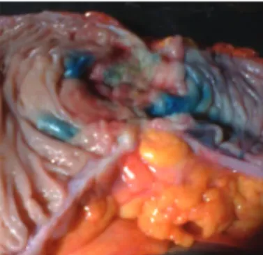
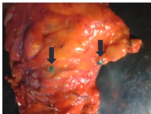
The procedure was general anesthesia, antisepsis, laparot-omy, and oncologic resection of the bowel segment and its mesentery containing the tumor. The surgical specimen was removed from the abdominal cavity, The searching process for sentinel lymph nodes was as follows: a) placing the speci-men removed in a surgical table; b) immediate opening of the intestinal lumen to locate the tumor; c) injection of 1 mL of patent blue dye 1% (manufactured by Citopharma Ltda. – Belo Horizonte, MG, Brazil), in the peritumoral submucosa, the vol-ume of dye was divided equally and applied to the four cardi-nal points around the lesion, using 1 mL syringe and needle 13 x 4.5 mm (Fig. 1); d) peritumoral massage for 5–10 minutes; e) identii cation by direct visualization of the i rst blue lymph nodes (Fig. 2), which are considered sentinel lymph nodes, and then marked with surgical thread.
All marked sentinel lymph nodes were removed from the mesentery and sent separately in numbered vials, along with the surgical specimen, to the pathology service.
Histological examination of all lymph node started with routine histology. Lymph nodes were embedded in parafi n blocks cut with a microtome, placed on slides, stained with hematoxylin-eosin and examined by light microscopy. In the absence of metastases by this method, only the sentinel lymph nodes were sent for multilevel section and immuno-histochemistry examination.
Fig. 2 – Intestinal mesentery with visual identifi cation of sentinel lymph nodes using patent blue dye.
The technique called multilevel section consists of mul-tiple cuts of lymph nodes included in parafi n block, stag-gered at intervals of 2-3 mm, which are then stained with hematoxylin-eosin and examined in more detail by light microscopy.
For immunohistochemistry, we used AE1/AE3 cytokera-tins (Biogenex®). The process phases were as follows:
cut-ting the lymph node to each scaled range of 50 microns; immunoperoxidase, streptavidin, biotin Supersensitive after antigen-induced heat with EDTA buffer, followed by staining control.
For staging, the pathological tumor-node-metastasis (TNM) criteria were followed.8
Tumors were located at the right colon, twelve (32%); transverse colon, one (3%); sigmoid colon, two (6%); and rec-tum, twenty-two (59%). The size ranged from 1–11 cm, with a mean of 3.5 cm (SD = 1.9).
Results
Identifi cation of lymph node using dye
In the 37 study patients, 415 lymph nodes were isolated, mean 11 (SD = 5.7) per patient. The patent blue sentinel lymph node was identii ed in 13 (35.1%) individuals. We identii ed 29 sentinel lymph nodes ex vivo using dye, with a mean of 2.2 (SD = 1.7) per patient (Table 1).
Dye and histology
Among the 29 sentinel lymph nodes identii ed using dye, histological examination of routine hematoxylin-eosin diag-nosed metastases in 2 (6.8%) and the other 27 (93.2%) had no signs of metastases. Of the 386 non-sentinel nodes, 31 (8%) had metastases, 355 (92%) were free of metastasis (Table 2).
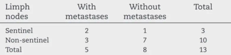
Considering the validity study, in 13 patients with sentinel lymph nodes identii ed using dye, routine histological exami-nation identii ed metastases in i ve (38.4%). Of this group of i ve patients, one (20%) had metastases exclusively located in sentinel lymph nodes, one (20%) had metastases in both sen-tinel and non-sensen-tinel lymph nodes, and three (60%) had me-tastases in non-sentinel lymph nodes, the latter i gure repre-sents the false negative. In the other eight patients, despite the identii cation of sentinel lymph nodes, no metastases
were diagnosed by routine histology with hematoxylin-eosin (Table 3).
Staging of patients
The routine histological examination did not diagnose me-tastasis in 23 (62.1%) of 37 patients, so they were considered N0 stage. In the other fourteen (37.9%), metastases were diag-nosed and classii ed in stage N +. Thus, staging (TNM) initial sample was: N0 = 23 patients (62.2%) and N + = 14 patients (37.8%). In the group of 23 patients without metastases by routine histology (stages I or II), additional tests was done in the sentinel lymph node, with multilevel section and im-munohistochemical, and metastasis was diagnosed in only one (4.3%), representing the ultra-staging. Therefore, the i nal staging (TNM) was stage N0 = 22 patients (59.5%) and N + = 15 patients (40.5%).
Discussion
Technique for ex vivo identifi cation of sentinel lymph node
The correct staging of the intestinal tumor is the major factor in patient survival, because when histology identii es lymph node metastasis, patients are referred for chemotherapy with known decreased recurrence and improved survival.3,4
Techniques to identify sentinel lymph node in intestinal tumors has been described for over half a century.9 But, the
i rst studies using the ex vivo technique in colorectal cancer were published only a little over ten years.10 In Brazil, there is
still no publication, a fact that motivated our study.
Authors who defend the ex vivo method describe its main advantages as simple execution; low cost; does not increase the operative time; obeys the principle of the cancer ‘no touch tumor’; and without risk of adverse effects such as anaphylaxis.11–13
In this study, the success rate in identifying sentinel lymph nodes in colorectal cancer was 35.1%. The literature credits worse results by: l aws in the injection; advanced stage of the lesion; mucinous histological type; location in the rectum, and prior radiotherapy.14–16 In this study, two
thirds of the patients were operated on for rectal tumors, and also received chemotherapy + radiotherapy preopera-tive.
Given the unfavorable results of the study, the investiga-tion of sentinel lymph node in ex vivo using patent blue dye is presented as an option to be held in rectal tumors, even with questionable benei ts, because it is technically unwork-able in rectal tumors, or in a complementary manner in co-lon tumors, when there has been failure with another tech-nique for identifying sentinel intraoperatively (in vivo).
Bookmarks
Vital staining has been used by most authors, because the radiopharmaceuticals add high costs (600 dollars each exam) and logistics complex.
Number of nodes
The number of lymph nodes examined is of great importance for staging, and directly inl uences treatment and prognosis of patients operated on for colorectal cancer. Current literature states that to obtain reliable pathology, it should be examined at least 10–12 lymph nodes. Publications found i ve-year sur-vival of approximately 73% when less than 10 lymph nodes were examined, 80% when 11–20 lymph nodes, and 87% with more than 20 lymph nodes examined.18 The American Joint
Committee on Cancer (AJCC) recommends that at least 7–14 lymph nodes should be examined.8
In 37 cases studied, we found a mean of 11.2 nodes per patient, in accordance with the recommended stage it is suf-i csuf-ient to relsuf-iably.19,20
It is worth noting that in surgical specimens who received ra-diotherapy, the work is much harder for researcher and patholo-gist in the search for lymph nodes,21 a fact clearly seen in this
study, as in one of the patients no lymph node could be studied since there were no remaining lymphoid in surgical specimen.
Sensitivity and false negative
In the specii c case of sentinel lymph node study for colorectal tumors, the adverse i ndings with high rates of false negative did not interfere with the results because the therapeutic radi-cal lymphadenectomy is always maintained, regardless of the presence or absence of metastases in regional lymph nodes.
In this study, the rate of 60% false negative results was found. According to the review work, the average overall rate of false negative rate is 33%, ranging from zero to 63%.22–24
According to the study of validity of this study, the sen-sitivity was 40%, while in the literature it ranges from 40 to 100%.25–27
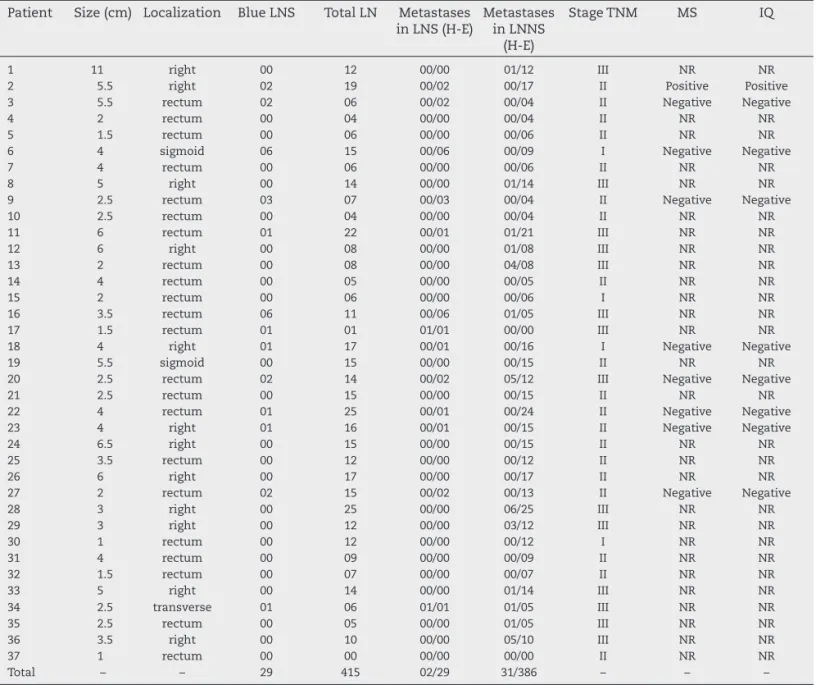
Table 1 – Overall outcome of the study.
Patient Size (cm) Localization Blue LNS Total LN Metastases in LNS (H-E)
Metastases in LNNS
(H-E)
Stage TNM MS IQ
1 11 right 00 12 00/00 01/12 III NR NR
2 5.5 right 02 19 00/02 00/17 II Positive Positive
3 5.5 rectum 02 06 00/02 00/04 II Negative Negative
4 2 rectum 00 04 00/00 00/04 II NR NR
5 1.5 rectum 00 06 00/00 00/06 II NR NR
6 4 sigmoid 06 15 00/06 00/09 I Negative Negative
7 4 rectum 00 06 00/00 00/06 II NR NR
8 5 right 00 14 00/00 01/14 III NR NR
9 2.5 rectum 03 07 00/03 00/04 II Negative Negative
10 2.5 rectum 00 04 00/00 00/04 II NR NR
11 6 rectum 01 22 00/01 01/21 III NR NR
12 6 right 00 08 00/00 01/08 III NR NR
13 2 rectum 00 08 00/00 04/08 III NR NR
14 4 rectum 00 05 00/00 00/05 II NR NR
15 2 rectum 00 06 00/00 00/06 I NR NR
16 3.5 rectum 06 11 00/06 01/05 III NR NR
17 1.5 rectum 01 01 01/01 00/00 III NR NR
18 4 right 01 17 00/01 00/16 I Negative Negative
19 5.5 sigmoid 00 15 00/00 00/15 II NR NR
20 2.5 rectum 02 14 00/02 05/12 III Negative Negative
21 2.5 rectum 00 15 00/00 00/15 II NR NR
22 4 rectum 01 25 00/01 00/24 II Negative Negative
23 4 right 01 16 00/01 00/15 II Negative Negative
24 6.5 right 00 15 00/00 00/15 II NR NR
25 3.5 rectum 00 12 00/00 00/12 II NR NR
26 6 right 00 17 00/00 00/17 II NR NR
27 2 rectum 02 15 00/02 00/13 II Negative Negative
28 3 right 00 25 00/00 06/25 III NR NR
29 3 right 00 12 00/00 03/12 III NR NR
30 1 rectum 00 12 00/00 00/12 I NR NR
31 4 rectum 00 09 00/00 00/09 II NR NR
32 1.5 rectum 00 07 00/00 00/07 II NR NR
33 5 right 00 14 00/00 01/14 III NR NR
34 2.5 transverse 01 06 01/01 01/05 III NR NR
35 2.5 rectum 00 05 00/00 01/05 III NR NR
36 3.5 right 00 10 00/00 05/10 III NR NR
37 1 rectum 00 00 00/00 00/00 II NR NR
Total – – 29 415 02/29 31/386 – – –
It is known that the more advanced the tumor, the higher will be the rate of false negative and less sensitivity. There-fore, another possible explanation for these results is that the vast majority of patients in this study was treated with prob-able late diagnoses, whereas 37.8% of patients were already in stage III (lymph nodes with metastases) after routine histol-ogy with hematoxylin-eosin.
Staging and micrometastases
Instead of a large number of lymph nodes examined un-der the microscope with few cuts, a detailed examination of the sentinel lymph nodes most likely to contain metas-tases could be performed. The commitment of the patholo-gist, who carried out multilevel section and/or immunohis-tochemical tests, increases the sensitivity of the method, promotes more adequate patient staging, and is more cost-effective.28,29
Less than 0.5% of the target tissue is removed for routine histology, hence the need for rethinking the care and patho-logical test techniques in colorectal malignancies.30,31
We believe that all professionals involved in the treat-ment of malignancies, especially pathologists, need to excel in their primary care, devoting more time to their analysis, so that patients are allocated in the correct staging. For it is known that more compliance with literature recommenda-tions enables to obtain excellent results without increasing costs, just increasing dedication.
In this study, one (4.3%) patient had micrometastases ex-clusively in sentinel lymph node, after performing addition-al histologicaddition-al examination, using multilevel section and im-munohistochemistry. According to authors, it may be a case where an individual would benei t from the investigation of
sentinel lymph node because the disease is diagnosed when the chances of providing a cure with adjuvant chemotherapy would be greater by attacking the tumor at its initial phase.32
In an attempt to avoid understaging, special histopatho-logical techniques with multilevel and immunohistochem-istry of sentinel lymph nodes may be used. Studies show ultra-staging with a wide range of 4%–50%.23,26,33
Therefore, we need more reliable work and with large samples to assess the actual role of micrometastases both in evolution and in survival, in addition to know if patients operated for colorectal cancer should or not receive adju-vant therapy in these specials situations.34,35
Conclusions
The ex vivo identii cation of sentinel lymph node using patent
blue dye in patients with colorectal cancer had questionable benei ts. Worse results are obtained when the work includes patients operated for rectal cancer, most notably in those who undergo radiotherapy.
Ultra-staging was possible by multilevel section and im-munohistochemistry of the sentinel lymph nodes. More re-search is needed to evaluate the role of micrometastases and the practical applicability of the method.
Conl ict of interest
The authors declare no conl ict of interest.
R E F E R E N C E S
1. World Health Organization (WHO), 2008. Available at: http:// www.who.int/cancer.
2. Morton DL, Wen DR, Wong JH, Economou JS, Cagle LA, Storm FK, et al. Technical details of intraoperative lymphatic mapping for early stage melanoma. Arch Surg 1992;127:392–9. 3. Gordon PH, Nivatvongs S. Principles and practice of surgery
for the colon, rectum, and anus, 2.ed. St. Louis, Missouri: Quality Medical Publishing, Inc., 1999.
4. Wolmark N, Rockette H, Mamounas EP. The relative efi cacy of 5-FU + leucovorin (LV-FU), 5-FU + levamisole (Lev-FU) and leucovorin, 5-FU + + levamisole (Lev-LV-FU) in Patients with Dukes B and C of the carcinoma colon: i rst report of NSABP C-04 [abstract 460]. Proc Am Soc Clin Oncol 1996;15:205. 5. Bilchick AJ, Nora DT. Lymphatic mapping of nodal
micrometastasis in colon cancer: putting the cart before the horse? Ann Surg Oncol 2002;9:529–31.
6. Bertoglio S, Sandrucci S, Percivale P, Goss M, Gipponi M, Moresco L, et al. Prognostic value of sentinel lymph node biopsy in the pathologic staging of colorectal cancer Patients. J Surg Oncol 2004;85:166–70.
7. Saha S, Seghal R, Patel M, Doan K, Dan A, Bilchick A, et al. A multicenter trial of sentinel lymph node mapping in colorectal cancer: Prognostic Implications for nodal staging and recurrence. Am J Surg 2006;191:305–10.
8. Greene FL, Balch CM, Fleming ID (eds.). AJCC Cancer Staging Handbook: TNM classii cation of Malignant Tumors. New York: Springer-Verlag, 2009. p. 129.
9. Braithwaite LR. Flow of lymph from the ileocecal angle. Br J Surg 1923;11:7.
Table 2 – Involvement of lymph nodes in 37 surgical specimens of colorectal cancer by routine histopathology with hematoxylin-eosin.
Lymph nodes
Metastases (+) Metastases (−) Total
Sentinel 2 (13.0%) 27 (87.0%) 29 (100.0%)
Non sentinel 31 (12.5%) 355 (87.5%) 386 (100.0%)
Total 33 382 415
(+), positive metastasis; (−), negative metastasis. P = 1.0 (Fisher test).
Table 3 – Histopathology of lymph nodes in patients with colorectal cancer in which sentinel lymph nodes were identifi ed.
Limph nodes
With metastases
Without metastases
Total
Sentinel 2 1 3
Non-sentinel 3 7 10
Total 5 8 13
CI, coni dence interval.
10. Wong JH, Steineman S, Calderia C, Bowles J, Namiki T.Ex vivo sentinel node mapping in carcinoma of the colon and rectum. Ann Surg 2001;233(4):515–21.
11. Bell SW, Mourra N, Fléjou JF, Parc R, Tiret E..Ex vivo sentinel lymph node mapping in colorectal cancer. Dis Colon Rectum 2005;48(1):74–9.
12. Demirbas S, Ince M, Baloglu H, Celenk T. Should sentinel lymph node mapping for colorectal cancer be performed? Turk J Gastroenterol 2004;5(1):39–44.
13. Smith J, Hwang H, Wiseman KW, Filipenko D, Phang PT. Ex vivo sentinel lymph node mapping in colon cancer: Improving the accuracy of pathologic staging? Am J Surg 2006;191:665–8.
14. Sommariva O, Donisi PM, Gnocato B, Vianello R, Pansa VS, Zaninotto G. Factors Affecting false-negative rates on ex vivo sentinel lymph node mapping in colorectal cancer. Eur J Surg Oncol 2010;36(2):130–4.
15. van der Zaag ES, Buskens CJ, Kooij N, Akol H, Peters HM, Bouma WH, et al. Improving staging accuracy in colon and rectal cancer by sentinel lymph node mapping: a comparative study. Eur J Surg Oncol 2009;35(10):1065–70. 16. Ceranic MS, Kecmanovic DM, Pavlov MJ, Nale DP, Micev
MT, Kovacevic PA, et al. Validation and feasibility of ex vivo sentinel lymph node “mapping” by methylene blue in colorectal cancer. Hepatogastroenterology 2010;57(102-103):1113–8.
17. Freitas AHA, Nunes TA, Wainstein AJA, Barroso AA, Ricardo Filho OP, Dias MA. Search sentinel lymph node in patients with colon cancer. Rev Bras Coloproct 2008;28(2):170–7. 18. Wong JH, Johnson DS, Namike P, Tauchi NP. Validation of
ex vivo lymphatic mapping in Hematoxylin-eosin node-negative carcinoma of the colon and rectum. Ann Surg Oncol 2004;11:772–7.
19. Benson AB, Schrag D, Someri eld MR, Cohen AM, Figueredo AT, Flynn PJ, et al. American Society of Clinical Oncology recommendations on adjuvant chemotherapy for stage II colon cancer. J Clin Oncol 2004;22:3408–19.
20. Rossi BM, Bepu P Jr, Ferreira FO, Santos EMM, Aguiar Jr S, Nakagawa WT, et al. Number of dissected lymph nodes in colorectal cancer Patients Submitted to radical surgery: the quality of oncology treatment. Applied Cancer Research 2006;26:27–33.
21. Quadros CA, Lopes A, Araujo I. Suggestion of optimal patient characteristics for sentinal lymph node mapping in colorectal adenocarcinoma. Arq Gastroentelol 2010;47(4):344–7.
22. Dess Guetz G, Uzzan B, Nicolas P, Cucherat M, Mestier P, Morere JF, et al. Is sentinel lymph node mapping in colorectal cancer a future Prognostic factor? A meta-analysis. World J Surg 2007;31:1304–12.
23. Tiffet O, Kaczmarek D, Chambonierre MR, Guillan T, Baccot S, Prevot N, et al. Dubois Combining radioisotopic and blue-dye technique does not Improve the false-negative rate in sentinel lymph node mapping for colorectal cancer. Dis Col Rectum 2007;50:962–70.
24. Joosten JJA, Strobbe JLA, Wauters CAP, Pruszczynski M, Wobbes T, Ruers TJM. Intraoperative lymphatic mapping and the sentinel node concept in colorectal carcinoma. Br J Surg 1999;86:482–6.
25. Doekhie FS, Peeters KCMJ, Kuppen PJK, Mesker WE, Tanke HJ, Morreau H, et al. The feasibility and reliability of sentinel node mapping in colorectal cancer. Eur J Oncol Soc 2005;31:854–62.
26. Tuech JJ, Pessaux P, Regenet N, Bergamaschi R, Colson A. Sentinel lymph node mapping in colon cancer. Surg Endosc 2004;18:1721–9.
27. Kitagawa Y, Watanabe M, Hasegawa H, Yamamoto S, Fujii H, Yamamoto K, et al. Sentinel node mapping for colorectal cancer with radioactive tracer. Dis Colon Rectum 2002;45:1476–80.
28. Wainstein AJA, Barroso AA, Belfort AF. Signii cance of sentinel lymph node in cancer of the digestive tract. Topics in gastroenterology 14. Publisher Medsi, Rio de Janeiro, RJ, 2004.
29. Steenbergen LN, van Lijnschoten G, Rutten HJ, Lemmens VE, Coebergh JW. Improving lymph node detection in colon câncer in community hospitals and their pathology department in southern Netherlands. Eur J Surg Oncol, 2010;36:135–40.
30. Paraf F, Sabourin JC. Optimal lymph node number and occult lymph node metastases in colorectal cancer: the pathologists view. Gastroenterol Clin Biol 2000;24:423–59. 31. Tschmelitsch J, Klimstra DS, Cohen AM. Lymph node
micrometastases not predict relapse in stage II colon cancer. Ann Surg Oncol, 2000;7:601–8.
32. Cohen AM. Tremiterra S, Candeh F. Adjuvant therapy for colorectal cancer. Curr Prob Cancer 1998;22:5–65. 33. Codignola C, Zorzi F, Zaniboni A, Mutti S, Rizzi A,
Padolecchia E, et al. Is there any role for sentinel node mapping in colorectal cancer staging? Personal experience and review of the literature. Jpn J Clin Oncol 2005;35:645–50. 34. Bembenek AE, Schineider U, Gretschel S, Fisher J. Detection
of lymph node micrometastases and isolated tumor cells in sentinel and nonsentinel lymph nodes of colon cancer patients. World J Surg 2005;29:1172–5.