CLINICAL |
CLÍNICO
RGO, Rev Gaúch Odontol, Porto Alegre, v.65, n.4, p. 376-379, out./dez., 2017 http://dx.doi.org/10.1590/1981-863720170002000143201
1
Universidade Estadual da Paraíba, Departamento de Odontologia, Programa de Pós-Graduação em Odontologia. Rua Baraúnas, 351, Bairro Universi-tário, 58429-500, Campina Grande, PB, Brasil. Correspondência para / Correspondence to: PM ALVES. E-mail: <[email protected]>. 2
Universidade Federal do Rio Grande do Norte, Programa de Pós-graduação em Patologia Oral. Natal, RN, Brasil. 3
Universidade Federal de Pernambuco, Departamento de Patologia. Recife, PE, Brasil.
Orthokeratinized odontogenic cyst associated with multinucleated giant
cell reaction: report of unusual indings
Cisto odontogênico ortoceratinizado associado à reação de células gigantes multinucleadas: relato de achados
incomuns
Lívia Natália Sales BRITO1
Francisco Jadson LIMA2
Pollianna Muniz ALVES1
Cassiano Francisco Weege NONAKA1
Gustavo Pina GODOY3
ABSTRACT
Orthokeratinized odontogenic cysts are developmental odontogenic cysts, presenting with low frequency, low rate of recurrence and their ethiopathogenesis is unknown. Radiographically, they show a radiolucent area in the mandibular posterior region. The aim of this report is to describe an unusual case of orthokeratinized odontogenic cysts, radiographically exhibiting radiopaque areas with an aspect of calciications in the lesion. Based on this, the clinical hypothesis of ameloblastic ibro-odontoma was suggested. After incisional biopsy and microscopic analysis, the conclusive diagnosis was orthokeratinized odontogenic cysts. The radiopaque foci were observed to be associated with a foreign body reaction. The patient was submitted to surgery under local anesthesia, with intraoral access for complete excision of the lesion and to re-establish esthetics. After follow-up of 24 months there were no signs of recurrence. Knowledge of this type of reaction is important because of the risk that the lesion may mimic a potentially more aggressive lesion, affecting the choice of treatment.
Indexing terms: Giant cells. Odontogenic cysts. Pathology.
RESUMO
Os cistos odontogênicos ortoceratinizados são cistos de desenvolvimento de origem odontogênica, de baixa frequência, etiopatogenia desconhecida e baixa taxa de recorrência. Radiograicamente, apresenta-se como uma área radiolúcida em região posterior de mandíbula. O objetivo deste artigo é descrever um caso não usual de cistos odontogênicos ortoceratinizados, que exibia, radiograicamente, focos radiopacos semelhantes à calciicações, no interior da lesão, sugerindo diagnóstico clínico de ibro-odontoma ameloblástico. Após a biópsia incisional e análise microscópica, o diagnóstico conclusivo foi de cistos odontogênicos ortoceratinizados. Observou-se que os focos radiopacos estavam associados, microscopicamente, a presença de uma reação de células gigantes multinucleadas por corpo estranho. O paciente foi tratado cirurgicamente, sob anestesia local, com acesso intrabucal para remoção total da lesão, com o restabelecimento da estética e sem sinais de recidiva após 24 meses. A importância do conhecimento deste tipo de reação dar-se pelo risco de mimetizar uma lesão potencialmente mais agressiva, direcionando a escolha do tratamento da lesão.
Termos de indexação: Células gigantes. Cistos odontogênicos. Patologia.
INTRODUCTION
The orthokerainized odontogenic cyst (OOC) was - for a long time - considered a less aggressive counterpart of the Keratocystic odontogenic tumor (KOT). However, as it presented less aggressive clinical, histopathological and behavioral characteristics1-2, according to the World Health
Organization (WHO) in 2005, it was reclassiied as a distinct clinical entity2-3.
The OOC is relatively rare, accounting for only 0.4% of cystic lesions of odontogenic origin4. Observed
predominantly in adult Caucasians of the male sex, its peak incidence is found between the third and fourth decades of
life2,4. This cyst generally presents as a single, asymptomatic,
radiolucent lesion, localized predominantly in the posterior region of the mandible2-3,5.
The histopathological characteristics it presents are an epithelium with an orthokeratinized lining, made up of a prominent granulous layer, and basal layer of lattened cuboidal cells that show no trend towards the palisaded nuclear arrangement1-2,6-7. Their treatment consists of
marsupialization or surgical enucleation2,7. These lesions
show a low level of aggressiveness and tendency towards recurrence at a rate lower than 4%2,8.
From this aspect, the purpose of the present article was to report a case of OOC with radiographic aspects not
RGO, Rev Gaúch Odontol, Porto Alegre, v.65, n.4, p. 376-379, out./dez., 2017 377
Orthokeratinized odontogenic cyst: unusual indings LNS BRITO et al.
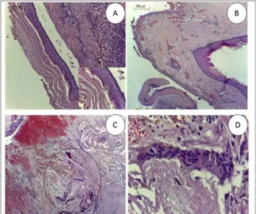
On microscopic examination, a fragment of cystic lesion was observed, lined with hyperorthokeratinized, stratiied, pavimentous epithelium, exhibiting a corrugated surface, an evident granulous layer and a lat epithelium-connective interface (igure 2A). The cystic capsule was usually reported in the literature, emphasizing the importance of distinguishing this lesion from other more aggressive types.
CASE REPORT
The patient, a 25-year-old man sought a private dental service with the intention of undergoing orthodontic correction. After accurate anamnesis and extraoral physical exam with the patterns of normality, the intraoral clinical exam was performed. During this exam, the presence of a tumefaction was found in the posterior region of the mandible on the left. This was asymptomatic and of a irm consistency on palpation. In the panoramic radiographic exam of the maxillae, a radiolucent, unilocular area was observed in the region of the mandibular body. This had an expansive aspect, covering the region from tooth 34 to tooth 38, with root resorption of the teeth involved. Inside the lesion the presence of radiopaque areas were evident and outstanding, compatible with calciied material deposition (Figure 1). Based on these indings, clinical diagnoses of cystic lesion of odontogenic origin, or odontogenic tumor of epithelial origin such as ameloblastic ibro-odontoma and calcifying epithelial odontogenic tumor were suggested. Therefore, incisional biopsy was performed, and the material removed was sent for histopathological analysis.
Figure 1. Panoramic radiographic image of the maxillae, showing an extensive, uniloc-ular radiolucent area, with areas of radiopaque foci (arrows), in the region of the mandibular body, covering the region from tooth 34 to tooth 38.
Figure 2. A) Note the cystic lesion fragment, lined with hyperorthokeratinized strati-ied pavimentous epithelium with a corrugated surface and lat epithelium-connective interface. (HE 100X). There was outstanding evidence of a thick layer of orthokeratin (HE 400x). B) Photomicrograph exhibiting the cystic capsule with slight chronic inlammatory iniltrate and moderate vascular-ization. (HE – 40X). C) Note the multinucleated giant cell reaction around the basophilic areas that were compatible with calciied material deposition (circle) (HE, 40X). D) At higher magniication, the multinucleated giant cells could be observed (HE, 400X).
shown to be composed of dense ibrous connective tissue, exhibiting slight chronic inlammatory iniltrate and moderate vascularization (igure 2B). An uncommon inding observed in the capsule as well, was the presence of basophilic areas compatible with calciied material deposition, surrounded by a multinucleated giant cell reaction (igures 2C 2D).
Therefore, in view of the histopathological characteristics observed, the lesion was diagnosed as an OOC. Complete surgical removal of the lesion was performed, and the patient was followed-up for 24 months, free of symptomatology and without signs of recurrence of the lesion. However, after this period, we lost touch with the patient because he moved to a different place.
DISCUSSION
The OOC is a relatively uncommon developmental cyst of an unknown etiology1-2. However, the
etiopathogenesis of OOCs is believed to be associated with a mutation of the Patched gene (PTCH)1,5. OOCs
comprise 10.5% of the cysts previously classiied as KOTs2.
378 RGO, Rev Gaúch Odontol, Porto Alegre, v.65, n.4, p. 376-379, out./dez., 2017
LNS BRITO et al. Orthokeratinized odontogenic cyst: unusual indings
entities, because KOTs exhibit a locally aggressive behavior and are predisposed to recurrence1-2,5,9, and have also been
found to be associated with the basal cell nevoid carcinoma syndrome, which is not observed in OOCs1-2,5,9.
With the purpose of comparing the proliferative activity of OOCs and dos KOTs, Dong et al.1 evaluated the immunohistochemical expression of Ki-67 and p63 in the epithelial lining of these lesions, and observed that in OOCs there was lower immunoexpression of both Ki67 and p63, thus inferring that OOCs were shown to be less aggressive than KOTs due to their lower level of proliferative activity.
These cysts generally present as single asymptomatic lesions10, in the posterior mandibular
region2,7-8. Nevertheless, some reports have shown painful
symptomatology associated with secondary infection7-8, and
extension between 1 and 3 centimeters. However, cases of large dimensions have been reported5,7, as in the case here
reported, the size of which at the largest diameters was 10.3 centimeters, covering the region from the premolars up to the body of the mandible.
Radiographically, OOCs present as well-delimited, and predominantly unilocular radiolucent lesions2,5,8 that
may be associated with the crown of an impacted tooth. It is frequently confused with the dentigerous cyst or with KOT2,8. The case reported here differed from those in the
reports found in the literature, relative to its extensive size and expansive radiolucent aspect, with the presence of radiopaque regions within it, and with the aspect of possible rupture of the cortical bone, which could make a differential diagnosis with the calcifying epithelial odontogenic tumor and ameloblastic ibroma-odontoma11. Furthermore, the
teeth involved showed areas of apical root resorption, a fact that justiied the hypotheses of ameloblastic ibroma-odontoma or calcifying epithelial odontogenic tumor. However, due to the radiographic similarities to these lesions, it is important to emphasize that the conclusive diagnosis must be based on careful clinical, radiographic exams and the histopathological indings4-5.
As histopathological characteristics, the OOC presents an epithelium with a uniform, orthokeratinized lining, made up of a prominent granulous layer and a basal layer of lattened cuboidal cells1-3,5-6 - aspects similar to those
that were found in the case here reported. However, inside the cystic capsule, the presence of multinucleated giant cells was observed; these are considered an uncommon histopathological inding in this type of lesion.
Multinucleated giant cells arise as a result of the fusion of macrophages, represent a terminal state of
differentiation, and play an important role in the chronic inlammatory response12. Foreign body multinucleated giant
cells arise from an inlammatory response resulting from the persistent presence of a non-phagocytable material13.
In spite of being rare, developmental cysts may present infections, as in the case reported by Carvalho et al.7, in which the secondary infection was associated with
an OOC, where rupture of the cortical bone served as the port of entry for the infection. In the case here reported, the radiographic images could suggest that rupture of the cortical bone could also have served as port of entry of the infection that generated an inlammatory response as a reaction to a foreign body. When reporting an uncommon case of root cyst with a mixed radiographic aspect, Ramos-Perez et al.3 considered one of the possibilities to be an
extravasation of illing material into the periapical region, thus causing an inlammatory reaction that triggered tissue necrosis and consequent formation of large dystrophic calciications. This would justify the mixed radiographic aspect of the case.
There is no completely established therapeutic strategy for treating the OOC5. Enucleation with or without
curettage and marsupialization followed by enucleation are the most recommended surgical interventions, however, a trend has been observed towards performing peripheral osteotomy in cases of multilocular and relatively extensive lesions. Due to the large extension of the lesion in the case here reported, the option was to perform surgical enucleation of the lesion, and placement of a surgical duct for local drainage, because the inlammatory content could maintain hydrostatic pressure on the injured tissue, and sometimes compromise the surgical repair. Therefore, the duct was maintained for seven days, and after its removal, the patient’s rapid recovery was observed.
CONCLUSION
RGO, Rev Gaúch Odontol, Porto Alegre, v.65, n.4, p. 376-379, out./dez., 2017 379
Orthokeratinized odontogenic cyst: unusual indings LNS BRITO et al
Collaborators
FJ LIMA was responsible for supplying the data
for the research. PM ALVES, CFW NONAKA, GP GODOY and FJ LIMA analysed the data and designed the research. LNS BRITO wrote the manuscript.
REFERENCES
1. Dong Q, Pan S, Sun LS, Li TJ. Orthokeratinized odontogenic cyst: a clinicopathologic study of 61 cases. Arch Pathol Lab Med. 2010;134(2):271-5. doi: 10.1043/1543-2165-134.2.271 2. MacDonald DS, Li TK. Orthokeratinized odontogenic cyst in a
Hong Kong community: the clinical and radiological features. Dentomaxillofac Radiol. 2010;39(4):240-5. doi: 10.1259/ dmfr/36547074
3. Ramos-Perez FM, Pontual AA, França TR, Pontual ML, Beltrão RV, Perez DE. Mixed periapical lesion: an atypical radicular cyst with extensive calciications. Braz Dent J. 2014 Oct; 25(5):447-50. doi: 10.1590/0103-6440201300235
4. Diniz MG, Galvão CF, Macedo PS, Gomes CC, Gomez RS. Evidence of loss of heterozygosity of the PTCH gene in orthokeratinized odontogenic cyst. J Oral Pathol Med. 2011;40(3):277-80. doi: 10.1111/j.1600-0714.2010.00977 5. Vignery A. Macrophage fusion: the making of osteoclasts and
giant cells. J Exp Med. 2005;202(3):337-40. doi: 10.1084/ jem.20051123
6. Boffano P, Gallésio C. Peculiar case of orthokeratinised odontogenic cyst: a peripheral counterpart of the intraosseous entity?. Br J Oral Maxillofac Surg. 2012;50(5):75-77. doi: 10.1016/j.bjoms.2011.09.019
7. Carvalho CH, Aquino AR, Nonaka CFW, Pereira SJS, Germano Rocha GA, Pereira PL. Infected orthokeratinized odontogenic cyst: a rare cause of facial cellulitis. Braz Dent J. 2012;23(5):612-6. doi: 10.1590/S0103-64402012000500025
8. McNally AK, Anderson JM. Foreign body-type multinucleated giant cells induced by interleukin-4 express select lymphocyte co-stimulatory molecules and are phenotypically distinct from
osteoclasts and dendritic cells. Exp Mol Pathol. 2011;91(3):673-81 doi: 10.1016/j.yexmp.2011.06.012
9. De Souza LB, Gordón-Nuñez MA, Nonaka CFW, De Medeiros MC, Torres TF, Emiliano GB. Odontogenic cysts: demographic proile in a Brazilian population over a 38-year period. Med Oral Patol Oral Cir Bucal. 2010;15(4):583-90. doi: 10.4317/ medoral.15.e583
10. Cheng YS, Liang H, Wright J, Teenier T. Multiple orthokeratinized odontogenic cysts: a case report. Head Neck Pathol. 2015;9(1):153-7. doi: 10.1007/s12105-014-0545-5
11. Silva Servato JP, Cardoso SV, Parreira da Silva MC, Cordeiro MS, Rogério de Faria P, Loyola AM. Orthokeratinized odontogenic cysts presenting as a periapical lesion: report of a case and literature review. J Endod. 2014;40(3):455-58. doi: 10.1016/j. joen.2013.09.044
12. Wright JM. The odontogenic keratocyst: orthokeratinized variant. Oral Surg Oral Med Oral Pathol. 1981;51(6):609-18. doi.10.1016/S0030-4220(81)80011-4
13. Selvamani M, Devi AY, Basandi PS, Madhushankari GS. Prevalence and clinicopathological comparison of kerotocystic odontogenic tumor and orthokeratinized odontogenic cyst in South Indian sample population: a retrospective study over 13 years. J Pharm Bioallied Sci. 2014;6(1):127-30. doi: 10.4103/0975-7406.137418