CLÍNICO |
CLINICAL
http://dx.doi.org/10.1590/1981-8637201700020001335421 Hospital Paulistano, Serviço de Cirurgia e Traumatologia Buco-Maxilo-Facial. São Paulo, SP, Brasil.
Mandible condyle osteochondroma: clinical case report
Osteocondroma do côndilo da mandíbula: relato de caso clínico
Denis Pimenta e SOUZA1
José Luiz Cintra JUNQUEIRA2
Ricardo RAITZ2
INTRODUCTION
Osteochondroma is one of the most common benign bone tumours. Although frequently found in the general skeleton, it is rarely seen to involve the facial bones1-4. It usually occurs early in life affecting young
adults between 10-30years of age but the craniofacial Osteochondroma are frequently seen in older women5.
The clinical presentation of Osteochondroma includes facial asymmetry, malocclusion, cross-bite on contra-lateral side and lateral open-bite on the affected side, deviation on opening, hypomobility, pain and clicking6. Pre-auricular swelling, TMJ pain, cliking
sound and recurrent joint dislocation are also common
manifestation of the tumour7. Differential diagnosis
of slow-growing tumours in the mandibular condyle Osteochondroma include giant cell tumor, condylar hyperplasia, ibro-osseous lesion, vascular malformation,
osteoma, chondroma, and osteochondroma8-9. Pre
operative computed tomography scans are (CT) scans is essential in the treatment planning of these tumours and depending on the symptoms and duration of the osteochondroma, the management ranges from excision of the tumor alone to condylectomy along with tumor excision1.
A recent paper10 reported that the treatment of
osteochondroma remains controversial because some authors have proposed conservative resection with
ABSTRACT
Osteochondroma is one of the most common benign bone tumours. However, it a rare in the mandibular condyle. Depending on the symptoms and duration of the osteochondroma, the management ranges from excision of the tumor alone to condylectomy along with tumor excision. This paper presents a case of Osteochondroma after surgical treatment affecting the mandible of a 39-year-old man and highlights the diagnosis, surgical procedures and 3-year follow-up period. At the clinical examination, the patient presented severe mandibular lateral deviation to the right, left side disocclusion with a slight oral opening limitation, dental abrasion compatible with bruxism and class III. After 3-year follow-up 3 years later revealed that the surgical management posed was successful for Osteochondroma and the patient is currently under regular review.
Indexing terms: Cone beam computed tomography. Diagnosis. Osteochondroma. Treatment.
RESUMO
Osteocondroma é um dos tumores ósseos mais comuns, mas raramente acomete o côndilo mandibular. Dependendo dos sintomas e duração do Osteocondroma o manejo da lesão pode variar desde a excisão do tumor até a realização da condilectomia com a remoção do tumor. O objetivo deste trabalho é relatar um caso clínico de Osteocondroma acometendo o côndilo da mandíbula em um paciente de 39 anos de idade, ressaltando as etapas para a realização do diagnóstico, procedimentos cirúrgicos e follow-up de 3 anos. Ao exame clínico observou-se severo desvio mandibular para o lado direito, desoclusão do lado esquerdo, abertura de boca reduzida, abrasão dentária compatível com bruxismo e classe III. Após a realização de tratamento cirúrgico e o período de acompanhamento de 3 anos foi possível observar a resolução do caso em imagens de tomograia computadorizada de feixe cônico. Conclusão: Pode-se concluir que a terapia proposta resultou em sucesso e o paciente continua em acompanhamento periódico.
preservation of most of the condyle by removing the tumour in multiple fragments. On the other hand, some authors proposed simultaneous condylar reconstruction and condylectomy. This paper presents a case of Osteochondroma after surgical treatment affecting the mandible of a 39-year-old man and highlights the diagnosis, surgical procedures and 3-year follow-up period.
CASE REPORT
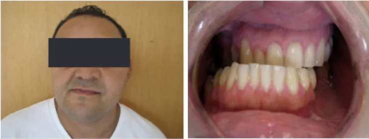
A dark-skin, 39 years old patient, from the city of Osasco, State of São Paulo, Brazil, attended the dental clinic complaining of left otalgia, left temporomandibular joint burst (TMJ), muscle fatigue during chewing, occasional tinnitus, limited mandibular opening, scapular and occipital pain and no previous temporal headache. The patient reported good general health and no chronic use of medications, either. At the clinical examination, the patient presented the following symptoms: severe mandibular lateral deviation to the right, left side disocclusion with a slight oral opening limitation, dental abrasion compatible with bruxism and class III (Figure 1A and B).
Figure 2. The tomographic examination revealed some mass-like dysformed condylar growth, extending medially from the left subcondylar region from where it approached the anatomical condyle right into the temporal bone man-dibular fossa.
Figure 1. A and B - At the clinical examination, the patient presented severe man-dibular lateral deviation to the right, left side disocclusion with a slight oral opening limitation, dental abrasion compatible with bruxism and class III.
At irst, condylar hyperplasia on the left side was hypothesized and orthodontic documentation was requested during which severe malocclusion was conirmed and growth in the subcondylar region on the left side was clear. The tomographic examination revealed some mass-like dysformed condylar growth, extending medially from the left subcondylar region from where it approached the anatomical condyle right into the temporal bone mandibular fossa (Figure 2). The patient was retrospectively evaluated concerning childhood trauma with mandibular impact, to which he initially denied but later recalled having had mild trauma when he was about 8 years old.
The bone scintigraphy examination showed active and proliferative condylar and subcondylar development and hyperstimulation was observed in the left condylar area, right knee and right shoulder.
The preoperative exames were normal and the surgery was perfomed in a surgical center, under anesthesia, naso-tracheal intubation, since the patient's limited dental occlusion was important in the intraoperative period. A pre-auricular incision was performed with a small extension to the retromandibular area. Surgical divulsion was perfomed following the facial nerve path in order to access the marginal mandibular branch, which was anatomically normal. A narrow base bone projection with progressive increase towards the mandibular fossa with appearance of irregular and hardened mass, similar to a bone, was observed through a subcondylar medial procedure. Due to the dificulties of access, a piezo tip cut of the normal subcondylar area was performed, the TMJ was devulsed and the normal condyle completely extirpated. The tumor resection-osteotomy was performed and immersed in isotonic solution, lactated ringer type.
The tumor was immediately subjected to a hospital pathological examination and no malignancy was found. Replacement of the healthy condyle was planned, with adequate marginal medial ostectomy, with a small margin followed by regularization and constant lactated ringer solution irrigation. The rigid internal ixation with titanium plates and screws, system 2.0 (Figures 3 A, B and C) was then performed.
Mandible condyle Osteochondroma
subjected to anatomopathological examination, aiming at inal diagnosis conirmation (Figure 4). During the 48-hour postoperative period the patient developed mild facial nerve neuro-apraxia, especially in the frontal and zygomatic branches, which were treated with low-power stimulating 40J / 808 laser therapy in 3 weekly sessions, for 6 weeks.
The pathological anatomy revealed osteochondroma, conirming the benign prognosis of the lesion and corroborating the conservative treatment given to the case.
The clinical changes have resulted in patient’s healthy control of function and aesthetic appearance, no facial-related disorders, restored functional occlusion and no pain complaints after a 3 year- follow-up (Figure 5 A, B and C).
Figure 3. Surgical procedures: Replacement of the healthy condyle was planned, with adequate marginal medial ostectomy, with a small margin followed by reg-ularization and constant lactated ringer solution irrigation. The rigid internal ixation with titanium plates and screws, system 2.0 was then performed.
Figure 4. The histological indings revealed cancellous bones covered in some parts by cartilaginous cap.
A progressively released diet and local massage stimulation physiotherapy in the left masticatory musculature, heat and ice thermotherapy as well as isometric exercises were oriented. A citoneurin-type medication was prescribed twice a day for 30 days, aiming at neurological improvement. There was gradual improvement of neuro-apraxia and muscle function, with
total regression of edema at 4 weeks. The dental
occlusion was reestablished, presenting no laterognatia or masticatory dificulties as it had been reported by the patient at the beginning of the treatment.
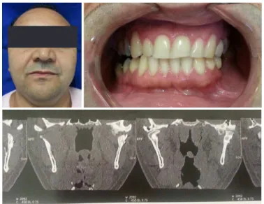
Figure 5. Extra (A), intraoral (B) and tomographic aspects (C) in a 3-year follow-up period. The clinical changes have resulted in patient’s healthy control of function and aesthetic appearance, no facial-related disorders and restored functional occlusion.
DISCUSSION
This report presents a case of osteochondroma in a 39-year-old male patient involving the left condyle which was accompanied by the main symptoms: otalgia, TMJ noise of the left side, mastication dificult and limited mouth opening. The patient presented the patient presented severe mandibular lateral deviation to the right, left side disocclusion with a slight oral opening limitation, dental abrasion compatible with bruxism and class III.
A previous study reported that ive patients with unilateral osteochondroma of the madibular condyle had the main complaint of pain in the temporomandibular joint and no patients were so disturbed by facial asymmetry. The sites of predilection were the coronoid process and the mandibular condyle3.
The etiology of osteochondroma is unclear12. For
osteochondroma.
According to Andrade et al.1 much confusion seems
to exist in the literature in differentiating osteochondroma from chondromas as well condylar hyperplasias. Clinico-pathological and radiological differentiation should be carried out between the two lesions. In addition, it is important to highlight that the treatment plan differs for both these conditions1. Radiographic appearance of osteochondroma of
the mandibular condyle can be pathognomonic, appearing as an irregularly shaped, mixed density and expansile lesion4.
Early diagnosis is essential in order to provide adequate treatment, which may affect the patient’s quality of life13.
In the present case lesion was surgically removed. According to the literature, the surgical treatment for osteochondroma of the mandibular condyle is condylectomia with or without simultaneous reconstruction of the condyle10.
Several reconstructive options are available in the literature. Zhou et al.10 followed up 10 patients with osteochondroma
who had the condyle resected or reconstructed. Three patients were treated with a free graft and seven with a pedicled graft. The authors concluded that both free grafts and pedicled graft seem to be alternative and promising approaches to condylar reconstruction in the treatment of osteochondroma. However, the pedicled graft results in less facial asymmetry,
less bone resorption, and better long-term clinical effects than the free graft. Condylectomy and condylar reconstruction, with simultaneous correction of the secondary dentofacial deformities adopting orthodontic procedures, might be a better alternative in order to manage osteochondroma accompanied by dentofacial deformities. Moreover, orthodontic treatment should be used for better improvement in facial esthetics and occlusion14.
It is important to highlight that surgical resection of osteochondroma can lead to complications such as: neuropraxia, fracture and arterial laceration9,11. Follow-up
was carried out in our case as the patient did not present any complication. Recurrence rate of the lesion after resection is considered extremely rare9 . However, due to these chances
of recurrence the patient is currently under regular review and there was no evidence of recurrence.
Collaborators
DP SOUZA assisted the patient in the case here reported. JLC JUNQUEIRA and R RAITZ made substantial contributions to all stages of this paper. All authors approved the inal version and its submission to this journal.
REFERENCES
1. Andrade NN, Gndhewar TM, Kapoor P, Thomas R. Osteochondroma of the mandibular condyle – Report of and atypical case and the importance of computed tomography. J Oral Biol Craniofac Res. 2014;4(3):208-13. doi: 10.1016/j.jobcr.2014.12.001
2. Dominguez MF, Castillo JL, Guerra MM, Sanchez RS, La Plata MM. Condylar osteochondroma treated with total condylectomy and preservation of the articular disc: a case report. Craniomaxillofac Trauma Reconstr. 2015;8(2):136-40. doi: 10.1055/s-0034-1393727
3. Jain M, Singhal S, Goyal M, Sharma B. Osteochondroma of maxilla posterior region: a unique case. J Clin Diagn Res. 2015;9(4):ZD14-5. doi: 10.7860/JCDR/2015/12478.5804
4. M H, Manjunatha BS, Kumar AN, Alavi YA. Osteochondroma (OC) of the condyle of left mandible: a rare case. J Clin Diagn Res. 2015;9(2):ZD15-6. doi: 10.7860/JCDR/2015/11451.5598 5. Utumi ER, Pedron IG, Perrela A, Zambon CE, Cecchetti MM,
Cavalcanti MG. Osteochondroma of the temporomandibular joint: a case report. Braz Dent J. 2010 ;21(3) :253-8.
6. Wolford LM, Mehra P, Franco P. Use of conservative condylectomy for treatment of osteochondroma of the mandibular condyle. J Oral Maxillofac Surg. 2002;60:262-e68. doi: 10.1053/ joms.2002.30570
7. Meng Q, Chen S, Long X, Cheng Y, Deng M, Cai H. The clinical and radiographic characteristics of condilar osteochondroma. Oral Surg Oral Med Oral Pathol Oral Radiol. 2012;114:e-66-74. doi: 10.1016/j.oooo.2012.01.016
8. Vezeau PJ, Fridrich KL, Vincent SD. Osteochondroma of the mandibular condyle: literature review and report of two atypical cases. J Oral Maxillofac Surg. 1995;53:640.
9. Kamble V, Rawat J, Kulkarni A, Pajnigara N, Dhok A. Osteochondroma of bilateral mandibular condyle with review of literature. J Clin Diagn Res. 2016;10(8):TD01-02. doi: 10.7860/ JCDR/2016/19877.8361
10. Zhou H, Liao C, Hu J, Fei W. Comparison of the clinical effects of treatment of osteochondroma by two types of vertical ramus osteotomy. Br J Oral Maxillofac Surg. 2017 Feb 26. pii: S0266-4356(16)30296-0. doi: 10.1016/j.bjoms.2016.09.023
11. Holmlund AB, Gynther GW, Reinholt FP. Surgical treatment of osteochondroma of the mandibular condyle in the adult. A 5-year follow-up. Int J Oral Maxillofac Surg. 2004;33(6):549-53. doi: 10.1016/j.ijom.2004.01.006
12. Yang XH, Zhang P, Xu JH, Hu YJ. An osteochondroma of the mandibular condyle. J Craniofac Surg. 2015 Mar;26(2):567-9. doi: 10.4103/0976-9668.116969
Mandible condyle Osteochondroma
deformity caused by bulky osteochondroma: report of an unusual case and the importance of cone beam computed tomography. Open Dent J. 2017 Apr 28;11:237-241. doi: 10.2174/1874210601711010237
14. Li H, Hu J, Luo E, Zhu S, Li J. Treatment of osteochondroma in the mandibular condyle and secondary dentofacial deformities using surgery combined with orthodontics in adults. J Oral
Maxillofac Surg. 2014 Nov;72(11):2295-317. doi: 10.1016/j. joms.2014.03.021