Artigo
Received 20 August 2007; Accepted 22 December 2007 18(2): 204-208, Abr./Jun. 2008
Puri
fi
cation of an antibacterial compound from
Lantana lilacina
Aline C. Pereira,
1Hudson W. P. Carvalho,
1Geraldo H. Silva,
2Denilson F. Oliveira,*
,1Henrique C. P. Figueiredo,
3Alberto J. Cavalheiro,
2Douglas A. Carvalho
41Departamento de Química, Universidade Federal de Lavras, Caixa Postal 3037,
37200-000 Lavras-MG, Brazil,
2Instituto de Química, Universidade Estadual Paulista, Caixa Postal 35, 14801-970
Araraquara-SP, Brazil,
3Departamento de Medicina Veterinária, Universidade Federal de Lavras, Caixa Postal 3037, 37200-00
Lavras-MG, Brazil,
4Departamento de Biologia, Universidade Federal de Lavras, Caixa Postal 3037, 37200-000 Lavras-MG, Brazil
RESUMO:“Purifi cação de um composto antibacteriano de Lantana lilacina”. Observou-se,
em estudo preliminar, que o extrato metanólico das folhas de L. lilacina, coletadas no município de Lavras (MG, Brasil), apresentava atividade antibacteriana. Em decorrência, buscou-se purifi car e identifi car a substância responsável por tal efeito, através de fracionamento do referido extrato direcionado por testes de difusão em agar com Aeromonas hydrophila, Bacillus subtilis,
Pseudomonas aeruginosa e Staphylococcus aureus. Após partições com solventes e vários processos cromatográfi cos, isolou-se o [β-3,4-diidroxifenil)etil]-(3’-O-α-L-ramnopiranosil)-(4’-O
-cafeoil)-β-D-glicopiranosídeo, que é conhecido como acteosídeo. A concentração inibitória mínima e a concentração bactericida mínima desta substância para A. hydrophila, B. subtilis, P. aeruginosa e S. aureus foram de 0,12, 1,00, 1,00 e 0,25 mg/mL, respectivamente.
Unitermos: Lantana lilacina, Verbenaceae, atividade antibacteriana, acteosídeo.
ABSTRACT: Since the methanol extract of Lantana lilacina leaves collected in the city of Lavras
(MG, Brazil) showed antibacterial properties in a preliminary study, a fractionation process guided by agar diffusion assays with Aeromonas hydrophila, Bacillus subtilis, Pseudomonas aeruginosa
and Staphylococcus aureus was carried out to purify and identify the active compounds. After solvent partition and several chromatographic steps, [β-3,4-dihydroxyphenyl)-ethyl]-(3’-O-α -L-rhamnopyranosyl)-(4’-O-cafeoyl)-β-D-glycopyranoside, known as acteoside, was isolated. The minimal inhibition concentration and the minimal bactericidal concentration of such substance against A. hydrophila, B. subtilis, P. aeruginosa and S. aureus were 0.12, 1.00, 1.00 and 0.25 mg/ mL, respectively.
Keywords:Lantana lilacina, Verbenaceae, antibacterial activity, acteoside.
INTRODUCTION
Although pharmaceutical industries have produced a large number of new antimicrobial agents, the emergence of resistant bacterial strains has become a public health problem all over the world. In the United States of America, for instance, each year, 70% of the two million cases of bacterial infections acquired in hospitals consist of strains resistant to at least one antibacterial compound (Alexandria, 2004). Moreover, adverse effects and the high cost of the antimicrobial substances make the search for new effective drugs extremely necessary.
As the biological activity of plants has been known since antiquity (Rios and Recio, 2005), a preliminary evaluation of local plant extracts was carried out to identify those with antimicrobial properties (Oliveira et al., 2007). During such study, a pronounced
in vitro antibacterial property was observed for the aerial parts of Lantana lilacina Desf. (Verbenaceae), a 50-120 cm height native Brazilian shrub, which produces pink or purple fl owers. Known as a weed and an ornamental plant (Lorenzi, 2000), it has been used in the traditional medicine to treat cold and bronchitis (Balbach, 1986). Although several substances have been identifi ed in other species of the Lantana genus (Begum et al., 2000; Siddiqui et al., 1995; Barbosa-Filho et al., 2006), only monoterpene glucoside esters were isolated from L. lilacina leaves (Dembitsky, 2004). No further studies concerning the chemical composition of this plant species have been found. Therefore, this research was aimed to purify and identify the antibacterial compounds present in the methanol extract of L. lilacina leaves.
Plant material
Leaves from Lantana lilacina Desf. (Verbenaceae) were collected in the city of Lavras, State of Minas Gerais (Brazil). Voucher specimens were identifi ed by Prof. Valéria E. G. Rodrigues and deposited in the Herbarium ESAL (ESAL 15.172), at Universidade Federal de Lavras, Lavras.
General experimental procedures
All reagents used were of recognized analytical grade. Acetic acid, acetonitrile, and methanol were HPLC-grade (Vetec, Brazil). During the purifi cation steps, solvent concentration was carried out in a rotatory evaporator at 35 ºC, followed by 24 h in a freeze-drier. Except when mentioned otherwise, all fractions were submitted to antibacterial diffusion assays to direct purification. Column chromatography was carried out on silica gel 60 (230-400 mesh, Merck). Mass spectra were obtained on an Agilent 1100 LC/MS Trap equipped with an electrospray interface. Samples (1.0 mg) were dissolved in MeOH (1.0 mL) and 20 L were directly injected into the interface at a fl ow rate of 5 L/ min. Deuterated dimethylsulphoxide (DMSO-d6) and deuterated methanol (CD3OD) were used as solvents for nuclear magnetic resonance (NMR) analyses performed on a Varian Unit 500 instrument using solvent peak as reference. 1H and 13C NMR spectra were obtained in both
solvents, while HMBC, HMQC and TOCSY analyses were carried out only in DMSO-d6.
Extraction and isolation
Fresh leaves of L. lilacina were exhaustively extracted with methanol at room temperature. Part of the crude extract (0.5 mg) was dissolved in 0.5 mL of an ethanol:H2O (7:3; v/v) solution and submitted to the antibacterial diffusion assay. Another part (0.5 mg) of such extract was dissolved in 0.5 mL of an aqueous 1% (g/mL) Tween 80 solution and used in the broth microdilution assay.
The crude extract (10.0 g) was subsequently washed with hexane (10 x 100 mL), ethyl acetate (AcOEt; 10 x 100 mL) and methanol (MeOH; 10 x 100 mL). Part (3.5 g) of the MeOH fraction (M1) was successively eluted with MeOH, H2O and HCl 0.1 M through a silica gel column (4 x 15 cm). Part (1.8 g) of the resulting MeOH fraction (M2) was eluted with MeOH through a C-18 column (1 x 5 cm), yielding fraction M3 (1.0 g). Then, M3 was fractionated on a C-18 column (Luna C-18, 200 x 21.2 mm, 10 M, Phenomenex, USA), using aqueous 0.1% acetic acid solution:MeOH (5% MeOH during 5 min, 5-100% MeOH during 60 min, 100% MeOH for 21 min), at a fl ow rate of 30 mL/min, as eluent. An UV detector set at 254 nm was employed to monitor the fractionation. One of the resulting fractions
(F11; 169 mg; eluted between 18-19 min) was purifi ed on the same column employing aqueous 0.1% acetic acid solution:MeOH (62:38) as eluent, at a fl ow rate of 20 mL/ min. In this case the UV detector was set at 320 nm. This procedure yielded fraction 6 as a pure compound (F6; 15 mg; eluted between 10.2-12.5 min), which was identifi ed as acteoside (Figure 1) by NMR and mass spectrometry analyses.
Antibacterial assays
Antibacterial activity was evaluated with four standard bacterial strains acquired from the American Type of Culture Collection (ATCC, USA): Bacillus subtilis ATCC 6633 and Staphylococcus aureus ATCC 25923 (Gram-positive), Aeromonas hydrophila ATCC 7966 and Pseudomonas aeruginosa ATCC 27853 (Gram-negative). Agar diffusion assays (NCCLS, 2003a) were carried out in duplicates. Briefl y, after bacterial growth in triptic soy agar (TSA, Acumedia, USA) during 24 h at 37 ºC, the resulting cultures were used to prepare cell suspensions in an aqueous 0.85% (g/mL) NaCl solution, at 0.5 turbidity according to MacFarland scale. Such suspensions were inoculated with a swab on the surface of Mueller-Hinton agar (Merck, Germany) Petri dishes and 40 L of each sample were deposited into 6 mm diameter holes made on the inoculated medium. After 24 h at 37 ºC, samples causing 7 mm or larger inhibition zone diameters around the holes were considered active.
Minimal inhibitory and minimal bactericidal concentrations (MIC and MBC) were determined by a broth microdilution assay (NCCLS, 2003b). A twofold serial dilution of the reference (chloramphenicol succinate: Sigma, USA; 400 g/mL) and samples were prepared using Mueller-Hinton broth (MHB: Biolife, Italy) supplemented with calcium and magnesium cations (Alderman and Smith, 2001). The crude extract was dissolved in an aqueous 1% (g/mL) Tween 80 solution at a concentration of 10 mg/mL and fi ltered through a 0.22 μm membrane (GV Durapore PVDF, Milipore, USA). 7.5 x 10⁴ CFU were poured into each well and the
initial extract concentration was 5.0 mg/mL. Aqueous 1% Tween 80 and chloramphenicol solutions were employed as negative and positive control, respectively. After 24 h at 37 oC, 10 L were withdrawn from each well with
no bacterial growth and subcultured in TSA during 24 h at the same temperature. MIC was considered the lowest concentration of the extract that prevented visible growth in the well during 24 h and MBC was defi ned as the lowest concentration yielding negative subcultures during 24 h. The isolated compound (acteoside, Figure 1) was also submitted to a broth microdilution assay. It (2 mg) was dissolved in 100 L of DMSO (P.A.) and diluted with MHB. The highest acteoside’s concentration in the well was 1.0 mg/mL.
1 2 3 4
5
6
7 8
9 O
HO
HO
O O
O
OH OH
O
OH
OH O
OH HO HO
H3C
1'
2' 3' 4'
5' 6' 7' 8'
6''' 5'''
4''' 3''' 2''' 1'''
1''
2'' 3'' 4'' 5''
6''
Figure 1. Structure of the acteoside.
Table 1. Inhibition zone diameter (IZD), minimal inhibitory concentration (MIC) and minimal bactericidal concentration (MBC)
for the crude methanol extract of Lantana lilacina aerial part: (-) no inhibition zone; (x) not performed.
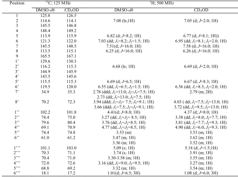
Table 2.13C- and 1H- NMR data for the isolated compound (acteoside) in DMSO-d6 and CD
3OD: δ ppm (mult; J = Hz; H).
Crude extract A. hydrophila B. subtilis P. aeruginosa S. aureus
IZD (mm) 12.5 10.0 - 8.0
M IC (mg/mL) 1.25 1.25 x 0.62
M BC (mg/mL) 1.25 1.25 x 2.50
Position 13C; 125 M Hz 1H; 500 MHz
DM SO-d6 CD3OD DM SO-d6 CD3OD
1 125.8 126.5
2 114.6 114.1 7.08 (ls;1H) 7.05 (d; J=2.0; 1H)
3 145.5 146.8
4 148.4 149.2
5 113.9 115.9 6.82 (d; J=8.2; 1H) 6.77 (d; J=8.1; 1H))
6 121.3 122.0 7.03 (dd; J1=8.2; J2=1.5; 1H) 6.95 (dd; J1=8.1; J2=2.0; 1H)
7 145.5 148.5 7.51(d; J=16.0; 1H) 7.58 (d; J=16.0; 1H)
8 113.5 115.1 6.25 (d; J=16.0; 1H) 6.26 (d; J=16.0; 1H)
9 165.5 167.1
1’ 129.6 130.3
2’ 116.2 115.3 6.68 (ls; 1H) 6.69 (d; J=2.0; 1H)
3’ 144.9 145.9
4’ 143.5 145.6
5’ 115.5 115.3 6.69 (d; J=6.5; 1H) 6.67 (d; J=8.3; 1H)
6’ 119.5 120.0 6.55 (dd; J1=6.5; J2=1.5; 1H) 6.56 (dd; J1=8.3; J2=2.0; 1H) 7’ 34.9 35.3 2.78 (ddd; J1=13.0; J2=J3=7.5; 1H)
2.73 (dd; J1=13.0; J2=7.5; 1H)
2.79 (m; 2H)
8’ 70.2 72.3 3.94 (ddd; J1=J2= 7.5; J3=9.1; 1H) 3.66 (ddd; J1=7.5; J2=J3=9.1; 1H)
4.03 ( dd; J1=7.5; J2=13.0; 1H) 3.72 (dd; J1=9.5; J2=13.0; 1H)
1’’ 102.2 101.8 4.41(d; J=8.0; 1H) 4.37 (d; J=8.0; 1H)
2’’ 74.4 75.0 3.27 (dd; J1=J2= 8.5; 1H) 3.38 (dd; J1=8.0; J2=7.7; 1H) 3’’ 79.6 80.4 3.76 (dd; J1=J2=8.5; 1H) 3.81 (dd; J1=7.7; J2=9.3; 1H) 4’’ 69.1 70.9 4.77 (dd; J1=J2=8.5; 1H) 4.90 (dd; J1=6.0; J2=9.3; 1H)
5’’ 74.4 74.8 3.53 (m) 3.53 (m; 1H)
6’’ 61.0 61.2 3.47 (m; 1H)
3.36 (m; 1H)
3.62 (m; 1H) 3.52 (m; 1H)
1’’’ 101.1 103.0 5.09 (s; 1H) 5.18 (d; J=1.5;1H)
2’’’ 70.3 71.1 3.74 (s; 1H) 3.91 (m; 1H)
3’’’ 70.4 71.0 3.30-3.58 (m; 1H) 3.55 (m; 1H)
4’’’ 72.0 72.6 3.16 (dd; J1=9.0; J2=9.5; 1H) 3.27 (m; 1H)
5’’’ 68.8 69.2 3.32 (m; 1H) 3.54 (m; 1H)
6’’’ 18.1 17.2 1.01(d; J=6.5; 3H) 1.08 (d; J=6.0; 3H)
As preliminarily observed (Oliveira et al., 2007),
L. lilacina methanol extract was able to inhibit the growth of Gram-positive and Gram-negative bacteria strains (Table 1).
During all purifi cation steps, only one active fraction was observed. Consequently, it seems that the antibacterial property of the crude extract was due only to the isolated substance, which amounted to 15 mg of a hygroscopic viscous oil that could get dark easily when exposed to light and air. In the 1H NMR spectrum (Table
2), obtained in DMSO-d6, it was clear that most signals belonged to groups linked to heteroatoms (2.7-5.0 ppm) or unsaturated carbons (6.2-7.5 ppm). Such result suggested aromatic rings linked to carbohydrate units. The 13C
NMR spectrum (Table 2) pointed to the same direction,
since most signals were in the carbon sp3-heteroatom and
carbon sp2 regions. Signals at 140 and 150 ppm suggested
carbon sp2 - heteroatom groups, while the one at 165
Table 3. Minimal inhibitory concentration (MIC) and minimal bactericidal concentration (MBC) of acteoside and chloramphenicol: Values in mg/mL; (x) not performed.
Substance A. hydrophila
MIC MBC
B. subtilis
MIC MBC
P. aeruginosa
MIC MBC
S. aureus
MIC MBC
Acteoside 0.12 0.12 >1.00 x 1.00 1.00 0.25 0.25
Chloramphenicol 0.02 0.05 0.10 0.10 >0.20 x 0.20 >0.20
interpretation of TOCSY 1D, 1H and 13C NMR spectra,
as well as heteronuclear correlations at short (HMQC) and long distances (HMBC), permitted to attribute the structure of the [β -3,4-dihydroxyphenyl)-ethyl]-(3’-O-α-L-rhamnopyranosyl)-(4’-O-cafeoyl)-β-D- glycopyranoside, known as acteoside or verbascoside (Figure 1), to the isolated compound. It is worth to mention that the stereochemistry of the caffeoyl moiety was clear in the 1H spectrum, since the coupling constant
(J) between H7 and H8 was 16 Hz, which is characteristic of a trans arrangement (Silverstein and Webster, 1998)
Although the spectra obtained in DMSO-d6 were in accordance with NMR data reported for the acteoside dissolved in the same solvent (Tanaka et al., 2004), CD3OD was also used, since it could afford a simpler 1H
NMR spectrum. As a consequence, the α-L-rhamnose moiety with OR (linked to C1’’’) and OH (linked to C2’’’) in the axial positions was confi rmed by the low J between H1’’’ and H2’’’ (J = 1.5 Hz, equatorial-equatorial). In the β-D-glucose unit, the trans-diaxial couplings (6.0 - 9.3 Hz) became clear for all hydrogen atoms linked to the ring. Moreover, NMR spectra obtained in CD3OD were totally in agreement with the fi ndings of Wu et al. (2004) and Owen et al. (2003) for the acteoside.
The mass spectrometry analysis of the isolated compound in the negative mode showed peaks at m/z 623 [M-H]− and 659 [M+Cl]−. Experiments inducing m/z 623 to fragmentation resulted in m/z 461 [M-H-162]− (MS2)
and m/z 315 [M-H-162-146]− (MS3), corresponding to
caffeoyl and rhamnose units loss, respectively. In the positive mode, as observed by Plaza et al. (2005), peaks were detected at m/z 647 [M+Na]+ and 501 [M+Na-146]+
(MS2), which was due to the rhamnose unit loss.
Once there was no doubt that the isolated compound was the acteoside, a microdilution assay was carried out to evaluate its antimicrobial activity. Specifi cally with A. hydrophila and S. aureus, MIC and MBC values were very close to those obtained with chloramphenicol (Table 3). These results are in agreement with those reported by Didry et al (1999), who observed
Proteus mirabilis and S. aureus growth inhibition by the acteoside at 0.128 mg/mL, during an agar dilution assay. Similarly, Lima et al. (2003) observed that a mixture of acteoside and isoacteoside showed MIC of 0.6 mg/mL against S. aureus and B. subtilis. According to Avila et al. (1999), acteoside inhibits S. aureus leucine admission, which stops protein synthesis and kills such bacterium. Unfortunately, P. aeruginosa, a bacterium resistant to several drugs (Barros, 2001), was able to grow in the presence of L. lilacina crude extract. Moreover,
acteoside’s MIC and MBC against this microorganism were both 1 mg/mL, which is a high value when compared to other substances (Ng et al., 1996; Ogundipe et al., 2001; Lima et al., 2003).
It is worth to mention that other biological activities have also been attributed to acteoside: protein kinase C inhibitor (Herbert et al.,1991); antitumor and immunosuppressive (Zhang et al., 2002; Ohno et al., 2002); antioxidant (Ono et al., 2005; Owen et al., 2003; Aligiannia et al., 2003); and antiinfl ammatory (Diaz et al., 2004).
In conclusion, for the fi rst time it is shown that the antibacterial activity of L. lilacina leaves methanol extract is due to the presence of acteoside, a compound largely distributed in the plant kingdom. As observed in this study and by other research groups, such substance should be better evaluated by pharmacological and chemical assays aiming at the pharmaceutical use, either in humans or animals.
ACKNOWLEDGMENTS
The authors gratefully acknowledge Fundação de Amparo à Pesquisa do Estado de Minas Gerais (FAPEMIG), Conselho Nacional de Desenvolvimento Científi co e Tecnológico (CNPq) and Coordenação de Aperfeiçoamento de Pessoal de Nível Superior (CAPES) for fi nancial support and fellowships.
REFERENCES
Alderman DJ, Smith P 2001. Development of draft protocols of standard reference methods for antimicrobial agent susceptibility testing of bacteria associated with fi sh diseases. Aquaculture196: 211-243.
Alexandria VA 2005. Statement of the Infectious Disease Society of America (IDSA) concerning ‘Bioshield II: Responding to an ever-changing threat’. IDSA, apud microbial activity of fl avonoids. Int J Antimicrob Agents26: 343-356.
Aligiannia N, Mikatu S, Tsardis ET, Harvala C, Tsarknis I, Lalas S, Haroutounian S 2003. Methanolic extract of Verbascum macrurum as a source of natural preservatives against oxidative rancidity. J Agric Food Chem51: 7308-7312.
Avila JG, Liverant JG, Martynez A, Martynez G, Munoz JL, Arciniegas A, De Vivar AR 1999. Mode of action of
Buddleja cordata verbascoside against Staphylococcus aureus.J Ethnopharmacol 66: 75-78.
Balbach A 1986. As plantas curam. Itaquaquecetuba (SP): Edel.
Athayde-Filho PF, Silva MS, Cunha EVL, Almeida JRGS, Quintans-Júnior LJ 2006. Natural products inhibitors of the enzyme acetylcholinesterase. Rev Bras Farmacogn 16: 258-285.
Barros E 2001. Antimicrobianos: consulta rápida. Porto Alegre (RS): Artmed.
Begum S, Wahab A, Siddiqui BS, Qamar F 2000. Nematicidal constituents of the aerial parts of Lantana camara.J Nat Prod 63: 765-767.
Dembitsky VM 2004. Chemistry and biodiversity of the biologically active natural glycosides. Chem Biodiversity1: 673.
Diaz AM, Abad MJ, Fernandez L, Silvan AM, De Santos J, Bermejo P 2004. Phenylpropanoid glycosides from
Scrophularia scorodonia: in vitro anti-infl ammatory activity. Life Sci74: 2515-2526.
Didry N, Seidel V, Dubreuil L, Tillequin F, Bailleul F 1999. Isolation and antibacterial activity of phenylpropanoid derivatives from Ballota nigra.J Ethnopharmacol67:
197-202.
Herbert JM, Maffrand JP,Taoubi K, Augereau JM, Fouraste I, Gleye J 1991. Verbascoside isolated from Lantana camara, an inhibitor of protein kinase C.J Nat Prod 54: 1595-1600.
Lima CSA, Amorim ELC, Fonseca KX, Ribeiro S, Chiappeta AA, Nunes XP, Agra MF, Cunha EVL, Silva MS, Barbosa-Filho JM 2003. Antimicrobial activity of a mixture of two isomeric phenylporpanoid glycosides from Arrabidaea Harleyi A.H. Gentry (Bignoniaceae).
Rev Bras Cienc Farm39: 77-81.
Lorenzi H 2000. Plantas daninhas do Brasil: terrestres, aquáticas, parasitas e tóxicas. Nova Odessa (SP): Instituto Plantarum.
NCCLS 2003a. Performance standards for antimicrobial disk susceptibility tests; approved standard. Wayne, Pennsylvania 23 (1) (document M2-A8).
NCCLS 2003b. Methods for dilution antimicrobial susceptibility tests for bacteria that grow aerobically; approved standard. Wayne, Pennsylvania 23 (2) (document M7-A6).
Ng TB, Ling JML, Wang ZT, Cai JN, Xu GJ 1996. Examination of coumarins, fl avonoids and polysaccharopeptide for antibacterial activity. Gen Pharmac27: 1237-1240. Ogundipe OO, Moody JO, Houghton PJ, Odelola HA 2001.
Bioactive chemical constituents from Alchornea laxifl ora (Benth) Pax and Hoffman. J Ethnopharmacol 74: 275-280.
Ohno T, Inoue M, Ogihara Y, Saracoglu I 2002. Antimetastic activity of acteoside, a phenylethanoid glycoside. Biol Pharm Bull25: 666-668.
Oliveira DF, Pereira AC, Figueiredo HC, Carvalho DA, Silva G, Nunes AS, Alves DS, Carvalho HWP 2007. Antibacterial activity of plant extracts from Brazilian southeast region. Fitoterapia78: 142-145.
Ono M, Morinaga H, Masuoka C, Ikeda T, Okawa M, Kinjo J, Nohara T 2005. New bisabolane-type sesquiterpenes from the aerial parts of Lippia dulcis. Chem Pharm Bull53: 1175-1177.
Owen RW, Haubner R, Mier W, Giacosa A, Hull W, Spiegelhalder B, Bartsch H 2003. Isolation structure elucidation and antioxidant potential of the major phenolic and
fl avonoid compounds in brined olive drupes. Food Chem Toxicol 41: 703-717.
Plaza A, Montoro P, Benavides A, Pizza C, Piacent S 2005. Phenylpropanoid glycosides from Tynanthus panurensis: caracterization and LC-MS quantitative analysis. J Agric Food Chem53: 2853-2858.
Pretsch E, Clerc T, Seibl J, Simon W 1989. Spectral data for structure determination of organic compounds. Berlin: Springer-Verlag.
Rios JL, Recio MC 2005. Medicinal plants and antimicrobial activity. J Ethnopharmacol 100: 80-84.
Siddiqui BS, Raza SM, Begum S, Siddiqui S, Firdous S 1995. Pentacyclic triterpenoids from Lantana camara. Phytochemistry 38: 681-685.
Silverstein RM, Webster FX 1998. Spectrometric identifi cation of organic compounds. New York: John Wiley & Sons, Inc., 6th edition.
Tanaka T, Ikeda T, Kaku M, Zhu XH, Okawa M, Yokomizo K, Uyeda M, Nohara T 2004. A new lignan glycoside and phenyletanoid glycosides from Strobilanthes cusia
Bremek. Chem Pharm Bull52: 1242-1245.
Wu J, Huang JS, Xiao S, Zhang S, Xiao ZH, Long LJ, Huang LM 2004. Espectral assignments and reference data - Complete assignments of H-1 and C-13 NMR data for 10 phenylethanoid glycosides. Magn Reson Chem 42: 659-662.