30 Radiol Bras. 2013 Jan/Fev;46(1):30–34
Prevalence of acute pyelonephritis and incidence of renal
scarring in children under the age of two with urinary tract
infection evaluated by
99m
Tc-DMSA renal scintigraphy:
the experience of a university hospital
*
Prevalência de pielonefrite aguda e incidência de cicatriz renal em crianças menores de dois anos de idade com infecção do trato urinário avaliadas por cintilografia renal com 99mTc-DMSA: a experiência de um hospital
universitário
Eduardo Herz Berdichevski1, Silvia Gelpi Mattos2, Sofia Bezerra3, Eduardo Rosito de Vilas4, Matteo Baldisserotto5
Objective: To calculate the frequencies of acute pyelonephritis and renal scarring in patients under the age of two, with first episode of urinary tract infection in a Brazilian university hospital, comparing with data reported in the international literature. Materials and Methods: Scintigraphic reports of children less than two years old submitted to 99mTc-DMSA
renal scintigraphy in a university hospital in Rio Grande do Sul between 2006 and 2009 were reviewed to investigate acute pyelonephritis/renal scarring. Additionally, the presence of vesicoureteral reflux, early use of antibiotics, and comorbidities were investigated on electronic records. The sample size calculation was based on a systematic review study and obtained a minimum of 147 patients. Patients whose electronic records were not available were excluded.
Results: One hundred and fifty-seven children met the inclusion criteria; among them 48 had acute pyelonephritis and 8 of these had renal scars. Neither age nor sex presented any significant association with acute pyelonephritis (p
= 0.405 and p = 0.124, respectively). No statistical significance was observed in the association between vesicoureteral reflux and acute pyelonephritis (p = 1.0) and other comorbidities (p = 0.470), and in relation to early use of antibiotics with acute pyelonephritis (p = 0.130) and renal scarring (p = 0.720). Conclusion: The frequencies found in the present study for acute pyelonephritis/renal scarring are in agreement with the results reported by most studies in the literature.
Keywords: Pyelonephritis; DMSA; Scar.
Objetivo: Calcular as frequências de pielonefrite aguda e cicatriz renal em pacientes menores de dois anos com cin-tilografia renal com 99m
Tc-DMSA com primeiro quadro de infecção do trato urinário em hospital universitário brasileiro, comparando com dados da literatura internacional. Materiais e Métodos: Foram revisados laudos cintilográficos de crianças menores de dois anos de idade que realizaram cintilografia renal com 99mTc-DMSA em um hospital
universi-tário no Rio Grande do Sul, entre 2006 e 2009, para pesquisa de pielonefrite aguda/cicatriz renal. Revisaram-se a presença de refluxo vesicoureteral, o uso precoce de antibiótico, e a presença de comorbidades que constassem nos prontuários eletrônicos. Calculou-se a amostra com base num estudo de revisão sistemática e obteve-se um mínimo de 147 pacientes. Excluíram-se pacientes sem registro eletrônico. Resultados: Cento e cinquenta e sete crianças preencheram critérios de inclusão do estudo, 48 tiveram pielonefrite aguda e 8 destas apresentaram cicatriz renal. Nem a idade nem o gênero dos pacientes apresentaram associação significativa com pielonefrite aguda (p = 0,405 e p = 0,124, respectivamente). Não houve diferença estatística nas associações de refluxo vesicoureteral e pielone-frite aguda (p = 1,0) e outras comorbidades (p = 0,470) e em relação ao uso precoce de antibiótico com pielonefrite aguda (p = 0,130) e cicatriz renal (p = 0,720). Conclusão: As frequências de pielonefrite aguda e cicatriz renal obtidas concordam com os resultados da maioria dos estudos publicados.
Unitermos: Pielonefrite; DMSA; Cicatriz.
Abstract
Resumo
* Study developed at Hospital São Lucas, Pontifícia Universi-dade Católica do Rio Grande do Sul (PUCRS), Porto Alegre, RS, Brazil.
1. Nuclear Physician, Service of Medicine, Hospital São Lu-cas, Pontifícia Universidade Católica do Rio Grande do Sul (PUCRS), Porto Alegre, RS, Brazil.
2. Graduate Student, Faculdade de Medicina da Pontifícia Universidade Católica do Rio Grande do Sul (PUCRS), Porto Ale-gre, RS, Brazil.
Berdichevski EH, Mattos SG, Bezerra S, Vilas ER, Baldisserotto M. Prevalence of acute pyelonephritis and incidence of renal scarring in children under the age of two with urinary tract infection evaluated by 99mTc-DMSA renal scintigraphy: the experience of a university hospital. Radiol Bras. 2013 Jan/Fev;46(1):30–34.
3. Graduate Student, Scientific Initiation Scholar in Radiology, Faculdade de Medicina da Pontifícia Universidade Católica do Rio Grande do Sul (PUCRS), Porto Alegre, RS, Brazil.
4. Fellow Master degree in Pediatrics and Child Health, Nu-clear Physician at Hospital São Lucas, Pontifícia Universidade Católica do Rio Grande do Sul (PUCRS), Porto Alegre, RS, Bra-zil.
5. PhD, Professor of Graduation and Post-Graduation, Facul-dade de Medicina, MD, Radiologist, Hospital São Lucas,
Ponti-fícia Universidade Católica do Rio Grande do Sul (PUCRS), Porto Alegre, RS, Brazil.
Mailing Address: Dr. Eduardo Herz Berdichevski. Avenida Car-los Von Koseritz, 744, ap. 802, São João. Porto Alegre, RS, Brazil, 90540-030. E-mail: [email protected].
INTRODUCTION
Urinary infection is a relatively frequent disease in children, and is the most serious bacterial infection in the childhood. When involving the upper urinary tract, such a disease is called acute pyelonephritis (APN) and it is defined as an acute suppu-rative bacterial infection of the kidney and of the renal pelvis, with suppurative necro-sis being its hallmark(1,2). If not early and
appropriately treated, such a condition may result in permanent renal scarring with se-quels of hypertension and chronic renal failure(3) – the most serious long term
com-plications(4). Approximately 6% to 13% of
the children with renal scarring will de-velop arterial hypertension, and in 5% to 10% of cases, renal scarring will cause chronic renal failure(5).
Besides late diagnosis (with associated late therapeutics), age under one year, pres-ence of vesicoureteral reflux (VUR) par-ticularly with high degree, presence of ob-structive lesions and occurrence of recur-rent APNs, constitute factors associated with the development of permanent renal damage(1,6).
Renal cortical scintigraphy with techne-tium-99m labeled dimercaptosuccinic acid (99mTc-DMSA) is known as the most sen-sitive method for detecting renal parenchy-mal lesions, either caused by APN or by scarring(7). It is a noninvasive method and
is highly sensitive and specific for detect-ing of renal inflammation and formation of scarring, allowing to assess the progression of renal damage and functional loss since the initial episode(APN)(6). Such methods
identifies infectious renal lesions even in patients with negative uroculture(8).
The described frequencies of APN and renal scarring are quite variable in the lit-erature, with some studies reporting renal scarring incidence between 10% and 40%(1) and others revealing frequencies of
37% and 15% in similar conditions. For oc-currence of APN, for example, some stud-ies report a frequency of 26%(9), while
oth-ers report 57% prevalence of renal infec-tion in the investigated populainfec-tion(10). Most
studies utilize renal scintigraphy with DMSA for the diagnosis of such condi-tions. A meta-analysis considering studies related to DMSA scintigraphy has
demon-strated an average of 46% of development of renal scarring after occurrence of APN, with a variation of 26% to 62%, depend-ing upon the region of the planet(11). In the
cases of high-degree VUR and febrile uri-nary tract infection (UTI), 90% of the pa-tients will present with APN(12). In spite of
the existence of several studies demonstrat-ing the prevalence of APN and incidence of renal scarring in PNA patients, no study was found in the Brazilian literature.
The present study is aimed at calculat-ing the prevalence of APN and incidence of renal scarring in patients under the age of two years, with clinically and laborato-rially confirmed diagnosis of UTI, referred to a large sized Brazilian university hospi-tal to undergo DMSA renal scintigraphy for evaluation of the presence of APN, and compare the findings with those reported in the international literature.
MATERIALS AND METHODS
Retrospective and descriptive study re-viewing scintigraphic reports of all the children under the age of two, referred to undergo DMSA renal scintigraphy in the unity of nuclear medicine of a large Brazil-ian university hospital in the Southern state of Rio Grande do Sul, in the period be-tween 2006 and 2009, for investigation of APN during a first episode of acute UTI. The patients originated from the outpatient sector, emergency service or had been ad-mitted to the hospital. Also, the authors reviewed reports from patients who were later re-examined since their initial assess-ment had suggested the presence of APN, in order to calculate the incidence of renal scarring in this population.
Besides the review of scintigraphic re-ports, the data collection comprised the review of the patients’ records with a view on the possibility of establishing associa-tions between APN and development of renal scarring with clinical variables. Be-sides age and sex of each patient, data were collected with respect to the presence of VUR and its degree of involvement, early utilization of antibiotics (upon the perfor-mance of the first scintigraphy), presence of comorbidities related to UTI and further comorbidities recorded in their respective electronic records. For such a reason, those
patients whose electronic records were not available were excluded from the study.
The calculation of the sample size was performed by means of the PEPI software, release 4.0 and based on the systematic review study developed by Shaikh et al.(10).
For a confidence level of 95%, an estimated APN prevalence of 57% and renal scarring incidence of 15%, and a margin of error of 5%, a minimum sample of 147 patients was obtained.
The quantitative variables were de-scribed by mean and standard deviation. The categorical variables were described by absolute and relative frequencies. In order to estimate the magnitude of APN prevalence and incidence of renal scaring, a 95% confidence interval was utilized. In the comparison of such ratios with those in the literature, the adjustment chi-squared test was applied.
The Student-t test (continuous vari-ables) or the chi-squared test of association (categorical variables) were applied to ana-lyze the associations between APN, renal scarring and other clinical data from the sample.
A multivariate Poisson regression model was utilized for controlling con-founding variables.
The adopted significance level was 5%, and the analyses were performed by utiliz-ing the SPSS software, release 18.0.
RESULTS
Between the years of 2006 and 2009, 157 children with under the age of two, whose electronic records were available, were referred to the unit of nuclear medi-cine of the hospital where the present study was undertaken, in order to be submitted to DMSA renal scintigraphy for investigation of APN during the first episode of acute UTI.
In the studied population, 75 patients were male individuals and 82 were female individuals, with mean age of approxi-mately 8 months, ranging from zero to 24 months. Ten patients with less than 30 days of life were referred to the nuclear medi-cine service for suspicion of APN.
of APN, as shown on Table 1, with three of them being less than one month old. Among those 48 patients, 13 did not repeat follow-up scintigraphy in the following months to confirm or rule out the presence of renal scarring. Of the patients who were investigated for development of renal scar-ring, only eight had their scintigraphic re-ports confirming the presence of the disor-der. Tables 2 and 3 summarize the results and provide the frequency percentages.
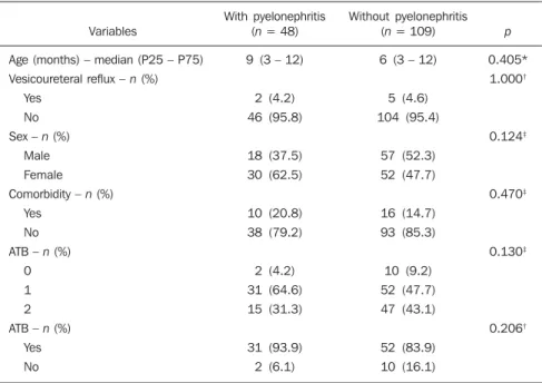
with APN. Only three patients presented congenital megaureter, and two of them had APN. Three patients presented pyelo-calyceal junction stenosis and another had a description of renal stenosis on his elec-tronic record, and it was not possible to understand whether such description meant stenosis of the renal artery or stenosis at some level of the urinary excretory system. One patient presented renal dysplasia and four patients had a history of phimosis, and none of them patients presented APN. No statistical significance was observed in re-lation to association between all such comorbidities and presence of APN (p = 0.470).
Eighty three patients were under antibi-otic therapy as the initial scintigraphy was performed for the diagnosis of APN. In 12 patients, antibiotic therapy was not admin-istered before the first scintigraphy and in the remaining ones it was not possible to confirm the utilization of antibiotics before initial scintigraphy. Among the 35 patients who were investigated for the presence of renal scarring, 24 were utilizing antibiotics before the first scintigraphy and two were not. In nine cases, it was not possible to identify, by their electronic records, whether the patients were under antibiotic treatment at the moment of the first scin-tigraphy. Neither in the whole studied
population, nor exclusively in the popula-tion under known early utilizapopula-tion of anti-biotics, there was statistical significance in relation to the APN diagnosis (p = 0.130 and p = 0.206, respectively). In the study sample, there was no statistical significance in the association between early utilization of antibiotics and development of renal scarring (p = 0.720). The median of renal scintigraphy repetition time for those pa-tients with renal scarring was 9.5 months (P25 = 3.5; P75 = 15.8), while the median for those who did not present renal scarring was 7 months (P25 = 3; P75 = 10), with p = 0.363. The results of the associations between clinical variables and APN and renal scarring are shown on Table 4.
DISCUSSION
Acute pyelonephritis is the most serious bacterial infection in the childhood, caus-ing severe symptoms, particularly in in-fants(13). In cases of urinary infection, the
presence of fever increases the chance of pyelonephritis and consequent develop-ment of renal scarring(14). Many studies
published in the international medical lit-erature have evaluated the prevalence of APN and the incidence of renal scarring. The prevalence varies from 26% to 60% in the case of APN, and it may reach 90% in
Table 2 Total incidence of renal scarring in APN patients. Renal parenchyma Scarring Normal Unknown* Total Frequency 8 27 13 48 % 16.7 56.3 27.1 100
APN, acute pyelonephritis; * Unknown refers to patient with APN who did not undergo scintigraphic follow-up. Table 1 Prevalence of APN.
UTI Lower UTI APN Total Frequency 109 48 157 % 69.4 30.6 100
UTI, urinary tract infection; APN, acute pyelonephritis.
Neither gender nor age of the patients presented a statistically significant associa-tion with the presence of APN (p = 0.124 and p = 0.405, respectively).
Only seven patients had VUR in their electronic records, of which five did not present APN, with no statistical difference in the association of VUR and APN (p = 1.0).
In the whole studied population, only 26 patients presented descriptions of some known comorbidity. Six patients from the study population presented hydronephrosis in some of the kidneys and only two of them had APN. Eight patients presented multicystic kidneys, half of them diagnosed
Table 3 Incidence of scarring observed at a sec-ond DMSA renal scintigraphy.
Renal parenchyma Scarring Normal Total Frequency 8 27 35 % 22.9 77.1 100
DMSA, dimercaptosuccinic acid.
Table 4 Association of variables with pyelonephritis.
Variables
Age (months) – median (P25 – P75) Vesicoureteral reflux – n (%)
Yes No Sex – n (%)
Male
Female Comorbidity – n (%)
Yes No
ATB – n (%) 0 1 2
ATB – n (%) Yes No
With pyelonephritis (n = 48)
9 (3 – 12)
2 (4.2) 46 (95.8) 18 (37.5) 30 (62.5) 10 (20.8) 38 (79.2) 2 (4.2) 31 (64.6) 15 (31.3) 31 (93.9) 2 (6.1) Without pyelonephritis (n = 109)
6 (3 – 12)
5 (4.6) 104 (95.4) 57 (52.3) 52 (47.7) 16 (14.7) 93 (85.3) 10 (9.2) 52 (47.7) 47 (43.1) 52 (83.9) 10 (16.1) p 0.405* 1.000† 0.124‡ 0.470‡ 0.130‡ 0.206†
cases of febrile UTI and high-degree VUR. On the other hand, the incidence of renal scarring ranges from 15% to 62%(9–13). In
the present review, the authors’ have not found in the Brazilian literature any study aimed at determining the prevalence of APN and incidence of renal scarring in the pediatric population. Such was the objec-tive of the present study.
The present study has found APN prevalence of 30.6%, while the incidence of renal scarring in the whole study popu-lation was 16.7% and, as the APN patients who did not have a second renal scintigra-phy for evaluation of renal scarring were excluded, the incidence was 22.9%. The values found in the present study are simi-lar to those reported by other published studies. Three 99mTc-DMSA scintigraphy
images, Figures 1, 2 and 3, respectively, il-lustrate three different cases as follows: normal result, APN and renal scarring.
In the present study, the clinical vari-ables such as presence of VUR, early utili-zation of antibiotics, presence of comor-bidities either related or not to the urinary system, besides patients age and gender, did not demonstrate statistical significance in the association with occurrence of APN and development of renal scarring. Consid-ering such results, one can infer that both investigative as well as therapeutic man-agement of APN and renal scarring in the authors’ environment are similar to those in large pediatric nephrology centers.
As regards treatment, the most recent guidelines determine that it is up to the nephrologist the immediate institution of antimicrobial therapy or the possibility of waiting for results of urine analysis and culture(15). In the present study, the authors
have observed that more than half of the patients were under antimicrobial treatment before undergoing renal scintigraphy.
The guidelines for imaging investiga-tion of APN and renal scarring highlight the use of DMSA renal scintigraphy. Accord-ing to Wong et al., in the presence of fever, besides girls, all boys under the age of two must be evaluated by means of DMSA(16).
Such protocol is the same utilized in the authors’ pediatric nephrology service. Re-cently, studies have proposed the utilization of magnetic resonance urography in the investigation of APN, renal scarring and
renal function, with encouraging initial results. However, such method requires general anesthesia, and its accuracy is yet to be better determined(17).
There is controversy about the utiliza-tion of prophylactic antibiotic therapy, in-cluding for those patients with VUR. Some studies demonstrate that there is no benefit in such preventive measure(18). For the
present study authors, such associations did not reveal statistical significance. The pres-ence of VUR is considered as being a known risk factor for APN and renal scar-ring(5), or at least, an aggravating factor for
both entities(8). In the present study, no
sta-tistical significance was found for such associations. This absence of statistical sig-nificance for such well known associations corresponds to a sample which was smaller than necessary to observe such signifi-cance, a direct consequence from the present study design.
The present study presents some limi-tations, for example, the fact of being ret-rospective, based on the review of scinti-graphic reports and electronic records. Such characteristics have limited the asso-ciations of clinical/demographic variables with APN and renal scarring. The measure-ments of APN prevalence and renal scar-ring incidence could performed because of the utilization of a sample of satisfactory size, a method based on a systematic review study developed by Shaikh et al.(10).
Finally, the present study results for APN and renal scarring frequencies are similar to those from other pediatric neph-rology centers. Renal scintigraphy still
plays a fundamental role in the investiga-tion of APN and its sequel in Brazil.
REFERENCES
1. Campos T, Mendes P, Maio J. Infecção urinária na criança. Acta Urológica. 2006;23:19–23. 2. D’Ippolito G, Abreu Junior L, Borri ML, et al.
Pielonefrite aguda: classificação, nomenclatura e diagnóstico por imagem. Rev Imagem. 2005;27: 183–94.
3. Majd M, Nussbaum Blask AR, Markle BM, et al. Acute pyelonephritis: comparison of diagnosis with 99mTc-DMSA, SPECT, spiral CT, MR im-aging, and power Doppler US in an experimen-tal pig model. Radiology. 2001;218:101–8. 4. Farhat W, Traubici J, Sherman C, et al.
Reliabil-ity of contrast enhanced sonography with har-monic imaging for detecting early renal scarring in experimental pyelonephritis in a porcine model: preliminary results. J Urol. 2002;168:1114–7. 5. Macedo CS, Riyuzo MC, Bastos HD. Cicatrizes
renais em crianças com refluxo vesicoureteral primário. J Pediatr (Rio J). 2003;79:355–62. 6. Oh MM, Cheon J, Kang SH, et al. Predictive
fac-tors for acute renal cortical scintigraphic lesion and ultimate scar formation in children with first
Figure 3.99mTc-DMSA renal scintigraphy demon-strating renal scarring. Image of left kidney present-ing focal uptake defects, with decrease in volume of functional parenchyma and clear size asymme-try in relation to the contralateral kidney. Figure 1. Normal 99mTc-DMSA renal scintigraphy.
Renal images with regular contours without the presence of focal defects radiotracer hypo-uptake.
febrile urinary tract infection. J Urol. 2010;183: 1146–50.
7. Zaki M, Badawi M, Al Mutari G, et al. Acute pyelonephritis and renal scarring in Kuwaiti chil-dren: a follow-up study using 99mTc DMSA re-nal scintigraphy. Pediatr Nephrol. 2005;20:1116– 9.
8. Jaksic E, Bogdanovic R, Artiko V, et al. Diagnos-tic role of initial renal corDiagnos-tical scintigraphy in chil-dren with the first episode of acute pyelonephri-tis. Ann Nucl Med. 2011;25:37–43.
9. Camacho V, Estorch M, Fraga G, et al. DMSA study performed during febrile urinary tract in-fection: a predictor of patient outcome? Eur J Nucl Med Mol Imaging. 2004;31:862–6.
10. Shaikh N, Ewing AL, Bhatnagar S, et al. Risk of renal scarring in children with a first urinary tract
infection: a systematic review. Pediatrics. 2010; 126:1084–91.
11. Faust WC, Diaz M, Pohl HG. Incidence of post-pyelonephritic renal scarring: a meta-analysis of the dimercapto-succinic acid literature. J Urol. 2009;181:290–8.
12. Koyle MA, Elder JS, Skoog SJ, et al. Febrile uri-nary tract infection, vesicoureteral reflux, and renal scarring: current controversies in approach to evaluation. Pediatr Surg Int. 2011;27:337–46. 13. Montini G, Tullus K, Hewitt I. Febrile urinary tract infections in children. N Engl J Med. 2011;365: 239–50.
14. Jakobsson B, Svensson L. Transient pyelone-phritic changes on 99mTechnetium-dimercapto-succinic acid scan for at least five months after infection. Acta Paediatr. 1997;86:803–7.
15. Subcommitte on Urinary Tract Infection, Steer-ing Committee on Quality and Management, Roberts KB. Urinary tract infection: clinical prac-tice guideline for the diagnosis and management of the initial UTI in febrile infants and children 2 to 24 months. Pediatrics. 2011;128:595–610. 16. Wong SN, Tse NKC, Lee KP, et al. Evaluating
different imaging strategies in children after first febrile urinary tract infection. Pediatr Nephrol. 2010;25:2083–91.
17. Smith EA. Pyelonephritis, renal scarring, and re-flux nephropathy: a pediatric urologist’s perspec-tive. Pediatr Radiol. 2008;38 Suppl 1:S76–82. 18. Lim R. Vesicoureteral reflux and urinary tract