p
+
Universidade NOVA de Lisboa
Instituto de Higiene e Medicina Tropical
Cohort study of associations between intestinal protozoa infection and
intestinal barrier function, nutritional status, and neurodevelopment
in infants from Republic of São Tomé
Marisol Garzon Lozano
Universidade NOVA de Lisboa
Instituto de Higiene e Medicina Tropical
Cohort study of associations between intestinal protozoa infection and
intestinal barrier function, nutritional status, and neurodevelopment
in infants from Republic of São Tomé
Ph.D. student: Marisol Garzon Lozano
Supervisor: Prof. Doutor Luis Pereira da Silva
Co-supervisor: Prof. Doutor Jorge Seixas
Doctoral dissertation complying with the requirements for Ph.D. degree in
Tropical Medicine
i Partial results of the PhD thesis published:
Association of enteric parasitic infections with intestinal inflammation and permeability in asymptomatic infants of São Tomé Island
Marisol Garzón, Luis Pereira-da-Silva, Jorge Seixas, Ana Luísa Papoila, Marta Alves, Filipa Ferreira & Ana Reis
Pathogens and Global Health
ISSN: 2047-7724 (Print) 2047-7732 (Online) Journal homepage: http://www.tandfonline.com/loi/ypgh20
iii
Dedicatory
To my love, my family, and my friends.
“All children should have the same opportunities to survive, develop and attain their full potential”
v
Acknowledgments
I would like to express immense gratitude to my supervisors Professors Sonia Lima, Jorge Atouguia and Jorge Seixas for the opportunity they gave me to come and work at IHMT. I am grateful for their insightful comments and ideas, and for lifting my spirits and motivation at the beginning of this journey.
I am deeply grateful to Professor Luis Pereira da Silva, without him, the completion of this thesis would have been impossible. I would love to thank him for clarifying many doubts, correcting countless mistakes with infinite patience and for his pragmatism that guided me through this final phase.
Additionally, I acknowledge the financial support received of Fundação de Ciências e Tecnologia.
I shall always remain indebted to all the staff of Institute Marques de Valle Flôr for their help in enabling my fieldwork in São Tomé. Always making me feel at home.
vii
Abstract
Cohort study of associations between intestinal protozoa infection and intestinal barrier function, nutritional status, and neurodevelopment in infants in Republic of São Tomé.
Marisol Garzón Lozano
Key words: infant growth, intestinal inflammation, intestinal parasites, intestinal permeability, neurodevelopment
Background
Giardia lamblia, Cryptosporidium and Entamoeba hystolitica are prevalent etiologic
agents of enteric infections in infants from low- and middle-income countries. Host-parasite interactions may lead to mucosal inflammatory response and increased intestinal permeability. Clinically this can result in a negative impact on growth and neurodevelopment. The effects of these subclinical enteric protozoa infections on infant health are poorly explored.
Aim
To analyze the associations between enteric parasitic infections and intestinal barrier function, nutritional status and neurodevelopment in asymptomatic infants in São Tomé.
Methods
A birth cohort study with a follow-up until 24 months of age was implemented. Anthropometry was assessed monthly and included attained growth (weight-for-length z-score, length-for-age z-score – LAZ, and length-for-age difference – LAD), growth velocity (weight velocity z-score – WAVZ, and length velocity z-score – LAVZ), and risk for undernutrition (wasting and stunting, using the <-1SD cut-off).
Neurodevelopment was screened at key ages using the “Bayley Infant
viii
months of age. Enteric protozoa and intestinal helminths were examined quarterly in stool samples using microscopic techniques. Different statistical models were used to explore associations between enteric parasitic infections and the three outcomes: intestinal barrier function, nutritional status, and neurodevelopment.
Results
A total of 475 neonates were enrolled, representing 8.6% of live-births in São Tomé; 280 (58.9%) infants completed 24 months of follow-up. Giardia lamblia and
helminths were the most prevalent parasites. The multivariable analysis showed that: 1) infants with Giardia lamblia and helminths infections had a tendency toward an
increase of 23.6 % and of 24.1 % in the inflammatory biomarker, respectively; those infected by any enteric parasite had a tendency toward an increase of 33.6% in the permeability biomarker; additionally, this biomarker was 100% higher in wasted infants and 50% higher in those stunted; 2) infants with Giardia lamblia and helminths
infections showed a significant association with a decrease in linear growth (by - 0.10 and -0.16 of LAZ and by -0.32 and -0,48 of LAD, respectively); those with
Cryptosporidium spp. infection displayed a significant association with a decrease in
weight and length velocities (-0.43 WAVZ and -0.55 LAVZ); 3) Giardia lamblia
infection and stunting were independently and significantly associated with a 1.69 and 2.37 increased risk of poor development, respectively.
Conclusions
This first birth cohort ever performed study in São Tomé is innovative in exploring associations between enteric parasitic infections and the intestinal barrier function, nutritional status and neurodevelopment in infants. The underestimated role of protozoa and helminths as etiologic agents of subclinical enteric infections was confirmed. These parasitic infections showed a tendency of association with intestinal barrier dysfunction and significant associations with decreased linear growth and risk of poor neurodevelopment. In the context of São Tomé, an endemic area for Giardia lamblia and helminths with a non-negligible proportion of marginally undernourished
ix
Resumo
Estudo de coorte sobre as associações entre infeções por protozoários intestinais e a função da barreira intestinal, o estado nutricional e o neurodesenvolvimento em lactentes da República de São Tomé.
Marisol Garzón Lozano
Key words: crescimento infantil, helmintas intestinais, inflamação intestinal, neurodesenvolvimento, permeabilidade intestinal, protozoários intestinais
Enquadramento
Em lactentes de países de baixo e médio rendimento, Giardia lamblia, Cryptosporidium e Entamoeba hystolitica são agentes prevalentes em infeções
intestinais. As interações hospedeiro-parasita podem levar a uma resposta inflamatória da mucosa e aumento da permeabilidade intestinal. Clinicamente, isto pode refletir-se por impacto negativo no crescimento e neurodesenvolvimento. Os efeitos destas infeções intestinais subclínicas na saúde infantil têm sido pouco estudado.
Objetivo
Analisar, em crianças assintomáticas de São Tomé, as associações entre infeções por parasitas intestinais e a função da barreira intestinal, o estado nutricional e o neurodesenvolvimento.
Métodos
Foi realizado um estudo coorte de nascimento com seguimento até aos 24 meses de idade. A antropometria foi avaliada mensalmente e incluiu o crescimento atingido ( z-scores para peso/comprimento, comprimento/idade – CIzs e diferença do
comprimento-para-idade – DCI), a velocidade do crescimento (z-scores para
velocidade ponderal – VPzs e linear – VLzs) e o risco de desnutrição (aguda e crónica, definida como <-1DP). O neurodesenvolvimento foi rastreado em idades-chave
x
avaliada trimestralmente por técnicas microscópicas Foram usados diferentes modelos estatísticos para estudar associações entre infecções por parasitas intestinais e os resultados das avaliações da função da barreira intestinal, estado nutricional e neurodesenvolvimento.
Resultados
Foram incluídos 475 recém-nascidos, representado 8,6% dos nados-vivos em São Tomé; 280 (58,9%) completaram os 24 meses de seguimento. Giardia lamblia e
helmintas foram os parasitas mais prevalentes. A análise multivariável revelou que: 1) lactentes infetados com Giardia lamblia e helmintas tiveram tendência para aumento
de 23,6% e 24,1% no marcador de inflamação intestinal, respectivamente; os infetados por qualquer parasita tiveram tendência para aumento de 33,6% no marcador de permeabilidade; além disso, os níveis de A1AT foram 100% superiores em lactentes com desnutrição aguda e 50% superiores nos com desnutrição crónica; 2) lactentes infetados com Giardia lamblia e helmintas tiveram associação significativa com
diminuição no crescimento linear (-0,10 e -0,16 CIzs; e -0,32 e -0,48 de DCI, respectivamente); os infetados com Cryptosporidium spp. tiveram associação
significativa com diminuição na velocidade de crescimento ponderal e linear (-0,43 VPzs e -0,55 VLzs); 3) a infecção por Giardia lamblia e a desnutrição crónica
associaram-se independentemente e significativamente com 1,69 e 2,37 maior probabilidade de atraso no desenvolvimento, respectivamente.
Conclusões
Este é o primeiro estudo de coorte de nascimento em São Tomé, pioneiro em estudar associações entre infeções por parasitas intestinais e a função da barreira intestinal, estado nutricional e neurodesenvolvimento. Foi confirmado o papel subestimado dos protozoários e helmintas como agentes etiológicos de infecções intestinais subclínicas. Estas infecções revelaram uma tendência para associação com a disfunção da barreira intestinal e associações significativas com restrição do crescimento linear e neurodesenvolvimento. Estas associações são problemáticas em São Tomé, endémico para Giardia lamblia e helmintas, em contexto de proporção não negligenciável de
xi
xiii
TABLE OF CONTENT
Page
Acknowledgements.……….………...v
Abstract……….……….…...vii
Abbreviations………...1
1. Introduction………...5
1.1.Prevalence of enteric parasites in infants in Africa and low and middle-income countries………..………....6
1.1.1. Giardia lamblia………...………....6
1.1.2. Cryptosporidiumspp………...11
1.1.3. Entamoeba histolytica………...13
1.1.4. Soil transmitted helminths………...14
1.2.Taxonomy and life cycle of enteric protozoa………....20
1.3.Host-parasite interaction: host damage.……….…...27
1.3.1. Apoptosis………...………27
1.3.2. Disruption of brush border.……….…………..30
1.3.3. Cytoskeleton remodeling and disassembly of tight junctions...32
1.3.4. Inflammation……….………34
1.4.Clinical picture……….……...39
1.5.Enteric protozoa infection and intestinal barrier ………...45
1.5.1. Intestinal barrier………45
1.5.2. Mucosal inflammatory response………..……….47
1.5.3. Assessment of intestinal barrier function...48
1.5.4. Enteric protozoa infection and intestinal barrier.………..56
1.6.Enteric protozoa infection and nutritional status ………...63
1.6.1. Infant growth ………...63
1.6.2. Assessment of nutritional status/anthropometry..……….………66
1.6.3. Enteric protozoa infection and nutritional status ……….70
1.7.Enteric protozoa infection and neurodevelopment status.………..……..83
xiv
1.7.2. Assessment of infant neurodevelopment……….……….………78
1.7.3. Enteric protozoa infection and infant neurodevelopment………….…79
2. Objectives………...87
3. Methods…..………...89
3.1.Study design……….…………...89
3.2.Ethical and legal issues.………....89
3.3.Setting………..89
3.4.Inclusion criteria ………...91
3.5.Sample size.……….……..91
3.6.Follow-up: points of assessment………...92
3.7.Data collection ………...93
3.7.1. Questionnaire for the first visit ………...93
3.7.2. Feeding practices.………..94
3.7.3. Acute infections and associated conditions.……….……….95
3.7.4. Nutritional status/anthropometry ……….95
3.7.5. Neurodevelopment assessment ………...97
3.7.6. Parasite examination techniques………...98
3.7.7. Fecal biomarkers of intestinal function………...99
3.8.Statistical analysis ………...………...100
4. Results………..103
4.1.Sample description.……….………103
4.1.1. Socio-demographic and socio-economic status.……….105
4.1.2. Obstetrical data and mothers’ anthropometry………….…...109
4.1.3. Infant feeding practices……….………..111
4.1.4. Acute infections and associated conditions.………..…………..112
4.1.5. Nutritional status/anthropometry.………...116
4.1.6. Neurodevelopment status.………...129
4.1.7. Enteric parasites……….….…………133
4.1.8. Fecal biomarkers of intestinal function……….……. ………138
4.2.Associations between enteric parasites and outcomes……….…...139
4.2.1. Enteric parasitic infection and intestinal barrier function…………...139
xv
4.2.3. Enteric parasitic infection and neurodevelopment.…….………155
5. Discussion………...159
5.1.São Tomé and Principe: a low and middle-income country.………...159
5.2.Enteric parasitic infections in infants from São Tomé………160
5.3.Intestinal barrier function in infants from São Tomé………...165
5.3.1. Intestinal inflammatory response ………...165
5.3.2. Intestinal permeability ………...167
5.4.Infants growth in São Tomé………….……...169
5.4.1. Attained growth.………..………169
5.4.2. Growth velocity.………..170
5.4.3. Wasting and stunting.………..171
5.5.Neurodevelopment screening in infants from São Tomé.…………..……...173
5.6.Association between enteric parasitic infection and intestinal barrier function………176
5.6.1. Association between enteric parasitic infection and intestinal inflammation.………..176
5.6.2. Association between enteric parasitic infection and intestinal permeability………..………..179
5.7.Association between enteric parasitic infection and infant growth………….180
5.7.1. Cofactors associated to growth faltering……….183
5.8.Association between enteric parasitic infection and poor neurodevelopment………...186
6. Strengths and limitations………...190
6.1.Strengths.………...190
6.2.Limitations.………...192
7. Conclusions ………...197 8. Gaps and future perspectives.………...………….199
9. References ………...203 Appendices Appendix 1. Informed consent ………...251
xvi
Appendix 3. Description of variables.……….………260
List of tables..………...xvii
List of figures.………..xx
xvii
LIST OF TABLES
Table 1. Prevalence of intestinal protozoa in children in African countries………...…7
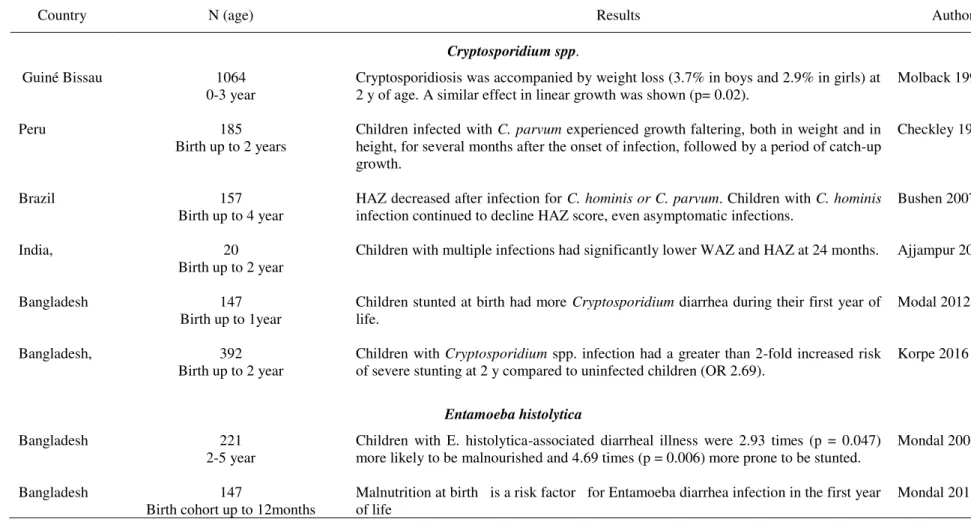
Table 2. Longitudinal studies of prevalence of Giardia lamblia, Cryptosporidium spp.
and Entamoeba histolytica in infants from low-and middle income countries...16
Table 3. Taxonomy, genotypes and life cycle of enteric protozoa Giardia lamblia, Cryptosporidium spp. Entamoeba histolytica...………..………..25
Table 4. Assessment of intestinal barrier function……….……...50 Table 5. Clinical studies of association between enteric protozoa infection and
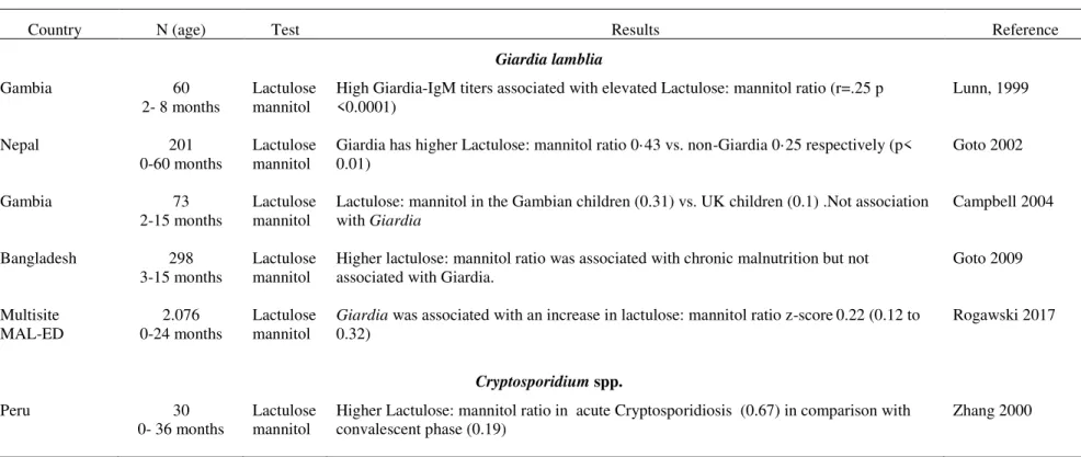
intestinal permeability in infants from developing countries………...58 Table 6. Clinical studies of association between enteric protozoa infection and
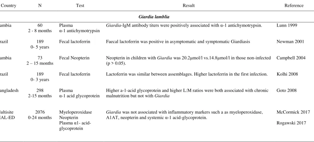
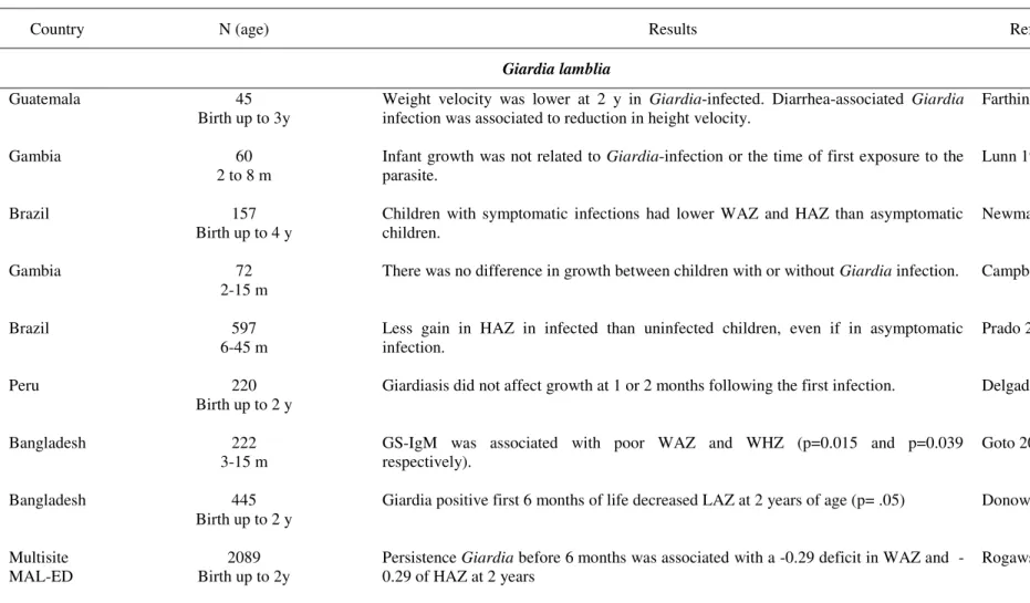
intestinal inflammatory response in infants from developing countries.………..61 Table 7. Longitudinal studies of association between enteric protozoainfection and
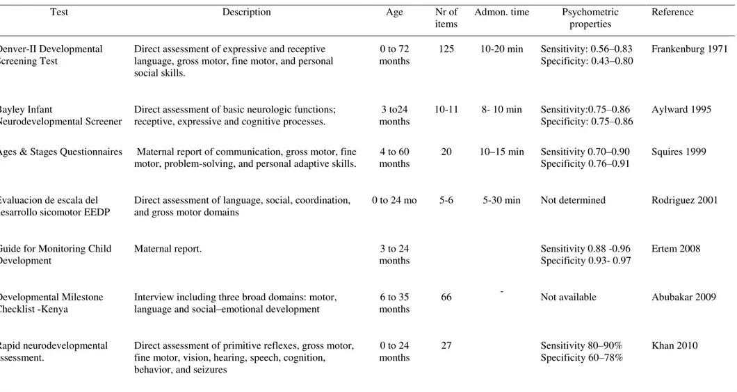
poor nutritional status in infants from developing countries.………...72 Table 8. Developmental screening tools in infants in low and middle-income countries
………...82 Table 9. Studies reporting association between enteric protozoa and neurodevelopment in infants from developing countries.………...84
Table 10. Newborns by district in São Tomé e Principe..………..………..92 Table 11. Follow-up point assessments...………..……...93 Table 12. Differences between infants who completed the study (at least 10 visits) and
those who did not………105
Table 13. Socio-demographic and household characteristics of the cohort…………106
Table 14. Multidimensional poverty index of the cohort……….……..108
Table 15. Obstetrical data and mothers’ anthropometry of the cohort.………...109
Table 16. Feeding practices of infants in the cohort………...111 Table 17. Acute infectious events and other conditions, by………...114 Table 18. Attained weight, length, head circumference, and mid-upper arm
circumference at target ages………117
Table 19. Attained weight, length, head circumference, by sex……….118 Table 20. Attained z-scores of weight-for-age, length-for-age, weight-for-length, body
circumference-xviii
for-age at target ages……….……..119 Table 21. Length- for- age difference by sex, at target ages………...120 Table 22. Weight and length increments in two-month intervals and respective
velocities z-scores……….………..………125
Table 23. Frequency of wasting and stunting during the study period, considering mild (<-1 SD) and moderate <-2 SD) and severe (<-3SD) degrees………...128
Table 24. BINS scores at each point of assessement………..130 Table 25. Proportion of infants at low and high risk for poor development at each
point of assessment.………....131
Table 26. Proportions of infants performing tasks in each BINS’s developmental
areas……….132
Table 27. Frequency of intestinal pathogenic parasites by age.……….….135 Table 28. Cumulative data on enteric parasitic infections, including age at first
detection, total number of infections, and number of episodes of infection………...136 Table 29. Molecular characterization of Giardia lamblia………..137
Table 30. Alpha 1 antitrypsin and S100A12 stool biomarkers………...138 Table 31. Fecal values of alpha1-anti-trypsin and S100A12 at 24 months of age,
considering sex, parasite agent, and nutritional status categories.………..…....139 Table 32. Univariable analysis for fecal alpha1-anti-trypsin and S100A12, considering sex, parasitic infection before and at 24 months of age, nutritional status, and feeding
practices………...141 Table 33. Multivariable regression models for alpha1-anti-trypsin and S100A12.
……….142 Table 34. Univariable analysis of attained measures………..144
Table 35. Univariable analysis for anthropometric velocity measures………...146 Table 36. Univariable analysis for wasting and stunting………...148 Table 37. Nutritional status/ anthropometry: multivariable models for attained growth
……….…153
Table 38. Nutritional status/ anthropometry: multivariable models for growth velocity
………...154 Table 39. Nutritional status/ anthropometry: multivariable models for wasting and
xix
Table 40. Descriptive analysis of BINS scores by categories of nutritional status and
xx
LISTOF FIGURES
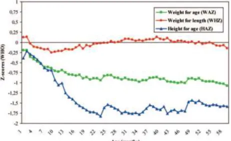
Figure 1. Host-parasite interactions in intestinal barrier……….……..27 Figure 2. Tight junction complex.………...46 Figure 3.Trends of anthropometric z-scores according to age relative to WHO
standards………63
Figure 4. Growth curves of attained growth and growth velocity………...66 Figure 5. The developmental course of human brain………77 Figure 6. Conceptual framework………...……87 Figure 7. The Democratic Republic of São Tomé and Príncipe………..….90 Figure 8. Time of recruitment and follow-up………92 Figure 9. Conceptual framework: hypotheses explored………..…...100
Figure 10. Flow chart………..104 Figure 11.Feeding practices by age……….112 Figure 12. Proportion of infants with acute diarrhea and acute respiratory infants…113 Figure 13. LOWESS-fitted curves applied to longitudinal data on weight, length and
head circumference curves………..121 Figure 14. LOWESS-fitted curves applied to longitudinal data of weight-for-age, length-for-age and weight-for-length z-scores…..………..122
Figure 15. LOWESS-fitted curves applied to longitudinal data on length for age
difference……….123
Figure 16. LOWESS-fitted curves applied to longitudinal data on weight increments and weight velocity z-scores………...124
Figure 17. LOWESS-fitted curves applied to longitudinal data on weight increments and weight velocity z-scores………...………124
1
ABBREVIATIONS
A1AT: alpha-1-antitrypsin AD: acute diarrhea
AF: attributable fraction APOE: apolipoprotein E ARI: acute respiratory infection
BINS: Bayley Infant Neurodevelopmental Screening BMI: body mass index
BSID: Bayley scales of infant development DDST: Denver development screening test EIA: enzyme immunosorbent assay
ELISA: Enzyme-Linked Immunosorbent Assay GBD: Global Burden Diseases
GEMS: Global Enteric Multicenter Study HAD: height-for-age differences
HAZ: height-for-age HC: head circumference
HIV: human immunodeficiency virus HLA: Human leukocyte antigen HR: hazard ratio
I-FABP: Intestinal fatty acid binding protein IFN-γ: Interferon gamma
Ig: immunoglobulin IL: interleukin
IQ: intelligent quotient
JAMs: junctional adhesion molecule LAD: length-for-age difference L:M: lactulose mannitol test LAVZ: length velocity z-score
LAZ: length-for-age z-score
2 LOWESS: locally weighted scatterplot smoothing LPS: lipopolysaccharide
MAL-ED: study of Malnutrition and Enteric Diseases MBL: mannose-binding lectin
MGRS: multicenter Growth Reference Study MHC: major histocompatibility complex MPI: multidimensional poverty index MPO: myeloperoxidase
MUAC: mid-upper arm circumference
NADPH: nicotinamide adenine dinucleotide phosphate NEO: neopterin
NET: neutrophil extracellular traps NF-κB: factor nuclear kappa B OR: odds Ratio
PCR: polymerase chain reaction PGE2: prostaglandin E2
RAGE: advanced glycation end products RR: relative Risk
SD: standard deviation
SGA: small-for-gestational age SSA: sub-Saharan Africa STH: soil-transmitted helminths STP: São Tomé and Principe
TGF β: Transforming growth factor-β
Th: T helper cells. TJs: tight junctions TLR: toll like receptors
TNF-α: tumor necrosis factor –alpha WAD: weight-for-age difference WAVZ: weight velocity z-score
WAZ: weight for age z-score
3 WLZ: weight-for-length z-score
WHZ: weight-for-length z-score
Introduction
5
1.
Introduction
Parasites found in the human gastrointestinal tract can be largely categorized into two groups, protozoa and helminths. Enteric protozoa Giardia lamblia, Cryptosporidium
spp. and Entamoeba histolytica are the protozoa of worldwide importance (WHO
2002a).
Infections by protozoa Giardia lamblia, Cryptosporidium spp. and Entamoeba histolytica are considered neglected tropical diseases, since they belong to a
heterogeneous group of infectious diseases that occur mostly in developing countries where climate, poverty, and lack of access to services are common (Savioli 2006). These diseases exhibit a considerable and increasing global burden and impair the ability of those infected to achieve their full potential, both developmentally and socio-economically (Savioli 2006). Whole populations will be geographically at risk, but children are observed to disproportionally carry the greatest burden of infection, this is particularly concerning, since the population structure of endemic regions is predominantly younger (Harnay 2010). Infection rates are highest in children living in Sub-Saharan Africa (SSA), followed by Asia, Latin America, and Caribbean (Harnay 2010).
Based on the conceptual model of the “cycle of poverty” (Guerrant 2008), enteric
infections have a central role leading to malnutrition, with long-term impact in conginitve development (Guerrant 2008). The classical burden of enteric infection measured in terms of diarrhea-associated mortality does not capture the true impact that enteric infections have on human development, neither the subtle morbidities on endemic communities (Guerrant 2002, Harnay 2010, McCormick 2016). For example, when long-term health effects of subclinical enteric infections are quantifying, the burden of stunting is substantially greater than reflected by diarrhea per se (Guerrant
Introduction
6
1.1. Prevalence of enteric parasites in infants in Africa and others low- and middle income countries
In Africa, several challenges such as the remoteness of communities, lack of transport, shortage of skilled health care workers, and lack of laboratory facilities make difficult to have reliable studies on prevalence of enteric protozoa (Squire 2017). Studies are mostly cross-sectional or case-control, addressed to children aged 0 to 16 years, at hospital or community settings, and using different diagnostic methods (Table 1). As a result, prevalence obtained from those studies is quite variable (Table 1).
Longitudinal studies conducted in infants from low- and middle-income countries (LMIC) are shown in Table 2.
1.1.1. Giardia lamblia
Giardia lamblia is ubiquitous and the initial infection is acquired in the first few
weeks of life (Muhsen 2012).In Guatemala, a birth cohort of infants was followed-up 3 years, with weekly stool samples. Giardia infected at very early ages (first week of
life) and every child has at least one episode in the first three years of life. The mean number of Giardia infections per child increased from 0.7 in the first year to 3.6 in the
third year. Prevalence reached 20.2% by the end of the third year (Mata 1978, Farthing 1986). In northeast Brazilian, infants were followed three times weekly for diarrheal surveillance up to 4 years (Newman 2001). Of 157 children followed, 27.4% were infected with Giardia lamblia with similar frequency in non-diarrhoeal (7.4%) and
diarrhoeal stools (9.7%)(Newman 2001). In Peru, a birth cohort of 220 infants was followed until 35 months of age, with weekly stool collection (Hollm-Delgado 2008). The overall prevalence of Giardia lamblia was 18.8% per child-week, and 85% of
children became infected during their follow-up (Hollm-Delgado 2008). In Bangladesh, Mondal et al. (2012) followed 147 infants since birth until the first year
of life with monthly stool surveillance for enteropathogens. Giardia lamblia was the
more frequent pathogen, infected 34% of all infants (Mondal 2012). The cumulative percentage of Giardia lamblia infection, increased from 30% in the first month to
Introduction
7
Table 1. Prevalence of intestinal protozoa in children in African countries
Country, author Study N Method Prevalence
Giardia lamblia Cryptosporidium E. histolytica
Angola
Gasparinho 2016 Etiology of Diarrhea in Children Younger Than 5 Years Attending the Bengo General Hospital in Angola
344
(0-5 y) Microscopy 21.6% 30.0% 0.9% E. histolityca/dis
par
Ethiopia
Wegayehu, 2013
Prevalence of Giardia and Cryptosporidium
among children and cattle in Ethiopia
384 (1-14y)
Microscopy 24.5
(1-5y) 12.3% (1-5y) 3.6% E his/dis Ethiopia
Mulatu, 2015 Intestinal parasitic infections among children < 5 y with diarrhoeal diseases in Health setting -Ethiopia.
158
(0-5 y) Microscopy 7% (2-7% 0- 24m) 3.8% (0-3% 0- 24m) 11.4% E. hist/ disp. (7% 0-24m)
Gambia
Sullivan, 1991 Prevalence and treatment of giardiasis in chronic diarrhea and malnutrition (6-36 m) 64 Microscopy IgM 45% cases 12 % control
Ghana
Addy, 2004 Prevalence of pathogenic E. coli and parasites in infants with diarrhea in Kumasi, Ghana (0-5 y) 284 Microscopy 3.7% cases 0% control 8% cases 0.8% control
Ghana
Reither, 2007 Acute childhood diarrhea in Ghana: epidemiological, clinical and microbiological characteristics
365
(0-12 y) EIA-PCR 3.7% cases 9.7% control 0.4 % cases 0.8% control
Guinea-Bissau Centeno-Lima, 2013
Giardia duodenalis and chronic malnutrition in children under five from a rural area of Guinea-Bissau.
109 (0 to 5 y)
Microscopy 29.0% cases
35.9% controls.
Kenya Mbae, 2013
Parasitic infections in children with diarrhea in outpatient and inpatient in Nairobi, Kenya
2112 (0 - 5 y)
Microscopy 5.8% outpatient
1.3% inpatient
6.7 % out 15.7% in-patient.
Introduction
8
Table 1. Prevalence of intestinal protozoa in children in African countries (continued)
Country, author Study N Method Prevalence
Giardia lamblia Cryptosporidium E. histolytica
Kenya
Thiongo, 2011 Spatial distribution of Giardia intestinalis in children up to 5 years old attending out-patient clinic at Provincial General hospital, Embu, Kenya
376
(0-5 y) Microscopy 12.8% 5.3%
Mozambique
Fonseca, 2014 Intestinal parasites in children hospitalized at the Central Hospital in Mozambique (0- 2 y) 93 Microscopy 6.5% 6.5 %
Mozambique
Nhampossa, 2015 Diarrheal Mozambique: burden, and Etiology of diarrhea 0–59 Months at Health Facilities (0-5 y) 2329 EIA 0-12m: 10% cases; 18% control 12-24m: 28% cases; 46% control. 0-12m: 20% cases; 10% control 12-24m: 19% cases; 9% control
E. his/dispar 0-12m: 9% cases-control. 12-24m: 11% cases; 10% control Rwanda
Ignatius, 2012 High Prevalence of
G.duodenalis Assemblage B
and association with Underweight in Rwandan Children.
583
(0-5 y) Microscopy PCR 60%
4.9% 1.1%
Senegal
Roger, 2013 Parasitic Infections among children <5y Senegal: Prevalence and Effect on Anemia and Nutritional Status
636
(0- 5 y) Microscopy 15.4%
São Tomé Lobo 2014
Cryptosporidium spp., G. duodenalis, E. bieneusi in young Children in STP
314
(1 -10 y) Microscopy PCR 7.5% community. 1.9% hospital 0% community 8.9% hospital E. histolytica /dispar 1.5%
São Tomé Ferreira 2015
G. duodenalis and soil-transmitted helminths
Introduction
9
Table 1. Prevalence of intestinal protozoa in children in African countries (continued)
Country, author Study N Method Prevalence
Giardia lamblia Cryptosporidium E. histolytica
São Tomé
Liao, 2016 Prevalence of intestinal parasitic infections among school children in Democratic Republic of São Tomé and Príncipe, West Africa.
252 School children
Microscopy 28.6% of
protozoan.
Tanzania
Moyo, 2011 Age specific of diarrhea in Hospitalized children < 5y In Salaam, Tanzania
280
(0-5 y) EIA 1.9% 18. %
Tanzania
Ngosso 2015 Pathogenic Intestinal Parasitic Protozoa in diarrhea < 5y Tanzania (0- 5 y) 720 Microscopy PCR 16.4% micro 35.6% PCR 4.4% Micro 7.8% PCR 20% microscopy 12% PCR Tanzania
Tellevick, 2015 Prevalence of
Cryptosporidium
, Entamoeba histolytica and Giardia among
Young Children with and without Diarrhea
1259
(0 - 2 y) PCR 3.4% cases 6 .1% control 16% cases 3.1% control 0%
Uganda Tumwine, 2003
Cryptosporidium parvum in children with
diarrhea in Mulago hospital, Kampala, Uganda. (0-5y) 2446 Microscopy 25% cases 8.5% control
Zambia,
Introduction
10
Also in Bangladesh, a birth cohort of 445 infants were followed until 2 years of the study with stool collected 3 times per week (Donowitz 2016). Seven percent of infants had a Giardia lamblia positive in the first 6 months of life, increasing to 74% by 2
years of age (Donowitz 2016). In Africa, only two longitudinal studies addressing
Giardia lamblia infection in infants have been conducted. In Gambia, 60 infants were
studied longitudinally between 2 and 8 months of age and followed-up with parasite-specific plasma immunoglobulins for Giardia (Lunn 1999). The median age for the
first Giardia lamblia exposure was between 3 and 4 months, and by 8 months of age
95% of infants had a positive titer in at least one occasion (Lunn 1999). In Guinea-Bissau, a birth cohort 200 infants was followed-up to 2 years with weekly stool specimen collection from enteropathogens (Valentiner-Branth 2003). In this study,
Giardia lamblia (cyst or trophozoites) was the most frequently detected protozoa, with
an incidence rate of 3.7 infections per child-year at risk, without association to
diarrhea (Odds ratio-OR 0.64) (Valentiner-Branth 2003). In São Tomé e Principe (STP), evidence came from two cross sectional studies. Lobo et al. (2014) studied 348
children between 2 months to 10 years using molecular techniques for protozoa detection. They found a prevalence of 7.5% for Giardia lamblia at community setting,
being the Assemblage A (60%) more common. Ferreira et al. (2015) studied 444
preschools children using microscopic technique for parasite detection. Giardia lamblia was the second more frequent parasite, infecting 41.7% (185 of 444) of the
children. A systematic review and meta-analysis of the association between Giardia lamblia and diarrhea from several case-control and cohort studies in children under 5
years in developing countries, concluded that Giardia did not cause acute diarrhea
among children in developing countries (OR 0.60; p = .03), although limited data suggest that infants in the first trimester of life may experience acute diarrhea in response to initial Giardia lamblia infections (Muhsen 2012). Additionally, Giardia
Introduction
11
protective for diarrhea (Relative Risk (RR) 0.95; 95% CI 0.90–1.00) suggesting that asymptomatic Giardia lamblia infection is common in children from developing
countries (Rogawski 2017). Regarding Giardia genotypes, assemblage B was more
common in African countries such as Tanzania (Forsell 2016), Uganda (Ankarklev 2012), Guinea Bissau (Ferreira 2012), Rwanda (Ignatius 2012) and Kenya (Mbae 2016). Assemblage A was far less frequent, reported only in STP (Lobo 2014) and Ethiopia (Gelanew 2007).
1.1.2. Cryptosporidium spp.
Cryptosporidium spp. is recognized globally as an important cause of diarrhea in
children either in self-limiting diarrhea in otherwise healthy children, or in chronic life-threatening illness in immunocompromised patients, most in those with human immunodeficiency virus -HIV or in malnourished young children (Mor 2008). In Peru, birth cohort of 185 infants was followed-up to 2 years with weekly stool samples. Forty eight percent of children aged 0-23 months became infected with
Cryptosporidium parvum during the study period, 60% of them asymptomatic
(Checkley 1998). In the same cohort, others authors reported prevalence near to 30%, being Cryptosporidium hominis the species most frequently detected (70%)(Cama
2008). Infections with Cryptosporidium hominis lasted longer and had higher parasite
excretion scores than other species (Cama 2008). In a prospective 4-year cohort study of 157 children in Brazil, (Newman 1999) Cryptosporidium spp. oocysts were
identified in 7.4% of all stools, more frequently in children with persistent diarrhea (16.5%) than in those with acute (8.4%) or no diarrhea (4.0%) (p< .001) (Newman1999). In the same cohort Cryptosporidiumhominis was identified in 57% of
cases, and Cryptosporidium parvum in 43 %. Cryptosporidium hominis infections
were associated with higher duration of diarrhea and heavier infections than stools from children with Cryptosporidium parvum (Bushen 2007). In a semi-urban
community in south India, a birth cohort of 452 infants was followed-up to 3 years in a twice-weekly basis (Ajjampur 2007) Cryptosporidium spp. alone accounted for 7.61%
of episodes of diarrhea.The two most common species were Cryptosporidiumhominis
Introduction
12
2007). In the same region, a weekly surveillance of 176 infants was conducted up to their second birthday. A total of 186 episodes of cryptosporidiosis, mostly asymptomatic, were observed in 67% of children during the follow-up period (Sarkar 2013). In Bangladesh, a birth cohort of 392 rural infants was followed-up to 2 years with monthly stool samples (Korpe 2016). In the first two years of life, 77% of children experienced at least one infection with Cryptosporidium spp. Non-diarrheal
infection (67%) was more common than diarrheal infection (6.3%)(Korpe 2016).
Cryptosporidium hominis was isolated from over 90% of samples (Korpe 2016). In
India, 97% of children acquired cryptosporidiosis by 3 years of age (Kattula 2017). In Africa, hospital and community-based studies document a high prevalence of cryptosporidiosis in younger children, particularly among those who are malnourished or positive for human immunodeficiency virus (HIV) infection and during rainy seasons (Mor 2008). In most SSA countries, cryptosporidiosis prevalence peaks among children aged 6–12 months and decreases thereafter and the majority of infections are caused by Cryptosporidium hominis (Mor 2008). In an open cohort
followed-up to 3 years in Guinea Bissau, Cryptosporidium spp. was found in 7.4% of
3215 episodes of diarrhea (Mølbak 1993).The parasite was most common in younger children (median age 12 months) at the beginning of the rainy seasons(Mølbak 1993). In Kenya, a prospective survey was conducted over a two-year study period, with analysis of 4899 stool samples in children (Gatei 2006). The overall prevalence of cryptosporidiosis was 4%, highest among children 13–24 months of age (5.2%), 87% of the Cryptosporidium isolates were Cryptosporidium hominis, and 9% Cryptosporidiumparvum (Gatei 2006). A prospective cohort of 108 women and their
infants in rural/semi-rural Tanzania were followed from delivery through six months of age (Pedersen 2014). Cryptosporidium spp. infection in infancy remained
undetected until 2 months of age and by 6 months, 33% of infants were infected. Maternal Cryptosporidium infection was associated with increased odds of infant
infection (unadjusted OR 3.18) (Pedersen 2014). In São Tomé, a prevalence of 8.9% was reported for Cryptosporidium spp. at hospital setting. From 19 isolates, 14
corresponded to Cryptosporidium hominis (6.5%) and 5 to Cryptosporidium parvum
Introduction
13
Regarding Cryptosporidium species, a geographically assessment of data derived from
a multisite study in infants from developing countries validated the dominance of
Cryptosporidium hominis (5.9 million) in infants and toddlers, in comparison with Cryptosporidium parvum (0.76 million) at SSA and South Asia countries (Sow 2016).
1.1.3. Entamoeba hystolitica
The true prevalence and incidence of infection and disease caused by Entamoeba histolytica is unknown for most areas of the world (Stauffer 2006). This can be
attributed to the fact that differentiation of Entamoeba histolytica from the identical
appearing non-pathogenic amebic species Entamoeba dispar is not possible based on
microscopic exam (Stauffer 2006). Most of the evidence came from studies from Bangladesh. In a study in 221 children aged 2-5 years who were followed-up for 3 years due to diarrheal illness (Mondal 2006), it was found that 53% of them had serum antibodies against the Entamoeba histolytica Gal/GalNac lectin (anti-lectin IgG) at the
time of enrollment (Mondal 2006). A cohort study of acute diarrhea included 289 preschool children in an urban slum of Dhaka, Bangladesh, Entamoeba histolytica was
identified in 8%of the diarrheal stool specimens based on antigen detection (Haque 2003). The incidence of Entamoeba histolytica associated diarrhea was 0.08 per
child-year, while the incidence of asymptomatic infection was 0.44 episodes per child-year.
Asymptomatic carrier state of Entamoeba histolytica infection increased by five-fold
the risk of Entamoeba histolytica-associated diarrhea (Haque 2003). In the same
cohort, 80% of children who completed 4.2 years of follow-up were infected with
Entamoeba histolytica at least once (Haque 2006). Also in Bangladesh, a birth cohort
study of 147 infants was followed-up to the first year of life (Mondal 2012).
Entamoeba histolytica was isolated in 3.8% of specimens using molecular techniques,
and children who were underweighted and stunted at birth were more colonized by
Entamoeba histolytica (Mondal 2012). In another cohort study among infants from
Bangladesh, approximately 80% of children were infected with Entamoeba histolytica
Introduction
14
derived from this study (Gilchrist 2016). To the best of our knowledge, there are not prevalence data of Entamoeba histolytica in children in African countries
1.1.4 Soil transmitted helmitnhs
Several studies addressing the prevalence of STH have been conducted in LMIC mostly cross-sectional studies in preschool and school children. Longitudinal studies of prevalence of STH in infants are scarce (Menzies 2014, LaBeaud 2015, MAL-ED 2015). In Peru, a survey of children of 7-9 and 12-14 months of age found that prevalence of any helminth infection increased linearly to approximately 37.0% by 14 months of age (Gyorkos 2011). In Kenya, STH infections were the most common among infants with a prevalence of 19% (Lebaud 2015). In MAL-ED study, only prevalence of Ascaris lumbricoides was mentioned: in diarrheal stools, it was of 5.3%
only detected in Brazil in infants aged 0-11 months, and ranged from 5.9% in Bangladesh to 6.3% in Brazil in those aged 12-24 months; in non-diarrheal stools, it was not detected in infants aged 0-11 months, and ranged from 4.7% in Brazil to 6.6% in India in those aged 12-24 month (Platts-Hills 2015).
Despite the aforementioned studies, to date, the best evidence of enteropathogens prevalence in younger children came from two major well-designed longitudinal studies carried out in developing countries. The Global Enteric Multicenter Study -GEMS of diarrheal disease, conducted in in developing countries, enrolled children aged 0-59 months with moderate-severe diarrhea. This study involved seven developing countries, Kenya, Mali, Mozambique and Gambia in sub-Saharan Africa, and Bangladesh, India and Pakistan in South Asia (Kotloff 2013). Potential pathogens were identified in 83% of children with diarrhea and 72% in controls.
Cryptosporidium spp. had the second highest attributed fraction during infancy at five
sites, persisting in importance, regardless of HIV prevalence. Furthermore,
Cryptosporidium spp. was associated with death during the ensuing 2–3 months in
toddlers aged 12–23 months (HR 2.3). Giardia lamblia was not significantly
positively associated with moderate to- severe diarrhea; to the contrary, in children aged 12–59 months Giardia lamblia was significantly more frequent in controls than
Introduction
15
was only reported in two sites (Bangladesh and Mali) and soil transmitted helminths (STH) were not found (Kotloff 2013).
The Global Network for the Study of Malnutrition and Enteric Diseases (MAL-ED) (Platts-Mills 2015) included a birth cohort at eight community sites in Africa (South Africa and Tanzania), Asia (Bangladesh, India, Nepal and Pakistan), and South America (Peru and Brazil). They assessed pathogen-specific burdens in diarrheal and non-diarrheal stool specimens from children aged 0–24 months. At least one pathogen was detected in 76.9% of diarrheal stools and 64.9% of non-diarrheal stools.
Cryptosporidium spp. (2.0%) exhibited one of the highest attributable burdens of
diarrhea in the first year of life and was associated with a higher severity score.
Giardia lamblia was not significantly associated with diarrhea for any age group, or
site, but it was common, around 30% among asymptomatic children aged 12–24 months. Entamoeba histolytica was not found. STH showed low prevalence in few
sites (Platts-Mills 2015).
These studies highlight the role of protozoan as etiologic agents of enteric infections in the first two years of age, particularly Cryptosporidium spp. associated to
moderate-to-severe diarrhea and Giardia lamblia in asymptomatic infected infants (Kotloff 2013,
Introduction
16
Table 2. Longitudinal studies of prevalence of Giardia lamblia, Cryptosporidium spp. and Entamoeba histolytica in infants from low-and middle income countries
Country (N) Study design Stool analysis Prevalence Reference
Giardia lamblia
Guatemala 45
Birth cohort up to 3 y Weekly stool Microscopy
Giardia episodes per child were 0.71 at 1 year to 3.60 at 3 year. Prevalence 20
% by the 3 years of age. Farting 1986
Kenya 84
Longitudinal 10-28 Weekly stool Microscopy
Giardia has a prevalence of 44.7%. New Giardia episodes per year per child
were 2.77. Chunge 1991
Gambia 60
Longitudinal 2-8m IgM anti-Giardia 95% with IgM positive at first year of life. Lunn 1999
Brazil 157
Birth cohort up to 4 y 3/weekly stool Microscopy 27.4% of infants were infected with
Giardia.Giardia was more common in
persistent (20.6%) than acute diarrhea (7.6%)(p= .002) Newman 2001
Guinea Bissau 200
Birth cohort up to 2 y Weekly stool Microscopy
Giardia incidence rate of 3.7 infections per child-year, without association to
diarrhea (OR 0.64) Valentiner-Branth 2003
Peru 220
Birth cohort up to 3 y Weekly stool Microscopy Prevalence of
Giardia was 18.8% per child-week, and 85% of children became
infected.High rate of reinfection (87%) Hollm-Delgado 2008
Bangladesh 147
Birth cohort up to 1 y Monthly stool PCR
Giardia infection increased from 30% in the 1 month to 90% at 12 months of
life. Mondal 2012
Bangladesh 445
Birth cohort up to 2 y 3/weekly stool EIA 7% of
Giardia at first 6 months of life, increasing to 74% by 2 years of age. Donowitz 2016
Multisite MAL-ED
2089
Birth cohort up to 2y Monthly EIA The incidence of at least one
Giardia episode ranged from 37.7% (Brazil) to
96.4% (Pakistan) and was higher in the second year of life. Repeated detections in 40% of the children.
Introduction
17
Table 2. Longitudinal studies of prevalence of Giardia lamblia, Cryptosporidium spp. and Entamoeba histolytica in infants from low-and middle income countries (continued)
Country (N) Study design Stool analysis Prevalence Reference
Cryptosporidium spp.
Guinea Bissau 1315
Open cohort up to 3y
Microscopy Ziehl-Neelsen
Cryptosporidium spp were found in 7.4% of episodes of diarrhea. Mølbak 1993
Peru 185
Birth cohort up to 2 y Weekly /stool Microscopy
Cryptosporidium in 48% of infants, 60% asymptomatic. Checkley 1998
Brazil 157
Birth cohort up to 5 y
Microscopy Ziehl-Neelsen
Cryptosporidium spp. was identified in 7.4%. Persistent diarrhea in 16.5% and
acute diarrhea 8.4 % (p < 0.001).
Newman1999
Kenya 4889
Longitudinal 24 m Microscopy Ziehl-Neelsen PCR
Prevalence was 4%, highest among children 13-24 months of age (5.2%). C. hominis in 87% - C. parvum 9%.
Gatei 2006
Peru 74
Birth cohort up to 2 y Weekly /stool Microscopy -PCR 77% of children had one or more episodes of cryptosporidiosis. Priest 2006
Brazil 157
Birth cohort up to 5 y Microscopy PCR Cryptosporidium was identified in 37% of children.
C. hominis 57% - C. parvum 43%
Bushen 2007
India 452
Birth cohort up to 3 y Weekly /stools Microscopy-PCR
Cryptosporidium spp. accounted for 7.61% of episodes of diarrhea. C. hominis in 81% - C. parvum in 12%.
Ajjampur 2007
Peru 533
Birth cohort up to 4 y
Microscopy
PCR Cryptosporidiosis was detected in 20%.
C. hominis in 70% - C. parvum in 13%
and C. meleagridis in 8%.
Cama 2008
India 2579
Open cohort 0-5 y Microscopy- PCR 2.7% of children had cryptosporidial diarrhea (75% in children <2 years)
. C. hominis in 80% of cases
Ajjampur 2010
India 176
Birth cohort up to 2 y Monthly /stool PCR Cryptosporidiosis in 67% of children; 64.4% asymptomatic and 10.2% symptomatic. Incidence rate was 0.59 episodes per child year
Introduction
18
Table 2. Longitudinal studies of prevalence of Giardia lamblia, Cryptosporidium spp. and Entamoeba histolytica in infants from low-and middle income countries (continued)
Country (N) Study design Stool analysis Prevalence Reference
Cryptosporidium spp
Tanzania 108
Birth cohort up 6 m Microscopy Ziehl-Neelsen 33% of infants were infected by
Cryptosporidium spp. Pedersen 2014
India 160
Birth cohort up to 2 y
PCR 62% of children had cryptosporidial infection during the follow-up. Lazarus 2015
India 497
Birth cohort up to 3 y Two weeks/ stool PCR Infection at 6 months 40.2%, by 3 years in 97%. Incidence of 0.86 infections per child-year. Reinfection in 81% of infants. Kattula 2016
Bangladesh 392
Birth cohort up to 2 y
Monthly/ stool PCR
77% of children had at least one Cryptosporidium infection. 67% asymptomatic
and 6.3% with diarrheal.
Korpe 2016
Entamoeba histolytica
Bangladesh 289
Longitudinal 2-5 y Monthly stool Stool antigen
E. histolytica was identified in 8% of the diarrheal stool. The incidence was 0.08 per child-year in diarrhea, and 0.44 episodes per child-year in asymptomatic
infection.
Haque 2003
Bangladesh 202
Longitudinal 2- 5 y Stool antigen
E. histolytica was detected in 80% of infants, and repeat infection in 53%.
Incidence of E. histolytica-associated diarrhea was 0.09 episodes/child.
Haque 2006
Bangladesh 221
Longitudinal 2-5 y
Stool antigen E. histolytica-associated diarrheal illness was 17%. High percentage 53% of
anti-lectin IgG at time of enrollment.
Mondal 2006
Bangladesh 147
Birth cohort up to 1y Monthly stool PCR
E. histolytica was isolated in 3.8%. Cumulative percentage was 40% at the end
Introduction
19
Table 2. Longitudinal studies of prevalence of Giardia lamblia, Cryptosporidium spp. and Entamoeba histolytica in infants from low-and middle income countries (continued)
Country (N) Study design Stool analysis Prevalence Reference
Bangladesh 147
Birth cohort up to 1 y 2 week stool PCR
E. histolytica was associated to diarrhea (OR 2.4) Taniuchi 2013
Bangladesh 226
Birth cohort up to 1y Monthly stool Antigen stool
E. histolytica occurred in 50% of infants. Korpe 2013
Bangladesh 392
Birth cohort up to 2y
Monthly stool ELISA -PCR
80% of children were infected with E. histolytica by the age of 2 years; 17%
were associated to diarrhea. Gilchrist 2016
Soil transmited diseases
Ecuador Birth cohort up to 3y 1697 Every three months- Microscopy
42.3% of children were infected east one STH infection during the first 3 years
of life.. A. Lumbricoides (33.2%) Menzies 2014
Kenya Birth cohort up to 3y 545 Every six months Microscopy STHs were the most common infection with 106 infections (19%) by age three
years. LaBeaud 2015
Introduction
20
1.2. Taxonomy and life cycle of enteric parasites
Parasite is defined as an organism that obtains its nutrients from one or a very few host individuals, normally causing harm but not causing death immediately (Tyler Miller 2009). The classification described by Anderson & May in 1991 enabled to elucidate
the principles governing the dynamics, epidemiology and courses of infection of pathogens that severely impair human health (WHO 2010a).
Microparasites: have simple life cycles and a tendency to replicate within the host.
Transmission may be either direct through environmental contamination, or indirect through a vector that may or may not be an intermediate host, or through blood transfusions or organ transplants. The infections by microparasites may vary from acute and recurrent to subclinical forms. The enteric protozoa such as Giardia lamblia, Cryptosporidium spp., and Entamoeba histolytica are examples of
microparasites (WHO 2010a).
Macroparasites: usually have complex life cycles involving intermediate and
reservoir hosts, and they grow but do not multiply in their host. The parasite produce specialized infective stages that are released to infect new hosts. Transmission may be either direct through ingestion from a contaminated environment or through skin penetration, or indirect, through ingestion of an infected intermediate host or tissues of a reservoir host. The infections caused by macroparasites tend to be chronic rather than acute, and mortality rates are considered low. The helminth worms such as soil-transmitted helminths (STH), are the major macroparasites (WHO 2010a).
Taxonomy and life cycle of Giardia lamblia, Cryptosporidium spp and Entamoeba histolytica are described in Table 3.
Giardia lamblia (also named Giardia duodenalis, or Giardia intestinalis) is a
Introduction
21
recognized in the genus, one in amphibians (G. agilis), two in birds (G. ardeae and G. psittaci), two in rodents (G. muris and G. microti), and one in mammals (G. lamblia)
(Certad 2017). Genetic studies have confirmed the division of Giardia lamblia human
isolates into two major genotypes, assemblages A and B (Adams 2001). There is evidence of phenotypic difference between assemblages A and B, which may be reflected in the duration, drug sensitivity and virulence of infections (Thompson 2009). Genetic studies showed amino acid identity of only 78% in coding regions between assemblages A and B parasites, sufficient to recognize them as different species (Jerlström-Hultqvist 2010). This highly infected protozoan has two life cycle
stages: the flagellated trophozoite that attaches to the intestinal microvilli and an infectious cyst that persists in the environment (Nosala 2015). The cysts are non-motile with a hardy cyst wall (consisting of 60% carbohydrate and 40% protein) that protects it from hypotonic lysis in the environment, being able to survive for several weeks outside the host (Ankarkev 2010). The trophozoite is the disease-causing stage. Infection occurs as a three-step process: excystation, attachment and multiplication, and encystation (Farthing 1997) (Table 2). Risk factors for Giardia are the high
intro-/peri-domicilliary concentration of domestic animals (Sackey 2003, Rogawski 2017), and living in crowed conditions or living in a house without access to a sewerage system (Silva 2009).
Cryptosporidium is a small obligate intracellular (but extracytoplasmic) protozoan that
infects epithelial cells and requires a host to multiply (Current 1991). Cryptosporidium
has been placed in the phylum Apicomplexa since it posses an apical complex in some invasive life cycle stage (sporozoites and merozoites) (Ryan 2003). Cryptosporidium
has several peculiarities that separate it from any other coccidian, including the location within the host cell, confined to the apical surfaces of the host cell (intracellular, but extracytoplasmic); the attachment of the parasite with a feeder organelle to facilitate the uptake of nutrients from the host cell; and the presence of two types of oocysts, thick-walled and thin-walled, with the latter responsible for the initiation of the auto-infective cycle in the infected host (Ryan 2003). Since
Cryptosporidium invades and resides in epithelial cells but does not usually invade
deeper mucosal layers, it can be viewed as a “minimally invasive” mucosal pathogen
Introduction
22
valid species of Cryptosporidium are recognized in fish, amphibians, reptiles, birds,
and mammals (Ryan 2014). Cryptosporidium hominis and Cryptosporidium parvum
are the species responsible for the majority of human infections (Xiao 2010). The genetic determinants that dictate the virulence and the host range of Cryptosporidium
species and genotypes are not fully understood (Bouzid 2013). It is widely accepted that successful parasite species should evolve to become less virulent over time, and maladapted novel parasites are initially more harmful and subsequently attenuating (Eber 1994). This highly infected protozoa parasite has a multistage life: oocyst is ingested, then it travels to the small intestine where the oocyst wall opens (excystation) and four motile sporozoites are released, then motile sporozoites attach to the intestinal epithelium and are enveloped by the host cell apical membrane. In these cells, the parasites undergo asexual multiplication (schizogony or merogony) and then sexual multiplication (gametogony)(Leitch 2012). After fertilization, the zygote can develop into two types of oocysts, a thick-walled oocyst that is excreted into the environment (80%), or a thin-walled oocyst that can auto-infects the host (20%)(Leitch 2012). The broad host range for Cryptosporidium together with the high
output of oocysts ensures a high level of contamination in the environment (Putignani 2010). Transmission of Cryptosporidium mainly occurs by ingestion of contaminated
water, food sources or by person-to-person contact. In SSA, transmission appears to occur predominantly through an anthroponotic cycle (Putignani 2010). Seasonal peaks of cryptosporidiosis have been reported in all regions of SSA, being more common in raining season (Newman 1999, Mølbak 1993).
Entamoeba histolytica is an invasive enteric protozoan parasite that causes amebiasis
(Ralston 2011). Molecular phylogeny places Entamoeba on one of the lowermost
branches of the eukaryotic tree (Haque 2003). Features of Entamoeba include the
presence of polyploid chromosomes, transposons and repetitive DNA that may facilitate genome rearrangements, and a novel GAAC - core promoter containing guanine (G), adenine (A), and cytosine (C) element important in transcriptional control in Entamoeba (Weedall 2011). Two morphologically identical species
colonize the human colon, Entamoeba dispar and Entamoeba histolytica, the latter
Introduction
23
found to be pathogenic in mice, and has been associatedtemporally with diarrhea in children (Shimokawa 2012). Entamoeba moshkovskii shared with Entamoeba histolytica the ability to infect mice indicating that they share virulence mechanisms,
which are not present in Entamoeba dispar (Shimokawa 2012). New specie Entamoeba Bangladeshi, isolated in Bangladesh community, appeared also to be
closer to Entamoeba histolytica (Gilchrist 2016). Entamoeba histolytica exhibits a
typical fecal–oral life cycle, consisting of infectious cysts passed into the feces and trophozoites that replicate within the large intestine. In many cases, the trophozoites remain confined to the intestinal lumen (noninvasive infection-asymptomatic carriers) or can invade the intestinal mucosa (invasive disease), or, through the bloodstream to extraintestinal sites such as liver, brain, and lungs (extra-intestinal disease) (Stanley 2003, Stauffer 2003). Transmission occurs mainly through the ingestion of fecally contaminated food and water containing cysts. Illness is spread by fecal oral route, person-to-person contact or fecally contaminated food and water (Stanley 2003). Soil-transmitted helminthiases (STH) is a term referring to a group of parasitic
diseases caused by nematode worms that are transmitted to humans by faecally- contaminated soil. The soil-transmitted helminths of major concern to humans are the roundworm (Ascaris lumbricoides), the whipworm (Trichuris trichiura) and
hookworms (Necator americanus and Ancylostoma duodenale) (WHO 2012). The
soil-transmitted helminths cause human infection through contact with parasite eggs or
larvae that thrive in the warm and moist soil of the world’s tropical and subtropical
countries (Bethony 2006). Adult hookwormsparasitise the upper part of the human small intestine, whereas ascaris roundworms parasitise the entire small intestine and adult trichuris whipworms live in the large intestine, especially the caecum (Bethony 2006). A. lumbricoides infects individuals through faecal–oral transmission. After
embryonated eggs are swallowed, first-stage larvae (L1) hatch, moult into second-stage larvae (L2), penetrate the intestinal mucosa, and migrate to the pulmonary circulation. Third-stage larvae (L3) migrate across the alveolar wall and traverse the tracheobronchial tree to the larynx and into the small intestine, to moult into fourth-stage larvae (L4) and adult worms. Adult female A. lumbricoides worms produce
Introduction
24
years. T. trichiura is transmitted through a faecal–oral cycle, with embryonated eggs
ingested via food or hands, and hatching into larvae that moult in the small intestine. Unlike ascaris, trichuris does not migrate through the lungs. The larvae attach to the intestinal villi and develop into adult worms, which reside in the caecum and ascending colon. Female worms lay thousands of eggs daily for several years. The eggs pass in the stool can survive for months (Bethony 2006). A.duodenale and N americanus larvae are free-living in the soil, and infect people by penetration of the
skin, typically bare feet. Larvae are transported to the pulmonary capillaries, where they penetrate the alveolar wall, pass to the larynx, and are swallowed. Larvae moult and develop into mature worms in thesmall intestine over 1–2 months, and can survive for months (A duodenale) or years (N americanus). A female worm releases thousands
Introduction
25
Table 3. Taxonomy, genotypes and life cycle of enteric protozoa Giardia lamblia, Cryptosporidium spp. Entamoeba histolytica
Giardia lamblia Cryptosporidium spp. Entamoeba histolytica
Taxonomy Genus Giardia is classified in the phylum
Metamonada, subphylum Trichozoa, super class Eopharyngia, class Trepomonadea, subclass Diplozoa and order Giardiida (Thompson 2009).
Genus Cryptosporidium is classified in the
phylum Apicomplexa, class Conoidasida and order Eucoccidiorida (Clode 2015).Recently, a closer affinity to gregarines, representing an early branch at the base of the phylum, has been demonstrated (Clode 2015).
Genus Entamoeba is classified in the phylum protozoa, superclass Rhizopoda and order Amoebida (Khan 2008).
Genotypes Based on the morphology criteria and host
specificity 6 species are recognized, one in amphibians (G. agilis), two in birds (G. ardeae and G. psittaci), two in rodents (G. muris and G. microti), and one in mammals (G.lamblia) (Adam
2001)
Eight groups of genetically related strains (assemblages A to H) have been identified within the Giardia lamblia species complex, of which two
(A and B) infect humans (Caccio 2008). Analysis highlighted large differences between assemblage A and B suggesting that are different Giardia
species (Ankarklev 2010).
Based on morphological, biological, and
molecular data, 31 valid species are recognized in fish, amphibians, reptiles, birds, and mammals (Ryan 2003).
In humans, C. hominis and C. parvum are the
major pathogens, causing >90% of cases (Xiao 2010).
The genus Entamoeba contains many species,
six of which E. histolytica, E. dispar, E. moshkovskii, E. polecki, E. coli and E. hartmanni reside in the human intestinal
lumen (Fotedar 2007).
New specie E. Bangladeshi appears to be
closer to E. histolytica (Royer 2012).
Morphology The parasite has two stages: the cyst and the trophozoite. Cysts: non-motile and oval shaped. Size: 7–10 μm wide.
Trophozoite: motile- and shape resembling a pear bisected. Size: 12–15 μm long and 5–9 μm wide.
Oocysts:
Morphology: round, oval. Size: 4-6 μm.
The parasite has two stages: the cyst and the trophozoite. Cyst: spherical. Size: 10-20 μm.
Trophozoite: highly motile, pleomorphic


