online | memorias.ioc.fiocruz.br
Conidial germination in
Scedosporium apiospermum
,
S. aurantiacum
,
S. minutisporum
and
Lomentospora prolificans
:
influence of growth conditions and antifungal susceptibility profiles
Thaís Pereira de Mello, Ana Carolina Aor, Simone Santiago Carvalho de Oliveira, Marta Helena Branquinha, André Luis Souza dos Santos/+
Universidade Federal do Rio de Janeiro, Instituto de Microbiologia Paulo de Góes,
Departamento de Microbiologia Geral, Laboratório de Investigação de Peptidases, Rio de Janeiro, RJ, Brasil
In the present study, we have investigated some growth conditions capable of inducing the conidial germina-tion in Scedosporium apiospermum, S. aurantiacum, S. minutisporum and Lomentospora prolificans. Germination in Sabouraud medium (pH 7.0, 37ºC, 5% CO2) showed to be a typically time-dependent event, reaching ~75% in S. minutisporum and > 90% in S. apiospermum, S. aurantiacum and L. prolificans after 4 h. Similar germination rate was observed when conidia were incubated under different media and pHs. Contrarily, temperature and CO2 tension modulated the germination. The isotropic conidial growth (swelling) and germ tube-like projection were evidenced by microscopy and cytometry. Morphometric parameters augmented in a time-dependent fashion, evi-dencing changes in size and granularity of fungal cells compared with dormant 0 h conidia. In parallel, a clear increase in the mitochondrial activity was measured during the transformation of conidia-into-germinated conidia. Susceptibility profiles to itraconazole, fluconazole, voriconazole, amphotericin B and caspofungin varied regarding each morphotype and each fungal species. Overall, the minimal inhibitory concentrations for hyphae were higher than conidia and germinated conidia, except for caspofungin. Collectively, our study add new data about the conid-ia-into-hyphae transformation in Scedosporium and Lomentospora species, which is a relevant biological process of these molds directly connected to their antifungal resistance and pathogenicity mechanisms.
Key words: Scedosporium - Lomentospora - conidial germination - growth conditions - antifungal susceptibility - morphological changes
doi: 10.1590/0074-02760160200
Financial support: FAPERJ, CNPq, CAPES. + Corresponding author: andre@micro.ufrj.br Received 13 May 2016
Accpeted 2 June 2016
Species belonging to the Pseudallescheria and Sce-dosporium genera are saprophytic fungi widely found in human-impacted environments, including soil, water and sediments, which have emerged as etiologic agents of localised and disseminated infections in both im-munocompromised and immunocompetent individuals (O’Bryan 2005, Cortez et al. 2008, Kaltseis et al. 2009, Tammer et al. 2011, Kantarcioglu et al. 2012, Lackner & Guarro 2013). Due to the morphological, biochemical and genetic features, some species of Pseudallescheria and Scedosporium were allocated in a fungal complex designated as Pseudallescheria/Scedosporium complex, which is currently composed by Pseudallescheria boydii, Scedosporium apiospermum, S. dehoogii, S. aurantia-cum and S. minutisporum (Gilgado et al. 2010, Lackner et al. 2014). S. prolificans (currently Lomentospora pro-lificans) is considered phylogenetically distant from the other species of Pseudallescheria and Scedosporium; as a consequence, it does not belong to the Pseudallesche-ria/Scedosporium complex (Lackner et al. 2014).
Scedosporium species are the second most fre-quently isolated fungi, just after Aspergillus fumigatus, recovered from patients with cystic fibrosis, which is characterised by defective mucociliary clearance that provides an ideal environment for the full development of airborne conidia in the lung of individuals carrying this genetically inherited disorder (Blyth et al. 2010, Lackner et al. 2012). Classically, Scedosporium spp. are mainly associated with white-grain mycetoma and subcutaneous infections in cartilage and joint areas, in which the most affected population are immunologically healthy individuals, who suffer traumatic inoculation of conidial cells and/or mycelial fragments. However, in recent years a growing number of invasive and dissemi-nated infections have been reported (Cortez et al. 2008, Kaltseis et al. 2009, Lackner & Guarro 2013). Invasive cases caused by Scedosporium usually start with inhala-tion of airborne conidia followed by their adhesion to the lung tissue. Subsequently, conidial cells differenti-ate into hyphae inside the respiratory tract of individu-als with predisposing conditions such as advanced hu-man immunodeficiency virus (HIV) infection, chronic granulomatous disease, hematological malignancies, transplantation recipients and near-drowning accident victims (O’Bryan 2005, Cortez et al. 2008, Tintelnot et al. 2008, Kaltseis et al. 2009, Tammer et al. 2011, Kant-arcioglu et al. 2012, Lackner & Guarro 2013).
develop-ment of a resting cell and its transformation to a mor-phologically different structure, which involves the con-version from a nonpolar cell into a polar germ tube-like projection, growing by extension at the tip. Three stages in the process may be visually distinguished: (i) a prelim-inary stage of swelling (isotropic growth), (ii) the estab-lishment of polarisation and the emergence of the germ tube-like projection, and (iii) the full hyphal development (Allen 1965, D’Enfert 1997, Osherov & May 2001). The morphological transition of conidia into hyphae is a criti-cal step during the life cycle and pathogenesis of filamen-tous fungi (van Burik & Magee 2001, Gow et al. 2002) and throughout this process several morphophysiological changes occur in the fungal cells (Wächtler et al. 2012, Gilmore et al. 2013). For example, in dormant conidia of A. fumigatus, the inner cell wall components (e.g., chitin and β-glucan) are masked by an inert hydrophobic rod let layer that is degraded upon swelling and germina-tion steps, exposing the underlying carbohydrate layers (Dague et al. 2008, Aimanianda et al. 2009). In S. apio-spermum, ceramide monohexoside (CMH) was found at the surface of mycelia, but it was not detected at the sur-face of conidial cells by means of immunofluorescence microscopy analysis using anti-CMH antibody (Pinto et al. 2002). The modulation on the expression/exposition of surface-located molecules (i) culminates in different ability to adhere on both abiotic and biotic surfaces, (ii) promotes the escape from host immune responses and (iii) induces changes concerning the susceptibility to antifungal drugs (Osherov & May 2001). Differences in minimum inhibitory concentration (MIC) values for filamentous fungi were reported when conidia (fungus in the lag phase) and hyphae (fungus in the log or sta-tionary growth phase) were tested (Guarro et al. 1997, Manavathu et al. 1999, Meletiadis et al. 2001, Osherov & May 2001, van de Sande et al. 2010, Lackner et al. 2012).
As a complex and multifaceted event, cellular ferentiation is finely orchestrated and controlled at dif-ferent cellular levels (D’Enfert 1997, Osherov & May 2001). It is well-known that some environmental con-ditions such as pH, temperature, nutrient availability, oxygen and carbon dioxide (CO2) are potent inducers of the differentiation process in fungi (Wächtler et al. 2012, Gilmore et al. 2013). In order to add new data on this rel-evant subject, in the present study we have investigated some physicochemical conditions able to induce the co-nidial germination in S. apiospermum, S. aurantiacum, S. minutisporum and L. prolificans, including culture medium composition, pH, temperature and CO2 tension. In addition, we have compared the susceptibility profile of conidia, germinated conidia and hyphae of these hu-man opportunistic filamentous fungi to classical anti-fungal drugs (itraconazole, fluconazole, voriconazole, caspofungin and amphotericin B).
MATERIALS AND METHODS
Microorganisms and growth conditions - S. ap-iospermum (strain HLBP) was provided by Dr Bodo Wanke (Hospital Evandro Chagas, Instituto Oswaldo Cruz, Rio de Janeiro, Brazil), L. prolificans (strain FMR 3569) was provided by Dr Josep Guarro
(Facul-tad de Medicina y Ciencias de la Salud, Reus, Spain), S. minutisporum (strain IHEM21148) and S. aurantiacum (strain IHEM21147) were provided by Dr Jean-Philippe Bouchara (Université d’Angers, Angers, France). The fungi were maintained on Sabouraud (2% glucose, 1% peptone, 0.5% yeast extract, pH 7.0) liquid culture me-dium for seven days at room temperature with orbital shaking (200 rpm) (Pinto et al. 2002, 2004, Silva et al. 2006). To obtain the conidial cells, each fungus was grown at room temperature on Petri dishes containing potato dextrose agar (PDA; Difco Laboratories, USA). After seven days in culture, conidia were obtained by washing the plate surface with phosphate-buffered sa-line (PBS; 10 mM NaH2PO4, 10 mM Na2HPO4, 150 mM NaCl, pH 7.2) and filtering them through a 40-µm nylon cell strainer (BD Falcon, USA) in order to remove the hyphal fragments (Hohl et al. 2005, Silva et al. 2006). The conidial cells were counted in a Neubauer chamber.
Conidial germination assay - Conidial suspension (5 × 10⁵ cells/µL, total volume of 20 µL) was transferred
into each well of a 96-well polystyrene microtiter plates (Corning®, Corning Incorporated, USA) containing 180
µL of Sabouraud medium (pH 7.0), up to 4 h at 37ºC with 5% CO2. After each time point (1, 2, 3 and 4 h), the num-ber of conidia and germinated conidia were counted in an inverted microscope (Zeiss, Germany). At least 200 fungal cells were counted per well in each system (Silva et al. 2011) and the results were expressed as percentage of germinated conidia in comparison to remaining co-nidial cells. In parallel, the fungal viability was assessed by the colorimetric assay that investigates the metabolic reduction of 2,3-bis (2-methoxy-4-nitro-5-sulfophenyl)-5-[(phenylamino) carbonyl]-2H-tetrazolium hydroxide (XTT; Sigma-Aldrich, St. Louis, MO, USA) to a water-soluble brown formazan product in mitochondria (Mowat et al. 2007, Peeters et al. 2008). In this sense, 100 µL of the XTT/menadione solution [4 mg XTT dissolved in 10 mL pre warmed PBS and supplemented by 100 µL men-adione stock solution (Sigma-Aldrich, St. Louis, MO, USA), which contained 55 mg menadione in 100 mL ac-etone] was added to all wells and incubated in the dark at 37ºC for 4 h. The contents of the wells were transferred to micro centrifuge tubes and centrifuged at 4,000×g for 5 min. A total of 100 µL of the supernatant from each well was transferred to a new microplate and the colorimet-ric changes were measured at 492 nm using a microplate reader (SpectraMax M3; Molecular Devices, USA).
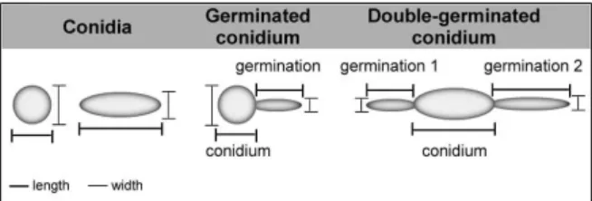
trypsin and 1 mM EDTA; Sigma-Aldrich, St. Louis, MO, USA) for 5 min at 37ºC. Then, the micro centrifuge tubes were harvested by centrifugation to remove the trypsin and added of a solution (1 mL) containing 0.01% Tween 80 (Sigma-Aldrich, St. Louis, MO, USA). The systems were vortexed for 1 min to release the adhered fungal cells. The cells were washed once with cold PBS and fixed in 4% paraformaldehyde at 4ºC for 30 min. Each fungal population was mapped (30,000 events) using a two-parameter histogram of forward-angle light scatter (FSC) versus side scatter (SSC), in order to evaluate size and granularity, respectively (Santos et al. 2012, Hayer et al. 2013). In parallel, the morphology of S. apiospermum, S. aurantiacum, S. minutisporum and L. prolificans cells were evaluated under light microscopy using a Zeiss Ax-ioplan 2 microscope with a 63´ objective lens and a final magnification of ´630 (Zeiss, Germany) (Silva et al. 2011). The dimensions (length and width) of 50 conidia and 50 germinated conidia of each species were measured as ex-emplified in Fig. 1. Considering the germinated conidia, the length and width of both conidium body and germi-nation projection were separately measured (Fig. 1). In the specific case of double-germinated conidia (Fig. 1), the final length and width of the germination projections were considered as the arithmetic mean of the individual measurements considering each analysed parameter.
Modulation of conidial germination by physico-chemical conditions - In this set of experiments, the fungal germination was evaluated by incubating the conidial cells under different growth conditions. In this way, conidia (104 cells) were incubated for 4 h in distinct
(i) culture media [Dulbecco’s modified Eagle’s medium (DMEM; Sigma-Aldrich, St. Louis, MO, USA), fetal bo-vine serum (FBS; Gibco, Life Technologies, USA) and Sabouraud], (ii) pH values (5.0, 7.0 and 9.0), tempera-tures (21ºC, 37ºC and 40ºC) and CO2 tensions (0.033% and 5%). The number of fungal morphotypes and viabil-ity were analysed as described above.
In vitro susceptibility testing - The in vitro antifun-gal susceptibility testing was performed using differ-ent fungal morphotypes (initial inoculum of 104
fun-gal cells). Conidia, 4 h-old-germinated conidia and 16 h-old-germinated conidia (in which just mycelia were observed) were used to investigate their susceptibility profiles to itraconazole (concentrations ranging from
0.03-128 µg/mL), fluconazole (0.03-256 µg/mL), vori-conazole (0.03-128 µg/mL), caspofungin (0.06-128 µg/ mL) and amphotericin B (0.03-128 µg/mL) (Sigma-Al-drich, St. Louis, MO, USA) by using broth microdilution method standardised for conidial cells of filamentous fungi according to the document M38-A2 published by the Clinical and Laboratory Standards Institute (CLSI 2008) and for hyphal cells as earlier proposed by Bez-jak (1985). The plates were then incubated for 48 h at 37ºC. The MICs were determined by visual inspection and confirmed with XTT-based reduction assay as the least concentration with no XTT reduction, which char-acterises the fungal cells with inactive metabolism. As recommended by CLSI, Candida krusei (ATCC 6258) and C. parapsilosis (ATCC 22019) were used as quality control isolates in each test. In all the susceptibility ex-periments, systems containing Roswell Park Memorial Institute (RPMI, Sigma-Aldrich, St. Louis, MO, USA) medium plus fungal suspension, RPMI plus tested an-tifungal drugs solutions (dissolved in dimethylsulfoxide - DMSO, Sigma-Aldrich, St. Louis, MO, USA), RPMI plus DMSO, RPMI plus DMSO plus fungal suspension and RPMI only were used as controls.
Statistics - All the experiments were performed in triplicate, in three independent experimental sets. The data were expressed as mean ± standard deviation. Re-sults were evaluated by Student’s t-test using Graphpad Prism 5 computer software. In all analyses, p values of 0.05 or less were considered statistically significant.
RESULTS AND DISCUSSION
Time-dependence of conidial germination - Germi-nation is a key event in fungal pathogenesis, because it allows the pathogen to be capable of adhering, spread-ing and invadspread-ing different cells and tissues in the host (D’Enfert 1997, Osherov & May 2001, van Burik & Ma-gee 2001, Gow et al. 2002). For instance, the susceptibil-ity of the insect larvae of Galleria mellonella to infection by A. fumigatus was directly dependent upon the stage of conidial germination, as follows: non-germinating (or resting) conidia < early stages of the germination < outgrowth phase of germination (Renwick et al. 2006). Furthermore, the examination of the immune response of G. mellonella to the fungal infection revealed that hemo-cytes were able to engulf non-germinating conidia and those in the early stages of the germination process, while conidia that reached the outgrowth stages of germination were not phagocytosed (Renwick et al. 2006). However, the mechanisms underlying this essential process remain poorly understood in filamentous fungi, especially in species belonging to the Scedosporium/Pseudallescheria complex as well as related species like L. prolificans.
The term germination usually implies the emergence of a definite germ tube-like projection from conidial cell (Allen 1965, D’Enfert 1997). Taking it into consideration, we initially analysed the time-dependence kinetics of the morphological transformation after incubation of S. apiospermum, S. aurantiacum, S. minutisporum and L. prolificans conidial cells in Sabouraud medium (pH 7.0) at 37ºC up to 4 h in an atmosphere of 5% CO2 (Fig. 2).
No conidial germination was detected in the first hour of incubation under the employed experimental conditions, while an extremely low germination rate (< 3%) was ob-served in the studied fungi after 2 h (Fig. 2). These rates significantly increased after 3 h of incubation to 30.5%, 59.4%, 36.6% and 32.0% in S. apiospermum, S. auran-tiacum, S. minutisporum and L. prolificans, respective-ly. After 4 h, the germination reached around 75% in S. minutisporum and more than 90% in S. apiospermum, S. aurantiacum and L. prolificans (Fig. 2).
Meletiadis et al. (2001) showed that the germina-tion of L. prolificans conidial cells started only after 4 h of incubation in Sabouraud and antibiotic medium 3 (AM3), after 5 h of incubation in RPMI and RPMI sup-plemented with 2% glucose and after 7 h of incubation in yeast nitrogen base (YNB). In addition, complete germi-nation was not achieved in any of the tested media even after 20 h of incubation at 37ºC in atmospheric concen-tration of CO2 (0.033%). In A. fumigatus, the germination of conidia started after 5 h of incubation in the five nu-trient media (AM3, YNB, Sabouraud, RPMI alone and RPMI with 2% glucose), although it was delayed by 1.5 h in YNB medium (Meletiadis et al. 2001). Ghamrawi et al. (2015) found around 30% of germinated conidia in S. boydii after incubation in yeast peptone dextrose for 4 h at 37ºC in atmospheric concentration of CO2. All these controversial results, including our own findings, could be explained due to the employment of different growth conditions such as culture medium composition and CO2 concentration, which are two relevant param-eters that modulate the differentiation process in several fungi (Allen 1965, D’Enfert 1997, Osherov & May 2001, Wächtler et al. 2012, Gilmore et al. 2013).
An early visual indicator of conidial germination in-volves the isotropic swelling of conidia before switch-ing to polarised growth, which results in the formation of a germ tube-like emergence and further mycelial development (Allen 1965, D’Enfert 1997). The results summarised in Fig. 2 suggested that in the first 2 h of
incubation, under the employed conditions in the pres-ent study, S. apiospermum, S. aurantiacum, S. minutis-porum and L. prolificans conidial cells did not develop the germ tube extension, probably because it was the pe-riod of conidial isotropic growth. In order to verify this hypothesis, the length and width of conidial cells were measured after 0, 1 and 2 h (Fig. 3). Our results showed that the conidial length increased around 15-30% and the conidial width around 15-20% after 2 h (Fig. 3). Howev-er, no significant differences on both morphological pa-rameters (length and width) were observed for any of the studied fungi at this time interval (Fig. 3). In contrast, during the conidial swelling of A. niger, Fusarium oxys-porum, Penicillium discolor and Verticillium fungicola, the diameter of the conidia increased two-fold or more and it involved water uptake and a decrease in the micro-viscosity of the cytoplasm (van Leeuwen et al. 2010).
Germination process was also monitored using flow cytometry through modifications in cell size (FSC) and granularity (SSC) as previously reported by A. niger, in which the first parameter provides quantifiable data on conidial swelling (Hayer et al. 2013). Corroborating the data exposed in Fig. 3, the FACS analysis revealed an augmentation around 15-40% in the conidia size dur-ing the pre-germinative stage (comparison between
Fig. 2: time-dependence kinetics of conidial germination in Scedospo-rium apiospermum, S. minutisporum, S. aurantiacum and Lomentospo-ra prolificans. Conidial cells were incubated in Sabouraud medium at 37ºC in an atmosphere of 5% CO2. After 0, 1, 2, 3 and 4 h of incubation,
the number of non-germinated and germinated conidial cells (please, see representative images in Fig. 1) were counted by using an inverted microscope. The results are expressed as percentage of germinated co-nidia in comparison to remaining coco-nidial cells. The results are shown as the mean ± standard deviation of three independent experiments.
0-2 h) (Fig. 4). However, in view of the whole germina-tion process (4 h), our results revealed that both mor-phometric parameters increased in a time-dependent manner (Fig. 4A-B), corroborating changes on the size and granularity (internal complexity) of fungal cells compared to dormant 0-h conidia. These results are in accordance with biological processes required for co-nidial germination such as increase in metabolic ac-tivities, including synthesis of new RNA, proteins and molecules that constitute the new membranes and cell wall being formed (Osherov & May 2001). Further on, changes in the expression of surface molecules and in the cellular architecture were reported. For example, the surface of A. fumigatus conidial cells contain hydropho-bins and melanin, while germinated conidia presented
a-1,3-glucan, galactomannan and
galactosaminogalac-tan exposed at the cell wall (Latgé & Beauvais 2014). The lipid composition of the plasma membrane also changes by the appearance of sterol-rich domains (van Leeuwen et al. 2010). Moreover, it is well-known that there is a rise in both endogenous respiratory rate and the rate of oxidation of carbon sources under conditions that permit germination. Conversely, if the external con-ditions needed for germination are removed, the rise in respiratory potential stops (Allen 1965, Osherov & May 2001). Supporting this statement, A. fumigatus was un-able to germinate under anaerobic conditions; contrari-ly, active mitochondria were evidenced by fluorescent mitotracker dye already at the stage of swollen conidia, which indicated that respiration is an early event during germination (Taubitz et al. 2007). In this sense, a clear time-dependent augmentation in the mitochondrial ac-tivity, as determined by the metabolic reduction of XTT, was evidenced during the transformation of conidia into germinated conidia of S. apiospermum, S. aurantiacum, S. minutisporum and L. prolificans (Fig. 4C).
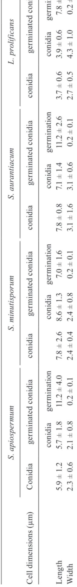
Subsequently, we performed an inspection of both conidial and germinated conidial cells by light micros-copy in order to better visualise these different fungal morphotypes (Fig. 5). The conidia of S. apiospermum were ovals in shape, measuring approximately 5.9 ± 1.2 µm x 2.3 ± 0.6 µm, and the germination projection was observed only from one site (pole) of each conidium, measuring around 11.2 ± 4.0 µm x 0.2 ± 0.1 µm (Fig. 5,
Fig. 4: morphological parameters and metabolic activity evaluated during the transformation from conidia to germinated conidia of Sce-dosporium apiospermum, S. minutisporum, S. aurantiacum and Lo-mentospora prolificans. In this set of experiments, conidial cells were incubated in Sabouraud medium at 37ºC in an atmosphere of 5% CO2 up to 4 h. After 0, 1, 2, 3 and 4 h, fungal cells were processed to esti-mate the size (forward scatter parameter) and granularity (side scatter parameter) by flow cytometry analysis and the results were expressed as fluorescence arbitrary units (FAU). In parallel, the mitochondrial activity was measured by monitoring the metabolic reduction of XTT at 492 nm using a microplate reader. The results are shown as the mean ± standard deviation of three independent experiments. p values were obtained comparing the dormant 0 h conidial cells to the other time-points in which the conidia were collected, and the diamond symbols indicate that p < 0.05 (Student’s t test).
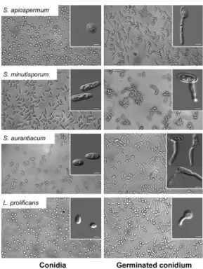
Fig. 5: light microscopies showing the dormant (0 h-conidia) and germi-nated conidial cells of Scedosporium apiospermum, S. minutisporum,
S. aurantiacum and Lomentospora prolificans after 4 h of incubation in Sabouraud medium at 37ºC in an atmosphere of 5% CO2. The insets highlighted the different morphologies regarding both conidia and
Table I). The conidia of S. minutisporum had ellipsoidal shape, with one of the straight edges and the other one rounded, measuring 7.8 ± 2.6 µm x 2.4 ± 0.4 µm and the germination was observed emerging from the central part of the conidial cell, measuring around 7.0 ± 1.6 µm x 0.2 ± 0.1 µm (Fig. 5, Table I). The conidia of S. auranti-acum were oval to cylindrical, measuring about 7.8 ± 0.8 µm x 3.1 ± 1.6 µm and the germination (11.2 ± 2.6 µm x 0.2 ± 0.1) can emerge from the middle, from one extrem-ity or from both conidial tips (Fig. 5, Table I). Germina-tion in both ends is called “bipolar germ-cell” and was also observed in other fungal species, such as Ashbya gossypii (Wendland & Philippsen 2001). Interestingly, this kind of morphology allows a more efficiently space exploration (Harris 2005). The conidia of L. prolificans had oval or globose shapes, measuring 3.7 ± 0.6 µm x 2.7 ± 0.5 µm, and the germination projection (7.8 ± 3.1 µm x 0.2 ± 0.1 µm) can appear in any part of the conidial surface (Fig. 5, Table I).
Effect of culture medium and pH on conidial germi-nation - It is well-known that conidial germination oc-curs in environments containing available water and the appropriate concentration of low molecular mass nutri-ents, like sugars, amino acids and inorganic acids (Os-herov & May 2001). For example, water supplemented with d-glucose was sufficient to enable conidial
ger-mination of A. niger (Hayer et al. 2013). A. fumigatus germination and hyphal growth in the mammalian lung, following the survival of resident pulmonary defenses, require the activation of nutrient-sensing, acquisition and biosynthetic pathways to obtain nutrients from the host environment (Dagenais & Keller 2009).
In order to evaluate the conidial germination of S. apiospermum, S. aurantiacum, S. minutisporum and L. prolificans under cultivation in different growth media, conidia were incubated in Sabouraud, DMEM and FBS at neutral pH at 37ºC for 4 h in an atmosphere of 5% CO2. In all fungal species, no significant difference re-garding the germination rate was observed among the culture media studied; however, S. minutisporum pre-sented a lower rate of differentiation (74.3%) compared to S. apiospermum (94.9%), S. aurantiacum (97.8%) and L. prolificans (87.2%) (Fig. 6). Sabouraud medium was selected for the further experiments because it is the cul-ture medium used to the growth of all these fungi.
Subsequently, the influence of pH on the germina-tion process was studied by incubating conidial cells in Sabouraud medium buffered at acidic, neutral and alka-line pH for 4 h at 37ºC with 5% CO2. It is important to highlight that fungal viability was not affected by the incubation under different pH values (data not shown). Conidia of each fungal species were able to germinate in very similar rates regardless of pH value (Fig. 7). We be-lieve that pH did not influence the conidial germination because all the nutrients required to this process were satisfactorily available in the studied media (Carlile et al. 2001). Once again, S. minutisporum showed the lowest percentage of germination compared to the other stud-ied fungal species concerning each analysed pH (Fig. 7). Several fungal species are also capable of differentiating
T A B L E I L e n g th a n d w id th o f c o n id ia a n d g e rm in at e d c o n id ia o f S c e d o sp or iu m a p io sp e rm u m , S . m in u ti sp or u m , S . a u ra nt ia c u m a n d L o me n to sp or a p ro li fi c a n s C el l d im e n si o n s ( µ m ) S . a pi o sp e rm u m S . m in u ti sp or u m S . a u ra nt ia c u m L . p ro li fi c a n s C o n id ia g e rm in at e d c o n id ia c o n id ia g e rm in at e d c o n id ia c o n id ia g e rm in at e d c o n id ia c o n id ia g e rm in at e d c o n id ia c o n id ia g e rmi n ati o n c o n id ia g e rmi n ati o n c o n id ia g e rmi n ati o n c o n id ia g e rmi n ati o n L e n g th 5
.9 ± 1
.2
5
.7 ± 1
.8
11
.2 ± 4
.0
7.
8 ± 2
.6
8.
6 ± 1
.3
7.
0 ± 1
.6 7.8 ± 0 .8 7.
1 ± 1
.4
11
.2 ± 2
.6
3
.7 ± 0
.6
3
.9 ± 0
.6
7.
8 ± 3
.1
Wi
d
th
2
.3 ± 0
.6
2
.1 ± 0
.8 0. 2 ± 0. 1 2 .4 ± 0 .4 2
.4 ± 0
.8 0. 2 ± 0. 1 3
.1 ± 1
.6
3
.1 ± 0
.6 0. 2 ± 0. 1 2
.7 ± 0
.5
4
.3 ± 1
in a broad range of pH; as an example, Potebniamyces pyri conidial cells were able to fully germinate in pH values ranging from 4.0-7.0 (Liu & Xiao 2005).
Collectively, these results demonstrated the ability of S. apiospermum, S. aurantiacum, S. minutisporum and L. prolificans conidia to differentiate at acid-to-basic environments as well as in different nutritional media, which can reflect their prodigious ability to colonise and to invade different sites of human body and natural envi-ronments (Cortez et al. 2008, Kaltseis et al. 2009).
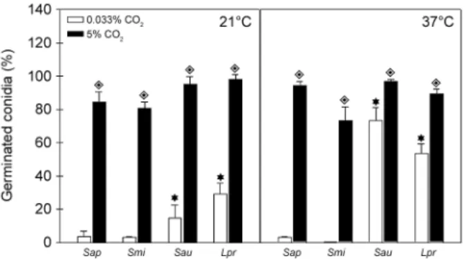
Effect of temperature and CO2 on conidial germina-tion - The ability of a microorganism to grow in human body temperature under both normal and fever condi-tions is an important requisite to cause systemic infec-tion (van Burik & Magee 2001). Another interesting parameter to be analysed is the CO2 level. In mamma-lian tissues, the concentration of CO2 is approximately 150-fold higher when compared to the atmospheric CO2 concentration; consequently, pathogens are exposed to drastic differences considering superficial infections in comparison to deep infections (Klengel et al. 2005). Herein, it was evaluated whether the conidia of S. ap-iospermum, S. aurantiacum, S. minutisporum and L. prolificans were able to differentiate under temperature conditions simulating environmental (21ºC), healthy hu-man body (37ºC) and fever condition (40ºC) in the pres-ence of 0.033% (atmospheric level) or 5% (concentration found in mammalian tissues) of CO2.
Firstly, we analysed the differentiation of conidia at temperatures of 21ºC, 37ºC and 40ºC in an environment containing 5% CO2, and the results demonstrated a com-parable ability of conidial cells to differentiate after 4 h of incubation either at 21ºC or 37ºC (Fig. 8); however, germination was not detected at 40ºC in any studied fungi (data not shown). Furthermore, conidia of S. apio-spermum and S. minutisporum did not differentiate even after incubation for 16 h at 40ºC in an environment with 5% of CO2; however, conidia of S. aurantiacum and L. prolificans were able to fully germinate under these con-ditions (data not shown). Secondly, a completely distinct profile was observed when the studied fungi were incu-bated in a lower concentration of CO2. In this sense, after 4 h of incubation at 21ºC under atmospheric concentra-tion of CO2, only 3.9% of S. apiospermum, 14.8% of S.
Fig. 6: evaluation of conidial germination of Scedosporium apiosper-mum, S. minutisporum, S. aurantiacum and Lomentospora prolificans
in different culture media. Conidial cells were incubated for 4 h in Sab-ouraud, Dulbecco’s modified Eagle’s medium (DMEM) or fetal bovine serum (FBS) at 37ºC with an atmosphere of 5% CO2. After incubation, fungal cells were counted by using an inverted microscope. The re-sults are shown as the mean ± standard deviation of three independent experiments. There was no statistical difference in the germination of each fungal species when cultured under these different culture media.
Fig. 7: evaluation of conidial germination of Scedosporium apiosper-mum, S. minutisporum, S. aurantiacum and Lomentospora prolifi-cans in different pH values. Conidial cells were incubated for 4 h at 37ºC with an atmosphere of 5% CO2 in Sabouraud medium in which
the pH was adjusted to 5.0, 7.0 and 9.0. After incubation, fungal cells were counted by using an inverted microscope. The results are shown as the mean ± standard deviation of three independent experiments. There was no statistical difference in the germination of each fungal species when cultured in different pH values.
Fig. 8: evaluation of conidial germination of Scedosporium apiosper-mum (Sap), S. minutisporum (Smi), S. aurantiacum (Sau) and Lomen-tospora prolificans (Lpr) in different temperatures and CO2 tensions. Conidial cells were incubated for 4 h on Sabouraud medium at 21ºC and 37ºC in an atmosphere of 5% CO2 or 0.033% CO2. After
incuba-tion, fungal cells were counted by using an inverted microscope. The results are shown as the mean ± standard deviation of three indepen-dent experiments. Diamonds represent the significant difference (p
< 0.05, Student’s t test) when the germination of each fungus was compared under different CO2 tensions (5% or 0.033% CO2), while
aurantiacum, 3.5% of S. minutisporum and 29.2% of L. prolificans conidia were capable of germinating (Fig. 8). When the temperature was changed to 37ºC (0.033% CO2), a considerable increase in the germination rate of S. aurantiacum (73.3%) and L. prolificans (53.5%) was detected, while no alteration regarding the germination level was observed in S. apiospermum and S. minutis-porum (Fig. 8). Finally, the fungi studied herein were not able to differentiate after incubation for 4 h at 40ºC with atmospheric concentration of CO2. Due to this later result, we decided to conduct an additional set of ex-periment in order to evaluate the conidial viability. By checking their mitochondrial activity, all the fungal spe-cies analysed presented similar ability to convert XTT in formazan after incubation under the atmospheric con-centration of CO2 for 4 h at 21ºC, 37ºC and 40ºC, being all these conditions able to sustain the fungal viability (data not shown). Kaur et al. (2015) demonstrated that both clinical and environmental strains of S. aurantia-cum presenting higher level of virulence also displayed flexibility and metabolic adaptability to different tem-peratures ranging from 28ºC to 37ºC. Several studies suggested that the germination rate of Aspergillus spp. at 37ºC correlate with pathogenicity in multiple animal models of invasive aspergillosis (Dagenais & Keller 2009). The germination rates of A. fumigatus, A. flavus and A. niger were similar at temperatures up to 30ºC, but differed at 37ºC and 42ºC (Araujo & Rodrigues 2004).
CO2 is long known to be an essential factor for the ger-mination of bacterial spores. Similarly, in Aspergillus co-nidia, CO2 is one of the essential factors for the initiation of germination (Yanagita 1957). Yanagita (1963) found that, after the germination of A. niger conidia had started, the incorporation of 14CO
2 proceeded actively without any
lag, and macromolecular substances, such as nucleic acids and proteins, were labeled very rapidly. The importance of 5% CO2 during the conidial germination process in S. apiospermum, S. aurantiacum, S. boydii, S. minutis-porum and L. prolificans is corroborated by studies done by other authors, who analysed conidial germination in L. prolificans (Meletiadis et al. 2001), S. apiospermum and S. boydii (Pinto et al. 2004, Santos et al. 2009, Lopes et al. 2010, Ghamrawi et al. 2015) after incubation at both room temperature and 37ºC under environmental atmospher-ic concentration of CO2. In all those works, the authors found low percentages of germinated conidia.
CO2 levels can change the physiology of fungal cells through changes in environmental acidity and those lev-els are used by fungal pathogens as a signal for modulat-ing the expression of virulence factors (Lang-Yona et al. 2013). For example, an atmosphere of 5% CO2 induced the pseudohyphae formation in C. albicans as well as capsule production in Cryptococcus neoformans (Klen-gel et al. 2005, Mitchell 2005). Conidia of Aspergillus were not able to germinate in the absence of CO2, even when cultivated in medium containing all the essential nutrients that allow full growth (Yanagita 1957). These results showed that the germination of conidia was high-ly induced by the presence of CO2 and thus favor dif-ferentiation in environments with this condition, such as mammalian tissues and bloodstream.
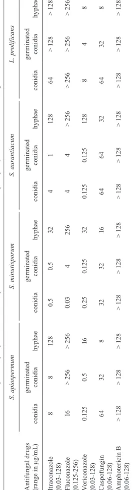
Antifungal susceptibility of conidia, germinated co-nidia and hyphae - Traditionally, standardised methods for in vitro susceptibility testing of filamentous fungi use exclusively inoculum of conidia, which are not the unique and/or prevalent morphological form found in the tissues/organs of the infected host. In this context, most infections caused by filamentous fungi are charac-terised by the presence of hyphal elements in tissue (van Burik & Magee 2001, Gow et al. 2002, Araujo & Ro-drigues 2004). Tests performed with hyphae could then predict the therapeutic potential of a drug, which could avoid treatment failures (van de Sande et al. 2010). Cor-roborating these findings, conidial and mycelial cells are usually found in human tissues infected by Scedo-sporium species (Cortez et al. 2008). Despite these data, almost nothing is known about the influence of different morphotypes on the susceptibility profiles to current an-tifungal drugs (Wetter et al. 2005). Aggravating this sce-nario, the few published studies comparing the suscep-tibility of conidia and hyphae of filamentous fungi are controversial. For instance, the results published in the literature with conidial and hyphal forms of A. fumigatus revealed that the MIC values were similar for ampho-tericin B, itraconazole, voriconazole and posaconazole (Bezjak 1985, Manavathu et al. 1999, Wetter et al. 2005). On the other hand, experiments conducted with species belonging to the Scopulariopsis, Paecilomyces, Clado-sporium and Cladophialophora genera demonstrated that MICs for hyphae were higher than for conidia when the antifungals amphotericin B, fluconazole, ketocon-azole, flucytosine, miconazole and itraconazole were employed (Guarro et al. 1997).
In this work, we performed the susceptibility test in order to identify possible differences among conidia, ger-minated conidia and hyphae of S. apiospermum, S. auran-tiacum, S. minutisporum and L. prolificans. Our results showed that the antifungal susceptibility profiles varied regarding each morphotype and each fungal species (Ta-ble II). In general, the MICs for hyphae were practically always substantially higher than for conidia and germi-nated conidia. The exception to this profile was caspofun-gin, for which the MIC values for hyphae were lower than for the remaining morphotypes in all tested fungi (Table II). Although in this work only one strain of each fungal species was studied, the MIC values found herein are in complete agreement with the MICs published by other authors, who worked with several strains (Gilgado et al. 2006, Wiederhold & Lewis 2009, Lackner et al. 2012). In this context, our data (Table II) confirm previously pub-lished results regarding the high degree of multidrug re-sistance of L. prolificans to antifungals irrespective of the method of detection used (Alvarez et al. 1995, Lackner et al. 2012), whereas S. minutisporum was the more suscep-tible species to azoles (Lackner et al. 2012).
be-T A B L E I I S u sc e p ti b il it y p ro fi le s ( m in im a l i n h ib iot ry c o n ce n tr at io n i n µ g /m L ) o f c o n id ia , g e rm in at e d c o n id ia a n d h y p h a e o f S c e d o sp or iu m a p io sp e rm u m , S . m in u ti sp or u m , S . a u ra nt ia c u m a n d L o me n to sp or a p ro li fi c a n s t o d if fe re n t a nt if u n g al s A n ti fu n ga l d ru gs (r a n g e i n µ g /m L ) S . a pi o sp e rm u m S . m in u ti sp or u m S . a u ra nt ia c u m L . p ro li fi c a n s c o n id ia g er m in ate d c o n id ia hy p h a e c o n id ia g er m in ate d c o n id ia hy p h a e c o n id ia g er m in ate d c o n id ia hy p h a e c o n id ia g er m in ate d c o n id ia hy p h a e It ra c o n a z ol e (0 .0 3 -1 2 8) 8 8 12 8 0. 5 0. 5 32 4 1 12 8 6 4 > 12 8 > 12 8 F lu c o n a z ol e (0 .1 25 -25 6 ) 16 > 2 56 > 2 56 0 .0 3 4 2 56 4 4 > 2 56 > 2 56 > 2 56 > 2 56 V o ri c on a z o le (0 .0 3 -1 2 8) 0 .1 2 5 0. 5 16 0. 2 5 0 .1 2 5 32 0 .1 2 5 0 .1 2 5 12 8 8 4 8 C a sp of u n g in (0 .0 6 -1 2 8) 6 4 32 8 32 32 16 6 4 6 4 32 6 4 32 8 A m p h ot e ri ci n B (0 .0 6 -1 2 8) > 12 8 > 12 8 > 12 8 > 12 8 > 12 8 > 12 8 > 12 8 > 12 8 > 12 8 > 12 8 > 12 8 > 12 8
cause MIC values above 2 µg/mL have been associated with the treatment failure of aspergillosis (Lass-Florl et al. 1998). Correlation of minimal effective concentration concerning the clinical outcome to caspofungin must be yet elucidated; however, we do assume that the three morphotypes of all fungal species tested in our work were resistant to this antifungal drug, because the pro-tocol published by CLSI document M38-A2 (CLSI 2008) recommends its use in a concentration range varying from 0.015-8 µg/mL and, in our records, the MICs were always equal or higher than 8 µg/mL. The same situation was observed for fluconazole, in which the concentration range recommended by CLSI is 0.125-64 µg/mL (CLSI 2008). Our results pointed out that hyphae of all fungal species were resistant to fluconazole (MICs ≥ 256 µg/ mL) as well as the germinated conidia of both L. pro-lificans and S. apiospermum (MIC > 256 µg/mL) and conidia of L. prolificans (MIC > 256 µg/mL), while the remaining species and conidia of S. apiospermum had a MIC lower than 16 µg/mL, and fungal strains with MICs lower than 64 µg/mL are considered susceptible to this drug (Saracli et al. 2003). Hyphae of all species as well as conidia and germinated conidia of L. prolificans and S. apiospermum were also considered resistant (> 8 µg/ mL) to itraconazole, whereas both conidial and germi-nated conidial cells of S. minutisporum, S. aurantiacum were susceptible to this drug. Voriconazol was the most effective drug against conidia and germinated conidia of S. apiospermum, S. aurantiacum, S. minutisporum and L. prolificans, which is in agreement with data found by other authors in the literature (Lackner et al. 2012, Biswas et al. 2013); however, hyphae of S. minutisporum (MIC 32 µg/mL) and S. aurantiacum (MIC 128 µg/mL) presented higher MIC than the maximum concentration (16 µg/mL) for this antifungal proposed by CLSI (2008). However, voriconazole has multiple drug interactions with medications used in immunosuppression of organ transplant recipients, which does not allow its use in those cases (O’Bryan 2005). Thereby, the optimal treatment to combat the infections caused by Scedosporium/Pseud-allescheria and Lomentospora in immunosuppressed pa-tients is still completely unknown (Lackner et al. 2012).
Concisely, our data are in concordance with Lackner et al. (2012), who showed that Scedosporium species do not have a normal MIC distribution, which generates a great difficulty to select a drug to be used in clinical settings. These findings point out to the necessity to the accurate differential diagnosis of these fungal species in order to permit a correct clinical treatment.
temperatures, which can reflect their abilities to colonise several sites of human body and natural environments. CO2 was a substantial inducer of the conidia-into-hy-phae transformation in S. apiospermum, S. aurantia-cum, S. minutisporum and L. prolificans. Furthermore, the susceptibility to antifungals was dependent of the cell morphotype, with hypha highly resistant to the ma-jority of the tested drugs. In brief, we can conclude that our results add novel data to clarify the complex phe-nomenon regarding the crucial transition of conidia into filamentous form in these fungal species. Finally, stud-ies on the cell differentiation mechanisms may also aid the elucidation of antifungal resistance of these relevant human opportunistic pathogens.
ACKNOWLEDGEMENTS
To Denise Rocha de Souza, for her technical assistance.
REFERENCES
Aimanianda V, Bayry J, Bozza S, Kniemeyer O, Perruccio K, Elluru SR, et al. Surface hydrophobin prevents immune recognition of airborne fungal spores. Nature. 2009; 460(7259): 1117-21. Allen PJ. Metabolic aspects of spore germination in fungi. Annu Rev
Phytopathol. 1965; 3: 313-42.
Alvarez M, Lopez PB, Rayon C, Garcia GJ, Roson PMC, Gonzalez M, et al. Nosocomial outbreak caused by Scedosporium prolificans
(inflatum): four fatal cases in leukemic patients. J Clin Microbiol. 1995; 33(12): 3290-5.
Araujo R, Rodrigues A. Variability of germinative potential among patho-genic species of Aspergillus. J Clin Microbiol. 2004; 42(9): 4335-7. Bezjak V. Standardization of a hyphal inoculum of aspergilli for
ampho-tericin B susceptibility testing. J Clin Microbiol. 1985; 21(4): 509-12. Biswas C, Sorrell TC, Djordjevic JT, Zuo X, Jolliffe KA, Chen SCA.
In vitro activity of miltefosine as a single agent and in combination with voriconazole or posaconazole against uncommon filamentous fungal pathogens. J Antimicrob Chemother. 2013; 68(12): 2842-6. Blyth CC, Harun A, Middleton PG, Sleiman S, Lee O, Sorrell TC, et
al. Detection of occult Scedosporium species in respiratory tract specimens from patients with cystic fibrosis by use of selective media. J Clin Microbiol. 2010; 48(1): 314-6.
Carlile MJ, Watkinson SC, Goody GW. The fungi. 2nd ed. London: Academic Press; 2001.
CLSI - Clinical Laboratory Standards Institute. Reference method for broth dilution antifungal susceptibility testing of filamen-tous fungi; approved standard, CLSI document M38-A2. 2nd ed. Wayne: CLSI; 2008. p. 52.
Cortez KJ, Roilides E, Quiroz-Telles F, Meletiadis J, Antachopoulos C, Knudsen T, et al. Infections caused by Scedosporium spp. Clin Microbiol Rev. 2008; 21(1): 157-97.
D’Enfert C. Fungal spore germination: insights from the molecular genetics of Aspergillus nidulans and Neurospora crassa. Fungal Genet Biol. 1997; 21: 163-72.
Dagenais TRT, Keller NP. Pathogenesis of Aspergillus fumigatus in invasive aspergillosis. Clin Microbiol Rev. 2009; 22(3): 447-65. Dague E, Alsteens D, Latgé JP, Dufrene YF. High-resolution cell
surface dynamics of germinating Aspergillus fumigatus conidia. Biophys J. 2008; 94(2): 656-60.
Ghamrawi S, Gastebois A, Zykwinska A, Vandeputte P, Marot A, Mabilleau G, et al. A multifaceted study of Scedosporium boydii
cell wall changes during germination and idenfitication of GPI-anchored proteins. PLoS ONE. 2015; 10(6): e0128680.
Gilgado F, Gené J, Cano J, Guarro J. Heterothallism in Scedospo-rium apiospermum and description of its teleomomorph Pseud-allescheria apiosperma sp. nov. Med Mycol. 2010; 48(1): 122-8. Gilgado F, Serena C, Cano J, Gené J, Guarro J. Antifungal
suscep-tibilities of the species of the Pseudallescheria boydii complex. Antimicrob Agents Chemother. 2006; 50(12): 4211-3.
Gilmore SA, Naseem S, Konopka JB, Sil A. N-acetylglucosamine (GlcNAc) triggers a rapid, temperature-responsive morphoge-netic program in thermally dimorphic fungi. PLoS ONE. 2013; 9(9): e1003799.
Gow NAR, Brown AJP, Odds FC. Fungal morphogenesis and host invasion. Curr Opin Microbiol. 2002; 5(4): 366-71.
Guarro J, Llop C, Aguiar C, Pujol I. Comparison of in vitro antifungal susceptibilities of conidia and hyphae of filamentous fungi. Anti-microb Agents Chemother. 1997; 41(12): 2760-2.
Harris SD. Morphogenesis in germinating Fusarium graminearum
macroconidia. Mycologia. 2005; 97(4): 880-7.
Hayer K, Stratford M, Archer DB. Structural features of sugars that trigger or support conidial germination in the filamentous fungus
Aspergillus niger. Appl Environ Microbiol. 2013; 79(22): 6924-31. Hohl TM, van Epps HL, Rivera A, Morgan LA, Chen PL, Feldmesser
M, et al. Aspergillus fumigatus triggers inflammatory responses
by stage-specific β-glucan display. PLoS Pathog. 2005; 1(3): e30.
Kaltseis J, Rainer J, de Hoog GS. Ecology of Pseudallescheria and
Scedosporium species in human-dominated and natural envi-ronments and their distribution in clinical samples. Med Mycol. 2009; 47(4): 398-405.
Kantarcioglu AS, de Hoog GS, Guarro J. Clinical characteristics and epidemiology of pulmonary pseudallescheriasis. Rev Iberoam Micol. 2012; 29(1): 1-13.
Kaur J, Pethani BP, Kumar S, Kim M, Sunna A, Kautto L, et al.
Pseudomonas aeruginosa inhibits the growth of Scedosporium aurantiacum, an opportunistic fungal pathogen isolated from the lungs of cystic fibrosis patients. Front Microbiol. 2015; 6: 866. Klengel T, Liang W, Chaloupka J, Ruoff C, Schroppel K, Naglik J, et
al. Fungal adenylyl cyclase integrates CO2 sensing with cAMP signaling and virulence. Curr Biol. 2005; 15(22): 2021-6. Lackner M, Guarro J. Pathogenesis of Scedosporium. Curr Fungal
Infect Rep. 2013; 7(4): 326-33.
Lackner M, Hoog G, Yang L, Moreno L, Ahmed S, Andreas F, et al. Proposed nomenclature for Pseudallescheria, Scedosporium and related genera. Fungal Diversity. 2014; 67(1): 1-10.
Lackner M, Hoog GS, Verweij PE, Najafzadeh MJ, Curfs-Breuker I, Klaassen CH, et al. Species-specific antifungal susceptibility patterns of Scedosporium and Pseudallescheria species. Antimi-crob Agents Chemother. 2012; 56(5): 2635-42.
Lang-Yona N, Levin Y, Dannemille KC, Yarden O, Peccia J, Rudich Y. Changes in atmospheric CO2 influence the allergenecity of
As-pergillus fumigatus. Glob Chang Biol. 2013; 19(8): 2381-8. Lass-Florl C, Kofler G, Kropshofer G, Hermans J, Kreczy A, Dierich
MP, et al. In vitro testing of susceptibility to amphotericin B is a reliable predictor of clinical outcome in invasive aspergillosis. J Antimicrob Chemother. 1998; 42(4): 497-502.
Latgé JP, Beauvais A. Functional duality of the cell wall. Curr Opin Microbiol. 2014; 20: 111-7.
Liu Q, Xiao CL. Influence of nutrient and environmental factors on conidial germination of Potebniamyces pyri. Phytopathology. 2005; 95(5): 572-80.
peptidor-hamnomannans of Scedosporium apiospermum enhance the patho-genecity of the fungus. PLoS Negl Trop Dis. 2010; 4(10): e853. Manavathu EK, Cutright J, Chandrasekar PH. Comparative study of
susceptibilities of germinated and ungerminated conidia of As-pergillus fumigatus to various antifungal agents. J Clin Micro-biol. 1999; 37(3): 858-61.
Meletiadis J, Meis JFGM, Mouton JW, Verweij PE. Analysis of growth characterisyics of filamentous fungi in different nutrient media. J Clin Microbiol. 2001; 39(2): 478-84.
Mitchell AP. Fungal CO2 sensing: a breath of fresh air. Curr Biol. 2005; 15(22): R934-6.
Mowat E, Butcher J, Lang S, Williams C, Ramage G. Development of a simple model for studying the effects of antifungal agents on multicellular communities of Aspergillus fumigatus. J Med Mi-crobiol. 2007; 56(Pt 9): 1205-12.
O’Bryan TA. Pseudallescheriasis in the 21st century. Expert Rev Anti Infect Ther. 2005; 3(5): 765-73.
Osherov N, May GS. The molecular mechanisms of conidial germina-tion. FEMS Microbiol Lett. 2001; 199(2): 153-60.
Peeters E, Nelis HJ, Coenye T. Comparison of multiple methods for quantification of microbial biofilms grown in microtiter plates. J Microbiol Methods. 2008; 72(2): 157-65.
Pinto MR, Limongi CL, Rozental S, Santos ALS, Barreto-Bergter E. Involvement of peptideorhamnomanana in the interaction of Pseu- dallescheria boydii and HEp2 cells. Microbes Infect. 2004; 6: 1259-67. Pinto MR, Rodrigues ML, Travassos LR, Haido RMT, Wait R, Bar-reto-Bergter E. Characterization of glucosylceramides in Pseu-dallescheria boydii and their involvement in fungal differentia-tion. Glycobiology. 2002; 12(4): 251-60.
Renwick J, Daly P, Reeves EP, Kavanagh K. Susceptibility of larvae of Galleria mellonella to infection by Aspergillus fumigatus is dependent upon stage of conidial germination. Mycopathologia. 2006; 161(6): 377-84.
Santos ALS, Bittencourt VCB, Pinto MR, Silva BA, Barreto-Bergter E. Biochemical characterization of potential virulence markers in the human fungal pathogen Pseudallescheria boydii. Med Mycol. 2009; 47(4): 375-86.
Santos ALS, Sodré CL, Valle RS, Silva BA, Abi-chacra EA, Silva LV, et al. Antimicrobial action of chelating agents: repercussions on the microorganism development, virulence and pathogenesis. Curr Med Chem. 2012; 19(17): 2715-37.
Saracli MA, Erdem U, Gonlum A, Yidiran ST. Scedosporium apio-spermum keratitis treated with itraconazole. Med Mycol. 2003; 41(2): 111-4.
Silva BA, Pinto MR, Soares RMA, Barreto-Bergter E, Santos ALS.
Pseudallescheria boydii releases metallopeptidases capable of cleaving several proteinaceous compounds. Res Microbiol. 2006; 157(5): 425-32.
Silva BA, Souza-Gonçalves AL, Pinto MR, Barreto-Bergter E, San-tos ALS. Metallopeptidase inhibitors arrest vital biological pro-cesses in the fungal pathogen Scedosporium apiospermum. My-coses. 2011; 54(2): 105-12.
Tammer I, Tintelnot K, Braun-Dullaeus RC, Mawrin C, Scherlach C, Schlüter D, et al. Infections due to Pseudallescheria/Scedospo-rium species in patients with advanced HIV disease - a diagnostic and therapeutic challenge. Int J Infect Dis. 2011; 15(6): e422-9. Taubitz A, Bauer B, Heesemann J, Ebel F. Role of respiration in the
germination process of the pathogenic mold Aspergillus fumiga-tus. Curr Microbiol. 2007; 54(5): 354-60.
Tintelnot K, Wagner N, Seibold M, de Hoog GS, Horré R. Re-identifi-cation of clinical isolates of the Pseudallescheria boydii-complex involved in near-drowning. Mycoses. 2008; 51(Suppl. 3): 11-6. van Burik JAH, Magee PT. Aspects of fungal pathogenesis in
hu-mans. Annu Rev Microbiol. 2001; 55: 743-72.
van de Sande WW, Tavakol M, van Vianen W, Bakker-Woudenberg IA. The effects of antifungal agents to conidial and hyphal forms of Aspergillus fumigatus. Med Mycol. 2010; 48(1): 48-55. van Leeuwen MR, van Doorn TM, Golovina EA, Stark J, Dijksterhuis J.
Water- and air-distributed conidia differ in sterol content and cyto-plasmic microviscosity. Appl Environ Microbiol. 2010; 76(1): 366-9. Wächtler B, Citiulo F, Jablonowski N, Förster S, Dalle F, Schaller
M, et al. Candida albicans-epithelial interactions: dissecting the roles of active penentration, induced endocytosis and host factors on the infection process. PLoS ONE. 2012; 7(5): e36952. Wendland J, Philippsen P. Cell polarity and hyphal morphogenesis
are controlled by multiple rho-protein modules in the filamentous ascomycete Ashbya gossypii. Genetics. 2001; 157(2): 601-10. Wetter T, Hazen K, Cutler J. Comparison between Aspergillus
fu-migatus conidia and hyphae susceptibilities to amphotericin B, itraconazole, and voriconazole by use of the mold rapid suscepti-bility assay. Med Mycol. 2005; 43(6): 525-32.
Wiederhold NP, Lewis RE. Antifungal activity against Scedosporium
species and novel asays to assess antifungal pharmacodynamics against filamentous fungi. Med Mycol. 2009; 47(4): 422-32. Yanagita T. Biochemical aspects on the germination of conidiospores
of Aspergillus niger. Arch Mikrobiol. 1957; 26(4): 329-44. Yanagita T. Carbon dioxide fixation in germinating conidiospores of