Maria Anice Mureb Sallum2,3, E L Peyton2, Bruce Arthur Harrison4 & Richard Charles Wilkerson2
ABSTRACT. Revision of the Leucosphyrus Group of Anopheles (Cellia) (Diptera, Culicidae). This is a comprehensive revision of 20 species of the Leucosphyrus Group of the Neomyzomyia Series of Anopheles (Cellia). Morphological description of the adults, male and female, male genitalia, pupa and fourth-instar larva are provided for each taxon in addition to bionomics, distribution data and systematic discussion for each species, including diagnostic characters. Identification keys for females and fourth-instar larvae are provided. When possible medical importance of each species is included. Illustrations of of the adults, fourth-instar larvae and pupae are provided. Distribution maps for each species are mainly based on the material examined; however, when possible published data were also used. Tables on adult character variations, fourth-instar larval and pupal setal branching are included as appendices. A neotype for An. takasagoensis Morishita and An. sulawesi Koesoemawinangoen, and a lectotype for An. balabacensis Baisas, are designated. The authorship of An. sulawesi previously cited as Waktoedi is corrected to Koesoemawinangoen. KEYWORDS. Anophelinae; Cellia; Distribution; Morphology; Revision; Taxonomy.
RESUMO. Revisão do Grupo Leucosphyrus de Anopheles (Cellia) (Diptera, Culicidae). Apresenta-se a revisão taxonômica de 20 espécies do Grupo Leucosphyrus da Série Neomyzomyia de Anopheles (Cellia). Apresentam-se as descrições morfológicas dos adultos, machos e fêmeas, genitália masculina, pupa e larva de quarto estádio, bem como dados de bionomia, distribuição e discussão sistemática para cada espécie, incluindo caracteres diagnósticos. Foram acrescentadas chaves dicotômicas de identificação para as espécies, utilizando caracteres dos adultos fêmeas e larvas de quarto estádio. Quando possível, foram incluídos dados sobre a importância médica de cada espécie. Foram também apresentadas ilustrações dos adultos, das larvas de quarto estádio e das pupas. Os mapas de distribuição das espécies foram elaborados com base no material examinado, no entanto, quando possível utilizou-se dados adicionais publicados na literatura. Fornecem-se tabelas com as variações dos caracteres dos adultos, das cerdas das larvas de quarto estádio das pupas. São designados os neótipos de An. takasagoensis Morishita e An. sulawesi Koesoemawinangoen, e o lectótipo de An. balabacensis Baisas. O nome do autor de An. sulawesi previamente citado como sendo Waktoedi foi corrigido para Koesoemawinangoen.
PALAVRAS CHAVE. Anophelinae; Cellia; Distribuição; Morfologia; Revisão; Taxonomia.
Species belonging to the genus Anopheles Meigen are vectors of parasites of the genus Plasmodium that cause human malaria, a primarily tropical disease that threatens 42 percent of the world population in 100 countries and territories. The World Health Organization estimates the annual global incidence of acute malaria cases at 300-500 million, resulting in an estimated 1.5 to 2.7 million deaths per year, of which approximately one million occur among children less than five years of age. In addition to their role as primary vectors of malaria, mosquitoes of the genus Anopheles additionally impact human health by transmitting lymphatic filariasis (Service 1993) and various mosquito-borne viruses (Gillies & Coetzee 1987).
The subgenus Cellia includes 224 of the 465 Anopheles
species and is divided into 6 informal series: Cellia, Myzomyia, Neocellia, Neomyzomyia, Paramyzomyia and Pyretophorus.
(Diptera, Culicidae)
11This research was performed under a Memorandum of Understanding between the Walter Reed Army Institute of Research and the Smithsonian
Institution, with institutional support provided by both organizations. The material to be published reflects the views of the authors and should be not construed to represent those of the Department of the Army or the Department of Defense. This publication is partially supported by FAPESP (05/00372-9), São Paulo, Brazil. / and U. S. NRC DAM D 17-00-2-0002 to MAMS.
2Department of Entomology, Walter Reed Biosystematics Unit, Museum Support Center, Smithsonian Institution, 4210 Silver Hill Road,
Suitland, MD, 20746, U.S.A.
3Núcleo de Pesquisa Taxonômica e Sistemática em Entomologia Médica, Faculdade de Saúde Pública, Universidade de São Paulo, Brazil.
Avenida Dr. Arnaldo 715, São Paulo, São Paulo, Brazil, 01246-904.
4NC DENR Public Health Pest Managment 585 Waughtown Street Winston-Salem, NC 27107, U.S.A.
The internal classification within the subgenus Cellia was first proposed by Edwards (1932) and later modified by several authors as seen in Harbach (2004). The subgenus Cellia was demonstrated to be monophyletic based on cladistic analyses of both morphological characters (Sallum et al. 2000) and DNA sequence data of several mitochondrial and nuclear genes (Krzywinski et al. 2001a,b; Sallum et al. 2002).
Leucosphyrus Group, its species, and geographical distribution are listed in Table 1.
This study of the Leucosphyrus Group is a product of over 25 years of planned collaborative efforts by many individuals. It started in 1977 when EL Peyton (deceased) of the U. S. Army’s Medical Entomology Project at the Smithsonian Institution, initiated a morphological study of the species in the Leucosphyrus Group. At that time the group was defined by the works of Colless (1956b, 1957) and Reid (1968), who recognized six species, two subspecies, and five geographical forms that contained several important malaria vectors in Southeast Asia. After six months of examining specimens available for study in the Smithsonian Institution, it became apparent that more species were probably involved, but additional specimens were needed to continue the study. In early 1978, Peyton and Harrison started a cooperative study with Harrison conducting field studies that targeted populations of the group, making larval and adult collections for rearing adults with associated larval and pupal exuviae, establishing colonies, conducting crossing studies, and shipping study specimens back to the Smithsonian Institution. The initial product of this joint arrangement was the discovery and description (Peyton & Harrison 1979) of a new species,
An. dirus Peyton and Harrison, masquerading as An. balabacensis Baisas, which until that time had been recognized as a primary vector of human malaria in an area stretching from east India to the Philippines. Shortly thereafter eggs were obtained and specimens reared of an uncommon species from Taiwan previously considered a synonym of An. balabacensis. Those specimens plus the few available pinned feral specimens revealed the Taiwan specimens to be a valid species, An. takasagoensis Morishita, which was distinct from An. balabacensis. These discoveries meant that An. balabacensis, considered at that time as a primary vector of malaria throughout Southeast Asia, was not a single species, but a complex of three or more species. These morphological findings were met with considerable opposition and not initially accepted by the WHO malaria staff and many malaria scientists and workers in the Southeast Asian Region. This reaction by malaria control professionals led to a reconsideration of approaches for conducting this study. It was decided that beside morphological studies other disciplines and approaches were needed to provide direction for morphological studies and to confirm or refute the current species based on morphological studies. Initially, these new approaches were conducted in Thailand and included genetic and cross-mating studies of the various members in the group, with specimens of suspected distinct species then shipped to the Smithsonian Institution and studied for morphological characters to differentiate the species.
By late 1979, Professor Visut Baimai, a geneticist at Mahidol University, Bangkok, had agreed to join the cooperative study. Initial studies on the members of the group (Baimai et al. 1980, 1981) included differentiation of the species by karyotypes, polytene chromosomes, and crossing studies. These findings and confirmation of the above morphological findings
instigated a number of additional multidisciplinary studies (Baimai et al. 1984a,b; Baimai & Green 1985; Baimai et al. 1987). The studies of Baimai and collaborators, as well as other independent workers, continued and eventually helped to distinguish additional members of the group.
Peyton (1989) corroborated the classification of Colless (1956b) and Reid (1968) for the Leucosphyrus Group and confirmed the Hackeri (as Elegans), Leucosphyrus, and Riparis Subgroups (Table 1). These three subgroups are distinguished based on the ratio of the length of the proboscis to the length of the forefemur (Fig. 2). The Leucosphyrus Group was demonstrated to be monophyletic and the earliest-diverged lineage within the subgenus Cellia (Sallum et al. 2000). However, phylogenetic relationships within the Leucosphyrus Group, such as the monophyly of the Hackeri, Leucosphyrus, and the Riparis Subgroups, and relationships among species have not been tested using phylogenetic methods. Furthermore, the systematics of the Leucosphyrus Group is problematic because it is a morphologically homogeneous group and identification of its species from a single life stage using morphological features is difficult and sometimes impossible. Consequently, in order to distinguish the species correctly it is necessary to use all life stages (adult, pupa, and fourth-instar larva), ultrastructure of eggs (Damrongphol & Baimai 1989), and also alternative methods for identification such as those developed by Baimai et al. (1987), Baimai et al.
(1988c,d), Sawadipanich et al. (1990), Walton et al. (1999), Huong et al. (2001), and Manguin et al. (2002).
The Leucosphyrus Subgroup includes the Dirus Complex, the Leucosphyrus Complex, An. baisasi Colless, and the geographical form named Con Son. The Dirus Complex comprises seven species (Table 1). Whereas Peyton & Harrison (1979) relied heavily on morphological characters of the adult, pupal, and larval stages to describe An. dirus, their elevation of An. takasagoensis to species status (Peyton & Harrison, 1980) and separation of this taxon from both An. dirus and An. balabacensis relied heavily on cross-mating and cytogenetic, as well as morphological evidence. Thereafter, the discovery of most members of the Dirus Complex was achieved primarily with crossing studies, cytogenetic studies, polytene chromosome banding patterns (Baimai et al. 1987; Baimai et al. 1988a,c,d; Sawadipanich et al. 1990; Poopittayasataporn & Baimai 1995), and allozyme data (Green
et al. 1992). More recently, polymerase chain reaction (PCR) based methods were developed in Thailand using species-specific primers to differentiate An. dirus, An. cracens Sallum and Peyton, An. scanloni Sallum and Peyton, and An. baimaii
demonstrated to be an unambiguous and reliable identification method to distinguish five species of the Dirus Complex in Thailand (Walton et al. 1999). Recently, a multiplex PCR method using paired-primers was developed to distinguish An. dirus, An. cracens, An. scanloni, and An. baimaii in Southeast Asia (Manguin et al. 2002). The Leucosphyrus Complex comprises four species (Table 1). Two species in the complex, An. latens
Sallum and Peyton and An. leucosphyrus Dönitz, were distinguished on the basis of mitotic chromosome karyotype and crossing experiments (Baimai et al. 1988c).
Several studies using cytogenetic and molecular tools were carried out to investigate species recognition, gene flow, and genetic population structure of the Leucosphyrus Subgroup (Baimai et al. 1987, 1988a,c,d; Sawadipanich et al. 1990; XU et al. 1998; Walton et al. 1999, 2000, 2001; Huong et al. 2001; Manguin et al. 2002). Few studies have addressed phylogenetic relationships among members of the Leucosphyrus Subgroup, except for the Dirus Complex. Baimai
et al. (1987), using polytene and mitotic chromosomes, found that An. dirus is closely related to An. scanloni. In contrast, Walton et al. (2000, 2001) found genetic evidence to support a sister group relationship between An. dirus and An. cracens, rather than between An. dirus and An. scanloni. More recently, Manguin et al. (2002) observed that An. scanloni shares SCAR fragment with An. dirus. In conclusion, evolutionary history and phylogenetic relationships among members of the Dirus Complex are not fully understood and remain unresolved.
Several species of the Leucosphyrus Group are of epidemiological importance as highly competent vectors of human malaria parasites in Southeast Asia: An. balabacensis
(White 1983; Schultz 1992; Barcus et al. 2002), An. latens
(Zulueta 1956; White 1983), An. leucosphyrus (Warren et al.
1963), An. baimaii (Rahman et al. 1977; Rosenberg &
Maheswary 1982b; Dutta et al. 1991; Prakash et al. 2001), An. dirus (Eyles et al. 1964; Scanlon & Sandhinand 1965; Sloof & Verdrager 1972; Ismail et al. 1974,1975; Wilkinson et al. 1978; Deng et al. 1982; Trung et al. 2004), and An. sulawesi
Koesoemawinangoen (Warren & Wharton 1963). Other species of the group are suspected to transmit simian malaria parasites (Warren & Wharton 1963; Coatney et al. 1971; Tsukamoto et al. 1978; Fooden 1994). Additional information regarding the role of each member of the Leucosphyrus Group in the transmission of either human or simian malaria parasites (or both) is provided below under each species.
The primary objectives of this study are: (1) to review the systematics of 20 species of the Leucosphyrus Group; (2) to provide morphological characters to identify the Leucosphyrus Group; (3) to provide morphological characters to differentiate species of the Leucosphyrus Group; (4) to provide indentification keys for both the fourth-instar larvae and adult females; and (5) to provide distribution data for each species included in the Leucosphyrus Group. The two geographical forms, Anophelesleucosphyrus Con Son form, and Anopheles leucosphyrus Negros form were not included in this revision because few specimens are available and they are in poor condition.
MATERIAL AND METHODS
The terminology adopted is that proposed by Harbach & Knight (1980, 1982) except for the wing veins and wing spots for which we used Belkin (1962) and Wilkerson & Peyton (1990), respectively (Fig. 3). The toothed margin index of the pupal paddle was defined by Colless (1956b) to express the ratio of the lengths, (a) from the tip of the inner basal tubercle of the paddle to the base of the most distant marginal tooth and (b) Table 1. Internal classification, list of species and geographical distribution of the Leucophyrus Group
Complex Leucosphyrus
-Dirus
-Subgroup
Leucosphyrus
Hackeri
Riparis
Species
An. leucosphyrus Dönitz
An. latens Sallum & Peyton
An. introlatus Colless
An. balabacensis Baisas
An. baisasi Colless
An. dirus Peyton & Harrison
An. cracens Sallum & Peyton
An. scanloni Sallum & Peyton
An. baimaii Sallum & Peyton
An. elegans (James)
An. takasagoensis Morishita
An. nemophilous Peyton & Ramalingam
An. mirans Sallum & Peyton
An. hackeri Edwards
An. pujutensis Colless
An. recens Sallum & Peyton
An. sulawesi Koesoemawinangoen
An. riparis King & Baisas
An. cristatus King & Baisas
An. macarthuri Colless
Distribution Indonesia, Sumatra
Indonesia, East Malaysia, West Malaysia, Thailand Indonesia, West Malaysia, Thailand
Brunei, Indonesia, East Malaysia, Philippines Philippines
Cambodia, China, Vietnam, Laos, Thailand Indonesia, West Malaysia, Thailand Thailand
Bangladesh, India, Thailand, Myanmar, China Southwestern India
China: Taiwan
West Malaysia, Thailand India, Sri Lanka
East and West Malaysia, Philippines, Thailand Indonesia, East and West Malaysia, Thailand Indonesia
Indonesia Philippines Philippines
from the same point to the base of seta 1-Pa (Fig. 31). The basal extension of the PSD spot on vein R as compared to the HD, PSP and PSD spots on the Costa defines the level of extension of the PSD (Peyton & RamalingaM 1988). Level 1 is defined as the PSD on vein R not extending basally beyond the PSD on the Costa, level 2 the PSD extends basally but not beyond 0.5 of the PSP, level 3 the PSD extends basally beyond 0.5 of the PSP but not to the level of the HD, level 4 the PSD extends basally to the level of the HD but not beyond 0.5 of the HD, level 5 the PSD extends basally beyond 0.5 of the HD but not beyond the HD, and level 6 the PSD extends basally beyond the HD (Fig. 3). As it was defined in Peyton & Ramalingam (1988) “the level of basal extension is determined by any dark scales occurring beyond the PSD spot of the vein C. It does not have to be a continuous uninterrupted extension of dark scales as it is often interrupted by one or more pale spots and these can occur beyond the PSP spot or, the most basal dark scales can be a small isolated spot of a few scales or rarely a few scattered dark scales.” The number of pale interruptions on PSD-PD spots is defined as the sum of pale interruptions along the area that includes the PSD and PD spots of vein R (Fig. 3).
Abbreviations and format used. Wing spots (Fig. 3): AD, apical dark spot; ASP, accessory sector pale spot; HD, humeral dark spot spot; HP, humeral pale spot; PD, posterior dark spot; PHP, prehumeral pale spot; PP, preapical pale spot; PSD, presector dark spot; PSP, presector pale spot; SCP, subcostal pale spot; SD, sector dark spot; SP, sector pale spot; ASA, allele-specific amplification; PCR, polymerase chain reaction; SCAR, sequence characterized amplified regions; RFLP, restriction fragment length polymorphism; mtDNA, mitochondrial DNA; ITS2, internal transcribed spacer 2 of the ribosomal DNA; H-banding, heterochromatin bands of the chromosome; L, larva; Le, larval exuviae; P, pupa; Pe, pupal exuviae; M, male; F, female; acc. no., colletion accession number; NHM, Natural History Museum, London, U.K.; NMNH, National Museum of Natural History, U.S.A.; AFRIMS, Armed Forces Research Institute of Medical Sciences, Bangkok. An asterisk (*) after the life stage means that at least part of the organism has been illustrated. Distribution records are listed in the following order and format: COUNTRY, Administrative division and locality name (latitude longitude). Species distribution maps are mainly based on the material examined, except for An. dirus, An. scanloni, An. baimaii and
An. nemophilous from which we also included published data by Walton et al. (1999), Huong et al. (2001), and Manguin et al. (2002).
LEUCOSPHYRUS GROUP
Other references: Strickland 1913, 1925; Roper 1914; Christophers 1916, 1924; Swellengrebel 1919, 1921; Swellengrebel & Swellengrebel de Graaf 1920a, 1920b; Senior-White 1925; Christophers et al. 1927; Covell 1927; Strickland & Choudhury 1927; Borel 1930; Brug & Edwards 1931: 251; Doorenbos 1931a,b; Swellengrebel & Rodenwaldt 1932;
Christophers 1933; Russell & Baisas 1934a, 1934b, 1936; Stoker 1934; King & Baisas 1936; Covell & Harbhagwan 1939; Covell 1944; McArthur 1946, 1947, 1948, 1949, 1950a, 1950b; 1954; Brug & Bonne-Wepster 1947; Reid 1949; Colless 1950, 1956b, 1957; Bonne-Wepster & Swellengrebel 1953; Stojanovich & Scott 1966; Reid 1968; Chow 1970; Reid 1970; Wilkinson et al.
1970; O’Corner & Sopa 1981; Harinasuta et al. 1982; Sandosham & Thomas 1982; Ramachandra Rao 1984; O’Conner & Arwati 1985; Prasittisuk 1985; Kondrashin 1986; Salazar et al. 1988; Jayaprakash 1990; Kondrashin et al. 1991; Marwoto & Arbani 1991; Sharma et al. 1991; Mya et al. 2002; Sithiprasasna et al. 2003.
The Leucosphyrus Group can be distinguished from the remaining members of Cellia by the presence of a broad, white-scaled apical band covering the tibio-tarsal joint of the hindleg (King & Baisas 1936; Colless 1956b; Reid 1968), and both the Cu1 and the M veins noticeably wavy, vein M slightly less wavy than Cu1 (Fig. 3). The Leucosphyrus Group contains 20 named species (Peyton 1989; Sallum et al. 2004) and two geographical forms (Peyton 1989) divided between the Leucosphyrus, Riparis and Hackeri Subgroups (Table 1; Fig. 1). Reid (1949) suggested that the Leucosphyrus Group consisted of six or seven distinct taxa. Previously, Colless (1950) listed six subspecies, An. l. leucosphyrus, An. l.
balabacensis, An. l. elegans, An. l.riparis, An. l.pujutensis
and An. l. hackeri, and An.cristatus, and the “hackeri-like form of Celebes” whose taxonomic status was unknown at that time. The “hackeri-like form of Celebes” is currently known as An.sulawesi. Colless (1956a) considered An.pujutensis,
An.riparis and An.hackeri valid species because they were sympatric and morphologically distinct from each other as well as from other members of the Leucosphyrus Group. The first comprehensive revision of the group was published by Colless (1956b), who included 13 taxa (four species, four subspecies and five geographical forms, An. l. leucosphyrus,
An. l. balabacensis, An. leucosphyrus Luzon Form (=An. baisasi), An. leucosphyrus Negros Form, An.leucosphyrus
Fraser’s Hill form (=An. nemophilous), An. leucosphyrus
Kepong Form (=An.introlatus), An.riparisriparis, An.riparis macarthuri, An.cristatus, An. pujutensis, An. elegans, An. hackeri and An. leucosphyrus Celebes Form (=An. sulawesi).
Peyton & Harrison (1979). Following this study, Peyton & Harrison (1980) noted that a multidisciplinary approach, including morphology, biology, cross-mating experiments, and cytogenetics had shown that An.balabacensis represents a species complex consisting of several morphologically similar species. They recognized, An. b. balabacensis, An. b.baisasi,
An. b.introlatus, An. dirus, and An. takasagoensis as members of the complex. Furthermore, using laboratory hybridization and cytogenetic data, they resurrected An. takasagoensis from synonymy of An.balabacensis. Baimai et al. (1981) using mitotic and meiotic karyotypes and polytene salivary chromosome banding pattern showed significant differences between An. dirus, An. takasagoensis, and An.balabacensis
(Perlis Form).Those differences were useful to both differentiate those taxa and to support their species status. Consequently, several other multidisciplinary studies were carried out to define and separate species within the An. balabacensis species complex, i.e. morphological studies (Sucharit & Choochote 1983; Hii 1986; Peyton & Ramalingam 1988; Damrongphol & Baimai 1989), cytogenetic and cross-mating experiments (Kanda et al. 1981, 1983; HII 1984b; Wibowo et al. 1984; Baimai et al. 1987, 1988a, 1988c, 1988d; Baimai 1988b; Sawadipanich et al. 1990; Poopittayasataporn & Baimai 1995), enzyme electromorphs (Takai 1986; Green et al. 1992), DNA hybridization (Audtho et al. 1995), and molecular makers (Xu & Qu 1997b; Xu et al. 1998; Walton et al. 1999; Huong et al. 2001; Manguin et al. 2002).
Members of the Leucosphyrus Group are primarily jungle breeders and have been incriminated as vectors of human
Plasmodium in widely separated areas throughout their geographical distribution in Malaysia, Thailand, Vietnam, Cambodia, Laos, Myanmar, Indonesia, the Philippines, China, Bangladesh, and India (Coatney et al. 1971; Rahman et al.
1977; Chang et al. 1995; Chareonviriyaphap et al. 2000; Mya
et al. 2002; Oo et al. 2003; Sithiprasasna et al. 2003; Kobayashi
et al. 2004; Trung et al. 2004). They are mostly primate feeders but also possibly bite small canopy mammals (Eyles et al.
1964), and at least seven species are involved in the transmission of non-human malarias in tropical broad-leaf evergreen forests and mangrove forests in Southeast Asia, as
well as in areas of southern India and Sri Lanka (Coatney et al.
1971). Anopheles mirans (as An. elegans) is a vector of macaque malaria in southwestern India and Sri Lanka, An.
cracens (as An. dirus), An. hackeri, An. latens (as An. leucosphyrus), and An. introlatus are vectors in west Malaysia,
An. balabacensis in Palawan, and An. takasagoensis is a possible vector in Taiwan. Furthermore, An. balabacensis
plays a major role in the epidemiology of filariasis (Cheong et al. 1984; Hii et al. 1985; Pokrovskii et al. 1986; Atmosoedjono
et al. 1993). Geographic distribution of non-human primate malarias, especially macaque malaria, is determined by the distribution of the Leucosphyrus Group, which is disjunct and coincident with that of tropical broad-leaf evergreen rain forest. The disjunct geographical distribution of the Leucosphyrus Group may be the result of either wind dispersal between Southeast Asia and southwestern India and Sri Lanka or disappearance of a former rain forest connection. Interestingly, some rain forest mammals show the same distribution pattern (Fooden 1994).
LEUCOSPHYRUS SUBGROUP
The Leucosphyrus Subgroup is comprised of the Leucosphyrus and the Dirus Complexes. The Leucosphyrus Complex includes An. leucosphyrus, An. latens, An. balabacensis, and An.introlatus. The Dirus Complex includes
An. dirus, An. cracens, An. scanloni, An. baimaii, An. elegans,
An. nemophilous, and An. takasagoensis. Anophelesbaisasi
and the Con Son form were placed outside both complexes (Table 1) (Peyton 1989). Members of the Leucosphyrus Subgroup can be distinguished from those of the Riparis and Hackeri Subgroups in having a proboscis as long as or longer than the forefemur, and the ratio of the length of proboscis to length of forefemur is 1.00-1.17 (Fig. 1; Table 2.1). Distinction among species of the Leucosphyrus Subgroup is not always possible using morphological characters. Members of the Leucosphyrus and the Dirus Complexes are morphologically similar in all life stages and no characters were found in the larval and pupal stages to separate the two complexes. Definitions of the Leucosphyrus and the Dirus Complexes are
Subgroup / Character Leucosphyrus
Elegans
Cristatus
P = proboscis - Fe-I = forefemur - Plp = maxillary palpus - R = range - MR = mean range - MG = mean subgroup
R M R M G
R M R M G
R M R M G
P length mm 1.62-2.60 1.81-2.38 2,07
2.17-3.04 2.50-2.59 2,54
1.34-2.00 1.48-1.80 1,63
Fe-I mm 1.44-2.35 1.61-2.20 1,89
1.69-2.43 1.87-2.08 1,98
1.37-2.12 1.59-1.89 1,73
Plp length mm 1.49-2.40 1.65-2.27 1,9
1.62-2.45 1.90-2.22 2,05
1.21-1.85 1.38-1.69 1,52
Ratio P/Fe-I 1.00-1.17 1.06-1.13 1,09
1.16-1.45 1.21-1.35 1,27
0.88-0.99 0.92-0.95 0,93
Ratio Plp/P 0.81-1.00 0.87-0.95 0,93
0.71-0.93 0.75-0.87 0,81
0.86-1.00 0.93-0.94 0,93
Ratio Plp/Fe-I 0.90-1.12 0.97-1.06 1,01
0.88-1.18 0.97-1.11 1,03
0.81-0.97 0.85-0.90 0,87
primarily based on characters of the adult stages. The Leucosphyrus Complex has the following combination of charaters that distinguish it from the Dirus Complex: 1) ASP spot extending onto vein C at least on one wing; 2) hindtarsomere 4 without pale scales at base; 3) hindtarsomere 5 without basal pale scales. Although An.balabacensis is placed in the Leucosphyrus Complex with the ASP spot usually present on vein C, infrequently it is absent on both veins C and subcosta. Also, hindtarsomere 4 always has a small patch of pale scales at the base on the dorsal surface, however this patch is usually less developed than in members of the Dirus Complex. Similarly, in An.introlatus the ASP spot is either present or absent on vein C, but the hindtarsomere 4 is always dark-scaled at base. The placement of An.baisasi within the Leucosphyrus Subgroup was based on the ratio of the length of the maxillary palpus and the length of the forefemur of the female. However, the assignment of this species to either of the two species complexes is not possible using morphological characters because An. baisasi exhibits characters that can place it in both. In common with species of the Dirus Complex,
An.baisasi has the ASP spot often absent on vein C (except for two individuals in which a short ASP spot (2,3 pale scales) is present on one wing); however, similar to members of the Leucosphyrus Complex, the hindtarsomere 4 is always dark-scaled at the base.
LEUCOSPHYRUS COMPLEX
The Leucosphyrus Complex can be distinguished from the Dirus Complex in having the ASP wing spot on the veins C, subcosta and R, and by the absence of pale scales at the base of hindtarsomere 4. In the Dirus Complex, the ASP spot usually does not extend onto vein C and hindtarsomere 4 is pale-scaled at base. Although generally useful to identify members of the Leucosphyrus Complex, An. balabacensis and An. introlatus are polymorphic for these characters and thus overlap with members of the Dirus Complex. Anopheleslatens
and An. leucosphyrus are morphologically identical. Differentiation of these two species is only possible using cytogenetic evidence and cross-mating experiments.
Anopheles latens has telocentric and subtelocentric sex chromosomes with a distinctive band of intercalary heterochromatin in the X chromosome, whereas An. leucosphyrus has submetacentric X and Y chromosomes (Baimai et al., 1988c). Members of the Leucosphyrus Complex occur in several countries throughout Southeast Asia, however no species has been found in India, China or Taiwan (Table 1). Members of the Leucosphyrus Complex are predominantly forest mosquitoes and also important vectors of human malaria parasites, An. leucosphyrus (Warren et al. 1963), An. latens
(Chang et al. 1995), An. balabacensis (Hii et al. 1985, 1988c; Schultz 1992; Barcus et al. 2002), non-human primate
Plasmodium, An. introlatus (Eyles et al. 1963; Warren & Wharton 1963) and An. balabacensis (Tsukamoto et al. 1978), and filariases, An. balabacensis (Cheong et al. 1984; Hii et al.
1984, 1985; Pokrovskii et al. 1986; Atmosoedjono et al. 1993).
Anopheles (Cellia) leucosphyrus Dönitz (Figs. 4, 7-10, 52-54)
Anopheles leucosphyrus Dönitz (1901:37) (F). Syntypes two females, 1 female deposited in the Zoology Museum Berlin, Germany, and 1 female in the NHM.
Anopheles leucosphyrus of Dönitz (1902:56) (F*); Stanton (1915a:254) (distribution); Bais (1920:1-137) (Plasmodium isolation); Carter (1921:354) (identification key); Edwards (1921:70) (taxonomic notes); Swellengrebel & Rodenwaldt (1932:193) (F*, M*, L*); Russell et al. (1943:116) (identification key); Reid (1949:48) (A*); Koesoemawinangoen (1954:63, 93) (A, identification key); Colless (1956b:55) (F*, P*, L*), (1957:133) (F, L*, in part, Sumatra); Chow (1961) (distribution, F*, identification key, in part, Sumatra); Reid (1968:386) (M*, F*, P*, L*, in part, Sumatra); Townsend (1990:94) (type information).
Anopheles leucosphyrus B of Baimai et al. (1988c:44) (karyotype, cross-mating experiments); Peyton (1989:197) (taxonomy). Anopheles leucosphyrus leucosphyrus of Taylor (1944:44) (A*, L*,
distribution, bionomics, A, L, identification keys); Puri (1949:488) (in part, Sumatra).
Anopheles leucophyrus of Theobald (1901:307) (F); Giles (1902:312) (F*); Theobald (1903:3) (F*).
Neomyzomyia leucosphyra of Swellengrebel (1919:1) (A, identification key); Swellengrebel et al. (1919:1-64) (vector competence); Doorenbos (1927:21) (Plasmodium isolation); Soesilo (1929a:1) (Plasmodium isolation); Soesilo (1929b:350) (Plasmodium isolation); Doorenbos (1931a:1228) (Plasmodium isolation); Doorenbos (1931b:1459) (Plasmodium isolation).
Nyssorhynchus leucosphyrus of Blanchard (1905:213) (F, in part, Sumatra).
spot of vein R extending basally from level 4 to level 6 (Fig. 4) on one or both wings (Table 4), PSD spot of vein R entirely dark or with 1-5 small pale interruptions (Table 5), SD spot of vein R with 1-4 pale interruptions (Table 6), sum of pale interruptions on PSD-PD spots of vein R varying from 5 to 12 for each wing, ratio of length of cell R2 to vein R2+3 1.49-2.10 (mean = 1.70), ratio of length of cell R2 to cell M1+2 1.22-1.42 (mean = 1.33). Legs (Fig. 8): femora, tibiae, and tarsomeres 1 dark-scaled, speckled with pale spots; foretarsomeres 2 and 3 with conspicuous basal and apical pale bands and 1,2 middle
pale spots, foretarsomere 4 with apical and basal pale bands, foretarsomere 5 with basal and apical pale bands, sometimes basal pale band absent or indistinct, apical pale band sometimes poorly distinct, pale bands on foretarsomeres 3-5 less distinct or absent on ventral surface; midtarsomeres 2,3 with apical and basal pale spots and 1,2 middle pale spots on dorsal surface, midtarsomere 4 with apical and basal pale spots on dorsal surface, basal pale spots absent or poorly distinct, midtarsomere 5 with indistinct apical pale band on dorsal surface; hindtarsomeres 2,3 with apical pale band, indistinct
Species / Character
LEUCOSPHYRUS SUBGROUP An. baisasi (n=8) LEUCOSPHYRUS COMPLEX An. leucosphyrus (n=14) An. latens (n=24) An. introlatus (n=20) An. balabacensis (n=22) DIRUS COMPLEX An. dirus (n=25) An. cracens (n=14) An. scanloni (n=10) An. baimaii (n=32) An. elegans (n=14) An. nemophilous (n=34) An. takasagoensis (n=16) HACKERI SUBGROUP An. mirans (n=18) An. hackeri (n=19) An. pujutensis (n=18) An. recens (n=10) An. sulawesi (n=14) RIPARIS SUBGROUP An. riparis (n=10) An. cristatus (n=9) An. macarthuri (n=29)
P = proboscis - Fe-I = forefemur - Plp = maxillary palpus - R = range - M = mean R M R M R M R M R M R M R M R M R M R M R M R M R M R M R M R M R M R M R M R M
P length mm
1.64-2.03 1.81 1.90-2.38 2.17 1.92-2.25 2.11 1.85-2.28 2.09 1.92-2.28 2.08 1.12-2.28 2.05 1.97-2.25 2.11 1.64-2.27 1.95 1.97-2.38 2.09 2.03-2.30 2.2 1.85-2.36 2.17 2.17-2.60 2.38 2.17-2.86 2.59 2.25-2.83 2.53 2.33-2.93 2.06 2.28-2.68 2.5 2.28-3.04 2.53 1.42-1.69 1.6 1.34-1.67 1.48 1.42-2.00 1.8 Fe-I mm 1.49-1.95 1.72 1.67-2.15 1.99 1.70-2.05 1.88 1.67-2.18 1.97 1.72-2.17 1.96 1.52-2.10 1.88 1.75-2.07 1.87 1.47-2.09 1.77 1.77-2.18 1.94 1.93-2.15 2.04 1.65-2.26 2.0 1.92-2.35 2.2 1.77-2.25 2.02 1.77-2.15 1.94 1.69-2.28 1.91 1.75-2.30 2.07 1.72-2.43 2.03 1.47-1.90 1.72 1.37-1.82 1.59 1.57-2.12 1.89
Plp length mm
1.49-1.83 1.65 1.67-2.15 1.97 1.62-2.15 1.89 1.64-2.10 1.91 1.62-2.15 1.91 1.49-2.13 1.93 1.82-2.10 1.96 1.52-2.15 1.82 1.77-2.18 1.96 1.93-2.20 2.06 1.55-2.29 1.96 1.97-2.40 2.27 1.85-2.45 2.22 1.69-2.02 1.9 1.92-2.58 2.11 1.80-2.33 2.1 1.62-2.38 1.97 1.21-1.64 1.49 1.26-1.64 1.38 1.34-1.85 1.69 Ratio P/Fe-I 1.03-1.12 1.06 1.06-1.14 1.09 1.06-1.16 1.12 1.02-1.11 1.06 1.01-1.11 1.06 1.03-1.15 1.09 1.09-1.17 1.13 1.07-1.14 1.1 1.01-1.14 1.08 1.03-1.11 1.08 1.00-1.15 1.09 1.04-1.14 1.08 1.19-1.31 1.25 1.22-1.38 1.31 1.29-1.45 1.35 1.16-1.30 1.21 1.23-1.30 1.25 0.88-0.96 0.92 0.89-0.98 0.93 0.90-0.99 0.95 Ratio Plp/P 0.88-0.94 0.91 0.88-0.94 0.91 0.81-0.95 0.89 0.89-0.94 0.92 0.86-0.95 0.92 0.91-0.96 0.94 0.89-0.97 0.93 0.90-0.98 0.93 0.88-1.00 0.94 0.90-0.99 0.94 0.84-0.99 0.9 0.89-1.00 0.95 0.84-0.93 0.87 0.72-0.81 0.75 0.76-0.88 0.82 0.79-0.86 0.83 0.71-0.84 0.78 0.86-0.97 0.93 0.88-1.00 0.93 0.88-0.98 0.94 Ratio Plp/Fe-I 0.93-1.00 0.97 0.98-1.01 0.99 0.91-1.07 1.00 0.93-0.98 0.97 0.90-1.00 0.97 0.99-1.10 1.03 1.01-1.12 1.05 1.00-1.10 1.03 0.96-1.05 1.00 0.98-1.02 1.01 0.93-1.01 0.98 0.99-1.08 1.03 1.02-1.15 1.1 0.93-1.04 0.98 1.06-1.18 1.11 0.97-1.05 1.01 0.88-1.06 0.97 0.81-0.89 0.85 0.81-0.93 0.87 0.82-0.97 0.9
basal pale band and 2,3 middle pale spots, middle spots sometimes reduced to 1,2 pale scales, hindtarsomeres 4 and 5 with apical pale bands (Fig. 8). Abdomen: tergum VI with few inconspicuous narrow, nearly transparent scales on posterolateral margin, occasionally without scales, tergum VII with few, sparse, nearly transparent narrow scales on posterior margin, tergum VIII covered with golden scales; sternum VI usually without scales, rarely with few dark scales on posteromedial margin, sternum VII with patch of dark scales posteromedially, sternum VIII with small, sparse lateral patches of whitish scales.
Male. Essentially as in female except for sexual characters. Wing generally paler with reduced scaling, pale spots usually longer than in female. Palpomere 2 with dorsal patch of pale
scales at middle, extending laterally, and few pale scales at base on dorsal surface, apex of palpomere 2 bare; palpomere 3 with long dorsal patch of pale scales at middle, extending laterally, apex of palpomere 3 with broad band of pale scales covering dorsal, lateral, and ventral surfaces except for narrow, ventrolateral spot of dark scales at apex; palpomeres 4 and 5 mainly pale-scaled with narrow basal band of dark scales. Abdomen: sternum VIII covered with pale cream-colored scales. Genitalia: gonocoxite covered with pale golden to yellowish scales ventrally, black scales dorsolaterally; 4,5 parabasal setae; ventral and dorsal clapette poorly separated, ventral claspette moundlike, more sclerotized laterally, membranous medially, with ventromedial spicules, 1 long, strong, apicolateral seta, approximately equal to length of club, Species / character
LEUCOSPHYRUS SUBGROUP An. baisasi (n=7) LEUCOSPHYRUS COMPLEX An. leucosphyrus (n=14) An. latens (n=24) An. introlatus (n=20) An. balabacensis (n=22) DIRUS COMPLEX An. dirus (n=18) An. cracens (n=14) An. scanloni (n=16) An. baimaii (n=30) An. elegans (n=28) An. nemophilous (n=28) An. takasagoensis (n=16)
HACKERI SUBGROUP An. mirans (n=18) An. hackeri (n=19) An. pujutensis (n=19) An. recens (n=10) An. sulawesi (n=16)
RIPARIS SUBGROUP An. riparis (n=10) An. cristatus (n=9) An. macarthuri (n=29)
Plp = palpomere - APB = apical pale band - BDB = basal dark band - R = range - M = mean R M R M R M R M R M R M R M R M R M R M R M R M R M R M R M R M R M R M R M R M
Plp 1-2 mm
0.58-0.68 0.62 0.61-0.76 0.68 0.61-0.76 0.67 0.58-0.73 0.65 0.56-0.73 0.67 0.56-0.77 0.68 0.68-0.78 0.71 0.56-0.73 0.64 0.61-0.68 0.65 0.66-0.73 0.7 0.53-0.76 0.66 0.73-0.83 0.79 0.68-0.83 0.76 0.63-0.76 0.7 0.68-0.86 0.73 0.68-0.81 0.76 0.61-0.86 0.7 0.48-0.61 0.55 0.51-0.61 0.54 0.51-0.66 0.62
Plp 3 mm
0.48-0.61 0.55 0.56-0.71 0.65 0.53-0.68 0.61 0.53-0.68 0.62 0.53-0.71 0.63 0.48-0.71 0.63 0.58-0.68 0.63 0.48-0.68 0.59 0.56-0.68 0.62 0.63-0.71 0.65 0.50-0.76 0.63 0.66-0.78 0.73 0.58-0.78 0.69 0.56-0.66 0.63 0.63-0.83 0.69 0.61-0.78 0.72 0.53-0.81 0.66 0.40-.56 0.51 0.4-0.58 0.47 0.45-0.63 0.58
Plp 4 mm
0.25-0.33 0.29 0.30-0.43 0.38 0.28-0.40 0.35 0.33-0.40 0.37 0.28-0.40 0.35 0.28-0.45 0.37 0.33-0.40 0.36 0.28-0.43 0.34 0.33-0.45 0.38 0.38-0.43 0.41 0.28-0.48 0.39 0.35-0.48 0.44 0.33-0.51 0.45 0.30-0.40 0.35 0.35-0.53 0.41 0.28-0.45 0.37 0.25-0.48 0.36 0.23-0.30 0.26 0.20-0.30 0.23 0.20-0.33 0.28
Plp 5 mm
0.18-0.25 0.21 0.20-0.33 0.26 0.20-0.30 0.25 0.20-0.30 0.27 0.20-0.30 0.26 0.20-0.31 0.26 0.23-0.28 0.26 0.20-0.30 0.24 0.023-0.30 0.27 0.25-0.33 0.28 0.20-0.33 0.26 0.20-0.33 0.29 0.25-0.38 0.31 0.18-0.25 0.22 0.25-0.35 0.28 0.20-0.30 0.24 0.20-0.28 0.24 0.12-0.20 0.18 0.13-0.18 0.15 0.18-0.25 0.21 Plp 5 APB mm 0.05-0.15 0.09 0.09-0.17 0.13 0.02-0.20 0.1 0.07-0.16 0.12 0.07-0.16 0.13 0.06-0.18 0.14 0.12-0.22 0.15 0.09-0.19 0.14 0.12-0.20 0.15 0.12-0.18 0.14 0.08-0.20 0.14 0.13-0.23 0.18 0.13-0.23 0.16 0.00-0.07 0.03 0.10-0.20 0.15 0.00-0.13 0.08 0.00-0.04 0.01 0.05-0.10 0.06 0.02-0.07 0.05 0.05-0.13 0.1 Plp 5 BDB mm 0.05-0.15 0.1 0.07-0.11 0.09 0.07-0.18 0.12 0.09-0.14 0.12 0.06-0.14 0.1 0.04-0.14 0.08 0.04-0.09 0.06 0.04-0.09 0.08 0.07-0.13 0.11 0.08-0.15 0.12 0.07-0.14 0.1 0.06-0.11 0.08 0.07-0.18 0.13 0.15-0.20 0.17 0.05-0.15 0.11 0.13-0.18 0.17 0.18-0.25 0.21 0.05-0.12 0.09 0.07-0.15 0.09 0.05-0.13 0.08
Table 3. Length, ratio, pale and dark scaled bands of female palpomeres 1-5 for species of the Leucosphyrus Group.
smaller short setae arising medially with transition to long lateral seta, all 3 setae inserted in sclerotized, lateral area; dorsal claspette sclerotized, with stout lateral club formed by 4,5 long, apically fused setae, setae from club separate at base; aedeagus narrow, elongate, dorsally curved, 7-9 leaflets arising on each side of tip, leaflets serrated on one or both edges; tergum IX with weakly sclerotized angulate lateral lobes, membranous, with minute spicules on median portion; proctiger cone-shaped, membranous, with minute spicules.
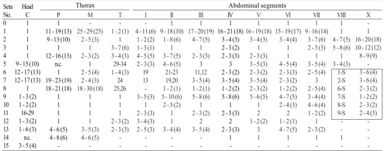
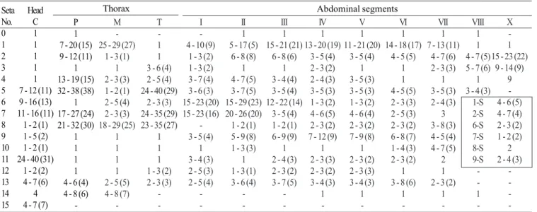
Pupa (Fig. 9). Position and development of setae as figured; range and modal number of branches in Table 7. All
measurements from 10-16 specimens. Integument without distinctive color pattern, mostly light brown to yellowish; sterna II-V with narrow dark band near anterior margin. Cephalothorax: setae 1-3-CT about equal in length, 4-CT usually triple, 5-CT with 5-7 branches, 6-CT double or triple, 7-CT usually triple, 8-7-CT single or double, 9-7-CT with 3-5 branches, 10-CT double or triple, 11-12-CT with 2-4 branches. Seta 9-II, III lightly pigmented, 9-IV-VIII pigmented light brown, slightly darker at base. Abdomen: seta 6-I usually double, 7-I with 4-6 branches; 1-II dendritic with 10-20 fine branches arising from 1-3 strong stem branches, 6-II usually double, 7-II with 2-6
f % f % f % f % f % f % f % f % f % f % f % f % f % f % f % f % f % f % f % f %
Species / Levels LEUCOSPHYRUS SUBGROUP An. baisasi (n=10) LEUCOSPHYRUS COMPLEX An. latens (n=74) An. leucosphyrus (n=42) An. introlatus (n=42) An. balabacensis (n=159) DIRUS COMPLEX An. dirus (n=236) An. cracens (n=58) An. scanloni (n=54) An. baimaii (n=180) An. elegans (n=42) An. nemophilous (n=132) An. takasagoensis (n=67) HACKERI SUBGROUP An. mirans (n=70) An. hackeri (n=51) An. pujutensis (n=36) An. recens (n=10) An. sulawesi (n=26) RIPARIS SUBGROUP An. cristatus (n=14) An. macarthuri (n=111) An. riparis (n=33) 1:1 0 -0 -0 -27/n 64.3 47/n 29.5 0 -0 -7/n 13 18/n 10 0 -96/n 72.7 40/n 59.7 33/n 47.1 23/n 45.1 24/n 66.7 0 -0 -14/n 100 111/n 100 27/n 81.9 1:2 0 -0 -0 -9/n 21.4 29/n 18.2 1/n 0.4 0 -1/n 1.8 18/n 10 0 -13/n 9.8 4/n 6 16/n 22.8 11/n 21.6 9/n 25 0 -0 -0 -0 -4/n 12.1 2:2 0 -0 -0 -0 -11/n 6.9 0 -4/n 6.9 2/n 3.7 6/n 3.3 0 -6/n 4.5 8/n 11.9 5/n 7.1 7/n 14 2/n 5.5 0 -0 -0 -0 -1/n 3 3:1 0 -0 -0 -3/n 7.1 18/n 11.3 3/n 1.3 1/n 1.7 6/n 11.1 17/n 9.4 0 -3/n 2.3 2/n 3 2/n 2.8 2/n 3.9 0 -0 -0 -0 -0 -1/n 3 3:2 0 -0 -0 -2/n 4.8 19/n 11.9 5/n 2.1 1/n 1.7 3/n 5.5 9/n 5 0 -8/n 6.1 5/n 7.5 3/n 4.3 0 -1/n 2.8 1/n 10 1/n 3.8 0 -0 -0 -3:3 2/n 20 0 -0 -1/n 2.4 21/n 13.2 10/n 4.2 9/n 15.5 12/n 22.2 28/n 15.5 0 -6/n 4.5 7/n 10.4 5/n 7.1 1/n 2 0 -0 -0 -0 -0 -0
-branches, 8-II rarely present with 3,4 -branches, 9-II very short, stout, arising distad from posterior margin of segment, length 0.010-0.013 mm (mean = 0.012 mm), 10,11-II absent; 1-III with 4-7 branches, 5-III with 5-4-7 branches, 6-III usually double, 9-III short, stout, length 0.01-0.02 mm (mean = 0.02 mm); 1-IV with 3,4 branches, 5-IV with 4,5 branches, 6-IV single or double, 9-IV short, length 0.02-0.04 mm (mean = 0.03 mm), ratios of length of 9-IV/III 1.28-2.67 (mean = 1.90) and 9-IV/V 0.21-0.40 (mean = 0.30); 1,6-V usually single, 5-V with 4-6 branches, 9-V long, length 0.09-0.11 mm (mean = 0.10 mm); 1-VI normally double, 5-VI with 4,5 branches, 6-VI single or double, 9-VI long, length 0.10-0.12 mm (mean = 0.10 mm) frequently simple, sometimes
with small spicules; 1-VII single or double, 5-VII with 2-5 branches, 6-VII usually double, 9-VII long, frequently simple, sometimes with small spicules, length 0.10-0.11 mm (mean = 0.10 mm); 9-VIII with 10-15 branches. Paddle: lightly tanned, buttress slightly darker, midrib faint, outer basolateral serration prominent, filamentous spicules on outer apical margin and most of inner margin prominent; seta 1-Pa strong, dark pigmented, 2-Pa with 1-3 branches, toothed margin index (= ratio of length a/b, see Fig. 31) 0.89-0.95 (mean = 0.91).
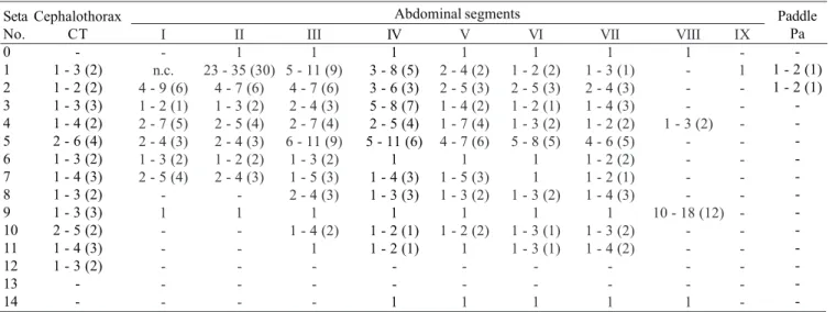
Larva (Figs. 9, 10, 52, 53). Position and development of setae as figured; range and modal number of branches in Table 8. All measurements from 11-22 specimens. Head: integument
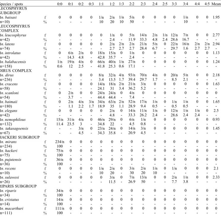
Species / spots LECOSPHYRUS SUBGROUP An. baisasi (n=10) LEUCOSPHYRUS COMPLEX An. leucosphyrus (n=42) An. latens (n=74) An. introlatus (n=42) An. balabacensis (n=158) DIRUS COMPLEX An. dirus (n=236) An. cracens (n=58) An. scanloni (n=54) An. baimaii (n=180) An. elegans (n=42) An. nemophilous (n=132) An. takasagoensis (n=67) HACKERI SUBGROUP An. mirans (n=234) An. hackeri (n=75) An. pujutensis (n=36) An. recens (n=10) An. sulawesi (n=26) RIPARIS SUBGROUP An. riparis (n=34) An. cristatus (n=14) An. macarthuri (n=111) 0:0 0 -0 -0 -0 -1/n 0.6 0 -0 -0 -0 -0 -15/n 11.4 -234/n 100 75/n 100 36/n 100 0 -0 -34/n 100 14/n 100 111/n 100 0:1 0 -0 -0 -6/n 14.3 19/n 12 0 -0 -2/n 3.7 2/n 1.1 0 -31/n 23.5 -0 -0 -0 -0 -0 -0 -0 -0 -0:2 0 -0 -0 -2/n 4.8 4/n 2.5 0 -0 -0 -4/n 2.2 0 -4/n 3 3/n 4.5 0 -0 -0 -0 -0 -0 -0 -0 -0:3 0 -0 -0 -0 -0 -0 -0 -0 -3/n 1.7 0 -0 -0 -0 -0 -0 -0 -0 -0 -0 -0 -1:1 1/n 10 0 -0 -18/n 42.8 66/n 41.8 8/n 3.4 14/n 24.1 24/n 44.4 34/n 18.9 0 -46/n 34.8 23/n 34.3 0 -0 -0 -1/n 10 0 -0 -0 -0 -1:2 2/n 20 1/n 2.4 2/n 2.7 15/n 35.7 40/n 25.3 32/n 13.5 18/n 31 24/n 44.4 63/n 35 2/n 4.8 29/n 22 24/n 35.8 0 -0 -0 -2/n 20 3/n 11.5 0 -0 -0
-Table 5. Frequency distribution (f) and percentage (%) of pale spots on PSD of vein R in females of species of the Leucosphyrus group.
light brown to yellowish without pattern of dark spots; length and width not measured; antenna length 0.27-0.29 mm (mean = 0.28 mm), ratio of distance base to 1A to antenna length 0.26-0.41 (mean = 0.33); seta 2-C long, single with sparse minute spicules on apical 0.5; 3-C single, length 0.07-0.09 mm (mean = 0.08 mm); distance between base of 2-C and 3-C 0.03-0.04 mm (mean = 0.04 mm); 4-C posterolaterad of 2-C, single to triple, length 0.06-0.09 mm (mean = 0.07 mm), usually not reaching base of 2-C, distance between basal insertions of 2-C and 4-C 0.07-0.10 mm (mean = 0.08 mm), ratio of length of 4-C to distance between the insertions of 2-C and 4-C 0.63-1.04 (mean = 0.80), distance between bases of 3-C and 4-C 0.05-0.09 mm (mean = 0.07 mm), 5-C longer than antennal shaft, reaching anterior
margin of head, 7-11 branches, 6-C with 10-17 branches, 7-C with 9-15 branches. Thorax: tubercles of all large setae light brown to yellowish; seta 1-P with 11-18 branches, stem stout, not noticeably expanded, flattened basally, arising from large tubercle, usually separate from tubercle of 2,3-P (Fig. 53), rarely narrowly joined to tubercle of 2,3-P, each tubercle either without or with small, inconspicuous apical lip arising from posterodorsal side and projecting forward over bases of 1,2-P, 14-P 5,6-branched; 4-M with 2-4 branches, 6-M usually triple, 14-M with 4-9 branches; 3-T (Fig. 52) poorly developed, palmate, with 5-8 narrow, nearly transparent lanceolate leaflets. Abdomen: seta 1-I small, somewhat palmate, with 4-7 narrow, nearly transparent, lanceolate leaflets, 2-I single or double,
3-Species / spots
LECOSPHYRUS SUBGROUP An. baisasi (n=10) LEUCOSPHYRUS COMPLEX An. leucosphyrus (n=42) An. latens (n=74) An. introlatus (n=42) An. balabacensis (n=158) DIRUS COMPLEX An. dirus (n=236) An. cracens (n=58) An. scanloni (n=54) An. baimaii (n=180) An. elegans (n=42) An. nemophilous (n=132) An. takasagoensis (n=67) HACKERI SUBGROUP An. mirans (n=234) An. hackeri (n=75) An. pujutensis (n=36) An. recens (n=10) An. sulawesi (n=26) RIPARIS SUBGROUP An. riparis (n=34) An. cristatus (n=14) An. macarthuri (n=111) 1:1 0 -0 -1/n 1.3 1/n 2.7 0 -0 -0 -14/n 25.9 7/n 3.9 2/n 4.8 57/n 43.2 0 -232/n 100 74/n 100 6/n 16.7 0 -0 -32/n 97.0 14/n 100 111/n 100 1:2 0 -3/n 7.3 5/n 6.7 5/n 13.5 6/n 3.8 1/n 0.4 1/n 1.7 21/n 38.9 20/n 11.1 9/n 21.4 35/n 26.5 2/n 3.0 0 -0 8/n 22.2 3/n 30.0 0 -0 -0 -0 -1:3 1/n 11.1 0 -0 -0 -4/n 2.5 1/n 0.4 0 -0 -2/n 1.1 2/n 4.8 2/n 1.5 1/n 1.5 0 -0 2/n 5.5 0 -0 -0 -0 -0 -2:2 2/n 22.2 12/n 28.2 28/n 7.8 20/n 54.0 53/n 33.5 24/n 10.2 12/n 20.7 14/n 25.9 62/n 34.4 20/n 47.6 31/n 23.5 9/n 13.4 0 -0 14/n 38.9 6/n 60.0 0 -1/n 3.0 0 -0 -2:3 4/n 44.4 10/n 25.6 23/n 1.1 8/n 21.6 62/n 39.2 64/n 27.1 18/n 31.0 5/n 9.2 50/n 27.8 7/n 16.7 7/n 5.3 21/n 31.3 0 -0 6/n 16.7 0 -3/n 11.5 0 -0 -0 -2:4 0 -1/n 2.6 3/n 4.0 0 -5/n 3.2 12/n 5.1 2/n 3.4 0 -2/n 1.1 0 -0 -3/n 4.5 0 -0 0 -0 -0 -0 -0 -0
-Table 6. Frequency distribution (f) and percentage (%) of pale spots on SD spot of vein R in females of species of the Leucosphyrus Group.
-I usually single, 9--I with 4-6 branches; 1--I-I small, less pigmented than 1-I, somewhat palmate, with 5-11 narrow, nearly transparent, lanceolate leaflets, basal stem narrow, poorly pigmented; 2-IV with 3,4 branches, 3-IV with 3,4 branches, 13-IV long with 3-5 branches, ratio of length to 10-13-IV 0.42-0.54 (mean = 0.47); 2-V with 3,4 branches; 1-VII smaller and less pigmented than 1-VI, with 11-17 moderately broad lanceolate leaflets with minute apicolateral serration and apical filaments; 1-X long, single, inserted on saddle; 12-14 pecten spines (Fig. 9), 4-6 long spines alternating with 7-9 short.
Type data. Syntypes, two females. Zoology Museum Berlin, Germany, and NHM, London, U.K. Type locality: INDONESIA, Sumatra Island, Tapanuli, Kajutanan (Kajoe Janam), north of Padang.
Material examined. One hundred twenty-eight specimens as follows: 48F, 20M, 28Le, 30Pe, 2L, derived from 20 separate collections from natural habitats (20 adult) and 7 progeny broods. INDONESIA. Sumatra: K. T. [Tapanuli, Kajutanan] (2o51’0"N 98o16’0"E), 1F. Labuhandeli
(3o45’0"N 98o41’60"E), [BonneWepster coll. nos., 1280, 81, 82,
-84, -86, -89, -90, -91, -92, -93, -96, -98], no dates, pre-1940, 14F; [17-12], no locality or date, 1F. Propinsi Sumatera Utara, Medan (3o35’N 98o40’E), 1F. Martapura (4o19’S 104o22’E), 26 Oct. 1977,
1F. Selatan, Baturaja (4o8’S 104o10’E), 19 Sept. 1975, 1F. Selatan,
Lebisbadak, 21 Dec. 1977, 1F. Propinsi Jambi: Bukit Baru (near Muarabungo) (1o28’S 102o7’E), [ID1-B], 7 Apr. 1986, 18F; [ID1-002,
-003, -005, -007, -011, -018, -019], 7 Apr. 1986, progeny, (002), 1F, 4M, 5LePe; (003), 1F, 4M, 4LePe; (005), 1M, 3LePe, 1 L; (007), 2F, 2M, 3LePe, 1 Pe, 2L; (011), 3M, 1LePe, 1Pe, 1L; (018), 3F, 6M, 6LePe, 2Pe; (019), 3F, 1M, 4LePe (V. Baimai and R.E. Harbach).
Distribution (Table 1; Fig. 54). Anophelesleucosphyrus
seems to be an endemic species from Sumatra, Indonesia. Most of the published literature on An. leucosphyrus refers to An. latens, which has a more widespread distribution (Sallum et al. 2004).
Bionomics and medical importance. Little is known about the bionomics of An. leucosphyruss.s. because this species has been largely misidentified as An. latens and most published
records refer to An. latens except for those from Sumatra, Indonesia. Adults of An.leucosphyrus were attracted to human bait in Bukit Baru, near Muarabungo, in Bungo Tebo Regency, Jambi Province, Sumatra. The collections were carried out inside and outside houses situated at the edge of secondary tropical rain forest in hilly areas (Baimai et al. 1988c). This species may be involved in the transmission of human malaria parasites in Sumatra (Warren et al. 1963). Adults were dissected and found infected with Plasmodium sp. in localities in Siantar Estate (Bais 1920), Kisiran (Doorenbos 1927, 1931a,b), Mandahiling (Swellengrebel et al. 1919) and Nies Island, Gawo River (Soesilo 1929a,b; Soesilo et al. 1935).
Systematics. Three allopatric populations of An. leucosphyrusl.s. from south Kalimantan, Sumatra and Thailand were investigated using karyotypes and cross-mating experiments. As a result, it was found that An. leucosphyrus l.s. included two distinct species, An.leucosphyrus A (= An. latens), which occurs in Thailand and south Kalimantan, and
An.leucosphyruss.s. which is confined to Sumatra. Genetic isolation between An. latens and An.leucosphyrus probably occurred either as a consequence of genetic shift or as by a product of genetic divergence within isolated populations on Sumatra. Based on the degree of synapsis in the polytene chromosomes of F1 hybrids, An. latens seems to be more closely related to An.balabacensis and members of the Dirus Complex than is An.leucosphyrus (Baimai et al. 1988c).
Anophelesleucosphyrus cannot be distinguished from An. latens based on morphological characters of the adult, pupa, and fourth-instar larva. Because these two species are allopatric, their geographical distribution should be considered when making an identification. Anopheles leucosphyrus can be distinguished from the remaining species by the following combination of characters. Adult: 1) proboscis slightly longer than forefemur, ratio of proboscis length to forefemur varying from 1.06-1.14; 2) apical pale band of palpomere 5 distinctly pale cream-colored, contrasting with silvery-white bands of
Seta No. 0 1 2 3 4 5 6 7 8 9 10 11 12 13 14 Cephalothorax CT -3 2 , 3 (2) 2 , 3 (3) 2 - 4 (3) 5 - 7 (6) 2 , 3 (3) 3 , 4 (3) 1 , 2 (1) 3 - 5 (3) 2 , 3 (2) 2 - 4 (3) 2 - 4 (3)
-I -n.c. 5 - 8 (6) 1 , 2 (1) 4 - 7 (5) 3 , 4 (3) 2 , 3 (2) 4 - 6 (5)
-1 -II 1 10 - 20 (10)
5 - 7 (5) 2 , 3 (3) 5 - 8 (6) 3 - 5 (4) 2 , 3 (2) 2 - 6 (4) 3 , 4 (3)
1 -III 1 4 - 7 (5) 5 - 7 (5) 3 - 5 (3) 4 - 6 (5) 5 - 10 (7)
2 , 3 (2) 2 - 4 (3) 2 - 4 (3)
1 3 , 4 (3)
1 -IV 1 3 , 4 (3) 3 , 4 (3) 5 - 8 (7) 1 - 4 (3) 4 - 5 (5) 1 , 2 (1) 2 - 4 (3) 2 , 3 (3)
1 2 1 -1 V 1 1 - 3 (1)
3 2 - 4 (3) 5 - 6 (5) 4 - 6 (5) 1 , 2 (1) 1 - 4 (3) 2 , 3 (2)
1 2 , 3 (2)
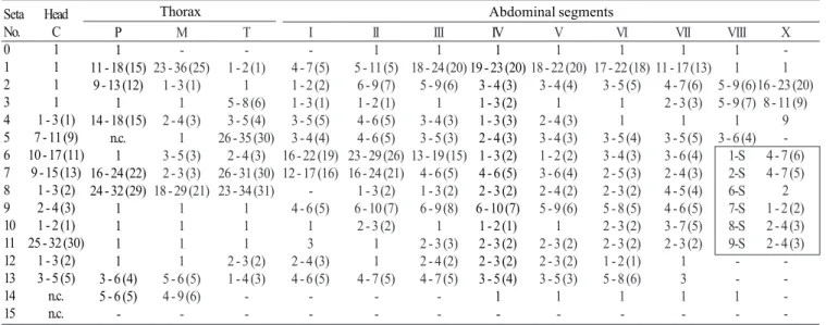
1 -1 Table 7. Number of branches for seta of pupae of An. leucosphyrus: range (mode).
VI 1 1 - 3 (2) 1 - 4 (3) 1 - 3 (2) 1 - 3 (2) 4 - 5 (5) 1 , 2 (1) 1 - 5 (1) 2 - 3 (3)
1 1 - 3 (2)
1 -1 VII 1 1 , 2 (2) 2 , 3 (3) 2 - 4 (3) 1 - 3 (2) 2 - 5 (5) 1 , 2 (2) 1 , 2 (1) 2 , 3 (2)
1 2 , 3 (3) 1 - 3 (2)
-1 VIII 1 -1 - 3 (3)
-10 - 15 (-10)
-1 IX -1 -Paddle Pa -1 , 2 (-1) 1 - 3 (1)
-Abdominal segments
palpomeres 2 and 3; 3) ASP spot usually extending onto the vein C on one or both wings, rarely absent on vein C on both wings; 4) hindtarsomere 4 without basal pale band; 5) hindtarsomere 5 without basal pale band; 6) hindtarsomere 5 with apical pale band; 7) PSD spot of vein R extending basally from level 4 to level 6; 8) PSD spot of vein R entirely dark or with 1-5 pale interruptions; 7) wing fringe with pale spot between veins 1A and Cu2; 10) fork of cell R2 in line or basal to fork of cell M1+2. Fourth-instar larva: 1) seta 3-C single; 2) seta 5-C conspicuously longer than antenna; 3) basal sclerotized tubercle of seta 1-P without prominent tooth or spine arising from posterolateral margin; 4) basal sclerotized tubercle of seta 1-P usually well separated from tubercle of 2-P, rarely both tubercles narrowly joined, tubercle of seta 1-P smaller than that of seta 2-P, distance between bases wide, equal or greater than basal width of tubercle of 1-P; 5) seta 1-II not fully developed, noticeably smaller than seta 1-III-VI. Pupa: 1) ratio of length of 9-IV to length of 9-V 0.21-0.40 (mean = 0.30); 2) seta 4-II with 5-8 branches; 3) seta 7-II with 2-4 branches; 4) seta 10-IV always double; 5) 9-V length 0.09-0.11 mm (mean = 0.10 mm); 6) 9-VI length 0.10-0.12 mm (mean = 0.10 mm); 7) toothed margin index 0.89-0.95 (mean = 0.91).
Anopheles (Cellia) latens Sallum & Peyton (Figs. 4, 7, 11, 12, 49, 52, 54)
Anopheles latens Sallum & Peyton, 2005, in Sallum et al. (2005) (F, M, P*, L*). Holotype male with associated slide mounted larval and pupal exuviae, deposited in the NMNH.
Anopheles leucophyrus of Leicester (1903:291) (bionomics). Anopheles leucosphyrus of Stanton (1912:6) (L); Bonne-Wepster &
Swellengrebel (1953:259) (F*, M*, L*, identification keys); Zulueta (1956:651) (vector human Plasmodium); Colless (1957:131) (F, L, P, in part, Sarawak, Borneo); Zulueta (1957:699) (vector W. bancrofti); Macdonald & Traub (1960:79) (bionomics notes); Chow (1961) (F* identification key, distribution, Malaya, Sarawak); Wharton et al. (1962:758) (vector P. inui); Ramalingam (1974:147) (distribution note); Kanda et al. (1981:321)
(hybridization, systematics); Kanda et al. (1983:193) (chromosomal polymorphism, phylogeny); Takai et al. (1984:145) (genetics); Takai (1986:45) (in part, electrophoresis, Sarawak); Harbach et al. (1987b:241) (bionomics); Chang et al. (1995:192) (bionomics); Seng et al. (1999:454) (bionomics).
Anopheles leucosphyrus species A of Baimai (1988b:667) (cytogenetics, distribution); Baimai et al. (1988c:44) (cytogenetics, crossing); Peyton (1989:197) (taxonomy); Harrison et al. (1990:202) (taxonomy, distribution notes).
Anopheles leucosphyrus leucosphyrus of Mendoza (1954) (A*, L*, identification keys); Colless (1956a:115) (bionomics); Bonne-Wepster (1963:705) (F, identification key, in part).
Myzomyia leucophyrus of Daniels (1908:1) (An. hackeri and An. leucosphyrus mixed, bionomics); Leicester (1908a:7) (bionomics); Leicester (1908b:18) (F, M, bionomics, probably mixed with An. hackeri); Leicester (1908c:267) (feeding behavior, probably mixed with An. hackeri).
Neomyzomyia elegans of James & Stanton (1912:59). Neomyzomyia eurysphyrus of de Meijere (1913:338) (in part). Neomyzomyia leucosphyra of Swellengrebel & Swellengrebel de Graff
(1920b:83) (L*).
Nyssorhynchus leucosphyrus of Blanchard (1905:213) (F, Borneo). Female (Figs. 4, 7). Head: proboscis uniformly dark-scaled, length 1.92-2.25 mm (mean = 2.11 mm), ratio of length to forefemur 1.06-1.16 (mean = 1.12), maxillary palpus (Fig. 7) length 1.62-2.15 mm (mean = 1.89 mm), ratio of length to proboscis 0.81-0.95 (mean = 0.89), ratio of length to forefemur 0.91-1.07 (mean = 1.00) (Table 2.2), ratio of length of palpomeres 3/4 1.62-2.08 (mean = 1.77), 3/5 2.17-2.87 (mean = 2.44), 4/5 1.22-1.62 (mean = 1.37), 4-5/3 0.84-1.08 (mean = 0.98), palpomeres 2 and 3 with narrow apical band of silvery-white scales, apical pale band of palpomere 4 variable, absent to larger than those of palpomeres 2 and 3, scales pale cream-colored to golden, slightly contrasting with those on palpomeres 2 and 3, palpomere 5 dark-scaled at base, with apical band of pale cream-colored to golden scales, length of apical pale band 0.17-4.00 (mean = 1.18) length of basal dark band of palpomere 5, ratio of length of apical pale band of palpomere 4 to length of basal dark band of palpomere 5 0.0-1.67 (mean = 0.51) (Table 3).
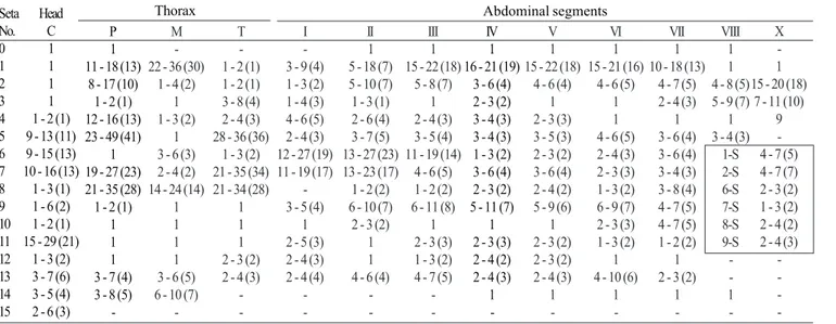
Seta No. 0 1 2 3 4 5 6 7 8 9 10 11 12 13 14 15 Head C 1 1 1 1 1 - 3 (1) 7 - 11 (9) 10 - 17 (11)
9 - 15 (13) 1 - 3 (2) 2 - 4 (3) 1 - 2 (1) 25 - 32 (30)
1 - 3 (2) 3 - 5 (5) n.c. n.c.
P 1 11 - 18 (15)
9 - 13 (12) 1 14 - 18 (15)
n.c. 1 16 - 24 (22) 24 - 32 (29)
1 1 1 1 3 - 6 (4) 5 - 6 (5)
-M -23 - 36 (25)
1 - 3 (1) 1 2 - 4 (3)
1 3 - 5 (3) 2 - 3 (3) 18 - 29 (21)
1 1 1 1 5 - 6 (5) 4 - 9 (6)
-T -1 - 2 (-1)
1 5 - 8 (6) 3 - 5 (4) 26 - 35 (30)
2 - 4 (3) 26 - 31 (30) 23 - 34 (31)
1 1 1 2 - 3 (2) 1 - 4 (3)
-I -4 - 7 (5) 1 - 2 (2) 1 - 3 (1) 3 - 5 (5) 3 - 4 (4) 16 - 22 (19) 12 - 17 (16)
-4 - 6 (5)
1 3 2 - 4 (3) 4 - 6 (5)
-II 1 5 - 11 (5)
6 - 9 (7) 1 - 2 (1) 4 - 6 (5) 4 - 6 (5) 23 - 29 (26) 16 - 24 (21) 1 - 3 (2) 6 - 10 (7)
2 - 3 (2) 1 1 4 - 7 (5)
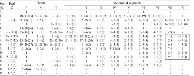
-Table 8. Number of branches for setae of fourth-instar larvae of An. leucosphyrus: range (mode).
III 1 18 - 24 (20)
5 - 9 (6) 1 3 - 4 (3) 3 - 5 (3) 13 - 19 (15)
4 - 6 (5) 1 - 3 (2) 6 - 9 (8)
1 2 - 3 (3) 2 - 4 (2) 4 - 7 (5)
-IV 1 19 - 23 (20)
3 - 4 (3) 1 - 3 (2) 1 - 3 (3) 2 - 4 (3) 1 - 3 (2) 4 - 6 (5) 2 - 3 (2) 6 - 10 (7)
1 - 2 (1) 2 - 3 (2) 2 - 3 (2) 3 - 5 (4)
1
-V 1 18 - 22 (20)
3 - 4 (4) 1 2 - 4 (3) 3 - 4 (3) 1 - 2 (2) 3 - 6 (4) 2 - 4 (2) 5 - 9 (6)
1 2 - 3 (2) 2 - 3 (2) 3 - 5 (3)
1
-VI 1 17 - 22 (18)
3 - 5 (5) 1 1 3 - 5 (4) 3 - 4 (3) 2 - 5 (3) 2 - 3 (2) 5 - 8 (5) 2 - 3 (2) 2 - 3 (2) 1 - 2 (1) 5 - 8 (6)
1
-Abdominal segments
n.c. = not counted
Thorax
VII 1 11 - 17 (13)
4 - 7 (6) 2 - 3 (3)
1 3 - 5 (5) 3 - 6 (4) 2 - 4 (3) 4 - 5 (4) 4 - 6 (5) 3 - 7 (5) 2 - 3 (2)
1 3 1 -VIII 1 1 5 - 9 (6) 5 - 9 (7)
1 3 - 6 (4)
1-S 2-S 6-S 7-S 8-S 9-S -1 -X -1 16 - 23 (20)
8 - 11 (9) 9 -4 - 7 (6) 4 - 7 (5)
2 1 - 2 (2) 2 - 4 (3) 2 - 4 (3)
-Thorax: pleural setae as follow: 1-4 upper proepisternal, 0-2 prespiracular, 5-8 prealar, 4-6 upper mesokatepisternal, 2,3 lower mesokatepisternal, 4-7 upper mesepimeral. Wing (Fig. 4): length 3.16-3.62 mm (mean = 3.46 mm), pale scales on vein C and subcosta light pale yellow, those on remaining veins white, not strongly contrasting with others, PHP spot of vein C small, always present, HP spot always present, usually prominent, rarely very small, PSP spot always present and prominent, rarely reduced, SP spot usually small, sometimes prominent, ASP spot usually present, occasionally reduced or absent, PP spot 0.55-1.54 (mean = 1.03) length of SCP spot, AD spot 1.17-3.00 (mean = 1.84) length of PP spot, PSD spot of vein R extending basally from level 4 to level 6 (Fig. 4) on both wings (Table 4), PSD spot of vein R with 1-5 small pale interruptions (Table 5), SD spot of vein R with 1-4 pale interruptions (Table 6), sum of pale interruptions on PSD-PD spots of vein R varying from 4-12 for each wing, ratio of length of cell R2 to vein R2+3 1.46-2.00 (mean = 1.69), ratio of length of cell R2 to cell M1+2 1.19-1.34 (mean = 1.28). Legs: femora, tibiae, and tarsomeres 1 dark-scaled, speckled with pale spots; foretarsomere 2 with apical and basal pale bands, middle region dark-scaled, sometimes with 1-4 pale spots on dorsal surface, foretarsomeres 3 and 4 mostly dark-scaled, with basal and apical pale bands, foretarsomere 4 sometimes mostly pale-scaled with inconspicuous middle band of light brown scales, foretarsomere 5 variable, mostly pale-scaled with inconspicuous patch of tan scales on dorsal surface, or with basal dark band, pale-scaled at apex, dark scales on ventral surface of foretarsomeres 2-5 light brown to tan; midtarsomeres 2-5 mostly dark-scaled with narrow apical bands of pale scales; hindtarsomeres 2-5 dark-scaled with narrow apical bands of pale scales, hindtarsomeres 2,3 sometimes with few pale spots in middle dark area. Abdomen: tergum VI frequently without scales, sometimes with 2,3 golden scales on posterolateral margin, tergum VII with few cream-colored to golden scales on posterior margin or rarely without scales, tergum VIII covered with yellowish to golden scales; sternum VI without scales, sternum VII with posteromedial patch of dark scales, sternum VIII with small lateral patch of whitish scales.
Male. Essentially as in female except for sexual characters. Wing generally paler with reduced scaling, pale spots usually longer than in female. Palpomere 2 with patch of pale scales at middle of dorsal surface, extending to lateral surface, apex of palpomere 2 bare; palpomere 3 dark-scaled, with long dorsal patch of pale scales at middle, extending to lateral surface, apex of palpomere 3 with broad band of pale scales interrupted by short patch of dark scales at apex of ventrolateral surface; palpomeres 4 and 5 mostly pale-scaled with narrow basal band of dark scales. Abdomen: sternum VIII covered with pale cream-colored scales. Genitalia: 3,4 parabasal setae; ventral claspette with 2 apical large setae, medial seta slightly shorter than lateral seta, 1 smaller subapical seta; dorsal claspette with 4 long setae fused apically into club, setae separate at base; aedeagus with 5-7 serrated leaflets at each side of tip.
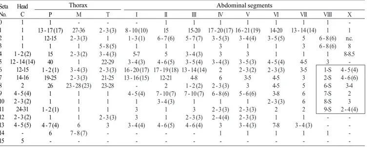
Pupa (Fig. 11). Position and development of setae as figured; range and modal number of branches in Table 9. All
measurements from 20-22 specimens. In general as described for An. leucosphyrus, except for the following characters. Cephalothorax: seta 5-CT with 3-8 branches, 7-CT usually with 4 branches, 8-CT single to triple, 9-CT with 2-5 branches, 10-CT with 2-4 branches. Abdomen: seta 6-I usually triple; 1-II dendritic, with >30 fine branches arising from strong basal stem; 7-II with 4-11 branches, 8-II always absent, 9-II very short, stout, length 0.010-0.014 mm (mean = 0.011), 10,11-II rarely present; 1-III with 5-10 branches, 5-III with 6-11 branches, 9-III short, stout, length 0.01-0.03 mm (mean = 0.02 mm); 1-IV with 3-7 branches, 5-IV with 4-8 branches, 9-IV short, length 0.02-0.05 mm (mean = 0.04 mm), ratio of length of 9-IV/III 1.25-4.0 (mean = 2.36) and 9-IV/V 0.19-0.52 (mean = 0.36); 1-V usually triple, 5-V with 3-5 branches, 9-V long, length 0.08-0.12 mm (mean = 0.10 mm); 1-VI usually triple, 5-VI with 3-7 branches, 6-VI single to triple, 9-VI long, length 0.09-0.13 mm (mean = 0.11 mm); 5-VII with 4-7 branches, 9-VII long, length 0.09-0.12 mm (mean = 0.11 mm); 9-VIII with 12-20 branches. Paddle: toothed margin index 0.86-0.94 (mean = 0.91).
Larva (Figs. 11, 12, 49, 52). Position and development of setae as figured; range and modal number of branches in Table 10. All measurements from 10-24 specimens. In general as described for An. leucosphyrus, except for the following characters. Head: integument light brown to yellowish, either with or without pattern of darkened spots, if dark spots present, as follow: 1 dark spot placed posteriorly on dorsal apoteme; 2 lateral spots on dorsal apoteme posterior to seta 8-C, posteriorly on frontal ecdysial line; 1 dark spot ventral on lateralia encircling seta 12-C; 2 dark spots on labiogula posterior to hypostomal suture; head length 0.60-0.70 mm (mean = 0.67 mm), head width 0.63-0.74 mm (mean = 0.68 mm), antenna length 0.25-0.30 mm (mean = 0.28 mm); ratio of distance base to 1-A to antenna length 0.31-0.42 mm (mean = 0.38 mm), 3-C length 0.07-0.09 mm (mean = 0.08 mm), distance between base of 2-C and 3-C 0.03–0.04 mm (mean = 0.04 mm), 4-C always single, length 0.04-0.07 mm (mean = 0.06 mm), distance between basal insertions of 2-C and 4-C 0.06-0.10 mm (mean = 0.08 mm), ratio of length of 4-C to distance between the insertions of 2-C and 4-C 0.51-0.88 (mean = 0.74), distance between base of 3-C and 4-C 0.06-0.09 mm (mean = 0.07 mm), 5-C extending beyond anterior margin of head, 6-C with 8-14 branches. Thorax: tubercles of all large setae light brown; tubercle of seta 1-P widely separated from that of 2,3-P or occasionally both tubercles joined at base, 14-P with 8 branches; 14-M with 5-11 branches; 3-T (Fig. 52) with 8-5-11 narrow, lanceolate nearly transparent leaflets. Abdomen: seta 1-I small, with 3-7 rigid branches, 2-I double or triple, 9-I with 3-5 branches; 1-II with 8-14 narrow, lanceolate branches; 3-IV single to triple, 13-IV long, ratio of length to 10-IV 0.46-0.64 (mean = 0.55); 11-15 pecten spines (Fig. 49), 4-6 long elements alternating with 6-10 short.