www.cbpv.com.br/rbpv ISSN 0103-846X (Print) / ISSN 1984-2961 (Electronic)
Doi: http://dx.doi.org/10.1590/S1984-29612014078
Serological detection of
Toxoplasma gondii, Leishmania
infantum
and
Neospora caninum
in cats from an area
endemic for leishmaniasis in Brazil
Detecção sorológica de
Toxoplasma gondii, Leishmania infantum
e
Neospora caninum
em gatos de uma área
endêmica para leishmaniose no Brasil
Keyla Carstens Marques de Sousa1; Heitor Miraglia Herrera2; Iara Helena Domingos3;
João Bosco Vilela Campos2; Isabela Maria Campanelli dos Santos1; Haroldo Henrique Neves1;
Rosangela Zacarias Machado1; Marcos Rogério André1*
1Universidade Estadual Paulista “Júlio de Mesquita Filho” – UNESP, Jaboticabal, SP, Brasil 2Universidade Católica Dom Bosco – UCDB, Campo Grande, MS, Brasil
3Centro de Controle de Zoonoses – CCZ, Campo Grande, MS, Brasil
Received April 29, 2014 Accepted August 5, 2014
Abstract
An investigation was made into the occurrence of antibodies to Toxoplasma gondii, Leishmania infantum and Neospora caninum in 151 domestic cats, based on the indirect fluorescent antibody test (IFAT). Serum samples were collected from 151 domestic cats (65 free-roaming and 86 domiciled cats; 55 males and 96 females) in Campo Grande, Mato Grosso do Sul, Brazil between January and April 2013. IgG antibodies to T. gondii, L. infantum and N. caninum were found, respectively, in 49 (32.5%), 34 (22.5%) and 10 (6.6%) sampled cats. A positive correlation was found between T. gondii and N. caninum, T. gondii and L. infantum, and N. caninum and L. infantum (p < 0.05) infections. Also, a significant interaction was identified between gender and area of activity on the probability of T. gondii (p = 0.0324) infection. However, no significant interaction was observed between gender and area of activity on infections by either N. caninum or L. infantum. This study showed that cats from an area endemic for visceral leishmaniasis in Brazil are exposed to three different protozoans, two of which are causal agents of important zoonosis.
Keywords: Cats, Leishmania infantum, Neospora caninum, Toxoplasma gondii, serology, Brazil.
Resumo
O presente estudo teve como objetivo investigar a ocorrência de anticorpos anti-Toxoplasma gondii, Leishmania
infantum e Neospora caninum, em 151 gatos, por meio da Reação de Imunofluorescência Indireta (RIFI). Entre os
meses de janeiro e abril de 2013, amostras de soro foram coletadas de 151 gatos domésticos (65 gatos errantes e 86 gatos domiciliados; 55 machos e 96 fêmeas), de Campo Grande, Mato Grosso do Sul, Brasil. Anticorpos IgG anti-T. gondii, anti-L. infantum e anti-N. caninum foram encontrados em 49 (32,5%) , 34 (22,5%) e 10 (6,6%) gatos amostrados, respectivamente. Verificou-se uma associação estatisticamente significativa entre as infecções por T. gondii e N. caninum, T. gondii e L. infantum e N. caninum e Leishmania infantum (p < 0,05). Além disso, foi observada uma interação significativa entre sexo, área de atividade na probabilidade de infecção por T. gondii (p = 0,0324). No entanto, não foi observada interação significativa entre sexo e área de atividade nas infecções por N. caninum e L. infantum. Este estudo mostrou que os gatos de uma área endêmica brasileira para leishmaniose visceral são expostos a três diferentes protozoários, sendo dois deles importantes agentes zoonóticos.
Palavras-chave: Gatos, Leishmania infantum, Neospora caninum, Toxoplasma gondii, sorologia, Brasil.
Introduction
Toxoplasma gondii and Neospora caninum are apicomplexan intracellular protozoal parasites that affect a wide range of animal species, including humans (DUBEY, 1986; DUBEY et al., 1988). Both organisms cause serious reproductive diseases as well as economic losses, mainly in ruminants (DUBEY et al., 1988; MASALA et al., 2003). Felines play an important role in the epidemiology of T. gondii, because they are the only animal group that excrete resistant oocysts into the environment (LINDSAY et al., 1997). On the other hand, the definitive hosts of N. caninum are domestic dogs, coyotes and Australian dingoes, which shed oocysts (DUBEY & Schares, 2011). Due to the importance of these parasites to public health, many surveys have been conducted worldwide showing the seroprevalence of T. gondii in felines (GARCIA et al., 1999; LANGONI et al., 2001; SILVA et al., 2002; DUBEY et al., 2004; PENA et al., 2006; LOPES et al., 2008; ESTEVES et al., 2014). However, there a few reports about the seroprevalence of N. caninum in cats (DUBEY et al., 2002; BRESCIANI et al., 2007; BRAGA et al., 2012).
Leishmaniasis, an infectious disease that affects humans and wild and domestic mammals worldwide, is caused by a kinetoplastid flagellate protozoan parasite of the genus Leishmania, transmitted mainly by sand flies of the genera Lutzomyia spp. and Phlebotomus spp. (SOLANO-GALLEGO et al., 2007). Although dogs are considered the main reservoirs of Leishmania spp. in urban areas of Brazil, the increase in cases of leishmaniasis among cats suggests the possibility that these animals participate in the epidemiology of the disease (MAROLI et al., 2007; VIDES et al., 2011). Recently, Leishmania spp. infection in domestic cats has been reported in several countries, including Brazil, where this zoonosis is endemic (POLI et al., 2002; SOLANO-GALLEGO et al., 2007; SOUZA et al., 2005; SOBRINHO et al., 2012).
This study was conducted to evaluate the serological occurrence of T. gondii, N. caninum and L. infantum in domestic cats living in an area endemic for leishmaniasis in the municipality of Campo Grande, Mato Grosso do Sul, Brazil. The risk factors associated with seropositivity for these selected agents in cats were also evaluated.
Materials and Methods
Sample collection
Between January and April 2013, whole blood samples were collected by convenience from 151 cats (54 males, 95 females and two without gender registration) in the city of Campo Grande, capital of the state of Mato Grosso do Sul, Brazil. Free-roaming non-domiciled cats (n = 65) were caught by technical staff from the local zoonosis control center (CCZ). These cats shared the same type of food and water, and received no health check-ups or vaccinations. Domiciled cats without outdoor access (n = 86) were sampled during preoperative procedures in a castration project at the CCZ; these animals were returned to their homes after surgery. Overall, the physical status of domiciled cats was better than that of the non-domiciled animals. Cat blood samples (6 mL) were collected by jugular venipuncture and stored at -20°C.
The project was approved by the Ethics Committee on Animal Use of São Paulo State University – UNESP at Jaboticabal, under Protocol no. 004987/13.
Serological tests for T. gondii, N. caninum and
L. infantum
The presence of T. gondii, N. caninum and L. infantum antibodies in the serum of each sampled animal was detected by the Indirect fluorescent Assay (IFAT), as described previously (ANDRÉ et al., 2010; OLIVEIRA et al., 2008). N. caninum NC-1 strain tachyzoites were used as an antigen (DUBEY et al., 1988; MINEO et al., 2009). Toxoplasma gondii RH strain tachyzoites were also used as an antigen, as described by Domingues et al. (1998). In this study, a L. infantum strain was used that was isolated in Jaboticabal (OLIVEIRA et al., 2008), in the state of São Paulo, and characterized as donovani complex, probably L. infantum, using molecular techniques described by Cortes et al. (2004). Promastigotes of L. infantum were maintained on RPMI-1640 medium (Sigma-Aldrich, St. Louis, USA) supplemented with 10% heat-inactivated fetal bovine serum (FBS; Gibco, Canyon City, USA) at 25°C. This medium was then subjected to seven freeze-thaw cycles, after which the medium was centrifuged (3000 × g for 30 min at 4°C). The resulting supernatant was harvested and used as crude antigen, and was used to prepare the crude antigens for IFAT (OLIVEIRA et al., 2009). Antigen slides were removed from storage and allowed to thaw at room temperature for 30 min. Ten microliters of sera at dilution of 1:50 (cut-off for N. caninum), 1:40 (cut-off for T. gondii) and 1:40 (cut-off for L. infantum) were placed in wells on antigen slides. Cat serum samples positive and negative for T. gondii,
N. caninum and L. infantum (BRAGA et al., 2012), obtained
from the serum bank of the Laboratory of Immunoparasitology, Department of Veterinary Pathology of UNESP at Jaboticabal, SP, were also used in the serological reactions. Slides were incubated at 37°C in a moist chamber for 45 min, washed three times in phosphate-buffered saline (pH 7.2) for 5 min, and air-dried at room temperature. Immunoglobulin G (IgG) anti-cat conjugate (whole molecule with fluorescein isothiocyanate, dilution of 1:32; Sigma®, St. Louis, Missouri) for domestic feline samples were diluted according to the manufacturer’s instructions and added to each well. These slides were incubated again, washed, dried, and overlaid with buffered glycerin (pH 8.7), covered with glass coverslips, and examined using an epifluorescence microscope (Olympus, Japan). The parasite strains used as antigens in the present study were also used in earlier serological surveys among wild and domestic animals in Brazil (MINEO et al., 2009; ANDRÉ et al., 2010; JUSI et al., 2011).
Statistical analysis
Yates continuity correction. All the analyses were performed using R 3.0.2 software (R DEVELOPMENT CORE TEAM, 2013).
Results
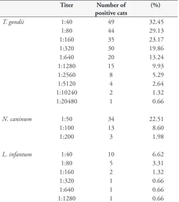
IgG antibodies to T. gondii, N. caninum and L. infantum were detected, respectively, in 49 (32.5%), 34 (22.5%) and 10 (6.6%) sampled cats. IgG antibody titers ranged from 1:40 (cut-off) to 1:20480 for T. gondii, 1:50 (cut-off) to 1:200 for N. caninum, and 1:40 (cut-off) to 1:1280 for L. infantum(Table 1). Five cats (3.3%) showed IgG antibodies to T. gondii, N. caninum and L. infantum. Seventeen cats (11.2%) were seropositive only for T. gondii and N. caninum; 2 (1.3%) were seropositive only for T. gondii and L. infantum, and 3 (1.9%) were seropositive for N. caninum and
L. infantum. Twenty-five cats (16.5%) showed antibodies to
T. gondii, and 9 (5.9%) were seropositive for N. caninum. None of the cats was seropositive only for L. infantum. Ninety cats (59.6%) were seronegative for all the parasites. The results showed a statistically positive correlation between infections by T. gondii and N. caninum, T. gondii and L. infantum, and N. caninum and L. infantum (p < 0.05) (Table 2).
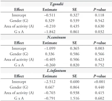
Logistic regression results revealed a significant interaction between gender and area of activity on the probability of infection by T. gondii (p = 0.0324) (Table 3), while neither of the predictor variables fitted in the logistic regression models had a significant effect on N. caninum and L. infantum infections (p = 0.7; p = 0.6 respectively) (Table 3). In the case of probability of infection by T. gondii, the results of subsequent regressions are presented
owing to the significant interaction between gender and area of activity, and these analyses are stratified by either gender or area of activity (Table 4). When the animals were stratified according to gender, male cats from the local CCZ exhibited a significantly higher probability of infection by T. gondii than domiciled cats, although this stratification showed no significant difference between females from the CCZ and domiciled females (Table 4). The results of the analyses stratified according to area of activity revealed that, among the domiciled cats, females showed a significantly higher probability of becoming infected by this agent than males, while the cats from the local CCZ showed no significant difference between males and females.
Discussion
This study revealed the occurrence of L. infantum antibodies in 6.6% of the sampled cats, which is higher than that found in previous studies conducted in another areas endemic for leishmaniasis in the state of São Paulo, Brazil, where seroprevalence ranging from 0.5 to 4.64% has been reported, using the same serological technique (IFAT) and cut-off (≥ 40) (SOBRINHO et al., 2012; CARDIA et al., 2013; BRESCIANI et al., 2010). On the other hand, Vides et al. (2011) reported a higher seroprevalence by IFAT (10.9%) among cats sampled in the city of Araçatuba, state of São Paulo, which was higher than that found in this study, probably because all the cats sampled in the former study showed dermatologic lesions, one of the most suggestive clinical signs of feline leishmaniasis (SOLANO-GALLEGO et al., 2007).
Sousa et al. (2013) recently found high seroprevalence (100%) and high levels of L. infantum antibody titers ranged from 1:40 to 1:20480 among dogs sampled in the same area where this study was conducted. Lower seropositivity to Leishmania spp. among cats compared to dogs in areas endemic for leishmaniasis have been reported in previous studies (SOLANO-GALLEGO et al., 2007; BRESCIANI et al., 2010; SOBRINHO et al., 2012; CARDIA et al., 2013). According to Solano-Gallego et al. (2007), the immune response in cats, mainly cellular immunity, is effective enough to control the infection and confer some natural resistance Table 1. Distribution of reciprocal titers measured by IFAT for
T. gondii, N. caninum and L. infantum in 151 cats of the city of Campo Grande, MS, Brazil.
Titer Number of positive cats
(%)
T. gondii 1:40 49 32.45
1:80 44 29.13 1:160 35 23.17 1:320 30 19.86 1:640 20 13.24 1:1280 15 9.93 1:2560 8 5.29 1:5120 4 2.64 1:10240 2 1.32 1:20480 1 0.66
N. caninum 1:50 34 22.51
1:100 13 8.60 1:200 3 1.98
L. infantum 1:40 10 6.62
1:80 5 3.31
1:160 2 1.32 1:320 1 0.66 1:640 1 0.66 1:1280 1 0.66
Table 2. Association between infections by T. gondii and N. caninum, T. gondii and L. infantum and N. caninum and L. infantum.
Agents Chi-square P-value
N.caninum
- +
T. gondii – 90 12 18.9716 0.0000133*
+ 27 22
L.infantum
- +
T.gondii – 99 3 5.1762 0.0229*
+ 42 7
L.infantum
- +
N.caninum – 115 2 16.908 0.000039*
+ 26 8
2012) or FeLV (SHERRY et al., 2011) and Leishmania spp. infection has been reported in cats.
The occurrence of T. gondii antibodies in 32.4% of the sampled cats (49/151) was higher than previously reported in the Brazilian states of São Paulo (25%) (BRESCIANI et al., 2007), Rio de Janeiro (24.39%) (GONÇALVES NETTO et al., 2003), and Santa Catarina (14.33%) (DALLA ROSA et al., 2010), but lower than those reported in the states of Maranhão (50.5%) (BRAGA et al., 2012), Rio Grande do Sul (37.9%) (PINTO et al., 2009) and Paraná (73%) (GARCIA et al., 1999)., Differences in specificity and sensitivity among different techniques used in earlier studies are likely the cause of disparate seroprevalence rates.
Although the production of immunoglobulins is not associated with oocyst shedding (DUBEY, 1986), the seroprevalence found in sampled felines may be an alternative to measure the spread of T. gondii in the environment (BRAGA et al., 2012). Titers lower than 1:64 in IFAT without clinical symptoms suggest chronic infection by T. gondii; on the other hand, titers exceeding 1024 usually indicate acute infection with or without clinical signs (MCKINNEY, 1973).
Several authors reported that they found no significant influence of gender on T. gondii infection (BRESCIANI et al., 2007; COELHO et al., 2011). In this study, we found that the we found that the correlation between gender and area of activity significantly influenced the probability of T. gondii infection (p = 0.0324). Male cats from the CCZ showed a significantly higher probability of becoming infected with T. gondii (OR= 7.77) than male domiciled cats, although no significant difference in terms of probability of T. gondii infection was found between female cats from different areas of activity (i.e., CCZ vs. domiciled). Environmental conditions and behavioral effects may have contributed to the animals’ exposure to T. gondii, since they live together in the same location (CCZ), share the same food and water, and may be in contact with other intermediate hosts, in addition to the fact that male cats fight with each other.
Among domiciled cats, females showed a higher probability of becoming infected with T. gondii than males, while among the cats from the CCZ, gender did not have a significant effect. Several studies have reported that gender does not significantly influence the probability of infection by T. gondii (LOPES et al., 2008; ESTEVES et al., 2014), although, unlike the present study, they did not investigate interactions between gender and other risk factors. As a general rule, males are more prone to acquiring infected parasites than females, owing to several endocrinal and behavioral differences (KLEIN, 2000). However, this seems to disagree with the higher probability of infection found for domiciled females in the present study. This discrepancy may be explained by the fact that some confounders (e.g., age, feeding habits, breed and contact with other intermediate hosts) were not controlled in this study, due to lack of information.
Although previous studies found no significant association between T. gondii and Leishmania spp. in cats (SHERRY et al., 2011; SOBRINHO et al., 2012), a positive statistical association between infections by T. gondii and L. infantum and N. caninum and L. infantum, which was indicative of coinfections, was detected in this study. Numerous studies have reported immunosuppression caused by Leishmania spp., which could enhance the susceptibility Table 3. Risk factors affecting the probability of infection by
Toxoplasma gondii, Neospora caninum and Leishmania infantum*.
T.gondii
Effect Estimate SE P-value
Intercept –0.511 0.327 0.118 Gender (G) 0.329 0.539 0.542 Area of activity (A) –0.210 0.435 0.630 G x A –1.842 0.861 0.032
N.caninum
Effect Estimate SE P-value
Intercept –1.099 0.365 0.003 Gender (G) 0.336 0.586 0.566 Area of activity (A) –0.405 0.506 0.423 G x A –0.260 0.820 0.752
L.infantum
Effect Estimate SE P-value
Intercept –2.512 0.600 <0.001 Gender (G) 0.667 0.864 0.440 Area of activity (A) –0.765 0.938 0.415 G x A –0.791 1.516 0.602
*Estimates and respective standard errors (SE) of logit coefficients obtained in multiple logistic regression analyses are presented, followed by the associated
P-values based on the Wald z-statistic.
Table 4. Estimated odds ratio of the probability of infection by
Toxoplasma gondii according to gender and area of activity*.
Grouping variable Risk factor OR 2.5% 97.5%
Gender Area of activity
Males Domiciled 1 - -CCZ 7.778 1.986 39.564 Females Domiciled 1 -
-CCZ 1.233 0.523 2.900 Area of activity Gender
Domiciled Males 0.220 0.048 0.731 Females 1 - -CCZ Males 1.389 0.479 4.019
Females 1 -
-*Estimated odds ratio (OR) and associated thresholds for the 95% confidence interval (2.5% and 97.5% represent lower and upper threshold, respectively) are presented for each level of the risk factors, obtained from stratified analyses, considering only one level of the grouping variable at each time. Levels of risk factor variables with OR values equal to 1 represent the reference level employed to compute the odds ratio in each situation. CCZ - Zoonosis control center.
of dogs to coccidian parasites (GENNARI et al., 2006). However, future studies are sorely needed to elucidate the real role of L. infantum in the epidemiology of N. caninum and T. gondii in cats. A positive correlation between the presence of N. caninum and T. gondii antibodies was also found in the present study, which corroborates the findings of previous studies, due to the feeding habits of cats (FRENKEL, 1973; HUANG et al., 2004; BRESCIANI et al., 2007). Recently, several authors reported that small birds and rodents living in synanthropic areas could act as intermediate hosts for T. gondii and N. caninum (MINEO et al., 2009; HUANG et al., 2004). Moreover, because of the mechanical transmission of oocysts, environmental contamination may have contributed to increase the exposure of cats to both agents.
There are few reports about the occurrence of N. caninum in cats. In this study, we found a seroprevalence to N. caninum of 22.5%, which is similar to previous reports in cats in the states of São Paulo (24.5%) and Maranhão in Brazil (27%) (BRESCIANI et al., 2007; BRAGA et al., 2012), and in Italy (24.8%) (FERROGLIO et al., 2005), but higher than that found by Dubey et al. (2002) among cats sampled in the cities of São Paulo and Guarulhos, Brazil (11.9%). The possibility of cross-reactions between T. gondii and N. caninum are remote, because cross-reactivity is observed only when soluble antigens are used, while surface antigens such as those used in IFA usually do not cause cross-reactions (DUBEY et al., 1996; HIGA et al., 2000).
The importance of N. caninum as a zoonotic agent remains unknown, but N. caninum antibodies have been detected in humans in the United States (TRANAS et al., 1999) and Brazil (LOBATO et al., 2006; BENETTI et al., 2009). In Brazil, IgG antibodies against N. caninum were detected in patients infected with human immunodeficiency virus (HIV) and in patients with neurological disorders, suggesting the possibility that neosporosis could be an opportunistic parasite in immunocompromised patients (LOBATO et al., 2006). Furthermore, the authors found a positive correlation between infections by N. caninum and T. gondii, and suggested that further studies are needed to evaluate N. caninum infections in humans.
Although the clinical status of the animals in this study was not evaluated, Dubey et al. (1990) observed encephalitis and myositis in tissues of cats experimentally infected with neosporosis similar to those of feline toxoplasmosis. Therefore, N. caninum should be included in the differential diagnosis of cats exhibiting neurological clinical symptoms.
The gender of cat was not a factor that could be correlated with N. caninum (p>0.05) infection, as also reported by other authors (FERROGLIO et al., 2005; BRESCIANI et al., 2007). Although environmental conditions seem to be an important factor for neosporosis infection, owing to the higher probability of contact with reservoirs of infection and final hosts (HORNOK et al., 2008), the results of this study did not reveal a significant correlation between the area of activity and the probability of infection with N. caninum.
In conclusion, this paper reported the occurrence of T. gondii, N. caninum and L. infantum antibodies in cats in an area endemic for canine leishmaniasis in Brazil. The real impact of infection by multiple pathogens in cats on the shedding of T. gondii oocysts or the acquisition of amastigotes by sand flies feeding on
L. infantum-infected cats is still unknown. The data presented herein may be useful in pointing out possible biological and/ or epidemiological interactions between these parasites in cats. Further studies should be conducted to investigate the real role of cats in the epidemiological cycle of leishmaniasis in Brazil. Preventive measures and improvements in the diagnostic assays for Leishmania spp. and T. gondii among cats should also be adopted, in view of their importance to public health. Lastly, it is advisable for veterinarians to include N. caninum in the differential diagnosis of cats displaying neurological clinical symptoms.
Acknowledgments
The authors thank FAPESP (São Paulo Research Foundation) for its financial support of this work (2013/09889-0), and the CCZ/MS for its technical assistance.
References
André MR, Adania CH, Teixeira RH, Silva KF, Jusi MM, Machado ST, et al. Antibodies to Toxoplasma gondii and Neosporacaninum in captive neotropical and exotic wild canids and felids. J Parasitol 2010; 96(5): 1007-1009. http://dx.doi.org/10.1645/GE-2502.1. PMid:20950109 Benetti AH, Schein FB, Santos TR, Toniollo GH, Costa AJ, Mineo JR, et al. Pesquisa de anticorpos anti-Neosporacaninum em bovinos leiteiros, cães e trabalhadores rurais da região Sudoeste do Estado de Mato Grosso. Rev Bras Parasitol Vet 2009; 18(Suppl 1): 29-33. http:// dx.doi.org/10.4322/rbpv.018e1005. PMid:20040187
Braga MS, André MR, Jusi MM, Freschi CR, Teixeira MC, Machado RZ. Occurrence of anti-Toxoplasma gondii and anti-Neosporacaninum
antibodies in cats with outdoor access in São Luís, Maranhão, Brazil.
Rev Bras Parasitol Vet 2012; 21(2): 107-111. http://dx.doi.org/10.1590/ S1984-29612012000200007. PMid:22832749
Bresciani KDS, Gennari SM, Serrano AC, Rodrigues AA, Ueno T, Franco LG, et al. Antibodies to Neosporacaninum and Toxoplasma gondii in domestic cats from Brazil. Parasitol Res 2007; 100(2): 281-285. http:// dx.doi.org/10.1007/s00436-006-0262-4. PMid:16941188
Bresciani KDS, Serrano ACM, Matos LVS, Savani ESMM, D’Auria SRN, Perri SHV, et al. Ocorrência de Leishmania spp. em felinos do município de Araçatuba, SP. Rev Bras Parasitol Vet 2010; 19(2): 127-129. http:// dx.doi.org/10.4322/rbpv.01902012. PMid:20624352
Cardia DFF, Camossi LG, Neto LS, Langoni H, Bresciani KDS. Prevalence of Toxoplasma gondii and Leishmania spp. infection in cats from Brazil. Vet Parasitol 2013; 197(3-4): 634-637. http://dx.doi. org/10.1016/j.vetpar.2013.07.017. PMid:23932640
Coelho WM, do Amarante AF, Apolinário JC, Coelho NM, de Lima VM, Perri SH, et al. Seroepidemiology of Toxoplasma gondii, Neospora caninum, and Leishmania spp. infections and risk factors for cats from Brazil. Parasitol Res 2011; 109(4): 1009-1013. http://dx.doi.org/10.1007/ s00436-011-2461-x. PMid:21626423
Cortes S, Rolão N, Ramada J, Campino L. PCR as a rapid and sensitive tool in the diagnosis of human and canine leishmaniasis using Leishmania donovani s.l.-specific kinetoplastid primers. Trans R Soc Trop Med Hyg
Dalla Rosa L, Moura AB, Trevisani N, Medeiros AP, Sartor AA, Souza AP, et al. Toxoplasma gondii antibodies on domiciled cats from Lages municipality, Santa Catarina State, Brazil. Rev Bras Parasitol Vet 2010; 19(4): 268-269. http://dx.doi.org/10.1590/S1984-29612010000400017. PMid:21184709
Domingues LM, Machado RZ, Costa MT, Carvalho CS, Costa AJ, Malheiros EB. Canine toxoplasmosis: A comparative evaluation of the detection of anti-Toxoplasma gondii antibodies by the indirect immunoenzymatic assay (ELISA) and indirect immunofluorescent reaction (IIF). Rev Bras Parasitol Vet 1998; 7(2): 79-85.
Dubey JP. Toxoplasmosis. J Am Vet Med Assoc 1986; 189(2): 166-170. PMid:3528098.
Dubey JP, Hattel AL, Lindsay DS, Topper MJ. Neonatal Neospora caninum infection in dogs: isolation of the causative agent and experimental transmission. J Am Vet Med Assoc 1988; 193(10): 1259-1263. PMid:3144521.
Dubey JP, Lindsay DS, Lipscomb TP. Neosporosis in cats. Vet Pathol 1990; 27(5): 335-339. http://dx.doi.org/10.1177/030098589002700505. PMid:2238386
Dubey JP, Lindsay DS, Adams DS, Gay JM, Baszler TV, Blagburn BL, et al. Serologic responses of cattle and other animals infected with
Neosporacaninum. Am J Vet Res 1996; 57(3): 329-336. PMid:8669764. Dubey JP, Lindsay DS, Hill D, Romand S, Thulliez P, Kwok OC, et al. Prevalence of antibodies to Neosporacaninum and Sarcocystis neurona in sera of domestic cats from Brazil. J Parasitol 2002; 88(6): 1251-1252. http://dx.doi.org/10.1645/0022-3395(2002)088[1251:POATNC]2.0 .CO;2. PMid:12537122
Dubey JP, Navarro IT, Sreekumar C, Dahl E, Freire RL, Kawabata HH, et al. Toxoplasma gondii infections in cats from Paraná, Brazil: seroprevalence, tissue distribution, and biologic and genetic characterization of isolates. J Parasitol 2004; 90(4): 721-726. http:// dx.doi.org/10.1645/GE-382R. PMid:15359466
Dubey JP, Schares G. Neosporosis in animals—the last five years.
Vet Parasitol 2011; 180(1-2): 90-108. http://dx.doi.org/10.1016/j. vetpar.2011.05.031. PMid:21704458
Esteves F, Aguiar D, Rosado J, Costa ML, de Sousa B, Antunes F, et al.
Toxoplasma gondii prevalence in cats from Lisbon and in pigs from centre and south of Portugal. Vet Parasitol 2014; 200(1-2): 8-12. http://dx.doi. org/10.1016/j.vetpar.2013.12.017. PMid:24418601
Ferroglio E, Guiso P, Pasino M, Accossato A, Trisciuoglio A. Antibodies to Neosporacaninum in stray cats from north Italy. Vet Parasitol 2005; 131(1-2): 31-34. http://dx.doi.org/10.1016/j.vetpar.2005.04.012. PMid:15919155
Frenkel JK. Toxoplasmosis: parasite life cycle, pathology, and immunology. In: Hammond DM, Long PL. The Coccidia Eimeria, Isospora, Toxoplasma and Related Genera. Baltimore: University Park Press; 1973; p. 343-410. Garcia JL, Navarro IT, Ogawa L, Oliveira RC. Soroprevalência do
Toxoplasma gondii, em suínos, bovinos, ovinos e eqüinos, e sua correlação com humanos,felinos e caninos, oriundos de propriedades rurais do norte do Paraná-Brasil. Ciênc Rural 1999; 29(1): 91-97. http://dx.doi. org/10.1590/S0103-84781999000100017.
Gennari SM, Cañon-Franco WA, Feitosa MM, Ikeda FA, Lima VMF, Amaku M. Presence of anti-Neosporacaninum and Toxoplasma gondii antibodies in dogs with visceral leishmaniosis from the region of Araçatuba, São Paulo, Brazil. Braz J Vet Res Anim Sci 2006; 43(5): 613-619.
Gonçalves Netto E, Munhoz AD, Albuquerque GR, Lopes CWG, Ferreira AMR. Ocorrência de gatos soropositivos para Toxoplasma gondii
Nicolle e Manceaux, 1909 (Apicomplexa: Toxoplasmatinae) na cidade de Niterói, Rio de Janeiro. Rev Bras Parasitol Vet 2003; 12(4): 145-149. Higa AC, Machado RZ, Tinucci-Costa M, Domingues LM, Malheiros EB. Evaluation of cross-reactivity of Toxoplasma gondii and Neospora caninum antigens in dogs sera. Rev Bras Parasitol Vet 2000; 9(2): 91-95. Hornok S, Edelhofer R, Joachim A, Farkas R, Berta K, Répási A, et al. Seroprevalence of Toxoplasma gondii and Neosporacaninum infection of cats in Hungary. Acta Vet Hung 2008; 56(1): 81-88. http://dx.doi. org/10.1556/AVet.56.2008.1.8. PMid:18401958
Huang CC, Yang CH, Watanabe Y, Liao YK, Ooi HK. Finding of
Neospora caninum in the wild brown rat (Rattus norvegicus). Vet Res
2004; 35(3): 283-290. http://dx.doi.org/10.1051/vetres:2004010. PMid:15210077
Jusi MMG, Starke-Buzetti WA, Oliveira TMFS, Tenório MS, Sousa LO, Machado RZ. Molecular and serological detection of Leishmania
spp. in captive wild animals from Ilha Solteira, SP, Brazil. Rev Bras Parasitol Vet 2011; 20(3): 219-222. http://dx.doi.org/10.1590/S1984-29612011000300008. PMid:21961752
Klein SL. The effects of hormones on sex differences in infection: from genes to behavior. Neurosci Biobehav Rev 2000; 24(6): 627-638. http:// dx.doi.org/10.1016/S0149-7634(00)00027-0. PMid:10940438 Langoni H, Silva AV, Cabral KG, Cunha ELP, Cutolo AA. Prevalence of toxoplasmosis in cats from Sao Paulo and Paraná States. Braz J Vet Res Anim Sci 2001; 38(5): 243-244.
Lindsay DS, Dubey JP, Butler JM, Blagburn BL. Mechanical transmission of Toxoplasma gondii oocysts by dogs. Vet Parasitol 1997; 73(1-2): 27-33. http://dx.doi.org/10.1016/S0304-4017(97)00048-4. PMid:9477489 Lobato J, Silva DAO, Mineo TWP, Amaral JDHF, Segundo GRS, Costa-Cruz JM, et al. Detection of immunoglobulin G antibodies to
Neosporacaninum in humans: high seropositivity rates in patients who are infected by human immunodeficiency virus or have neurological disorders. Clin Vaccine Immunol 2006; 13(1): 84-89. http://dx.doi. org/10.1128/CVI.13.1.84-89.2006. PMid:16426004
Lopes AP, Cardoso L, Rodrigues M. Serological survey of Toxoplasma gondii infection in domestic cats from northeastern Portugal. Vet Parasitol 2008; 155(3-4): 184-189. http://dx.doi.org/10.1016/j. vetpar.2008.05.007. PMid:18571327
Maroli M, Pennisi MG, Di Muccio T, Khoury C, Gradoni L, Gramiccia M. Infection of sandflies by a cat naturally infected with Leishmania infantum. Vet Parasitol 2007; 145(3-4): 357-360. http://dx.doi. org/10.1016/j.vetpar.2006.11.009. PMid:17174035
Masala G, Porcu R, Madau L, Tanda A, Ibba B, Satta G, et al. Survey of ovine and caprine toxoplasmosis by IFAT and PCR assays in Sardinia, Italy. Vet Parasitol 2003; 117(1-2): 15-21. http://dx.doi.org/10.1016/j. vetpar.2003.07.012. PMid:14597274
McKinney HR. A study of toxoplasma infections in cats as detected by the indirect fluorescent antibody method. Vet Med Small Anim Clin
1973; 68(5): 493-495. PMid:4575329.
Mineo TW, Carrasco AO, Marciano JA, Werther K, Pinto AA, Machado RZ. Pigeons (Columba livia) are a suitable experimental model for
Babesia canis and Ehrlichia canis in enzyme-linked immunosorbent assay and indirect fluorescent antibody test. Rev Bras Parasitol Vet 2008; 17(1): 7-11. PMid:18554433.
Oliveira TM, Mineo TW, Bason M, Day MJ, Machado RZ. IgG subclass profile of serum antibodies to Leishmaniachagasi in naturally infected and vaccinated dogs. Vet Parasitol 2009; 162(1-2): 16-22. http://dx.doi. org/10.1016/j.vetpar.2009.02.018. PMid:19345019
Pena HFJ, Soares RM, Amaku M, Dubey JP, Gennari SM. Toxoplasma gondii infection in cats from São Paulo state, Brazil: seroprevalence, oocyst shedding, isolation in mice, and biologic and molecular characterization. Res Vet Sci 2006; 81(1): 58-67. http://dx.doi. org/10.1016/j.rvsc.2005.09.007. PMid:16289158
Pennisi MG, Venza M, Reale S, Vitale F, Lo Giudice S. Case report of leishmaniasis in four cats. Vet Res Commun 2004;28(S1, Suppl 1): 363-366. http://dx.doi.org/10.1023/B:VERC.0000045447.96444.be. PMid:15372998
Pinto LD, Araújo FAP, Stobb NS, Marques SMT. Soroepidemiologia de Toxoplasma gondii em gatos domiciliados atendidos em clínicas particulares de Porto Alegre, RS, Brasil. Ciênc Rural 2009; 39(8): 2464-2469. http://dx.doi.org/10.1590/S0103-84782009005000185. Poli A, Abramo F, Barsotti P, Leva S, Gramiccia M, Ludovisi A, et al. Feline leishmaniosis due to Leishmaniainfantum in Italy. Vet Parasitol 2002; 106(3): 181-191. http://dx.doi.org/10.1016/S0304-4017(02)00081-X. PMid:12062507
R Development Core Team. R: A language and environment for statistical computing. Vienna: R Foundation for Statistical Computing; 2013. Available from: http://www.R-project.org/.
Sherry K, Miró G, Trotta M, Miranda C, Montoya A, Espinosa C, et al. A serological and molecular study of Leishmaniainfantum infection in cats from the Island of Ibiza (Spain). Vector Borne Zoonotic Dis 2011; 11(3): 239-245. http://dx.doi.org/10.1089/vbz.2009.0251. PMid:20804432 Silva JCR, Gennari SM, Ragozo AM, Amajones VR, Magnabosco C, Yai LEO, et al. Prevalence of Toxoplasma gondii antibodies in sera of
domestic cats from Guarulhos and São Paulo, Brazil. J Parasitol 2002; 88(2): 419-420. PMid:12054029.
Sobrinho LS, Rossi CN, Vides JP, Braga ET, Gomes AA, de Lima VM, et al. Coinfection of Leishmaniachagasi with Toxoplasma gondii, Feline Immunodeficiency Virus (FIV) and Feline Leukemia Virus (FeLV) in cats from an endemic area of zoonotic visceral leishmaniasis.
Vet Parasitol 2012; 187(1-2): 302-306. http://dx.doi.org/10.1016/j. vetpar.2012.01.010. PMid:22285010
Solano-Gallego L, Rodríguez-Cortés A, Iniesta L, Quintana J, Pastor J, Espada Y, et al. Cross-sectional serosurvey of feline leishmaniasis in ecoregions around the Northwestern Mediterranean. Am J Trop Med Hyg
2007; 76(4): 676-680. PMid:17426169.
Sousa KC, André MR, Herrera HM, Andrade GB, Jusi MM, Santos LL, et al. Molecular and serological detection of tick-borne pathogens in dogs from an area endemic for Leishmaniainfantum in Mato Grosso do Sul, Brazil. Rev Bras Parasitol Vet 2013; 22(4): 525-531. http://dx.doi. org/10.1590/S1984-29612013000400012. PMid:24473877
Souza AI, Barros EM, Ishikawa E, Ilha IM, Marin GR, Nunes VL. Feline leishmaniasis due to Leishmania (Leishmania) amazonensis in Mato Grosso do Sul State, Brazil. Vet Parasitol 2005; 128(1-2): 41-45. http:// dx.doi.org/10.1016/j.vetpar.2004.11.020. PMid:15725531
Sukhumavasi W, Bellosa ML, Lucio-Forster A, Liotta JL, Lee AC, Pornmingmas P, et al. Serological survey of Toxoplasma gondii, Dirofilaria immitis, Feline Immunodeficiency Virus (FIV) and Feline Leukemia Virus (FeLV) infections in pet cats in Bangkok and vicinities, Thailand.
Vet Parasitol 2012; 188(1-2): 25-30. http://dx.doi.org/10.1016/j. vetpar.2012.02.021. PMid:22497870
Tranas J, Heinzen RA, Weiss LM, McAllister MM. Serological evidence of human infection with the protozoan Neosporacaninum. Clin Diagn Lab Immunol 1999; 6(5): 765-767. PMid:10473533.
Vides JP, Schwardt TF, Sobrinho LS, Marinho M, Laurenti MD, Biondo AW, et al. Leishmaniachagasi infection in cats with dermatologic lesions from an endemic area of visceral leishmaniosis in Brazil. Vet Parasitol