Multicenter survey on the use of device-assisted
enteroscopy in Portugal
Rolando Pinho
1,2, Miguel Mascarenhas-Saraiva
1, Susana Ma˜o-de-Ferro
3,
Sara Ferreira
3, Nuno Almeida
4,5, Pedro Figueiredo
4,5, Ade´lia Rodrigues
2,
He´lder Cardoso
6, Margarida Marques
6, Bruno Rosa
7, Jose´ Cotter
7,8,9,
Germano Vilas-Boas
10, Carla Cardoso
10, Marta Salgado
11and
Ricardo Marcos-Pinto
11Abstract
Background:Device-assisted enteroscopies (DAEs) are recent endoscopic techniques that enable direct endoscopic small-bowel evaluation.
Objective:The objective of this article is to evaluate the implementation of DAEs in Portugal and assess the main indications, diagnoses, diagnostic yield, therapeutic yield and complication rate.
Methods:We conducted a multicenter retrospective series using a national Web-based survey on behalf of the Portuguese Small-Bowel Study Group. Participants were asked to fill out two online databases regarding procedural data, indications, diagnoses, endoscopic therapy and complications using prospectively collected institutional data records.
Results:A total of eight centers were enrolled in the survey, corresponding to 1411 DAEs. The most frequent indications were obscure gastrointestinal bleeding (OGIB), inflammatory bowel disease and small-bowel tumors. The pooled diagnostic yield was 63%. A relation between the diagnostic yield and the indications was clear, with a diagnostic yield for OGIB of 69%
(p¼0.02) with a 52% therapeutic yield. Complications occurred in 1.2%, with a major complication rate of 0.57%.
Perforations occurred in four patients (0.28%).
Conclusion:DAEs are safe and effective procedures, with complication rates of 1.2%, the most serious of which is perfor-ation. Most procedures are performed in the setting of OGIB. Diagnostic and therapeutic yields are dependent on the indication, hence appropriate patient selection is crucial.
Keywords
Enteroscopy, double-balloon enteroscopy, single-balloon enteroscopy, spiral enteroscopy, small-bowel, survey, diagnosis, therapy, complications
Received: 25 May 2015; accepted: 15 August 2015
Introduction
In recent years, two major innovations revolutionized small-bowel endoscopic exploration: capsule endoscopy
(CE) and deep enteroscopy. CE was developed in 20011
and rapidly evolved as the ideal technique for the endo-scopic evaluation of small-bowel pathology, as it is a safe, easy and noninvasive technique that enables com-plete small-bowel visualization. However, it has some major limitations, mostly lack of movement control and therapeutic capabilities. Deep enteroscopy, on the other hand, allows direct endoscopic visualization and control, as well as most diagnostic and therapeutic
endoscopic techniques. The concept of deep
1
ManopH, Instituto CUF, Portugal
2
Servic¸o de Gastrenterologia, Centro Hospitalar de Gaia/Espinho, Portugal
3Servic¸o de Gastrenterologia, Instituto Portugueˆs de Oncologia de Lisboa
Francisco Gentil, EPE, Portugal
4Faculdade de Medicina da Universidade de Coimbra, Portugal
5Servic¸o de Gastrenterologia, Centro Hospitalar e Universita´rio de Coimbra,
Portugal
6Servic¸o de Gastrenterologia, Centro Hospitalar de Sa˜o Joa˜o, Portugal 7Servic¸o de Gastrenterologia, Centro Hospitalar do Alto Ave, Portugal 8Instituto de Cieˆncias da Vida e Sau´de (ICVS), Escola de Cieˆncias da Sau´de,
Universidade do Minho, Portugal
9
ICVS/3B’s, Laborato´rio Associado, Braga/Guimara˜es, Portugal
10Servic¸o de Gastrenterologia, Hospital Pedro Hispano, Portugal 11
Servic¸o de Gastrenterologia, Centro Hospitalar do Porto, Portugal
Corresponding author:
Rolando Taveira Pinho, Servic¸o de Gastrenterologia—Centro Hospitalar de Vila Nova de Gaia, Rua Conceic¸a˜o Fernandes, 4434-502—Vila Nova de Gaia, Portugal.
Email: [email protected]
United European Gastroenterology Journal 0(0) 1–11
!Author(s) 2015 Reprints and permissions:
enteroscopy was introduced with the development of
double-balloon enteroscopy (DBE) in 2003 by
Yamamoto et al.2 Soon, single-balloon enteroscopy3
(SBE) and spiral enteroscopy4 (SE) were also
intro-duced. These techniques are grouped under the desig-nation of device-assisted enteroscopy (DAE).
The experience with DAE mostly relies on single-center series from experienced referral single-centers, and on multicenter studies also usually from referral cen-ters focusing on specific pathologies or technical
comparisons. There is limited evidence on the
national use of DAEs in the community, particularly in Portugal.
Hence, the authors aimed to evaluate the
Portuguese experience with DAEs, focusing on two different aspects: technical ones, related to the equip-ment, logistics and training capabilities; and clinical ones such as indications, diagnoses, therapeutic
pro-cedures, diagnostic and therapeutic yield and
complications.
Methods
We conducted a multicenter retrospective descriptive
study of medical centers performing DAEs in
Portugal. A national survey of centers performing DAEs was performed, on behalf of the Portuguese
Small-Bowel Study Group (GEPID—‘‘Grupo de
Estudos Portugueˆs do Intestino Delgado’’), a section
of the Portuguese Society of Gastroenterology
(SPG—‘‘Sociedade Portuguesa de Gastrenterologia’’). A formal invitation letter to participate in the study was sent from the SPG to all Portuguese
gastro-enterology departments. This invitation letter
explained the nature of the multicenter survey and the purpose of data collection. The invitation letter also contained links to two online questionnaires for data collection.
Questionnaires
Centers enrolled in the survey were asked to fill out two online questionnaires. The links sent in the invitation letter gave access to online forms for which only data entry was possible. The deadline for data entry was set to May 7, 2014. Participants were not able to see the questionnaires from other centers. Patients were inserted anonymously by the participants entering data. Access to the database was granted only to inves-tigators directly involved in data analysis.
The first questionnaire (Form #1) aimed to charac-terize individual centers performing DAEs and the second questionnaire (Form #2) aimed to collect data from individual patients undergoing DAEs in each center (Table 1).
DAE procedures
Participants were invited to include all procedures per-formed until the deadline for data inclusion. All DAE procedures were performed according to each institu-tion’s individual protocol, reflecting standard community practice. Each patient gave informed consent for DAE, complementary diagnostic and therapeutic procedures and sedation. The survey was approved by the ethics committees and both the SPG and GEPID boards.
Statistical analysis
Data from the online forms were exported as an ‘‘.xls’’-file spreadsheet and imported to SPSS v19.0 for data analysis. Data were reviewed for homogeneity, and inconsistencies and some answers were grouped into different categories when appropriate.
Descriptive statistics were used to summarize data: Fisher’s exact test to compare discrete variables (diag-nostic yield, complications); Spearman’s correlation coefficient to correlate the satisfaction score with the total number of procedures; and the Mann-Whitney test was used to compare the satisfaction score with the type of enteroscope.
Results
Individual centers and DAE techniques
Eight centers, from 11 centers performing DAE in Portugal, agreed to participate in the survey and answered the online forms (Table 2). All centers are located in the Northern region of Portugal, except for one center in the Central and one center in the Southern regions. Five centers used exclusively SBE, two centers exclusively DBE and one center both DBE and SE. One center performing DBE used the diagnostic enteroscope (EN-450P5/20) and the remaining the therapeutic enteroscope (EN-450T5). One of these centers recently upgraded their DBE to the new EN-580T. Only one of the responding centers (ManopH-ICUF) is located in an outpatient facility and the remaining are hospital-based centers.
Two of these centers started DAEs in 2005, one in 2007, 2008, 2009, 2010 and two in 2013. The first three centers implementing DAEs adopted DBE and all the remaining centers from 2008 onward selected SBE.
A total of 23 endoscopists have performed DAEs in these eight centers. Currently, 20 of them routinely per-form DAEs, ranging from two to three in each center.
Use of DAE
Half (n¼4) of the centers reported using the entero-scope for other purposes than small-bowel enteroscopy (ManopH-ICUF, CHGE, CHUC and HPH), mainly to perform difficult colonoscopies and push enteroscopies.
Only two centers (CHGE and HPH) reported its use to perform endoscopic retrograde colangiopancreatogra-phy (ERCP) in patients with surgically altered anatomy.
Table 2. Summary of the characteristics of individual centers
Center Region
Type of DAE
Start of DAE
Number of procedures
Number of procedures/ month
Number of endoscopists performing DAE
Availability of fluoroscopy
Satisfaction score
Training of fellows
Training of specialists
ManopH-ICUF North DBE, SE 2005 528 4.7 2 No 7 4 2
CHUC Center DBE 2005 215 1.9 3 No 6 5 1
IPOLFG South DBE 2007 316 4.0 2 No 6 5 2
CHAA North SBE 2008 25 0.4 3 No 7 4 –
CHGE North SBE 2009 155 2.9 2 No 8 6 4
CHSJ North SBE 2010 135 2.9 3 Yes 7 3 –
HPH North SBE 2013 21 1.4 2 No 3 – –
CHP North SBE 2013 13 1.3 2 Yes 5 – –
ManopH-ICUF: ManopH, Instituto CUF, Porto; CHUC: Centro Hospitalar da Universidade de Coimbra; IPOLFG: Instituto Portugueˆs de Oncologia de Lisboa, Francisco Gentil; CHAA; Centro Hospitalar do Alto Ave; CHGE: Centro Hospitalar de Vila Nova de Gaia/Espinho; CHSJ: Centro Hospitalar de Sa˜o Joa˜o; HPH: Hospital Pedro Hispano; CHP: Centro Hospitalar do Porto; DAE: device-assisted enteroscopy; DBE: double-balloon enteroscopy; SBE: single-balloon enteroscopy; SE: spiral enteroscopy.
Table 1. Questionnaires sent to gastroenterology departments performing device-assisted enteroscopy in Portugal
Form #1
- Names of individual physicians performing DAE in your center - Type of DAE used (DBE, SBE or SE).
- The date when DAE was started in your center - Number of procedures preceded by CE
- Number of urgent procedures for active small-bowel bleeding - Number of therapeutic procedures
- Number of procedures where the purpose of the examination was considered fulfilled - Degree of satisfaction with the technique
- Use of DAE for procedures other than small-bowel enteroscopy - Availability of fluoroscopy in the examination room
- Availability of an anesthesiology team during the procedure
- Type of sedation used: mild sedation, deep sedation, general anesthesia
- Training capacity for residents or specialists from other centers without experience in DAE Form #2:
- Anonymous patient identification - Age
- Sex
- Indication for DAE
- Type of DAE used (DBE, SBE or SE). - Route of insertion
- Depth of insertion
- Reason to interrupt the progression - Duration of the procedure
- Type of sedation - Diagnosis
- Additional diagnostic or therapeutic procedures performed during DAE - Complications
- Identification of the physician filling the questionnaire
Satisfaction with DAE was evaluated on a subjective analog scale ranging from 0 (no satisfaction) to 10 (complete satisfaction). The median satisfaction score was 6.5 ranging from 3 to 8 and was not correlated with
the total number of procedures (p¼0.3) or the type of
enteroscope used (p¼0.9).
Only two centers (CHSJ and CHP) have fluoroscopy available in the examination room used to perform DAEs, although all centers (except ManopH-ICUF) have fluoroscopy available in other locations on request.
All participating centers declared having an anes-thesiologist during the procedure. In all centers, most cases were performed under deep sedation with pro-pofol without orotracheal intubation. In three
cen-ters, 24 (1.7%) patients were examined under
general anesthesia and in one center, 55 (3.9%)
patients were examined under mild midazolam
sedation.
Regarding the capacity to train gastroenterology fellows and specialists from other centers without experience in DAE, six centers reported training cap-acity. The two centers whose activity started only in 2013 declared no training capacity at their current stage. A total of 27 gastroenterology fellows have already been trained in DAE in the six centers with training capacity. A total of five centers have already trained nine gastroenterology specialists from other centers (CHGE: four, ManopH-ICUF: two, IPOLFG: two and CHUC: one).
Case series
A total of 1411 cases were recorded in the online Form #2, from the eight participating centers. From these, 1054 were DBE, 351 SBE and six SE.
The mean age was 57.6 years (SD 18.3), ranging from 9 to 98 years old. Males accounted for 53.3% of patients. Of the 1411 procedures, 16 (1.1%) were
pedi-atric patients (<18 years old) and 567 (40.2%) were
elderly patients (65 years old).
The indications for DAE are summarized in Table 3. As expected, the main indications were OGIB, sus-pected/confirmed small-bowel inflammatory bowel dis-ease and suspected small-bowel tumors.
The route of insertion according to the different types of DAE and indications is detailed in Table 3. Predictably, the oral route of insertion was more fre-quently used with all DAE techniques.
Total enteroscopy was rarely attempted as the goal of most procedures was dictated by the findings of a
previous examination, mostly CE (n¼1033, 73.2%).
The rate of total enteroscopy could not be determined as the database was not designed to evaluate the limit of insertion in the uncommon procedures in which it
was attempted. However, total enteroscopy was reported in 22 procedures (DBE: 19, SBE: two, SE: one).
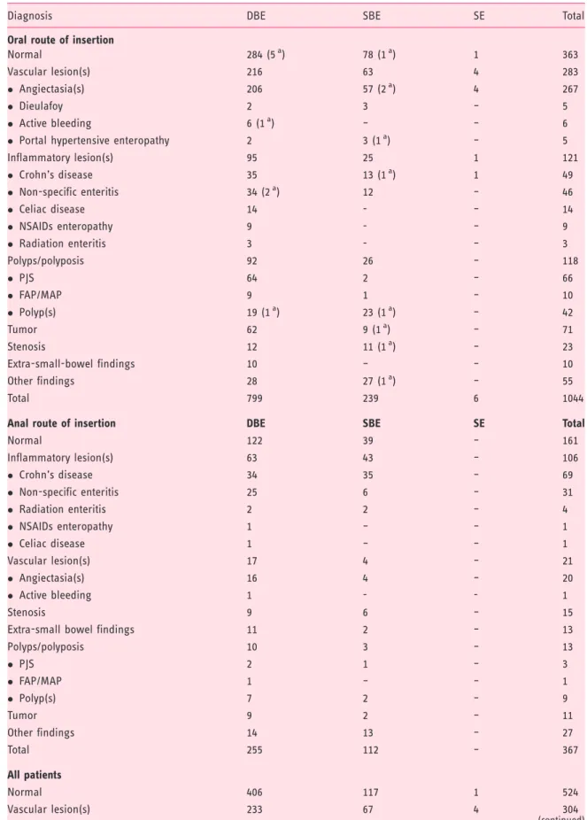
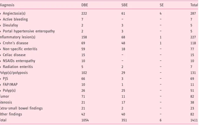
The main diagnoses established in each enteroscopy according to the type of DAE and the route of insertion are detailed in Table 4. No significant findings were established in 37.1% of patients. As expected from other case series, angiectasias, Crohn’s disease and tumors/polyps were the most frequent diagnoses, accounting for 43.8% of all diagnoses.
The diagnostic and therapeutic procedures per-formed during DAEs according to the type of DAE used are detailed in Table 5. In 595 (42.2%) patients no additional endoscopic procedures other than occa-sional tattooing were performed and in 380 (26.9%) of patients only biopsies were taken. Therapeutic
proced-ures were performed in 436 (30.9%) patients.
As detailed in Table 4, argon-plasma coagulation and polypectomy represented the most frequently used therapeutic techniques.
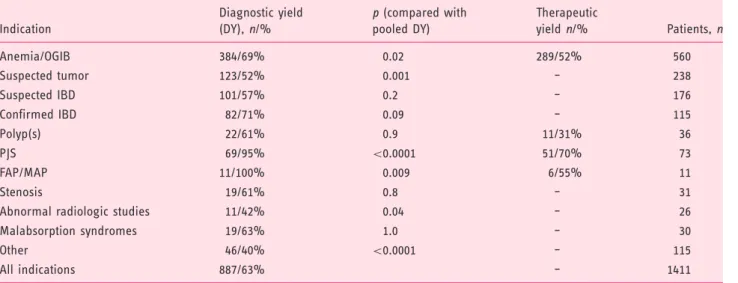
The diagnostic and therapeutic yield according to the indication is detailed in Table 6. A pooled diagnos-tic yield for all indications of 63% was found. Compared to this pooled diagnostic yield, the
diagnos-tic yield was higher for anemia/OGIB (69%, p¼0.02),
Peutz-Jeghers Syndrome (PJS) (95%, p<0.0001) and
familial adenomatous polyposis (FAP)/MUTYH
-asso-ciated polyposis (MAP) (100%, p¼0.009) and lower
for suspected tumors (52%,p¼0.001), abnormal
radio-logic studies (42%,p¼0.04) and miscellaneous
less-fre-quent indications (40%, p<0.0001).
Major complications are reported in Table 7. Anesthetic complications requiring interruption of the procedures were reported in nine (0.6%) patients, all under deep propofol sedation, six of them age 65 or
older (p>0.05) and seven of them performing oral
DAEs (p>0.05). Perforation was reported in 0.28%,
including one diagnostic procedure in a patient with radiation enteritis and three therapeutic procedures: a balloon-dilation, argon-plasma coagulation of an angiectasia and a direct percutaneous endoscopic
jeju-nostomy (DPEJ).5 In a patient with PJS undergoing
polypectomy of a large polyp, snare entrapment
occurred during polypectomy requiring surgical
intervention.
Discussion
DAE was introduced early in Portugal, shortly after the description of the procedure. In 2005, two centers were already performing the technique and, a few years later, DAE was introduced in a larger number of cen-ters. The later widespread dissemination consisted
mostly of SBE, as in Portugal a greater proportion of endoscopy centers use Olympus equipment, hence lim-iting acquisition costs to the enteroscope itself. According to the organization of the Portuguese national health system, most enteroscopy centers are Table 3. Indications for DAE, according to the type of DAE and route of insertion
Indications DBE SBE SE Total
Global
Anemia/OGIB 436 120 4 560
Suspected tumor 189 48 1 238
Suspected IBD 121 55 – 176
Confirmed IBD 88 26 1 115
Polyp(s) 9 27 – 36
PJS 69 4 – 73
FAP/MAP 10 1 – 11
Stenosis 19 12 – 31
Abnormal radiologic studies 14 12 – 26
Malabsorption syndromes 18 12 – 30
Other 81 34 – 115
Total 1054 351 6 1411
Oral route of insertion
Anemia/OGIB 361 (4a) 90 (4a) 4 455
Suspected tumor 139 (1a) 36 (2a) 1 176
Suspected IBD 73 (2a) 25 (3a) 0 98
Confirmed IBD 45 7 1 53
Polyp(s) 9 (1a) 24 0 33
PJS 65 3 0 68
FAP/MAP 9 1 0 10
Stenosis 13 7 0 20
Abnormal radiologic studies 11 11 0 22
Malabsorption syndromes 16 7 0 23
Other 58 (1a) 28 0 86
Total 799 239 6 1044
Anal route of insertion
Anemia/OGIB 75 30 – 105
Suspected tumor 50 12 – 62
Suspected IBD 48 30 – 78
Confirmed IBD 43 19 – 62
Polyp(s) – 3 – 3
PJS 4 1 – 5
FAP/MAP 1 – – 1
Stenosis 6 5 – 11
Abnormal radiologic studies 3 1 – 4
Malabsorption syndromes 2 5 – 7
Other 23 6 – 29
Total 255 112 – 367
aNumber of patients in whom oral and anal DAE was performed the same day. These procedures are recorded as a single procedure, and detailed in the
Table 4. Main diagnostic finding established in all enteroscopies according to the route of insertion and type of enteroscope used
Diagnosis DBE SBE SE Total
Oral route of insertion
Normal 284 (5a) 78 (1a) 1 363
Vascular lesion(s) 216 63 4 283
Angiectasia(s) 206 57 (2a) 4 267
Dieulafoy 2 3 – 5
Active bleeding 6 (1a) – – 6
Portal hypertensive enteropathy 2 3 (1a) – 5
Inflammatory lesion(s) 95 25 1 121
Crohn’s disease 35 13 (1a) 1 49
Non-specific enteritis 34 (2a) 12 – 46
Celiac disease 14 - – 14
NSAIDs enteropathy 9 - – 9
Radiation enteritis 3 - – 3
Polyps/polyposis 92 26 – 118
PJS 64 2 – 66
FAP/MAP 9 1 – 10
Polyp(s) 19 (1a) 23 (1a) – 42
Tumor 62 9 (1a) – 71
Stenosis 12 11 (1a) – 23
Extra-small-bowel findings 10 – – 10
Other findings 28 27 (1a) – 55
Total 799 239 6 1044
Anal route of insertion DBE SBE SE Total
Normal 122 39 – 161
Inflammatory lesion(s) 63 43 – 106
Crohn’s disease 34 35 – 69
Non-specific enteritis 25 6 – 31
Radiation enteritis 2 2 – 4
NSAIDs enteropathy 1 – – 1
Celiac disease 1 – – 1
Vascular lesion(s) 17 4 – 21
Angiectasia(s) 16 4 – 20
Active bleeding 1 - - 1
Stenosis 9 6 – 15
Extra-small bowel findings 11 2 – 13
Polyps/polyposis 10 3 – 13
PJS 2 1 – 3
FAP/MAP 1 – – 1
Polyp(s) 7 2 – 9
Tumor 9 2 – 11
Other findings 14 13 – 27
Total 255 112 – 367
All patients
Normal 406 117 1 524
Vascular lesion(s) 233 67 4 304
public inpatient facilities. In each center, two or three endoscopists are dedicated to performing DAEs. The rate of complete enteroscopy, diagnostic and thera-peutic yields and the complication rate of DBE and
SBE were compared in two recent meta-analyses.6,7
They were found to be comparable regarding all aspects except the rate of complete enteroscopy, which was found to be slightly higher with DBE. This is, however,
Table 4. Continued
Diagnosis DBE SBE SE Total
Angiectasia(s) 222 61 4 287
Active bleeding 7 – – 7
Dieulafoy 2 3 – 5
Portal hypertensive enteropathy 2 3 – 5
Inflammatory lesion(s) 158 68 1 227
Crohn’s disease 69 48 1 118
Non-specific enteritis 59 18 – 77
Celiac disease 15 – – 15
NSAIDs enteropathy 10 – – 10
Radiation enteritis 5 2 – 7
Polyp(s)/polyposis 102 29 – 131
PJS 66 3 – 69
FAP/MAP 10 1 – 11
Polyp(s) 26 25 – 51
Tumor 71 11 – 82
Stenosis 21 17 – 38
Extra-small bowel findings 21 2 – 23
Other findings 42 40 – 82
Total 1054 351 6 1411
a
Number of patients in which oral and anal DAE was performed the same day. These procedures are recorded as a single procedure, and detailed in the oral route of insertion section. DAE: device-assisted enteroscopy; SBE: single-balloon enteroscopy; SE: spiral enteroscopy; NSAIDs: nonsteroidal anti-inflammatory drugs; PJS: Peutz-Jeghers syndrome; FAP: familial adenomatous polyposis; MAP.MUTYH-associated polyposis.
Table 5. Diagnostic and therapeutic procedures performed during DAE. Only one technique is reported per procedure. In some cases, more than one technique has been used – for instance, in procedures where polypectomy has been performed, injection and tattooing could also have been used
Procedures DBE SBE SE Total
None/tattooing 461 130 4 595
Only biopsies 271 109 – 380
Hemostatic/ablative therapies 235 64 1 300
Argon plasma coagulation 210 57 1 268
Only adrenaline injection 20 2 – 22
Hemostatic clips 5 5 – 10
Polypectomy 72 24 – 96
Balloon dilation 6 10 1 17
Foreign body removal 9 5 – 14
DPEJ – 8 – 8
Stenting – 1 – 1
Total 1054 351 6 1411
of arguable clinical significance as the goal of each examination can usually be reached using both tech-niques, as expected by their similar diagnostic and therapeutic yields.
The use of DAE in the emergency setting is still uncommon. This may result from organizational diffi-culties, as the two or three endoscopists experienced in DAEs in each center may not be assigned to emergency procedures at that time; and logistical obstacles, since the procedure is time and resource consuming requiring a long allotment of time and significant staff including an anesthesiologist, for the entire duration of the pro-cedure. Furthermore, in some centers CE is not rou-tinely performed in the emergency setting to guide the use of DAE. This has important clinical implications
since the use of CE and DAE8early in the presentation
of overt OGIB improves both the diagnostic and thera-peutic yields.
Only half the centers involved reported the use of DAE for purposes other than small-bowel evaluation and only 25% reported its use for ERCP (DAE-ERCP)
in patients with surgically modified anatomy. This may be explained in part by the different areas of expertise of endoscopists performing DAE, as most of them may not be proficient in other techniques such as ERCP. DAE-ERCP enables access to the biliary system and most conventional biliary therapeutics in the great majority of this subset of patients.9–11 Nevertheless, it is very demanding, time consuming, and limited by the
availability of specific accessories,12 which also adds
technical and logistic difficulties to its widespread implementation.
Only two centers rated the subjective satisfaction with DAE with a score equal to or below 5 (0–10). In these two centers the technique was introduced in the year previous to the survey, hence these centers had performed a limited number of procedures (13 and 21). The remaining centers rated the satisfac-tion between 6 and 8, probably reflecting greater experience, although a correlation between the satis-faction score and the total number of procedures was not found.
Table 6. Diagnostic and therapeutic yield according to the indication
Indication
Diagnostic yield
(DY),n/%
p(compared with
pooled DY)
Therapeutic
yieldn/% Patients,n
Anemia/OGIB 384/69% 0.02 289/52% 560
Suspected tumor 123/52% 0.001 – 238
Suspected IBD 101/57% 0.2 – 176
Confirmed IBD 82/71% 0.09 – 115
Polyp(s) 22/61% 0.9 11/31% 36
PJS 69/95% <0.0001 51/70% 73
FAP/MAP 11/100% 0.009 6/55% 11
Stenosis 19/61% 0.8 – 31
Abnormal radiologic studies 11/42% 0.04 – 26
Malabsorption syndromes 19/63% 1.0 – 30
Other 46/40% <0.0001 – 115
All indications 887/63% – 1411
OGIB: obscure gastrointestinal bleeding; IBD: inflammatory bowel disease; PJS: Peutz-Jeghers syndrome; FAP: familial adenomatous polyposis; MAP: MUTYH-associated polyposis.
Table 7. Complications of device-assisted enteroscopy
Complications DBE (n/%) SBE (n/%) SE (n/%) Total (n/%)
Anesthetic complicationsa 9/0.85% – –/– 9/0.64%
Perforation 3/0.28% 1/0.28% –/– 4/0.28%
Pancreatitis 2/0.19% –/– –/– 2/0.14%
Post-polypectomy syndrome –/– 1/0.28% –/– 1/0.07%
Snare entrapment 1/0.09% –/– –/– 1/0.07%
Total 14/1.33 % 2/0.57% –/– 17/1.20%
a
Significant findings were found in 63% of patients, a
diagnostic yield similar to a recent systematic review13
of 12,823 DBEs in which a pooled diagnostic yield of 68.1% was found. In this review, the most frequent findings were inflammatory lesions (either Crohn’s dis-ease or non-specific inflammatory lesions), vascular lesions and tumoral lesions (either benign or malig-nant). In our series these were also the most frequent findings, accounting for 84% (745/887) of all positive findings and 52.8% of all diagnoses (745/1411). This high rate of positive findings is derived from the fact that DAE is considered a secondary procedure after an initial positive diagnostic test, as recommended by
cur-rent European guidelines.14
Small-bowel polyposis syndromes accounted for 6% (84/1411) of the examinations. Most (86.9%; 73/84) consisted of PJS referred for endoscopic polypectomy of polyps larger than 10 mm detected in surveillance CE. Snare entrapment in a large polyp was reported in one procedure (1.4%), requiring surgical interven-tion to remove the snare and resect the polyp. No per-forations were recorded after polypectomy in contrast to high rates of post-polypectomy perforations
previ-ously reported in PJS.15,16 A multicenter series of 46
procedures comprising a survey wherein polypectomy
was performed was recently published,17 in which 214
polyps with a median size of 30 mm were removed. In six of these 46 procedures, surgical intervention was required (jejunal neoplasia: one case, polyps locally concentrated in large numbers: two cases, bulky polyps: two cases with a size up to 60 mm and invagi-nated polyp: one case). All remaining polyps were man-aged endoscopically.
DAE is frequently used for therapy of small-bowel
lesions detected in other procedures.14,18–20 This is
reflected in a high percentage of therapeutic procedures (30.9%). As expected from the main indications and
other series,13,15,18,21 hemostatic therapies, namely
argon-plasma coagulation, were the most frequently employed endoscopic therapies. Polypectomy, fre-quently in the setting of PJS, was also commonly performed.
The diagnostic yield was high in all indications, reflecting an appropriate patient selection, since in most cases DAE followed a positive CE as in other
series.18In patients with OGIB, the most frequent
indi-cation for CE and DAE,13,16,22,23 the diagnostic yield
was 69%, similar to other series,15,21 and the
thera-peutic yield was also high at 52%. In patients with polyposis syndromes, the diagnostic yield was near 100% (95% and 100% for PJS and FAP/MAP) and the therapeutic yield was also high (70% for PJS and 55% for FAP/MAP). Although not directly evaluated in the present study, since in Portugal the surveillance of these patients is typically performed with CE these
data also point to a good accuracy of CE for screening these patients. Although several reports show that CE24,25 and cross-sectional radiological modalities26 may miss significant mass lesions, both have a role for small-bowel screening.25,27
The diagnostic yield for suspected small-bowel tumors (52%) was below the pooled diagnostic yield
(p¼0.001). Although DAE is regarded as the gold
standard for the diagnosis of small-bowel tumors,28
this lower diagnostic yield reflects a significant portion of false-positive tumors in CE, probably resulting from bulges due to compressions from adjacent organs or bowel loops.29
Most examinations were performed under anesthe-siologist-administered propofol sedation, a common practice in deep sedation for endoscopic procedures in
Portugal.30Anesthetic complications occurred in 0.6%
of DAEs and consisted mainly of oxygen-desaturation and less frequently bradycardia and vomiting. All cases were reversed after the interruption of the procedure. Minor oxygen desaturation not requiring the interrup-tion of the procedure was not regarded as a complica-tion. In a recent study, Lara et al.31reported anesthetic adverse events in 15 of 432 procedures (3.5%) per-formed under deep sedation, mostly oxygen desatur-ation. Based on this high rate of anesthetic adverse events, the authors currently perform endotracheal intubation for antegrade DAEs. However, this seda-tion-related complication rate is superior to previous
reports.15,32 A recent large multicenter study from
Germany, a country where non-anesthesiologist propo-fol-sedation is common practice, reported 11 (0.5%)
sedation-related adverse events in 2245 procedures.15
No more sedation-related adverse events were found in elderly patients, as previously reported.33
Complications were reported in 1.2% (17/1411),
similar to the multicenter German study15 in which a
complication rate of 1.2% (27/2245) was also found. Major complications were reported in 0.57% (eight of 1411), similar to a recent systematic review where major complications where reported in 0.72% of
examin-ations.13Only two cases of acute pancreatitis were
rec-orded. However, this complication is probably
underreported, as it occurs frequently several hours after the procedure and most centers perform DAEs
for patients from other institutions. Perforation
occurred in four patients (0.28%), most of them after
therapeutic procedures, as previously reported.15,16
The authors acknowledge some limitations.
a simple database and the required fields were easy to follow, some inconsistencies in the records may have occurred. However, the database was checked for pos-sible inconsistencies by two authors (RP, MMS). Most centers perform procedures for other institutions and the institution with the highest volume is an outpatient center performing procedures exclusively for other institutions. Hence patient follow-up and recording of delayed complications such as acute pancreatitis may be compromised.
Conclusions
This is the first published survey on the use of DAEs in Portugal. It shows that DAE is widely available in Portugal, mostly in Northern centers, with good per-formance and training capabilities. Earlier centers adopted DBEs whereas the centers that recently intro-duced DAE elected SBE. The diagnostic and thera-peutic yields are high as most procedures were performed to manage abnormalities detected in other examinations, mostly CE. The complication rate is low, mostly anesthetic complications promptly reversed after the interruption of the procedure. Perforations are rare (0.28%) and occur mainly in therapeutic procedures.
Funding
This research received no specific grant from any funding agency in the public, commercial, or not-for-profit sectors.
Conflicts of interest
None declared.
Acknowledgments
Ma´rio Dinis-Ribeiro: ManopH, Instituto CUF; Servic¸o de Gastrenterologia, Instituto Portugueˆs de Oncologia do Porto Fernando Castro-Poc¸as: ManopH, Instituto CUF; Servic¸o de Gastrenterologia, Centro Hospitalar do Porto
Sandra Lopes: Servic¸o de Gastrenterologia, Centro Hospitalar e Universita´rio de Coimbra
Carlos Sofia: Servic¸o de Gastrenterologia, Centro Hospitalar e Universita´rio de Coimbra
Ana Rita-Lopes: Servic¸o de Gastrenterologia, Centro Hospitalar e Universita´rio de Coimbra
Diogo Branquinho: Servic¸o de Gastrenterologia, Centro Hospitalar e Universita´rio de Coimbra
Iolanda Ribeiro: Servic¸o de Gastrenterologia, Centro Hospitalar de Gaia/Espinho
Ana Ponte: Servic¸o de Gastrenterologia, Centro Hospitalar de Gaia/Espinho
Joana Silva: Servic¸o de Gastrenterologia, Centro Hospitalar de Gaia/Espinho
Armando Ribeiro: Servic¸o de Gastrenterologia, Centro Hospitalar de Sa˜o Joa˜o
Filipe Vilas-Boas: Servic¸o de Gastrenterologia, Centro Hospitalar de Sa˜o Joa˜o
Joa˜o Antunes: Servic¸o de Gastrenterologia, Centro Hospitalar de Sa˜o Joa˜o
Maria Joa˜o Moreira: Servic¸o de Gastrenterologia, Centro Hospitalar do Alto Ave
Jose´ Soares: Servic¸o de Gastrenterologia, Hospital Pedro Hispano
Aˆngela Rodrigues: Servic¸o de Gastrenterologia, Centro Hospitalar do Porto
References
1. Appleyard M, Glukhovsky A and Swain P. Wireless-capsule diagnostic endoscopy for recurrent small-bowel bleeding.N Engl J Med2001; 344: 232–233.
2. Yamamoto H, Yano T, Kita H, et al. New system of double-balloon enteroscopy for diagnosis and treatment of small intestinal disorders.Gastroenterology2003; 125: 1556 ; author reply 1557.
3. Tsujikawa T, Saitoh Y, Andoh A, et al. Novel single-balloon enteroscopy for diagnosis and treatment of the small intestine: Preliminary experiences.Endoscopy2008; 40: 11–15.
4. Akerman PA, Agrawal D, Cantero D, et al. Spiral entero-scopy with the new DSB overtube: A novel technique for deep peroral small-bowel intubation.Endoscopy2008; 40: 974–978.
5. Pinho RT, Rodrigues MA and Proenc¸a ML. Overtubes and fluoroscopy for direct percutaneous endoscopic jeju-nostomy: Useful, although not always needful and some-times harmful.Dig Endosc2015; 27: 399–400.
6. Wadhwa V, Sethi S, Tewani S, et al. A meta-analysis on efficacy and safety: Single-balloon vs. double-balloon enteroscopy.Gastroenterol Rep (Oxf)2015; 3: 148–155. 7. Lipka S, Rabbanifard R, Kumar A, et al. Single versus
double balloon enteroscopy for small bowel diagnostics: A systematic review and meta-analysis. J Clin Gastroenterol2015; 49: 177–184.
8. Pinto-Pais T, Pinho R, Rodrigues A, et al. Emergency single-balloon enteroscopy in overt obscure gastrointes-tinal bleeding: Efficacy and safety. United European Gastroenterol J2014; 2: 490–496.
9. Skinner M, Popa D, Neumann H, et al. ERCP with the overtube-assisted enteroscopy technique: A systematic review.Endoscopy2014; 46: 560–572.
10. Pais TP, Pinho R, Proenc¸a L, et al. Single-balloon entero-scopy assisted endoscopic retrograde cholangiopancrea-tography with the rendezvous technique.GE Portuguese Journal of Gastroenterology2015; 22: 39–41.
enteroscopy assisted ERCP—overcoming limitations of current accessories. Rev Esp Enferm Dig 2013; 105: 561–564.
13. Xin L, Liao Z, Jiang YP, et al. Indications, detectability, positive findings, total enteroscopy, and complications of diagnostic double-balloon endoscopy: A systematic review of data over the first decade of use. Gastrointest Endosc2011; 74: 563–570.
14. Pennazio M, Spada C, Eliakim R, et al. Small-bowel cap-sule endoscopy and device-assisted enteroscopy for diag-nosis and treatment of small-bowel disorders: European Society of Gastrointestinal Endoscopy (ESGE) Clinical Guideline.Endoscopy2015; 47: 352–386.
15. Mo¨schler O, May A, Mu¨ller MK, et al. Complications in and performance of double-balloon enteroscopy (DBE): Results from a large prospective DBE database in Germany.Endoscopy2011; 43: 484–489.
16. Mo¨schler O, May AD, Mu¨ller MK, et al. Complications in double-balloon-enteroscopy: Results of the German DBE register [article in German].Z Gastroenterol 2008; 46: 266–270.
17. Serrano M, Ma˜o-de-Ferro S, Pinho R, et al. Double-bal-loon enteroscopy in the management of patients with Peutz-Jeghers syndrome: A retrospective cohort multi-center study.Rev Esp Enferm Dig2013; 105: 594–599. 18. Sethi S, Cohen J, Thaker AM, et al. Prior capsule
endos-copy improves the diagnostic and therapeutic yield of single-balloon enteroscopy. Dig Dis Sci 2014; 59: 2497–2502.
19. Pinho R. The vanishing frontiers of therapeutic entero-scopy. GE Portuguese Journal of Gastroenterology2015; 22: 137–142.
20. Fernandes C, Pinho R, Rodrigues A, et al. Argon plasma coagulation of a bleeding angioectasia in a jejunal diver-ticulum by single-balloon enteroscopy. Endoscopy 2015; 47(Suppl 1 UCTN): E62.
21. May A, Nachbar L and Ell C. Double-balloon entero-scopy (push-and-pull enteroentero-scopy) of the small bowel: Feasibility and diagnostic and therapeutic yield in patients with suspected small bowel disease.Gastrointest Endosc2005; 62: 62–70.
22. Frantz DJ, Dellon ES, Grimm IS, et al. Single-balloon enteroscopy: Results from an initial experience at a U.S. tertiary-care center. Gastrointest Endosc 2010; 72: 422–426.
23. Ribeiro I, Pinho R, Rodrigues A, et al. Obscure gastro-intestinal bleeding: Which factors are associated with positive capsule endoscopy findings? Rev Esp Enferm Dig2015; 107: 334–339.
24. Soares J, Lopes L, Vilas Boas G, et al. Wireless capsule endoscopy for evaluation of phenotypic expression of small-bowel polyps in patients with Peutz-Jeghers syn-drome and in symptomatic first-degree relatives.
Endoscopy2004; 36: 1060–1066.
25. Ribeiro I, Pinho R, Rodrigues A, et al. The importance of alternative diagnostic modalities in the diagnosis of small bowel tumors after a negative capsule endoscopy.
GE Portuguese Journal of Gastroenterology 2015; 22: 112–116.
26. Louro-da-Ponte AI, Taveira-Pinho R, Rodrigues MA, et al. Advances and pitfalls in the management of small bowel polyps in Peutz-Jeghers syndrome.Rev Esp Enferm Dig2015; 107: 390–391.
27. van Lier MG, Wagner A, Mathus-Vliegen EM, et al. High cancer risk in Peutz-Jeghers syndrome: A systematic review and surveillance recommendations. Am J Gastroenterol2010; 105: 1258–1264 ( author reply 1265. 28. Almeida N, Figueiredo P, Lopes S, et al. Double-balloon
enteroscopy and small bowel tumors: A South-European single-center experience.Dig Dis Sci2009; 54: 1520–1524. 29. Girelli CM, Porta P, Colombo E, et al. Development of a novel index to discriminate bulge from mass on small-bowel capsule endoscopy. Gastrointest Endosc 2011; 74: 1067–1074; quiz 1115.e1-e5.
30. Ferreira AO, Torres J, Dinis-Ribeiro M, et al. Endoscopic sedation and monitoring practices in Portugal: A nationwide web-based survey. Eur J Gastroenterol Hepatol2015; 27: 265–270.
31. Lara LF, Ukleja A, Pimentel R, et al. Effect of a quality program with adverse events identification on airway management during overtube-assisted enteroscopy.
Endoscopy2014; 46: 927–932.
32. Sethi S, Thaker AM, Cohen J, et al. Monitored anesthe-sia care without endotracheal intubation is safe and effi-cacious for single-balloon enteroscopy.Dig Dis Sci2014; 59: 2184–2190.