Immediate and Late Results of Percutaneous Mitral Valve Repair in
Patients with Mitral Stenosis
Luiz Francisco Cardoso,
Carlos Vinetou Ayres, André Moreira Bento, Flávio Tarasoutchi, Marcelo Luiz Vieira,
Max Grinberg
Instituto do Coração do Hospital das Clínicas da Faculdade de Medicina da Universidade de São Paulo (InCor)
Abstract
Background: The analysis of immediate and long-term results of mitral valvoplasty by balloon catheter (MVRBC) are still lacking in literature, mainly in the national context.
Objective: To assess the immediate and late results of patients submitted to mitral valve repair by balloon catheter.
Method: A total of 330 consecutive patients were followed-up by 47 ± 36 months (up to 126 months). Univariate and multivariate analyses assessed the factors associated with the success of the procedure, restenosis and late events (death or necessity of new intervention on mitral valve). Kaplan-Meier method estimated survival without events.
Results: The procedure was successful in 305 cases (92.4%). The mitral valve anatomy was the main predictor for immediate success for the procedure. During the procedure, restenosis occurred in 77 (23.3%) patients and was associated with smaller mitral valve area and with larger calcification before the process. In a mean period of 38 ± 26-month follow-up, 67 events occurred. The probability of survival without events was of 95% in one year, 75% in five years and 61% in ten years. The predictors of survival without events were: age, echocardiography score and immediate result of the procedure.
Conclusion: Mitral valve repair by balloon catheter is an effective procedure, as 60% patients did not present events after the follow-up. The anatomical condition of the mitral valve and the patient’s age were the best predictors of survival without events, and should be taken into account when selecting patients for the mentioned procedure. (Arq Bras Cardiol 2010; 94(3):383-390)
Key words: Balloon dilatation; mitral valve stenosis; outcome and process assessment (health care).
Mailing address: Luiz Francisco Cardoso •
Rua Barata Ribeiro, 237 cj 26 - Bela Vista - 01308-000 - São Paulo, SP, Brazil E-mail: [email protected]
Manuscript received December 08, 2008; revised manuscript received April 29, 2009; accepted July 01, 2009.
Introduction
Since the introduction of balloon dilatation technique in mitral stenosis, in 19841, the mitral valvuloplasty by balloon
catheter (MVBC) has showed to be an effective treatment for mitral stenosis in selected patients. The possibility of its accomplishment, even in situation with high surgical risk, as avoiding the complications intrinsic to the couple thoracotomy/extracorporeal circulation, has made MVBC a therapeutic option concerning surgical treatment. Reduction of hospitalization period, reduction of hospital costs and low mortality are other advantages that made this method extremely attractive and alternative to surgery. After the procedure, the immediate improvement of the valvar area and the survival without medium and long-term events2-4 are
comparable to the surgicalcommissurotomy5,6. Many factors
have been suggested as determinants of late MVBC success, including: age, morphological aspects, cardiac rhythm, cardiac debt, mitral valve area (MVA) and experience of the
intervention center7-18. However, there are still uncertainties
with regard to these variables’ predictive power, for the results are not homogeneous and some researchers did not found association of the abovementioned factors with long-term results7,9-19. Regional differences in the patients’ profile may
justify the variation of the results in the method used for the morphological analysis of mitral valve and in the technique used for the procedure. The possibility of demonstrating the experience of one single Brazilian center has motivated us to develop an analysis of the evolution at short and long term in patients submitted to MVBC in order to identify the factors related to the immediate or late success of this procedure.
Methods
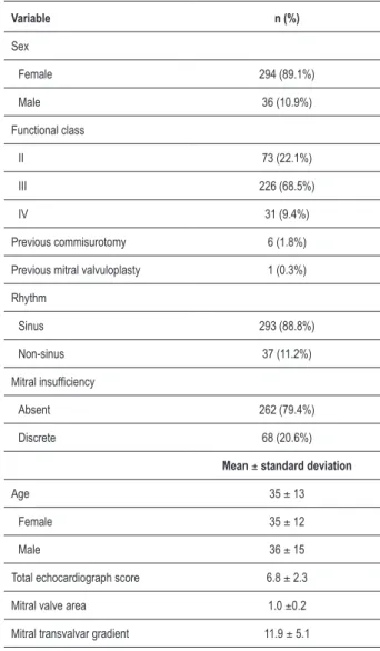
Variable n (%)
Sex
Female 294 (89.1%)
Male 36 (10.9%)
Functional class
II 73 (22.1%)
III 226 (68.5%)
IV 31 (9.4%)
Previous commisurotomy 6 (1.8%)
Previous mitral valvuloplasty 1 (0.3%)
Rhythm
Sinus 293 (88.8%)
Non-sinus 37 (11.2%)
Mitral insuficiency
Absent 262 (79.4%)
Discrete 68 (20.6%)
Mean ± standard deviation
Age 35 ± 13
Female 35 ± 12
Male 36 ± 15
Total echocardiograph score 6.8 ± 2.3
Mitral valve area 1.0 ±0.2
Mitral transvalvar gradient 11.9 ± 5.1
Table 1 – Clinical characteristics and complementary exams in basal condition of patients submitted to mitral valvuloplasty by balloon-catheter
MVBC and patients with score superior to the ideal, but with high surgical risk, were included in the study. Patients with mitral insufficiency (MI) of grades higher than discrete and carriers of other cardiopathies with surgical indication were excluded from the sample. The study was approved by the Ethics Committee of the institution.
All patients went through a clinical assessment based on history and physical or complementary exams in the following moments: immediately before MVBC (henceforth PRE), after MVBC (henceforth POI) and before hospital discharge, 30 days after hospital discharge, 6 months after MVBC (henceforth PO6M), 12 months after MVBC (henceforth PO12M) and then every 12 months, till the limit of 120 months.
Clinical assessment was performed in all the aforementioned moments and consisted of complete history and physical exam. The functional class and the presence of immediate postoperative complications were particularly quantified
according to NYHA, and the events were defined as follows: cardiovascular obit, non-cardiovascular obit, new intervention on mitral valve through surgery or new MVBC. Forty-six patients (13.9%) were clinically reassessed in other institutions and their clinical data were obtained by letter or phone call. The echocardiograph evaluation was performed in all aforesaid moments, except for the assessment 30 days after hospital discharge. The images obtained from ECO enabled the assessment of the morphological patterns used in the graduation of the valve damage and the quantification of mitral valve reflux grade. The variables taken into account were: morphological study of mitral valve (echocardiograph score), medium mitral transvalvar gradient (G) in mmHg, mitral valve area (MVA) in cm2 and MI. The mitral valve morphological
study was based in semi-quantitative analysis, as established grade 1 (discrete damage) to 4 (severe damage) for mobility, thickness and calcification of the cuspids, as well as the fiberoptic damage of subvalvar system. The sum of these grades resulted in the total echocardiography score according to the criteria proposed by Wilkins et al10. With continuum
Doppler and color flux mapping from the apical window, the best curve of mitral flux velocity was obtained for the MVA calculation and of medium mitral G20,21. Pulsing Doppler
and/or color flux mapping were used for the detection and semi-quantitative assessment of MI grade22.
MVBC was performed exclusively by the same interventionist doctor by means of right-heart catheterization technique and transseptal approach. The single or double-catheter approach was used according to the availability of the material. The double-balloon technique was used in 83 (25.2%) patients. Unifoil, bifoil and trefoil balloons were used in 4 (1.2%), 30 (9.1%) and 8 (2.4%) patients, respectively. The Inoue balloon was used in the remaining 205 patients (62.1%). Hemodynamic measures were made, including left atrium pressure, left ventricle final diastolic pressure, pressure G by mitral valve and cardiac debt (through thermal dissolution) after and before the balloon dilation. Dilation by balloon was repeated till a satisfactory drop in mitral G, improvement of cardiac auscultation, mitral regurgitation appearance or other complications were achieved.
MVA after the procedure, calculated by ECO, superior or equal to 1.5 cm2 or 50% gain in relation to the measured area
before the procedure was considered successful.
During the follow-up, the occurrence of MVA, calculated by ECO, inferior to 1.5 cm2 or loss of 50% of the initial obtained
gain in the first ECO post-procedure (POI) were defined as restenosis.
The necessity of new intervention on the mitral valve (surgery or MVBC) and the occurrence of obit by cardiovascular or non-cardiovascular causes were considered to be the main events.
Statistical analysis
Variables Success (n=305) Fail (n=25) p-value
Sex 0.334*
Female 273 (92.9%) 21 (7.1)
Male 32 (86.1%) 4 (11.1%)
Functional class 0.814†
II 67 (91.8%) 6 (8.2%)
III or IV 238 (92.6%) 19 (7.4%)
Cardiac Rhythm 0.386*
Non-sinus 33 (89.2%) 4 (10.8%)
Sinus 272 (92.8%) 21 (7.2%)
Total echocardiograph score <0.001*
≤8 242 (97.2%) 7 (2.8%)
>8 37 (82.2%) 8 (17.8%)
Valve morphology
Subvalvar 0.002†
1 to 2 218 (97.3%) 6 (2.7%)
3 to 4 61 (87.% 9 (12.9%)
Mobility 0.034*
1 to 2 267 (95.7%) 12 (4.3%)
3 to 4 12 (80.0%) 3 (20.0%)
Thickness 0.019*
1 to 2 261 (96.0%) 11 (4.0%)
3 to 4 18 (81.8%) 4 (18.2%)
Calciication 0.064*
1 to 2 263 (95.6%) 12 (4.4%)
3 to 4 16 (84.2%) 3 (15.8%)
Previous valvar insuficiency 0.663*
Absent 243 (92.8%) 19 (7.2%)
Discrete 62 (91.2%) 6 (8.8%)
Mean ± standard deviation
Age 35 ± 12 43 ± 14 0.001‡
Previous valve area 0.99 ± 0.23 1.07 ± 0.21 0.249‡
Previous mitral transvalvar
gradient 11.0 ± 5.2 12.2 ± 4.6 0.803‡
Fisher’s Exact test; †Chi-square test; ‡Student’s t-test.
Table 2 – Results from the univariate analysis for the success predictor factors
and 2 versus 3 and 4), calcification (1 and 2 versus 3 and 4), subvalvar (1 and 2 versus 3 and 4), MVA, medium mitral G and MI grade in the pre-procedure period.
The predictive factors for restenosis and events in the follow-up of patients were also studied through univariate and multivariate analysis. Besides the variables used in the analysis of success, the measures of MVA and medium mitral G obtained in the ECO performed during POI were included.
To estimate the probability of survival without the occurrence of events combined throughout time, the Kaplan-Meier method was applied.
The chi-square or Fisher’s exact tests were used to compare the qualitative variables, while the Student’s t-test was applied in the comparison of quantitative variables.
Through multivariate logistic regression, the probability of success, restenosis and events was correlated to the predetermined variables from univariate analysis.
The significance level applied in the tests was 5%.
Results
Immediate
MVBC was considered successful in 305 patients (92.4%). Technical difficulties occurred in seven patients and in the others the obtained MVA did not reach the success criteria. MVA increased from 1.00 ± 0.23 cm2 to 2.03 ± 0.41 cm2
after MVBC (p<0.001). Following this findingm médium transmitral G decreased from 12 ± 5 mmHg to 5 ± 3 mmHg (p<0.001). After the procedure, MI was considered absent in 171 patients, discrete-grade in 106 patients, moderate in 38 patients and severe in 15 patients. Other complications related to the procedure occurred in 14 (4.2%) patients, as 9 (2.7%) patients presented cardiac tamponing, 4 (1.2%) patients presented embolism in the central nervous system, and one presented (0.3%) peripheral embolism.
The results of the univariate analysis for the predictive factors for success are presented in Table 2. Mean age of patients that had successful procedure was significantly lower (p=0.001). The following factors were considered predictive of success for MVBC: less subvalvar system fiberoptic damage (p=0.002), less thickness (p=0.019) and more valvar cuspids mobility (p=0.034), as well as total echocardiograph score inferior to 8 (p<0.001).
Through the multivariate analysis, only echocardiograph score inferior to 8 was identified as predictive factor for MVBC success. Besides, the chance of failing was determined as approximately 7.47 times higher in individuals with value superior to 8 (confidence interval 95%, 2.559 to 21.830, p=0.002).
Late
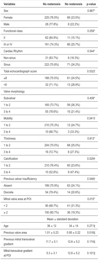
Variables No restenosis No restenosis p-value
Sex 0.867*
Female 225 (76.5%) 69 (23.5%)
Male 28 (77.8%) 8 (22.2%)
Functional class 0.059*
II 62 (84.9%) 11 (15.1%)
III or IV 191 (74.3%) 66 (25.7%)
Cardiac Rhythm 0.544*
Non-sinus 31 (83.7%) 6 (16.3%)
Sinus 222 (75.8%) 71 (24.2%)
Total echocardiograph score 0.532†
≤8 188 (75.5%) 61 (24.5%)
>8 32 (71.1%) 13 (28.9%)
Valve morphology
Subvalvar 0.409*
1 to 2 165 (73.7%) 59 (26.3%)
3 to 4 55 (78.6%) 15 (21.4%)
Mobility 0.541†
1 to 2 210 (75.3%) 12 (24.7%)
3 to 4 10 (66.7%) 3 (33.3%)
Thickness 0.813*
1 to 2 204 (75.0%) 68 (25.0%)
3 to 4 16 (72.7%) 6 (27.3%)
Calciication 0.029†
1 to 2 210 (76.4%) 65 (23.6%)
3 to 4 10 (52.6%) 9 (47.4%)
Previous valvar insuficiency 0.548†
Absent 199 (75.9%) 63 (24.1%)
Discrete 54 (79.4%) 14 (20.6%)
Mitral valve area at POI 0.015*
< 2 90 (68.7%) 41 (31.3%)
≥ 2 150 (80.7%) 36 (19.3%)
Mean ± standard deviation
Age 36 ± 12 34 ± 14 0.271‡
Previous valve area 1.01 ± 0.23 0.95 ± 0.22 0.018‡
Previous mitral transvalvar
gradient 11.7 ± 5.1 12.6 ± 5.2 0.174‡
Mitral transvalvar gradient
at POI 5.3 ± 3.1 12.6 ± 5.2 0.101‡
Table 3 – Results from the univariate analysis for the restenosis predictor factors
*Chi-square test; †Fisher’s Exact test; ‡Student’s t-test.
mean follow-up period of 54 ± 34 months (varying from 2 to 126 months).
Restenosis
During the follow-up, 77 (23.3%) patients presented echocardiography criteria for restenosis in a mean period of 62 ± 32 months after MVBC. The mean valvar area and the mitral gradient of these patients were: 1.07 ± 0.19 cm2 and
9.5 ± 4.1 mmHg, respectively.
Through the multivariate analysis, three variables related to a higher probability of restenosis during procedure were identified (Table 3). Patients who developed restenosis presented higher intensity of mitral valve calcification (p=0.029), MVA significantly smaller at PRE (p=0.018) and inferior to 2.0 cm2 at POI (p=0.015).
The multivariate analysis, performed through logistic regression, identified only MVA at PRE and higher valve calcification grade as main predictive factors for restenosis.
Clinical events
Sixty-seven events (20.3%) were observed during follow-up, with a mean period of 38 ± 26 months after MVBC.
There were 17 death occurrences (4.84%) within 33 ± 19 months after procedure. The causes of obits are presented in Table 4.
Forty-three patients (13.0%) were submitted to surgical treatment during follow-up. In 18 patients (5.45%) it was necessary to implant cardiac prosthesis 30 ± 28 months after MVBC. Mitral commissurotomy was performed in 23 patients (6.96%) within 48 ± 25 months, and mitral valve plastic surgery in other 2 patients (0.6%) within 6 ± 3 months after MVBC. New mitral valvuloplasty was performed in 7 patients (2.12%) within 64 ± 22 months after the first MVBC.
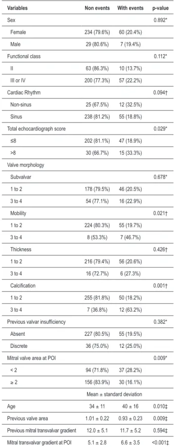
The probability of survival without events (surgery, valvuloplasty or obit) was 94.99 ± 1.31% within 1 year, 75.14 ± 3.03% within 5 years and 61.44 ± 4,36 within 8 years, as maintaining it till 10 years of follow-up (Figure 1). No events occurred after the eighth year of follow-up. Through the multivariate analysis, seven variables related to the probability of events during follow-up were identified (Table 5). The patients who developed any event presented significantly higher mean age (p=0.010), lower MVA at PRE (p=0.009), reduced mobility of cuspids (p=0.021), higher valvar calcification grade (p=0.001) and echocardiograph score superior to 8 (p=0.029) at PRE. At POI, the most elevated mean transvalvar G (p<0.001) and MVA smaller than 2.00 cm2 (p=0.009) were also predictors of events, and
was associated with long-term events rate.
Through the multivariate analysis, the following variables were identified as the most strongly related to the occurrence of long-term events: advanced age, smaller MVA at PRE and higher medium mitral transvalvar G value at POI.
Discussion
(n)
Cardiovascular obit 8
Cardiac insuficiency 5
Sudden death 1
Myocardial infarction 1
Vascular brain accident 1
Non-cardiovascular obit 9
Unknown 3
Pulmonary infection 2
Trafic accident 1
Morbid obesity 1
Pulmonary neoplasm 1
Hepatic insuficiency 1
Table 4 – Death causes during follow-up
years7-18, however, the studied populations differ in their
socioeconomic, ethnic, cultural and aging profiles, besides clinical and echocardiograph characteristics, from those found in our sample. All these particularities stick out the importance of knowing the long-term results of MVBC in the national context, which is represented by scarce but valuable studies7,23.
Mean age of the patients herein presented was 35 ± 13 years old, a value that is intermediate to those of developed countries24, where these patients are younger, and to the
mean ages of patients from European countries25,26 and from
the United States of America8,27, which are considered to be
more elevated. In North American studies by Palácios et al27
and Cohen et al8, mean ages of patients were 55 and 59 years
old, respectively. This observation is justified by the fact that
Variables Non events With events p-value
Sex 0.892*
Female 234 (79.6%) 60 (20.4%)
Male 29 (80.6%) 7 (19.4%)
Functional class 0.112*
II 63 (86.3%) 10 (13.7%)
III or IV 200 (77.3%) 57 (22.2%)
Cardiac Rhythm 0.094†
Non-sinus 25 (67.5%) 12 (32.5%)
Sinus 238 (81.2%) 55 (18.8%)
Total echocardiograph score 0.029*
≤8 202 (81.1%) 47 (18.9%)
>8 30 (66.7%) 15 (33.3%)
Valve morphology
Subvalvar 0.678*
1 to 2 178 (79.5%) 46 (20.5%)
3 to 4 54 (77.1%) 16 (22.9%)
Mobility 0.021†
1 to 2 224 (80.3%) 55 (19.7%)
3 to 4 8 (53.3%) 7 (46.7%)
Thickness 0.426†
1 to 2 216 (79.4%) 56 (20.6%)
3 to 4 16 (72.7%) 6 (27.3%)
Calciication 0.001†
1 to 2 255 (81.8%) 50 (18.2%)
3 to 4 7 (36.8%) 12 (63.2%)
Previous valvar insuficiency 0.382*
Absent 227 (80.5%) 55 (19.5%)
Discrete 36 (75.0%) 12 (25.0%)
Mitral valve area at POI 0.009*
< 2 94 (71.8%) 37 (28.2%)
≥ 2 156 (83.9%) 30 (16.1%)
Mean ± standard deviation
Age 34 ± 11 40 ± 16 0.010‡
Previous valve area 1.01 ± 0.22 0.93 ± 0.23 0.009‡
Previous mitral transvalvar gradient 12.0 ± 5.1 11.7 ± 5.2 0.594‡
Mitral transvalvar gradient at POI 5.1 ± 2.8 6.6 ± 3.5 <0.001‡
Table 5 – Results from the univariate analysis for the predictor factors of events occurrence
*Chi-square test; †Fisher’s Exact test; ‡Student’s t-test. 0,0
0,1 0,2 0,3 0,4 0,5 0,6 0,7 0,8 0,9 1,0
Tempo (anos)
P
robabi
lidade de s
obrev
ida l
iv
re de ev
entos
0 1 2 3 4 5 6 7 8 9 10
Figure 1 – Curve of survival without events after mitral valvuloplasty by
rheumatic disease is endemic and cardiac assault happens more precociously in developed countries.
This paper reports MVBC in a population formed primarily by young women, with valves considered to be ideal for the MVBC proceeding, though some patients with less favorable profile are also presented in our research. The predominance of females (89%) in this study suits other studies7,8,25-28.
The mean echocardiograph score in the presented sample was 6.77, and only 14 patients, who had higher risk for surgical treatment, had a value superior to 9. Similar proportions of patients with unfavorable valvar morphology were studied by other authors7,27.
Immediate results
By means of the aforementioned criteria, MVBC was successful in 92.4% of the cases followed by clinical improvement immediately after the procedure and at long term. This success incidence is similar to what was previously reported11,29-31, although the definition of success may have
varied among case reports.
In the present study, the univariate analysis detected the following items as success predictors: age, echocardiograph score, subvalvar thickness and cuspids mobility. The multivariate analysis demonstrated that the most powerful variable was the echocardiograph score inferior to 9. The MVBC results, similarly to those observed in mitral commissurotomy, are directly related to the involvement grade of the valvar system. Though the echocardiography analysis of mitral anatomy by means of score may present certain grade of variability among observers, it has showed to be a sensitive and effective method for quantifying the alterations on the mitral structure. Several studies on MVBC3,11,23,25,26 that used the same analysis criteria for valvar
morphology have also showed that the echocardiograph score is the most important success predictor. Adverse effects of MVBC were not frequent. We found 14 cases (4.23%) of non-valvar complications (cardiac tamponing and embolism) and 15 cases (4.5%) of MI importance. This incidence of complications is similar to what has been already described by other authors3,32.
Late results
Restenosis
The progression of the valvar lesion in rheumatic disease may result from a subclinical rheumatic activity and/or from the turbulent flux generated in an already deformed valve. Such conditions may contribute to an increase in commissural fusion, thickness and calcification of a previously deformed valve system. They may occur in valves without intervention as much as in valves that previously went through such procedures. Restenosis is an ambiguous term that may include the inadequate result of MVBC, the inaccuracy of MVA determination, the true restenosis and the disease progress. The definition for
restenosis is not homogeneous among many studies, as it may be based on clinical aspects, MVA, absolute loss of valvar area, loss percentage or absence of increase in this area. All this variability of factors and employed criteria may justify the great difference of restenosis rate among the various reports, varying from 3 to 70% within a period of 1 to 3 years33,34. Echocardiograph score superior to
824,33,34 and the presence of calcification35 before MVBC and
reduced MVA26,36 after MVBC have already been reported
as restenosis predictors in other studies. In this 10-year study, restenosis rate was 23.3% and happened after a mean period of 62 ± 32 months. The importance of restenosis study is based on the fact that this is the more frequent cause for a new intervention. Among the 43 operated patients, restenosis was diagnosed in 26 cases. Our results support the hypothesis that the echocardiograph analysis is essential to determine patients with high probability of developing restenosis at long term and, therefore, is useful to select the best therapeutic option.
Events
The valvar anatomy is an important predictor of dissatisfactory long-term results in patients submitted to surgical treatment for mitral stenosis37,38. From the
introduction of MVBC, the echocardiograph score had already been identified as a predictor for immediate and medium-term results11,25,30,32. The results herein presented
support the hypothesis that a score superior to 8 is strongly related to the occurrence of events in long-term follow-up. Although the majority of patients do not present elevated calcification score (3 or 4), when it was presented, was related to the development of late events. In this analysis, MVA before the procedure was significantly lower in the patients who presented late events. Such finding was reported in other studies26,29,30,35 and may be associated with
the higher restenosis rate in this group of patients.
Two other variables related to immediate success of MVBC, the mitral transvalvar gradient and MVA at POI, were also late events predictors. This result in compliance with other authors’ report11,26,28,29 and emphasizes the
importance of the immediate result of MVBC in the long-term evolvement. Likewise the present study, age was also described as a predictor factor for late events in other studies25,27-29,35.
Therefore, the immediate result is also related to long-term prognostic. The analysis of mitral anatomy is essential to determine the patients who have greater chances of achieving good results with MVBC, for it may predict immediate and long-term results.
Conclusion
References
predictors of survival without events. This should be taken into account when selecting patients for percutaneous mitral commissurotomy.
Potential Conflict of Interest
No potential conflict of interest relevant to this article was reported.
Sources of Funding
There were no external funding sources for this study.
Study Association
This article is part of the thesis of doctoral submitted by Carlos Vinetou Ayres, from Faculdade de Medicina de São Paulo.
1. Inoue K, Owaki T, Nakamura T, Kitamura F, Miyamoto N. Clinical application of transvenous mitral commissurotomy by a new balloon catheter. J Thorac Cardiovasc Surg. 1984; 87 (3): 394-402.
2. Multicenter experience with balloon mitral commissurotomy. NHLBI Balloon Valvuloplasty Registry Report on immediate and 30-day follow-up results. The National Heart, Lung, and Blood Institute Balloon Valvuloplasty Registry Participants. Circulation. 1992; 85 (2): 448-61.
3. Vahanian A, Michel PL, Cormier B, Vitoux B, Michel X, Slama M, et al. Results of percutaneous mitral commissurotomy in 200 patients. Am J Cardiol. 1989; 63 (12): 847-52.
4. McKay RG, Lock JE, Keane JF, Safian RD, Aroesty JM, Grossman W. Percutaneous mitral valvuloplasty in an adult patient with calcific rheumatic mitral stenosis. J Am Coll Cardiol. 1986; 7 (6): 1410-5.
5. Turi ZG, Reyes VP, Raju BS, Raju AR, Kumar DN, Rajagopal P, et al. Percutaneous balloon versus surgical closed commissurotomy for mitral stenosis: a prospective, randomized trial. Circulation. 1991; 83 (4): 1179-85.
6. Kirklin JW. Percutaneous balloon versus surgical closed commissurotomy for mitral stenosis. Circulation. 1991; 83 (4): 1450-1.
7. Borges IP, Peixoto EC, Peixoto RT, Oliveira PS, Netto MS, Labrunie P, et al. Percutaneous mitral balloon valvotomy: long-term outcome and assessment of risk factors for death and major events. Arq Bras Cardiol. 2005; 84 (5): 397-404.
8. Cohen DJ, Kuntz RE, Gordon SP, Piana RN, Safian RD, McKay RG, et al. Predictors of long-term outcome after percutaneous balloon mitral valvuloplasty. N Engl J Med. 1992; 327 (19): 1329-35.
9. Herrmann HC, Ramaswamy K, Isner JM, Feldman TE, Carroll JD, Pichard AD, et al. Factors influencing immediate results, complications, and short-term follow-up status after Inoue balloon mitral valvotomy: a North American multicenter study. Am Heart J. 1992; 124 (1): 160-6.
10. Wilkins GT, Weyman AE, Abascal VM, Block PC, Palacios IF. Percutaneous balloon dilatation of the mitral valve: an analysis of echocardiographic variables related to outcome and the mechanism of dilatation. Br Heart J. 1988; 60 (4): 299-308.
11. Herrmann HC, Wilkins GT, Abascal VM, Weyman AE, Block PC, Palacios IF. Percutaneous balloon mitral valvotomy for patients with mitral stenosis. Analysis of factors influencing early results. J Thorac Cardiovasc Surg. 1988; 96 (1): 33-8.
12. Reid CL, Chandraratna PA, Kawanishi DT, Kotlewski A, Rahimtoola SH. Influence of mitral valve morphology on double-balloon catheter balloon valvuloplasty in patients with mitral stenosis: analysis of factors predicting immediate and 3-month results. Circulation. 1989; 80 (3): 515-24.
13. Abascal VM, Wilkins GT, O’Shea JP, Choong CY, Palacios IF, Thomas JD et al. Prediction of successful outcome in 130 patients undergoing percutaneous balloon mitral valvotomy. Circulation. 1990; 82 (2): 448-56.
14. Hung JS, Chern MS, Wu JJ, Fu M, Yeh KH, Wu YC, et al. Short- and long-term results of catheter balloon percutaneous transvenous mitral commissurotomy. Am J Cardiol 1991; 67(9): 854-62.
15. The National Heart, Lung and Blood Institute Balloon Valvuloplasty. Registry Participants. Complications and mortality of percutaneous balloon mitral commissurotomy. A report from the National Heart, Lung, and Blood Institute Balloon Valvuloplasty Registry. Circulation. 1992; 85 (6): 2014-24.
16. Pan M, Medina A, Suarez DL, Hernandez E, Romero M, Pavlovic D, et al. Factors determining late success after mitral balloon valvulotomy. Am J Cardiol. 1993; 71 (13):1181-5.
17. Herrmann HC, Feldman T, Isner JM, Bashore T, Holmes DR Jr, Rothbaum DA, et al. Comparison of results of percutaneous balloon valvuloplasty in patients with mild and moderate mitral stenosis to those with severe mitral stenosis. The North American Inoue Balloon Investigators. Am J Cardiol. 1993; 71 (15): 1300-3.
18. Tuzcu EM, Block PC, Palacios IF. Comparison of early versus late experience with percutaneous mitral balloon valvuloplasty. J Am Coll Cardiol. 1991; 17 (5): 1121-4.
19. Feldman T, Carroll JD, Isner JM, Chisholm RJ, Holmes DR, Massumi A, et al. Effect of valve deformity on results and mitral regurgitation after Inoue balloon commissurotomy. Circulation. 1992; 85 (1): 180-7.
20. Hatle L, Angelsen B, Tromsdal A. Noninvasive assessment of atrioventricular pressure half-time by Doppler ultrasound. Circulation. 1979; 60 (5): 1096-104.
21. Holen J, Simonsen S. Determination of pressure gradient in mitral stenosis with Doppler echocardiography. Br Heart J. 1979; 41 (5): 529-35.
22. Helmcke F, Nanda NC, Hsiung MC, Soto B, Adey CK, Goyal RG, et al. Color Doppler assessment of mitral regurgitation with orthogonal planes. Circulation. 1987; 75 (1): 175-83.
23. Cardoso LF, Ratti MA, Grinberg M, Medeiros CC, Tarasoutchi F, Rossi EG, et al. Mitral valvuloplasty by balloon catheter. Early results and one-year follow-up. Arq Bras Cardiol. 1992; 58 (6): 445-51.
24. Ben Farhat M, Betbout F, Gamra H, Maatouk F, Ben Hamda K, Abdellaoui M, et al. Predictors of long-term event-free survival and of freedom from restenosis after percutaneous balloon mitral commissurotomy. Am Heart J. 2001; 142 (6): 1072-9.
25. Iung B, Garbarz E, Michaud P, Helou S, Farah B, Berdah P, et al. Late results of percutaneous mitral commissurotomy in a series of 1024 patients: analysis of late clinical deterioration: frequency, anatomic findings, and predictive factors. Circulation. 1999; 99 (25): 3272-8.
26. Hernandez R, Banuelos C, Alfonso F, Goicolea J, Fernandez-Ortiz A, Escaned J, et al. Long-term clinical and echocardiographic follow-up after percutaneous mitral valvuloplasty with the Inoue balloon. Circulation. 1999; 99 (12): 1580-6.
27. Palacios IF, Tuzcu ME, Weyman AE, Newell JB, Block PC. Clinical follow-up of patients undergoing percutaneous mitral balloon valvotomy. Circulation. 1995; 91 (3): 671-6.
29. Pavlides GS, Nahhas GT, London J, Gangadharan C, Troszak E, Barth-Jones D, et al. Predictors of long-term event-free survival after percutaneous balloon mitral valvuloplasty. Am J Cardiol. 1997; 79 (10): 1370-4.
30. Orrange SE, Kawanishi DT, Lopez BM, Curry SM, Rahimtoola SH. Actuarial outcome after catheter balloon commissurotomy in patients with mitral stenosis. Circulation. 1997; 95 (2): 382-9.
31. Goswami KC, Bahl VK, Talwar KK, Shrivastava S, Manchanda SC. Percutaneous balloon mitral valvuloplasty using the Inoue balloon: analysis of echocardiographic and other variables related to immediate outcome. Int J Cardiol. 1999; 68 (3): 261-8.
32. Palacios I, Block PC, Brandi S, Blanco P, Casal H, Pulido JI, et al. Percutaneous balloon valvotomy for patients with severe mitral stenosis. Circulation. 1987; 75 (4): 778-84.
33. Palacios IF, Block PC, Wilkins GT, Weyman AE. Follow-up of patients undergoing percutaneous mitral balloon valvotomy: analysis of factors determining restenosis. Circulation. 1989; 79 (3): 573-9.
34. Trevino AJ, Ibarra M, Garcia A, Uribe A, de la Fuente F, Bonfil MA, et al. Immediate and long-term results of balloon mitral commissurotomy for rheumatic mitral stenosis: comparison between Inoue and double-balloon techniques. Am Heart J. 1996; 131 (3): 530-6.
35. Zhang HP, Ruiz CE, Allen JW, Lau FY. A novel prognostic scoring system to predict late outcome after percutaneous balloon valvotomy in patients with severe mitral stenosis. Am Heart J. 1997; 134 (4): 772-8.
36. Langerveld J, Thijs Plokker HW, Ernst SM, Kelder JC, Jaarsma W. Predictors of clinical events or restenosis during follow-up after percutaneous mitral balloon valvotomy. Eur Heart J. 1999; 20 (7): 519-26.
37. Gross RI, Cunningham JN Jr, Snively SL, Catinella FP, Nathan IM, Adams PX, et al. Long-term results of open radical mitral commissurotomy: ten year follow-up study of 202 patients. Am J Cardiol. 1981; 47 (4): 821-5.