Dental Materials
Carla Rodrigues Mazzo(a) Andréa Cândido dos Reis(b) Antonio Carlos Shimano(c) Mariana Lima da Costa Valente(a)
(a) School of Dentistry of Ribeirão Preto, University of São Paulo - USP, Ribeirão Preto, SP, Brazil.
(b)Department of Dental Materials, School of Dentistry of Ribeirão Preto, University of São Paulo - USP, Ribeirão Preto, SP, Brazil. (c)Department of Biomechanics and
Orthopaedics, School of Medicine of Ribeirão Preto, University of São Paulo - USP, Ribeirão Preto, SP, Brazil.
Corresponding author:
Andréa Cândido dos Reis E-mail: [email protected]
Received for publication on Sep 29, 2011 Accepted for publication on Mar 26, 2012
In vitro
analysis of the influence of
surface treatment of dental implants on
primary stability
Abstract: Surface treatment interferes with the primary stability of den-tal implants because it promotes a chemical and micromorphological change on the surface and thus stimulates osseointegration. This study aimed to evaluate the effects of different surface treatments on primary stability by analyzing insertion torque (IT) and pullout force (PF). Eight samples of implants with different surface treatments (TS - external hexa-gon with acid surface treatment; and MS - external hexahexa-gon, machined surface), all 3.75 mm in diameter × 11.5 mm in length, were inserted into segments of artiicial bones. The IT of each sample was measured by an electronic torquemeter, and then the pullout test was done with a univer-sal testing machine. The results were subjected to ANOVA (p < 0.05), followed by Tukey’s test (p < 0.05). The IT results showed no statistically signiicant difference, since the sizes of the implants used were very simi-lar, and the bone used was not highly resistant. The PF values (N) were, respectively, TS = 403.75 ± 189.80 and MS = 276.38 ± 110.05. The im-plants were shown to be different in terms of the variables of maximum force (F = 4.401, p = 0.0120), elasticity in maximum lexion (F = 3.672, p = 0.024), and relative stiffness (F = 4.60, p = 0.01). In this study, exter-nal hexagoexter-nal implants with acid surface treatment showed the highest values of pullout strength and better stability, which provide greater indi-cation for their use.
Descriptors: Dental Implantation; Osseointegration; Tensile Strength; Torque.
Introduction
In the 1960s, Brånemark1 discovered that titanium was biocompatible
with bone and could attach to it. Since then, many studies have been con-ducted to determine the nature of the interaction that occurs between the metal and the tissue2-5 called osseointegration, in which the bone-implant
unit becomes an anatomical structure6 and supports patient function
without causing injury.
Currently, the dental implant is an object of study by several research-ers7-11 whose goals are to develop design innovations and application
techniques to optimize the implant’s physical-chemical and mechani-cal properties.7 One current area of research involves the treatment of
implant surfaces for osseointegration stimulation due to a chemical or micromorphological alteration of the implant9 that can facilitate early
stallation of the prosthesis.12
These innovations are designed to improve the technical conditions for implant insertion and im-prove osseointegration, which is possible only if pri-mary stability of the implants is attained. Pripri-mary stability is deined as the resistance to micromove-ment of the implant in the surgical site immediately after its insertion, and is one of the main factors re-quired for osseointegration, since stability facilitates the formation of bone cells around the implants. Pri-mary stability is dependent on factors such as bone type, and on characteristics related to implants, such as design and surface topography.13
Primary stability of implants can be measured by the analysis of resonance frequency14 or through
the pullout assay.15,16 Another property that must be
considered is the implant’s resistance to the forces to which it is subjected, such as the insertion tech-nique, bone resistance, and chewing forces (in cases of immediate loading). Therefore, in vitro tests have been performed to evaluate the behavior of implants subjected to such forces.17 In these tests, it was
ob-served that the implants with treated surfaces be-haved differently than the machined implants.18,19
This study aimed to evaluate the insertion torque and the pullout resistance of titanium dental im-plants with treated and untreated surfaces before osseointegration, and to evaluate the effects of these treatments on primary stability.
Materials and methods
In this study, 16 cylindrical external hex-agonal implants, measuring 3.75 mm in diame-ter × 11.5 mm in length (Conexão, Arujá, Brazil), were evaluated. They were divided into two groups of samples, one with treated surfaces (TS) and an-other with machined surfaces (MS). Each implant was inserted into a femur of synthetic material (Symbone, Malans, Switzerland),20,21 since artiicial
polyurethane bones have greater standardization of density, preventing the interference of this factor in the values of insertion torque and pullout resistance.
To obtain the portion of the synthetic femur with the desired characteristics, we sawed the upper portion of the femur just below where the implants were to be inserted. Next, we prepared an oriice for
each implant using an electric motor under torque of 25N/cm and 1470 RPM (revolutions per minute), following the bur sequence recommended by the manufacturer. The implants were then inserted ver-tically by means of a ratchet key, and the insertion torque was measured by an electronic torquemeter.
To verify pullout strength, we developed a shield that was welded to the slap implant and attached to a piece to adapt to the load cell. This device was ixed to the base of a universal testing machine (model Emic DL-10000; Emic - São José dos Pin-hais, Brazil) through a circular hole.
The test was performed under a load cell of 200 kgf, and the results were collected with Tesc 3.13 software (Emic - São José dos Pinhais, Brazil). In addition to the values of maximum force (Fmax), we also obtained values for maximum delection (Def max), relative stiffness (RS), and modulus of elasticity in maximum lexion (EF max).
At the end of the test, results were subjected to statistical ANOVA to detect statistically signiicant differences between and among the different im-plants tested. Statistically signiicant variables were subjected to multiple comparisons with Tukey’s HSD test. The signiicance level was 5%, and analy-sis was performed with SPSS Statistical Software, version 17 (SPSS, Chicago, USA).
Results
The medium insertion torques obtained were 11.39 N cm for the TS implants and 10.34 N cm for the MS implants. ANOVA showed that there was no signiicant difference (p < 0.05).
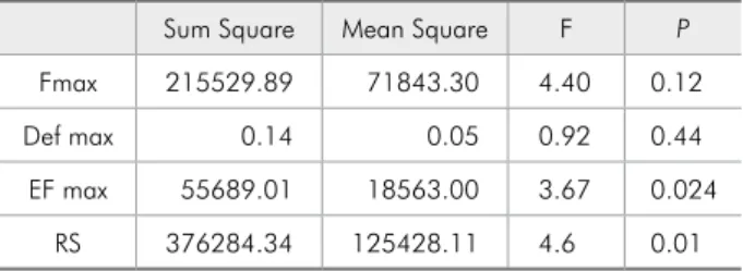
The pullout resistance tests were performed with the dependent variables maximum pullout strength, Def max, RS, and EF max (Tables 1, 2, and 3).
The results obtained by the ANOVA tests
(mul-Table 1 - ANOVA.
Sum Square Mean Square F P Fmax 215529.89 71843.30 4.40 0.12
Def max 0.14 0.05 0.92 0.44
tivariate analysis of variance) showed signiicant differences between the two groups of implants in terms of pullout tests (Wilks’ Lambda = 0.312, p = 0.001).
Discussion
Because of the great progress that has been made in the development of dental implants, these materi-als ill the market with different options regarding geometry, size, and surface characteristics. Given this variety, the dentist may choose an implant that favors rehabilitation, by increasing the surface avail-able for bone-implant contact, and promotes good primary stability, osseointegration, and distribution of forces.22,23
However, this extensive range of available op-tions can also generate doubts about the real advan-tages and beneits of each product.
With the aim of evaluating the insertion torque and pullout resistance of different implants, we opted to use synthetic bone, because its mechani-cal properties are similar to those of natural bones, except for trials involving twisting.20,21 During
pro-cessing, synthetic bone showed uniformity in den-sity and geometry, favoring a standard analysis of the variable types of bones.
Primary stability is undoubtedly one of the main factors required for the occurrence of osseointegra-tion; however, changes in the shapes and types of implants in the quest to increase this stability can often lead to necrosis of the bone surface24 when
the threshold of bone strength is crossed due to very high torque. In this study, when we measured inser-tion torque, there was no signiicant difference be-tween the groups. It is believed that this is due to the lack of difference in diameter between the implants used.
Given the diversity of rehabilitation methods, mediated or delayed, a further dificulty may arise in the selection of the implant, since each requires
correlation of bone type, implant type, and primary stability.
Some paradigms exist wherein the implant sur-face treatment inluences mainly the primary sta-bility of implants. This study showed that surface treatment also inluences primary stability, which is extremely positive, since the cylindrical implants with treated surfaces showed superior results of pull-out strength compared with those with machined surfaces. This result suggests that the roughness caused by surface treatment increases the friction between the implant surface and bone, inluencing primary stability.9,18,19 Moreover, when the surface
treatment increased bone-implant contact, the like-lihood of osseointegration increased.11 However,
the high pullout resistance of implants with treated surfaces compared with those having machined sur-faces suggests that the surface inluences initial sta-bility even when osseointegration does not occur.
It is suggested that the correlation between primary stability and pullout resistance is biome-chanical, since the higher stability of the screw in the bones suggests that the pullout resistance of the same is greater.16
The results demonstrated the great eficiency of implants with treated surfaces, and the litera-ture shows that treating implants with acids, as in the present study, leads to the formation of surface roughness, allowing for greater contact with osteo-blastic cells.9,18,25
When pullout tests are performed, the implant should be completely vertical, so that its pullout
re-Implant Fmax Def max EF max RS
Treated Surface 403.7 ± 189.8 0.91 ± 0.23 198.77 ± 102.37 660 ± 187.44 Machined Surface 276.38 ± 110.05 0.77 ± 0.21 123.91 ± 62.95 518.1 ± 142 Table 2 - Average values of
maximum force, maximum deformation, maximum relative stiffness, and maximum EF obtained for each implant.
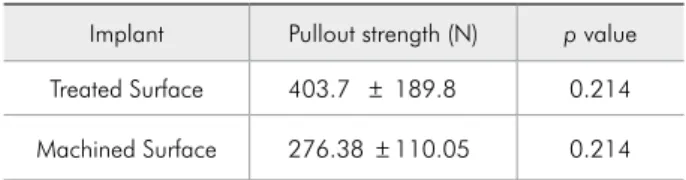
Table 3 - Means (± SD) of pullout strength (N) of implants (n = 8).
Implant Pullout strength (N) p value Treated Surface 403.7 ± 189.8 0.214
sistance would be inluenced only by the surround-ing bone, since if the implant was inclined, part of the bone would be around the implant, resulting in a resistance higher than that obtained. To prevent this, we developed a shield with a circular hole with a diameter smaller than that of the upper portion of the femur, and ixed it to a universal testing ma-chine in which the sample was positioned with the implant, so that, when pulled, the implant would be positioned as vertically as possible.
The pullout tests simply evaluated the screw re-sistance after the application of axial force, which does not correspond to physiological forces on the implant. This was the most practical way to
eval-uate this variable, which is complex and, as men-tioned before, is related to various factors such as bone quality and implant characteristics.26
Conclusion
The surface treatment of implants increases their primary stability. This suggests a greater indication for the use of surface-treated implants for oral reha-bilitation and implantology.
Acknowledgments
This study was supported by the National Coun-cil for Scientiic and Technological Development (Identiication number: 149531/2010-9).
References
1. Hagberg K, Brånemark R. One hundred patients treated with osseointegrated transfemoral amputation prostheses – reha-bilitation perspective. J Rehabil Res Dev. 2009;46(3):331-44. 2. Brentel AS, Vasconcellos LMR, Oliveira MV, Graça MLA,
Vasconcellos LGO, Cairo CAA, et al. Histomorphometric analysis of pure titanium implants with porus surface versus rough surface. J Appl Oral Sci. 2006 Jun;14(3): 213-8. 3. Castilho GAA, Martins MD, Macedo WAA. Surface
char-acterization of titanium based dental implants. Braz J Phys. 2006 Sep;36(3):1004-8.
4. Gahlert M, Röhling S, Wieland M, Sprecher CM, Kni-ha H, Milz S. Osseointegration of zirconia and titanium dental implants: a histological and histomorphometrical study in the maxilla of pigs. Clin Oral Implants Res. 2009 Nov;20(11):1247-53.
5. Gebran MP, Wassal T. Evaluation in vitro of the adhesion of osteoblasts on osseointegrated implant with treated surface (Titamax II). Implant News. 2007 Jan-Feb;4(1):79-84.
6. Fathi MH, Azam F. Novel hydroxiapatite/tantalum sur-face coating for metallic dental implant. Mater Lett. 2007 Feb;61(4-5):1238-41.
7. Citeau A, Guicheux J, Vinatier C, Layrolle P, Nguyen TP, Pilet P, et al. In vitro biological effects of titanium rough surface obtained by calcium phosphate grid blasting. Biomaterials. 2005 Jan;26(2):157-65.
8. De Maetzu MA, Braceras I, Alava JI, Gay-Escoda C. Im-provement of osseointegration of titanium dental implant surfaces modified with CO ions: a comparative histomor-phometric study in beagle dogs. Int J Oral Maxillofac Surg. 2008 May;37(5):441-7.
9. Le Guéhennec L, Soueidan A, Layrolle P, Amouriq Y. Surface treatments of titanium dental implants for rapid osseointegra-tion. Dent Mater. 2007 Jul;23(7):844-54.
10. Li DH, Liu BL, Zou JC, Xu KW. Improvement of osseointegra-tion of titanium dental implants by a modified sandblasting surface treatment: an in vivo interfacial biomechanics study. Implant Dent. 1999;8(3):289-94.
11. Sollazzo V, Pezzetti F, Scarano A, Piattelli A, Bignozzi CA, Massari L, et al. Zirconium oxide coating improves implant osseointegration in vivo. Dent Mater. 2008 Mar; 24(3): 357-61.
12. Akça K, Chan TL, Tekdemir I, Fanuscu M. Biomechanical aspects of initial intraosseous stability and implant design: a quantitative micromorphometric analysis. Clin Oral Implants Res. 2006 Aug;17(4):465-72.
13. Amarante ES, Lima LA. [Optimization of implant surfaces: titanium plasma spray and acid-etched sandblasting—cur-rent state]. Pesqui Odontol Bras. 2002 Apr-Jun;15(2):166-73. Portuguese.
14. Kahraman S, Bal BT, Avsar NV, Turkyilmaz I, Tözüm TF. Clinical study on the insertion torque and wireless resonance frequency analysis in the assessment of torque capacity and stability of self-tapping dental implants. J Oral Rehabil. 2009 Oct;36(10):755-61.
15. Defino HL, Rosa RC, Silva P, Shimano AC, Volpon JB, De Paula FJ, et al. The effect of repetitive pilot-hole use on inser-tion torque and pullout strength of vertebral system screws. Spine (Phila Pa 1976). 2009 Apr;34(9):871-6.
16. Kim JW, Baek SH, Kim TW, Chang YI. Comparison of stabil-ity between cylindrical and conical type mini-implants. Angle Orthod. 2008 Jul;78(4):692-8.
18. González JMM, Sabán FG, Bernal JF, Lafuente JCG, Sánchez JC, Dorado CB. Removal torque and physico-quimical char-acteristics of dental implants etched with hydrofluoric and nitric acid. An experimental study in beagle dogs. Med Oral Patol Oral Cir Bucal. 2006 May;11(3):281-5.
19. Stüker RA, Teixeira ER, Beck JCP, Costa NP. Preload and torque removal evaluation of three different abutment screws for single standing implant restorations. J Appl Oral Sci. 2008 Jan-Feb;16(1):55-8.
20. Cristofolini L, Viceconti M, Capello A, Toni A. Mechanical validation of whole bone composite femur models. J Biomech. 1996 Apr;29(4):525-35.
21. Cristofolini L, Viceconti M. Mechanical validation of whole composite tibia models. J Biomech. 2000 Mar;33(3):279-88. 22. Hsu SH, Liu BS, Lin WH, Chiang HC, Huang SC. Charac-terization and biocompatibility of a titanium dental implant with a laser irradiated and dual-acid etched surface. Biomed Mater Eng. 2007 Jan;17(1):53-68.
23. Misch CE. Density of bone: effect on treatment plans, surgical approach, healing, and progressive bone loading. Int J Oral Implantol. 1990;6(2):23-31.
24. Lim SA, Cha JY, Hwang CJ. Insertion torque of orthodon-tic miniscrews according to changes in shape, diameter and length. Angle Orthod. 2008 Mar;78(2):234-40.
25. Langhoff JD, Voelter K, Schanweber D, Schnabelrauch M, Scholotting F, Hefti T, et al. Comparison of chemically and pharmaceutically modified titanium and zirconia implant surfaces in dentistry: a study in sheep. Int J Oral Maxillofac Surg. 2008 Dec;37(12):1125-32.