Abstract
Submitted: November 23, 2016 0RGL¿HG)HEUXDU\ Accepted: February 22, 2017
Effects of continual intermittent
administration of parathyroid
hormone on implant stability in the
presence of osteoporosis: an
in vivo
study using resonance frequency
analysis in a rabbit model
Objective: This study aimed to evaluate the effects of continual intermittent administration of parathyroid hormone (PTH) on implant stability in the presence of osteoporosis, using rabbit models. Material and Methods: Fifteen female New Zealand white rabbits underwent ovariectomy and were administered glucocorticoids to induce osteoporosis, following which they were
PTH for 4 weeks until implant placement (PTH1), while the second and third groups received PTH (PTH2) and saline (control), respectively, for 4 weeks before and after implant placement. After intermittent administration of PTH or saline, titanium implants were inserted into the left femoral epiphyses of all animals, and the implant stability quotient (ISQ) was measured immediately after placement to assess the primary stability and at 2 and 4 weeks after implant placement to assess osseointegration. At 4 weeks after implant placement, histological and histomorphometric evaluations were conducted and the bone area around the implant socket was measured as a ratio of the total bone area to the total tissue area. Results: Regarding primary stability,
those for the control group (p<0.05). Concerning osseointegration, the ISQ
for the PTH1 and control (p<0.05) groups. Histological assessments showed a thicker and more trabecular bone around the implant sockets in the PTH2 specimens than in the PTH1 and control specimens. The bone area around the
and control groups (p<0.05). Conclusions: Our results suggest that continual intermittent PTH administration before and after dental implant placement is effective for the achievement of favorable stability and osseointegration in the presence of osteoporosis.
Ke y w or ds: Dental implants. Parathyroid hormone. Osseointegration.
Yoshifumi OKI1
Kazuya DOI1
Yusuke MAKIHARA1
Reiko KOBATAKE1
Takayasu KUBO1
Kazuhiro TSUGA1
1Hiroshima University, Graduate School of Biomedical & Health Sciences, Department of Advanced
Prosthodontics, Division of Dental Sciences, Hiroshima, Japan.
Corresponding address: Kazuya Doi Division of Dental Sciences - Department of Advanced
Prosthodontics - Graduate School of Biomedical & Health Sciences - Hiroshima University,
1-2-3 - Kasumi - Minami-ku. Hiroshima, 734-8553 - Japan. Phone: +81 82 257 5677 - Fax: +81 82 257 5679
Introduction
Successful implant therapy depends on the achievement of favorable implant stability, which can be divided into primary stability and secondary stability or osseointegration9. Both primary stability
and osseointegration are affected by different factors, including bone quantity and quality, implant design, and surgical protocols24. In particular, the most
important factor is the condition of the bone at the site of implant placement19. Primary stability decreases
at sites with a low bone density, which may result in implant failure25.
Osteoporosis is a skeletal disease that causes the systematic loss of bone regarding density and quantity. As mentioned above, the condition of the bone at the implant placement site is strongly correlated with the implant failure rate. Patients with osteoporosis who undergo implant treatment show less favorable outcomes compared with patients exhibiting healthy bone28. The most common secondary form
of osteoporosis is that induced by glucocorticoid
autoimmune disorders17. Glucocorticoids affect the
bone quality mainly by decreasing bone formation by a decrease in osteoblastogenesis and an increase in osteoblast and osteocyte apoptosis. Therefore, glucocorticoid-induced osteoporosis is an unfavorable factor regarding implant stability. In a previous study, we showed that glucocorticoid-induced osteoporosis decreased the primary stability of implants and the mechanical strength of the femur in a rabbit model23.
The phenomenon of poorly primary stability was caused by reduction of cortical bone thickness and mechanical strength.
Currently, the intermittent administration of parathyroid hormone (PTH) for enhancing bone formation and improving bone quantity is clinically approved. Some animal studies have reported that intermittent PTH administration is effective in promoting bone remodeling and increasing the trabecular bone mass10,11. PTH affects cancellous bone
remodeling by promoting the formation of osteoblasts and suppressing their apoptosis2,16. Furthermore, it
increases the thickness of not only trabecular bone, but also cortical bone15. Therefore, intermittent PTH
administration can be effective in improving the bone density at the implant placement site and achieving favorable primary stability and osseointegration in
patients with severe osteoporosis, including that induced by glucocorticoids. Corsini, et al.5 (2008)
reported that intermittent PTH administration enhanced secondary stability in normal healthy rabbits. Almagro, et al.1 (2013) reported that osseointegration could be
improved by intermittent PTH administration in rabbit models with osteoporosis.
In these studies, however, intermittent PTH administration was initiated after implant placement; furthermore, only secondary implant stability or osseointegration was evaluated. Therefore, the effects on primary stability remained unclear, considering the bone quality at the implant placement site was not improved by prior intermittent PTH administration. On the other hand, our previous study assessed the effects of intermittent PTH administration initiated before implant placement in rabbit models with osteoporosis21. Thus, the bone condition was
improved before implant placement and favorable primary stability was achieved. However, secondary stability was not evaluated. Therefore, few studies have evaluated the effects of PTH therapy on osseointegration after the achievement of favorable primary stability. This study aimed to evaluate the effects of continual intermittent PTH administration before and after dental implant placement on primary stability and secondary stability in the presence of osteoporosis induced in rabbit models by ovariectomy and glucocorticoid administration.
Material and methods
Ethics
All animal experiments were conducted in accordance with the current version of the Japan Law on the Protection of Animals. This study was approved by the Research Facilities Committee (A16-3). All surgeries were performed under general anesthesia, and all efforts were made to minimize suffering during the experimental period.
Animals and experimental design
Fifteen 17-week-old female New Zealand White rabbits (3.0-3.5 kg body weight) were used in this study. The experimental design is shown in Figure 1. All animals initially underwent ovariectomy, and, 2 weeks later, they received intramuscular injections of methylprednisolone acetate (0.5 mg/kg/day)
(Depo-Medrol®
4 consecutive weeks to induce osteoporosis3,4. Seven
weeks after ovariectomy, the animals were divided into
PTH (40 μg/day, 5 days/week) (Forteo®, Eli Lilly,
Indianapolis,Indiana, USA) for 4 weeks (PTH1 group) until implant placement, then saline was administrated for 4 weeks. The second group received subcutaneous PTH for 4 weeks before and after implant placement (PTH2 group), and the third group received saline vehicle solution for 4 weeks before and after implant placement as osteoporosis (control group). The study end point was at 4 weeks after implant placement.
Implantation procedure
All procedures were performed under anesthesia with sodium pentobarbital (10 mg/kg, i.v.; Somnopentyl®,
Kyoritsu Seiyaku Corporation, Chiyoda-ku, Tokyo, Japan). Implant sockets were prepared in the distal epiphysis (knee joint) of the left femur according to the GC protocol in the manufacturer’s instructions.
surgical system (iChiropro, Bien-air, Bienne, Bern, Switzerland) with a rotary speed not exceeding 800 rpm was used for consecutive applications of a 2.0-mm round drill, 2.0-2.0-mm twist drill, 3.0-2.0-mm pilot drill, 3.0-mm twist drill, and countersink drill. Following the socket preparation procedures, implants (3.8 mm in diameter, 6.5mm in length; SETiO®, GC, Itabashi-ku,
Tokyo, Japan) were inserted until the color indicator was level with the bone ridge (Figure 2).
Measurement of the implant stability quotient
(ISQ)
Resonance frequency analysis (RFA) was performed using an Osstellp device (Osstell AB, Gothenburg, Figure 1- Study design
Figure 2- Implant placement in a rabbit model of osteoporosis.
The socket is created in the distal epiphysis (knee joint) of the left femur. After the knee joint was exposed, an implant surgical system with a rotary speed not exceeding 800 rpm was used for consecutive applications of a 2.0-mm round drill, 2.0-mm twist drill, 3.0-mm pilot drill, 3.0-mm twist drill, and countersink drill. Following the socket preparation procedures, implants were inserted until the color indicator was level with the bone ridge
Figure 3- ,PSODQW VWDELOLW\ TXRWLHQW ,64 PHDVXUHPHQWV
stability quotient (ISQ) immediately and 2 and 4 weeks after implant placement for the evaluation of primary stability and secondary stability, respectively (Figure 3).
Measurements were performed three times from two different directions, and the values obtained for each implant were averaged. All measurements were obtained using procedures described in a previous study6,7.
Histological analysis
Four weeks after implant placement, the animals
and tissue blocks were collected. The tissue blocks were trimmed and cut using a diamond saw system
(400CS, EXAKT Apparatebau, Norderstedt, Land Schleswig-Holstein, Germany) at the center of the implant socket. The Observation section was set at
hydrochloride solution (KC-X®, FALMA, Shibuya-ku,
Tokyo, Japan) for 5 days, dehydrated by a graded ethanol series, cleared with xylene, and embedded
obtained from each block and stained with hematoxylin and eosin. Histological analysis was performed using light microscopy (BZ-9000, Keyence, Osaka, Osaka, Japan). Histological images were digitized and histomorphometrically analyzed using NIH ImageJ software (National Institutes of Health, Bethesda, Maryland, USA), and the bone area around the implant
socket was measured as a ratio of the total bone area to the total tissue area. The regions of interest for the calculation of this ratio were set in the area around the implant socket, at 1.5 mm from its side and at half the vertical distance from the top of the implant shoulder. These regions were selected in accordance with previous studies21.
Statistical analysis
The data obtained were expressed as means ± standard deviations. The values obtained were statistically analyzed using one-way analysis of variance and Tukey’s HSD test for multiple comparisons, with
Results
Results of the RFA
Figure 4 shows the ISQ values obtained immediately after implant placement. The values for the PTH1 (73.9±3.9) and PTH2 groups (75.6±7.1) were
(47.7±12.7; p<0.05).
Figure 5 shows the ISQ values obtained 2 weeks after implant placement. The values for the control, PTH1, and PTH2 groups were 70.0±6.0, 74.4±2.5, and
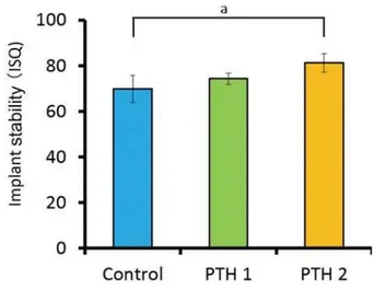
between the control and PTH2 groups (p<0.05). Figure 6 shows the ISQ values obtained 4 weeks after implant placement. At this point, the value for Figure 4- ,PSODQW VWDELOLW\ TXRWLHQW ,64 YDOXHV LPPHGLDWHO\
DIWHU LPSODQW SODFHPHQW D S 7KH YDOXH IRU WKH 37+ JURXSLVVLJQL¿FDQWO\KLJKHUWKDQWKHYDOXHIRUWKHFRQWUROJURXS ES 7KHYDOXHIRUWKH37+JURXSLVVLJQL¿FDQWO\KLJKHU than the value for the control group
Figure 5- ,PSODQW VWDELOLW\ TXRWLHQW ,64 YDOXHV ZHHNV DIWHU
LPSODQWSODFHPHQWDS 7KHYDOXHIRUWKH37+JURXSLV VLJQL¿FDQWO\KLJKHUWKDQWKHYDOXHIRUWKHFRQWUROJURXS
than the values for both the control (68.1±5.1) and PTH1 (69.4±8.3) groups (p<0.05).
Histological observations and histomorphometric
analyses
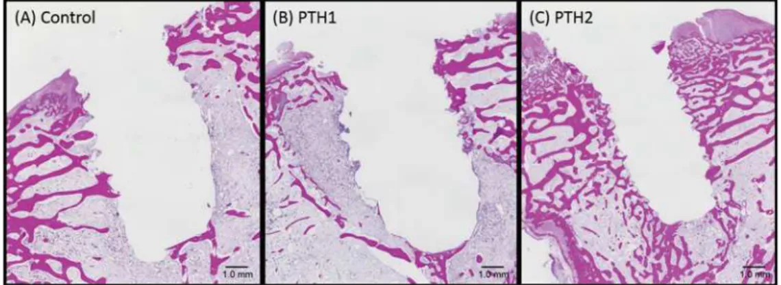
Figure 7 shows the findings of histological evaluation. In the control and PTH1 specimens, the trabecular bone structure was limited to the upper portion around the implant socket; the lower portion
PTH2 specimen, on the other hand, trabecular bone structure was detected in the upper and lower portions around the implant socket (near the marrow).
The bone area around the implant socket was
than in the PTH1 (30.8±7.7%) and control groups (25.5±3.8%; Table 1).
Discussion
Low bone density, such as that observed in patients with osteoporosis, results in poor primary implant stability because of decreased mechanical bone strength at the placement sites. Furthermore, osseointegration is barely achieved at such sites because of the suppression of bone remodeling. In this study, we found that continual intermittent PTH administration before and after implant placement can improve both primary stability and secondary stability, as determined by ISQ values.
suppressing bone formation through the inhibition of osteoblastogenesis and promotion of osteoblast and osteocyte apoptosis2,16. In our study, ISQ
Figure 6- ,PSODQW VWDELOLW\ TXRWLHQW ,64 YDOXHV ZHHNV DIWHU
LPSODQWSODFHPHQWDS ES 7KHYDOXHIRUWKH 37+JURXSLVVLJQL¿FDQWO\KLJKHUWKDQWKHYDOXHIRUWKH37+ and control group
Figure 7- Histological analyses of specimens from the (A) Control as osteoporosis. (B) PTH1: PTH administration for 4 weeks before
implant placement. (C) PTH2: PTH administration for 4 weeks before and after implant placement. In the PTH1 and control specimens, WUDEHFXODUERQHLVOLPLWHGWRWKHXSSHUSRUWLRQDURXQGWKHLPSODQWVRFNHWWKHORZHUSRUWLRQLVSULPDULO\RFFXSLHGE\PDUURZRU¿EURXV tissue. In the PTH2 specimen, trabecular bone is detected in the upper and lower portions (near the marrow) around the implant socket. (hematoxylin and eosin staining)
Bone area % (SD) Tukey’s HSD test
&RQWUROQ 25.5 (3.8)a S
37+Q 30.8 (7.7)b S
37+Q 41.7 (6.2)
SD: standard deviation
a6WDWLVWLFDOO\VLJQL¿FDQWGLIIHUHQFHFRPSDULQJ&RQWURODQG37+JURXSV b6WDWLVWLFDOO\VLJQL¿FDQWGLIIHUHQFHFRPSDULQJ37+DQG37+JURXSV
measurements were used to evaluate primary stability and secondary stability. Implant stability is considered an important measurement for evaluating the success of an implant therapy30. RFA is a noninvasive method
for continuously measuring implant stability in clinical cases8,18, and we used an Osstell® device to perform
it. ISQ values are derived on a scale from 1 to 100, and those for successfully stabilized implants are considered to range from 57 to 828. This device
measures ISQ using RFA, which measures the emitting frequency by a vibration transducer attached to the
14
an effective amount of bone is surrounding the implant and whether the bone and implant surfaces have integrated or not12,26.
adaptation between the implant surface and the surrounding bone27. In this study, ISQ values for
(4 weeks before implant placement) and PTH2 (4 weeks before and after implant placement) groups than for the control group. The ISQ value for the control group was only 47.7±12.7, which indicated unfavorable
of osteoblast differentiation and suppression of osteoclasts by intermittent PTH administration in poor
our previous study21. In another study, bone density
glucocorticoid administration, as assessed by dual-energy X-ray analysis4. In addition, our previous
study showed that the mechanical bone strength was lower in rabbit models with osteoporosis induced by ovariectomy and glucocorticoid administration than in a healthy rabbit model. Accordingly, we believe that the primary stability in both PTH groups of our study increased because of an improvement in the bone condition at the implant placement site caused by intermittent PTH administration before implant placement. The aspects consider that intermittent PTH administration before implant placement inhibits osteoclast activity and enhances osteoblast activity, hence the trabecular structure increase at the implant placement portion. Thus, primary stability of PTH1 and PTH2 groups was improved.
as the integration between the implant surface and the surrounding bone. The newly formed bone and bone remodeling at the bone-implant interface and in the
surrounding area correlate with RFA measurement18.
Several studies have reported that the bone condition
measurements are related to the supported length of the bone stiffness around the implant socket8,12,18,22.
In our study, the ISQ value 4 weeks after implant placement was significantly higher for the PTH2 group than for the other two groups. Histological assessments showed a thicker and more trabecular bone in the PTH2 specimens than in the PTH1 and control specimens. In the PTH2 group, newly formed bone was detected not only in the upper portion but also in the lower portion near the bone marrow. In addition, the implant socket could be clearly visualized in this group. In the other two groups, the newly formed bone was limited to the upper portion around the implant socket. Histomorphological analyses indicated that the bone area around the implant socket
the PTH1 and control groups. The ISQ is considered to increase in condition to the stiffness of the bone-implant interface13,26.
Also, Miyamoto, et al.20
correlation detected between the ISQ and bone cortical thickness. This result was in accordance with the ISQ values at 4 weeks, which were high for the PTH group because of new bone formation around the placed implants, caused by the effects of PTH administration during the healing period. The aspects consider that intermittent PTH administration after implant placement accelerate bone formation around the placed implant enhances osteoblast activity. Thus, the secondary stability of PTH2 group was increased. We
the PTH1 and PTH2 groups at 2 weeks after implant placement. Castañeda, et al.3 (2008) reported that the
effects of glucocorticoids persisted for more than 3 months after discontinuation in a rabbit study. Thus, we considered that the behavior of glucocorticoids persists during the term in this study. On the other hand, the half-life of PTH is relatively short. Therefore, we believe that the bone remodeling caused by PTH administration continues for 2 weeks and ceases at 4 weeks. We observed favorable primary stability in the PTH1 group, although secondary stability was
indicate that continual intermittent administration of PTH after implant placement is necessary to promote osseointegration. Various reports have documented
the appropriate dosage of PTH for achieving such an effect5,29. In our study, the PTH dose rate was set at
μ
is within the range used for in vivo rabbit studies
(15-μ 3,21.
In conclusion, the results of our study suggest that continual intermittent PTH administration is effective for achieving favorable primary and secondary stability in the presence of osteoporosis. It is the authors’ intention to conduct further studies comparing normal healthy models to investigate the detailed effects of PTH on implant stability.
The authors declare no competing financial interests.
Acknowledgments
Grant (No. 15K11160 and 16K11595) from the Japan Society for the Promotion of Science.
References
1- Almagro MI, Roman-Blas JA, Bellido M, Castañeda S, Cortez R, Herrero-Beaumont G. PTH [1-34] enhances bone response around titanium implants in a rabbit model of osteoporosis. Clin Oral Implants Res. 2013;24:1027-34.
2- Canalis E, Giustina A, Bilezikian JP. Mechanisms of anabolic therapies for osteoporosis. N Engl J Med. 2007;357:905-16.
3- Castañeda S, Calvo E, Largo R, González-González R, de la Piedra C, Díaz-Curiel M, et al. Characterization of a new experimental model of osteoporosis in rabbits. J Bone Miner Metab. 2008;26:53-9. 4- Castañeda S, Largo R, Calvo E, Rodríguez-Salvanés F, Marcos ME, Díaz-Curiel M, et al. Bone mineral measurements of subchondral and trabecular bone in healthy and osteoporotic rabbits. Skeletal Radiol. 2006;35:34-41.
5- Corsini MS, Faraco FN, Castro AA, Onuma T, Sendyk WR, Shibli JA. Effect of systemic intermittent administration of human parathyroid hormone (rhPTH[1-34]) on the resistance to reverse torque in rabbit tibiae. J Oral Implantol. 2008;34:298-302.
6- Doi K, Kajihara S, Morita K, Makihara Y, Okada S, Akagawa Y. The
implants using resonance frequency analysis and Periotest M®: a study in a dog. Br J Oral Maxillofac Surg. 2014;52:29-33.
7- Doi K, Oue H, Morita K, Kajihara S, Kubo T, Koretake K, et al. Development of implant/interconnected porous hydroxyapatite complex as new concept graft material. PLoS One. 2012;7:e49051.
measurements of one-stage Brånemark implants during healing in mandibles. A clinical resonance frequency analysis study. Int J Oral Maxillofac Surg. 1999;28:266-72.
9- Gómez-Polo M, Ortega R, Gómez-Polo C, Martín C, Celemín A, Del Río J. Does length, diameter, or bone quality affect primary and secondary stability in self-tapping dental implants? J Oral Maxillofac Surg. 2016;74:1344-53.
10- Hirano T, Burr DB, Turner CH, Sato M, Cain RL, Hock JM. Anabolic effects of human biosynthetic parathyroid hormone fragment (1-34), LY333334, on remodeling and mechanical properties of cortical bone in rabbits. J Bone Miner Res. 1999;14:536-45.
11- Hock JM, Gera I, Fonseca J, Raisz LG. Human parathyroid hormone-(1-34) increases bone mass in ovariectomized and orchidectomized rats. Endocrinology. 1988;122:2899-904.
resonance frequency of dental implants. J Oral Maxillofac Surg. 2003;61:1184-8.
13- Hui E, Chow J, Li D, Liu J, Wat P, Law H. Immediate provisional for single-tooth implant replacement with Brånemark system: preliminary report. Clin Implant Dent Relat Res. 2001;3:79-86.
14- Isoda K, Ayukawa Y, Tsukiyama Y, Sogo M, Matsushita Y, Koyano K. Relationship between the bone density estimated by cone-beam computed tomography and the primary stability of dental implants. Clin Oral Implants Res. 2011;23:832-6.
15- Jiang Y, Zhao JJ, Mitlak BH, Wang O, Genant HK, Eriksen EF. Recombinant human parathyroid hormone (1-34) [teriparatide] improves both cortical and cancellous bone structure. J Bone Miner Res. 2003;18:1932-41.
16- Kraenzlin ME, Meier C. Parathyroid hormone analogues in the treatment of osteoporosis. Nat Rev Endocrinol. 2011;7:647-56. 17- Mazziotti G, Angeli A, Bilezikian JP, Canalis E, Giustina A. Glucocorticoid-induced osteoporosis: an update. Trends Endocrinol Metab. 2006;17:144-9.
18- Meredith N, Alleyne D, Cawley P. Quantitative determination of the stability of the implant-tissue interface using resonance frequency analysis. Clin Oral Implants Res. 1996;7:261-7.
stability in patients with osteoporosis. Clin Implant Dent Relat Res. 2016;18:253-60.
cortical bone thickness and implant length on implant stability at the time of surgery-clinical, prospective, biomechanical, and imaging study. Bone. 2005;37:776-880.
21- Oki Y, Doi K, Makihara Y, Kubo T, Oue H, Tsuga K. Intermittent administration of parathyroid hormone enhances primary stability of dental implants in a bone-reduced rabbit model. J Oral Sci. 2016;58:241-6.
22- Ostman PO, Hellman M, Wendelhag I, Sennerby L. Resonance frequency analysis measurements of implants at placement surgery. Int J Prosthodont. 2006;19:77-83.
of implant surface topography on primary stability in a standardized osteoporosis rabbit model study. J Funct Biomater. 2015;6:143-52. 24- Quirynen M, Naert I, van Steenberghe D. Fixture design and
Brånemark system. Clin Oral Implants Res. 1992;3:104-11. 25- Roos J, Sennerby L, Albrektsson T. An update on the clinical documentation on currently used bone anchored endosseous oral implants. Dent Update. 1997;24:194-200.
26- Tawse-Smith A, Perio C, Payne AG, Kumara R, Thomson WM. One-stage operative procedure using two different implant systems: a prospective study on implant overdentures in the edentulous mandible. Clin Implant Dent Relat Res. 2001;3:185-93.
27- Trisi P, De Benedittis S, Perfetti G, Berardi D. Primary stability, insertion torque and bone density of cylindric implant ad modum Brånemark: is there a relationship? An in vit ro study. Clin Oral Implants Res. 2011;22:567-70.
29- Tsunori K. Effects of parathyroid hormone dosage and schedule on bone regeneration. J Oral Sci. 2015;57:131-6.
30- Zarb GA, Schmitt A. Osseointegration and the edentulous predicament. The 10-year-old Toronto study. Br Dent J. 1991;70:439-44.