Sérgio Augusto Fernandes(a) Flávio Vellini-Ferreira(a) Helio Scavone-Junior(a) Rívea Inês Ferreira(a)
(a)Department of Pediatric Dentistry and Orthodontics, University of São Paulo City (UNICID), São Paulo, SP, Brazil.
Corresponding author: Rívea Inês Ferreira
E-mail: [email protected]
Received for publication on Mar 16, 2011 Accepted for publication on Jun 14, 2011
Crown dimensions and proximal
enamel thickness of mandibular
second bicuspids
Abstract: To achieve proper recontouring of anterior and posterior teeth, to obtain optimal morphology during enamel stripping, it is im-portant to be aware of dental anatomy. This study aimed at evaluating crown dimensions and proximal enamel thickness in a sample of 40 ex-tracted sound, human, mandibular, second bicuspids (20 right and 20 left). Mesiodistal, cervico-occlusal and buccolingual crown dimensions were measured using a digital caliper, accurate to 0.01 mm. Teeth were embedded in acrylic resin and cut along their long axes through the proximal surfaces to obtain 0.7 mm-thick central sections. Enamel thick-ness on the cut sections was measured using a perilometer. Comparative analyses were carried out using the Student’s-t test (α = 5%). The mean mesiodistal crown widths for right and left teeth were 7.79 mm (± 0.47) and 7.70 mm (± 0.51), respectively. Mean cervico-occlusal heights ranged from 8.31 mm (± 0.75) on the right to 8.38 mm (± 0.85) on the left teeth. The mean values for the buccolingual dimension were 8.67 mm (± 0.70) on the right and 8.65 mm (± 0.54) on the left teeth. The mean enamel thickness on the mesial surfaces ranged from 1.35 mm (± 0.22) to 1.40 mm (± 0.17), on the left and right sides, respectively. On the distal surfaces, the corresponding values were 1.44 mm (± 0.21) and 1.46 mm (± 0.12). No signiicant differences were found between measurements for right and left teeth. However, enamel thickness was signiicantly greater on the distal surfaces, compared with the mesial surfaces.
Descriptors: Bicuspid; Tooth Crown; Dental Enamel; Orthodontics.
Introduction
Interproximal enamel stripping may represent a suitable alternative to tooth extraction; and, it has also been associated with short treatment time and favorable inal results.1-5 According to a recent study,1 clinical follow-up examinations 3.5 to 7 years after orthodontic treatment gen-erally showed healthy dentitions with good occlusion, no signs of iat-rogenic effects, and normal periodontal conditions with intact gingival papillae between all teeth in the maxillary and mandibular arches.
Enamel stripping has been used routinely worldwide, including the United States of America,4,6 Europe,1,7 Turkey5 and Brazil8 because, while providing correction of tooth-arch discrepancies, it increases treatment stability6-8 and holds the transversal dental arch dimensions and anterior inclinations constant.1,5,9 Nevertheless, it is important to estimate proxi-Declaration of Interests: The authors
mal enamel thickness before performing the strip-ping procedure to avoid damaging the adjacent den-tal tissues.
Considering that previous studies10,11 have re-ported that about ifty percent of proximal enamel thickness can be safely removed, accurate measure-ments of tooth crown dimensions and enamel thick-ness are useful to guide the orthodontist during stripping. Thus, the aim of this experimental inves-tigation was to assess the values relative to mesio-distal, cervico-occlusal and buccolingual crown di-mensions, as well as the proximal enamel thickness at the contact area of mandibular second bicuspids.
Methodology
This experimental study was approved by the Institution Committee for Research Ethics and complies with the Brazilian resolution regulating re-search involving humans (196/96).
Sample
An in vitro model consisting of 20 standardized repetitions would be statistically adequate for a tri-dimensional analysis of the dental crown and to es-timate the enamel thickness of teeth from patients who were born in the same geographical area.8 Based on this methodological premise, 40 sound, human, mandibular, second bicuspids were col-lected from a human teeth bank associated with a public university located in Goiânia, Goiás, Brazil. The teeth were divided into two groups, 20 right mandibular second bicuspids and 20 left mandibu-lar second bicuspids, and randomly numbered from one to twenty.
Dental crown measurements
A well-trained examiner performed the dental crown measurements using a digital caliper accurate to 0.01 mm (Mitutoyo Sul Americana Ltda., Suza-no, Brazil), measuring the teeth (right and left) in a sequence from one to 20. During the measurements, each tooth was placed on a lat surface and kept in a ixed position. The mesiodistal width was measured from the most central point in the contact area of the mesial surface to the respective point on the dis-tal surface. Afterwards, the cervico-occlusal
dimen-sion was measured from the most extreme point of the occlusal edge to the dentinoenamel junction on the buccal surface. The buccolingual dimension rep-resented the distance measured between the middle points of the crown on the buccal and lingual sur-faces (Figure 1).
Specimen preparation
To estimate the proximal enamel thickness, the teeth were ixed in plastic containers with utility wax. The buccal surface of each tooth was kept in contact with the wax at the bottom of the contain-er. Then, the teeth were embedded in acrylic resin (ARAZYN 1.0, Redelease, São Paulo, Brazil). All the specimens were taken out of the containers and the occlusal and apical points were determined on the resin, which served as the reference for tracing the long axes of the teeth.
The specimens were cut along their long axes through the proximal surfaces, parallel to the buc-cal surfaces, to obtain 0.7 mm-thick central sec-tions. During sectioning, the occlusal surfaces of the specimens were kept facing the operator. Each cut section corresponded to the central area of the proximal surface, in that it encompassed the greater crown width and the thicker enamel portion. The cut sections were obtained using a 4-inch diameter, high concentration diamond wafering blade (Extec Corp., Enield, USA), mounted onto a high precision Lab Cut 1010 saw (Extec Corp., Enield, USA), under water cooling to prevent organic component loss (Figure 2). To avoid damaging the specimens, the diamond disc speed was set to 225 rotations per minute.
mesiodistal cervico-occlusal buccolingual
Enamel thickness estimation
Enamel thickness on the proximal surfaces was measured using millesimal precision equipment, the perilometer (Mitutoyo, Kawasaki, Japan). The cut sections were manipulated, to allow standardization of the measurements, by aligning the Cartesian axes (X and Y) of the perilometer with the long axis of the tooth. The Y axis was placed in the mesial direc-tion, up to the most external point of the enamel, es-tablishing “point A”. The caliper of the perilometer was set to the zero position, and then placed paral-lel to the most external point of the dentin on the mesial surface, establishing “point B”. Sequentially, readouts were made of the mesial enamel thickness, expressed in millesimal parts of millimeters. To per-form the estimates on the distal surfaces, the Y axis was placed up to the most external point of the den-tin, establishing “point C”. Next, it was placed par-allel to the most external point of the enamel on the distal surface, establishing “point D”. The measured distances were A-B and C-D, representative of the enamel thickness on the mesial and distal surfaces, respectively.
Statistical analyses
Some measures of central tendency and disper-sion were calculated for the mesiodistal, cervico-occlusal and buccolingual crown dimensions, as
well as proximal enamel thickness, of right and left mandibular second bicuspids. Possible differences between mean crown dimensions of the studied teeth, considering the side of the dental arch, were assessed using the non-paired Student’s-t test. Mean enamel thickness values on the mesial and distal surfaces were compared using the paired Student’s-t
test. The signiicance level was set at 5%. Statistical analyses were carried out using IBM SPSS Statistics 17.0 (IBM, Chicago, USA).
Results
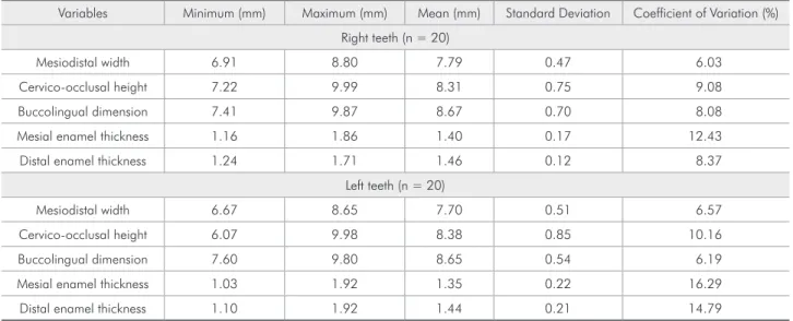
Measures of central tendency and dispersion for dental crown dimensions and proximal enam-el thickness are shown in Table 1. The differences between the mean values of the crown dimensions for right and left teeth were approximately 0.1 mm. The mean mesiodistal width ranged from 7.7 mm to 7.8 mm. The mean values of cervico-occlusal height varied from 8.3 mm to 8.4 mm. Variation along the buccolingual dimension was within the range from 8.6 mm to 8.7 mm. Mean enamel thickness esti-mates on the mesial surfaces were 1.40 mm for right teeth and 1.35 mm for left teeth. The values corre-sponding to the distal surfaces were 1.46 mm and 1.44 mm for right and left teeth, respectively.
Based on the coeficients of variation, it may be assumed that there was greater variability of data in
relation to the mean values for the left teeth, mainly with regard to the proximal enamel thickness. On the other hand, data for the right teeth appeared to show more homogeneity as the coeficients of varia-tion were below 10%, except for the mesial enamel thickness (12.4%). Although the coeficients of vari-ation for the proximal enamel thickness of the left teeth ranged from 14.8% to 16.3%, they may still be considered relatively low.
For all the comparisons shown in Table 2, the critical value of the two-tailed “t” test was 2.024394 / −2.024394. The calculated values of “t” were within the range of critical “t”, resulting in non-signiicant p values. Hence, data in Table 2 af-irm the homogeneity of the measurements obtained
and indicate that there were no signiicant differ-ences between the crown dimensions and enamel thicknesses for right and left mandibular second bi-cuspids. However, the mean values relative to enam-el thickness were signiicantly higher on the distal surfaces in comparison with the mesial surfaces, for both sides of the dental arch (Table 3).
Discussion
Acknowledgement of dental crown dimensions and enamel thickness on proximal surfaces is really useful to establish diagnosis and proper planning for orthodontic treatment; speciically, with regard to decisions about performing either tooth extrac-tion or interproximal enamel stripping. This
proce-Table 1 - Measures of central tendency and dispersion relative to crown dimensions and proximal enamel thickness of man-dibular second bicuspids.
Variables Minimum (mm) Maximum (mm) Mean (mm) Standard Deviation Coefficient of Variation (%)
Right teeth (n = 20)
Mesiodistal width 6.91 8.80 7.79 0.47 6.03
Cervico-occlusal height 7.22 9.99 8.31 0.75 9.08
Buccolingual dimension 7.41 9.87 8.67 0.70 8.08
Mesial enamel thickness 1.16 1.86 1.40 0.17 12.43
Distal enamel thickness 1.24 1.71 1.46 0.12 8.37
Left teeth (n = 20)
Mesiodistal width 6.67 8.65 7.70 0.51 6.57
Cervico-occlusal height 6.07 9.98 8.38 0.85 10.16
Buccolingual dimension 7.60 9.80 8.65 0.54 6.19
Mesial enamel thickness 1.03 1.92 1.35 0.22 16.29
Distal enamel thickness 1.10 1.92 1.44 0.21 14.79
Table 2 - Comparative analysis of the measurements ob-tained for right (n = 20) and left (n = 20) mandibular sec-ond bicuspids.
Variables t value* p value Significance
Mesiodistal width 0.64 0.26 Not significant
Cervico-occlusal height −0.28 0.39 Not significant
Buccolingual dimension 0.10 0.46 Not significant
Mesial enamel thickness 0.72 0.24 Not significant
Distal enamel thickness 0.46 0.33 Not significant
*Critical value of two-tailed “t” test (38 degrees of freedom), tcritical (0.05;38) = 2.024394
Table 3 - Comparative analysis of the mean values for me-sial and distal enamel thickness on right (n = 20) and left (n = 20) mandibular second bicuspids.
Enamel thickness t value* p value Significance
Right side
Mesial surface versus
distal surface −2.42 0.03
Significant (p < 0.05)
Left side
Mesial surface versus
distal surface −3.90 0.0009
Highly significant (p < 0.01)
dure may avoid extractions, reduce treatment time and yield more favorable inal results.2-5 From a clin-ical point of view, many patients may not be willing to undergo tooth extraction, which should be taken into account.12
Interproximal enamel stripping may be used to solve cases of mild to moderate crowding.1,2,4,8 This therapeutic alternative may be performed in cases of Class l arch-length discrepancies with orthognathic proiles; minor Class ll dental malocclusions, espe-cially those in which patients have stopped growing; and Bolton tooth-size discrepancies.12 A space gain of 8.9 mm may be achieved in the dental arches by using enamel stripping techniques, not limited to the anterior teeth.13 The estimate of the amount of enamel to be removed must depend on the degree of dental discrepancy.4,10,14 Enamel removal can be substantial on teeth with deviating morphology; whereas, incisors with parallel proximal surfaces, screwdriver-shaped teeth and round bicuspids might not be candidates for any stripping.1
Periapical and bite-wing radiographs, as well as computed tomography images, are suitable ad-juncts to the clinical assessment of dental crown dimensions and proximal enamel thickness.15,16 Nevertheless, interestingly, one of the restrictions with computed tomography was the blurred limits for enamel thicknesses smaller than 1.1 mm. This makes it dificult to determine the point from which the measurement begins, despite the high resolution of this imaging modality.16 The present study pro-vided data pertaining to proximal enamel thickness of mandibular second bicuspids (Table 1). Mean values for enamel thickness on the mesial and dis-tal surfaces ranged from 1.35 mm to 1.40 mm, and from 1.44 mm to 1.46 mm, respectively. Proximal enamel was signiicantly thicker on the distal sur-faces, in comparison with the mesial surfaces (Table 3). This inding is in agreement with a similar Bra-zilian study8 and other investigations that suggested thresholds for proximal enamel reduction, in the in-terval of 0.4-0.5 mm, irrespective of the side.5,6,9
A study in which dental crown dimensions were measured directly in the patient’s mouth with a compass reported that aligned teeth had smaller mesiodistal widths and larger buccolingual
mea-surements.17 In another study, an index to assess morphological deviations of teeth, for eliminating crowding in the mandibular incisors region, was de-veloped.4 The sample comprised young, white adult women, assigned to two groups: one group present-ing satisfactory alignment of mandibular incisors; the second group consisting of patients diagnosed with crowding (control). Measurements were also made directly in the patients’ mouths. When com-paring the mesiodistal width with the buccolingual dimension of the same tooth, it could be observed that the irst measurement was always smaller than the second one.4 The indings of these clinical in-vestigations are in agreement with the results of the present study, since the mean mesiodistal width (7.70-7.79 mm) corresponded to the smallest crown dimension of the right and left mandibular second bicuspids, compared with the buccolingual (8.65-8.67 mm) and cervico-occlusal (8.31-8.38 mm) measurements (Table 1). However, it should be pointed out that this study design provided greater idelity because the measurements were performed with an accurate digital caliper directly on the ex-tracted teeth. Factors that could have interfered with the measurements of crown dimensions in previous studies, such as proximal restorations, alterations of axial inclination, misalignments and movement of the patients during registrations, were eliminated.
enamel due to masticatory forces is seen more fre-quently in elderly patients.18-20
Concerning sexual dimorphism, a study report-ed that mesiodistal crown measurements were larger for men, compared with the dimensions obtained for women.21 Some authors assessed the mesiodis-tal and buccolingual crown dimensions in North Americans, Egyptians and Mexicans.22 All these populations showed signiicant differences between the measurements for men and women, which cor-roborated the indings of that previous study.21 Men had larger canines and irst molars.22 Other authors found that dentin thickness appeared to be great-er in men.9,15 Accordingly, sexual dimorphism in crown mesiodistal width might presumably be due to dentin thickness. In the present study, the crown mesiodistal, buccolingual and cervico-occlusal di-mensions, as well as the proximal enamel thickness-es, were assessed irrespective of the gender. More-over, the mandibular second bicuspids selected were donated by patients from the Midwestern region of Brazil, where there is a highly miscegenetic popula-tion. However, for all of the measurements taken, there were no statistically signiicant differences be-tween the right and left teeth. This suggests sym-metry in dental crown morphology and proximal enamel thickness of the mandibular second bicus-pids studied (Table 2).
Morphological imaging modalities, such as ra-diography, tomography and particularly cone-beam computed tomography, may be used in clinical prac-tice for estimating the amount of proximal enamel
that can be removed safely, considering each pa-tient’s individual variations.8 Nevertheless, it is also clinically relevant to carry out experimental studies that provide accurate estimate numbers relative to the crown dimensions and proximal enamel thick-ness for, at least, the anterior teeth and bicuspids. These values may serve as parameters for perform-ing strippperform-ing durperform-ing orthodontic treatment. After stripping, the use of precision measuring devices is recommended equally for estimating the magnitude of dental reduction.23
In spite of the valid contribution represented by the measurements of the mandibular second bicus-pids, some caution is needed when interpreting these results because of possible ethnic differences. There-fore, the marked inluence of intrinsic factors on tooth formation justiies the conduct of future stud-ies involving tooth crown measurements in different ethnic groups.
Conclusions
1. Based on the estimated mean values, the mesio-distal width (7.7-7.8 mm) was the smallest di-mension of the dental crown in comparison with the cervico-occlusal height (8.3-8.4 mm) and buccolingual dimension (8.6-8.7 mm).
2. The mean enamel thickness was signiicant-ly greater on the distal surfaces (right side, 1.46 mm; left side, 1.44 mm) in comparison with the mesial surfaces (right side, 1.40 mm; left side, 1.35 mm).
References
1. Zachrisson BU, Minster L, Ogaard B, Birkhed D. Dental health assessed after interproximal enamel reduction: caries risk in posterior teeth. Am J Orthod Dentofacial Orthop. 2011 Jan;139(1):90-8.
2. Ballard ML. Asymmetry in tooth size: a factor in the etiol-ogy, diagnosis and treatment of malocclusion. Angle Orthod. 1944 Jul-Oct;14(3):67-70.
3. Neff CW. Tailored occlusion with the anterior coefficient. Am J Orthod. 1949 Apr;35(4):309-13.
4. Peck H, Peck S. An index for assessing tooth shape deviations as applied to the mandibular incisors. Am J Orthod. 1972 Apr;61(4):384-401.
5. Germeç D, Taner TU. Effects of extraction and nonextraction therapy with air-rotor stripping on facial esthetics in postado-lescent borderline patients. Am J Orthod Dentofacial Orthop. 2008 Apr;133(4):539-49.
6. Rossouw PE, Tortorella A. Enamel reduction procedures in orthodontic treatment. J Can Dent Assoc. 2003 Jun;69(6):378-83.
8. Macha AC, Vellini-Ferreira F, Scavone-Junior H, Ferreira RI. Mesiodistal width and proximal enamel thickness of maxil-lary first bicuspids. Braz Oral Res. 2010 Jan-Mar;24(1):58-63. 9. Stroud JL, Buschang PH, Goaz PW. Sexual dimorphism in
mesiodistal dentin and enamel thickness. Dentomaxillofac Radiol. 1994 Aug;23(3):169-71.
10. Demange C, François B. Measuring and charting interproxi-mal enamel removal. J Clin Orthod. 1990 Jul;24(7):408-12. 11. Jarjoura K, Gagnon G, Nieberg L. Caries risk after inter-proximal enamel reduction. Am J Orthod Dentofacial Orthop. 2006 Jul;130(1):26-30.
12. Stroud JL, English J, Buschang PH. Enamel thickness of the posterior dentition: its implications for nonextraction treat-ment. Angle Orthod. 1998 Apr;68(2):141-6.
13. Sheridan JJ. A ir rotor stripping. J Clin Orthod. 1985 Jan;19(1):43-59.
14. Tuverson DL. Anterior interocclusal relations. Parts I and II. Am J Orthod. 1980 Oct;78(4):361-93.
15. Harris AF, Hicks JD. A radiographic assessment of enamel thickness in human maxillary incisors. Arch Oral Biol. 1998 Oct;43(10):825-31.
16. Spoor CF, Zonneveld FW, Macho GA. Linear measurements of cortical bone and dental enamel by computed
tomogra-phy: applications and problems. Am J Phys Anthropol. 1993 Aug;91(4):469-84.
17. Doris JM, Bernard BW, Kuftinec MM, Stom D. A biometric study of tooth size and dental crowding. Am J Orthod. 1981 Mar;79(3):326-36.
18. Begg PR. Stone age man’s dentition. Am J Orthod. 1954 Apr;40(4):298-312.
19. Molnar S, Gantt DG. Functional implications of primate enamel thickness. Am J Phys Anthropol. 1977 May;46(3):447-54.
20. Macho GA, Berner ME. Enamel thickness of human max-illary molars reconsidered. Am J Phys Anthropol. 1993 Oct;92(2):189-200.
21. Ghose LJ, Baghdady VS. Analysis of the Iraqi dentition: me-siodistal crown diameters of permanent teeth. J Dent Res. 1979 Mar;58(3):1047-54.
22. Bishara SE, Jakobsen JR, Abdallah EM, Fernandez Garcia A. Comparisons of mesiodistal and buccolingual crown dimen-sions of the permanent teeth in three populations from Egypt, Mexico, and the United States. Am J Orthod Dentofacial Or-thop. 1989 Nov;96(5):416-22.