RBCCV 44205-1662 DOI: 10.5935/1678-9741.20150044
Relationship between pre-extubation positive
end-expiratory pressure and oxygenation after coronary
artery bypass grafting
Relação entre a pressão expiratória positiva inal pré-extubação e a oxigenação após revascularização
cirúrgica do miocárdio
Reijane Oliveira Lima
1, PT; Daniel Lago Borges
1, PT; Marina de Albuquerque Gonçalves Costa
1, PT;
Thiago Eduardo Pereira Baldez
1, PT; Mayara Gabrielle Barbosa e Silva
1, PT; Felipe André Silva Sousa
1,
PT; Milena de Oliveira Soares
1, PT; Jivago Gentil Moreira Pinto
2, PT
1Hospital Universitário da Universidade Federal do Maranhão (HUUFMA),
São Luís, MA, Brazil.
2Universidade Estadual do Piauí (UESPI), Teresina, PI, Brazil.
This study was carried out at University Hospital of the Federal University of Maranhão (HUUFMA), São Luís, MA, Brazil.
Financial support.
Correspondence address: Daniel Lago Borges
Hospital Universitário da Universidade Federal do Maranhão (HUUFMA) Rua Barão de Itapary, nº 227, Centro - São Luis, MA, Brazil
Zip code: 65020-070
E-mail: [email protected]
Article received on February 15th, 2015
Article accepted on June 22nd, 2015 Abstract
Introduction: After removal of endotracheal tube and artii -cial ventilation, ventilatory support should be continued, offer-ing oxygen supply to ensure an arterial oxygen saturation close to physiological.
Objective: The aim of this study was to investigate the ef-fects of positive-end expiratory pressure before extubation on the oxygenation indices of patients undergoing coronary artery bypass grafting.
Methods: A randomized clinical trial with seventy-eight pa-tients undergoing coronary artery bypass grafting divided into three groups and ventilated with different positive-end expirato-ry pressure levels prior to extubation: Group A, 5 cmH2O (n=32); Group B, 8 cmH2O (n=26); and Group C, 10 cmH2O (n=20). Oxygenation index data were obtained from arterial blood gas samples collected at 1, 3, and 6 h after extubation. Patients with chronic pulmonary disease and those who underwent off-pump, emergency, or combined surgeries were excluded. For statistical analysis, we used Shapiro-Wilk, G, Kruskal-Wallis, and analysis
of variance tests and set the level of signiicance at P<0.05.
Results: Groups were homogenous with regard to de-mographic, clinical, and surgical variables. There were no statistically significant differences between groups in the first 6 h after extubation with regard to oxygenation indices and oxygen therapy utilization.
Conclusion: In this sample of patients undergoing coronary artery bypass grafting, the use of different positive-end expi-ratory pressure levels before extubation did not affect gas
ex-change or oxygen therapy utilization in the irst 6 h after endo -tracheal tube removal.
Descriptors: Oxygenation. Positive-Pressure Respiration, Intrinsic. Coronary Artery Bypass.
Resumo
Introdução: Após a remoção do tubo endotraqueal e
venti-lação artiicial, o suporte ventilatório deve ser continuado, ofe -recendo suprimento de oxigênio para garantir uma saturação
INTRODUCTION
Coronary artery bypass grafting (CABG) is a therapeutic modality widely used to treat coronary artery disease, mini-mize symptoms, improve cardiac function, and improve sur-vival[1,2].
Intraoperative conditions, such as general anesthesia, manual compression of the left lower lung lobe during ex-posure of the posterior heart surface, manual compression of the right lung during cannulation of the inferior vena cava, manual compression of lungs during dissection of the inter-nal mammary artery and apnea during cardiopulmonary
by-pass (CPB) may impair pulmonary function[3]. Thus,
pulmo-nary complications occur in up to 60% patients undergoing
CABG[4].
Invasive mechanical ventilation (IMV) is essential during
the irst few hours after CABG to allow recovery from anes
-thesia and reestablish homeostasis[5]. Typical restoration of
hemodynamic stability occurs 5–6 h after surgery in uncom-plicated CABG. This interval also correlates with regaining
of consciousness and IMV weaning[6].
When IMV is no longer required, the most appropriate
method for its discontinuation must be determined[7]. The
spontaneous breathing trial (SBT) is a simple method using
Abbreviations, acronyms & symbols
BMI Body mass index
CABG Coronary artery bypass grafting FiO2 Inspired oxygen fraction
FiO2N Necessary inspired oxygen fraction
ICU Intensive Care Unit
IMV Invasive mechanical ventilation
NIV Noninvasive ventilation
PaO2 Arterial oxygen partial pressure
PaO2I Ideal arterial oxygen partial pressure PEEP Positive end-expiratory pressure PSV Pressure support ventilation SaO2 Arterial oxygen saturation SBT Spontaneous breathing trial
pressure support ventilation (PSV) to determine whether a patient would tolerate IMV interruption. This ventilation
mode consists of a pressure support of 7 cm H2O (the
mini-mum level to overcome circuit resistance), positive
end-expi-ratory pressure (PEEP) of 5-8 cm H2O (nearest to
physiolog-ical values), and inspired oxygen fraction (FiO2) < 40%. This
trial lasts 30-120 min and is helpful in identifying patients
who are able to maintain spontaneous breathing[8].
Following endotracheal tube and artiicial ventilation re -moval, respiratory support should be provided with oxygen
to ensure arterial oxygen saturation (SaO2) close to
physio-logical levels (95%). Oxygen therapy can be offered using a
nasal catheter, nebulization mask, or Venturi system[6].
In this study, we investigated the effects of different PEEP levels applied during SBT on oxygenation indices in patients undergoing CABG.
METHODS
We performed a randomized clinical trial with 78 pa-tients undergoing CABG between August 2013 and March 2014 who were admitted to the Cardiovascular Intensive Care Unit (ICU) at Hospital Universitário da Universidade Federal do Maranhão, in São Luís, Maranhão, Brazil. We
Objetivo: O objetivo deste estudo foi investigar os efeitos da
pressão expiratória positiva inal antes de extubação nos índices
de oxigenação de pacientes submetidos à cirurgia de revascula-rização miocárdica.
Métodos: Ensaio clínico randomizado com 78 pacientes sub -metidos à cirurgia de revascularização do miocárdio, divididos
em três grupos e ventilados com diferentes níveis de pressão ex
-piratória positiva inal antes da extubação: Grupo A, 5 cmH2O
(n=32); Grupo B, 8 cm H2O (n=26); e grupo C, 10 cmH2O
(n=20). Dados do índice de oxigenação foram obtidos a partir de amostras de gases sanguíneos arteriais coletados em 1, 3 e 6
h após a extubação. Pacientes com doença pulmonar crônica e aqueles que foram submetidos à cirurgia sem circulação
extra-corpórea, de emergência ou combinadas foram excluídos. Para a análise estatística, foram utilizados Shapiro-Wilk, G, Kruskal -Wallis, e análise dos testes de variância e deinição do nível de signiicância em P<0,05.
Resultados: Os grupos foram homogêneos em relação às
va-riáveis demográicas, clínicas e cirúrgicas. Não houve diferen
-ças estatisticamente signiicativas entre os grupos nas primeiras 6 h após extubação no que diz respeito aos índices de oxigenação
e a utilização de oxigenoterapia.
Conclusão: Nesta amostra de pacientes submetidos à revas
-cularização do miocárdio, o uso de diferentes níveis de pressão expiratória positiva inal antes da extubação não afetou as tro -cas gasosas ou utilização de oxigenoterapia nas primeiras 6h após a remoção do tubo endotraqueal.
Descritores: Oxigenação. Respiração por Pressão Positiva
excluded patients with chronic obstructive pulmonary disease and those undergoing emergency, off-pump, or combined surgeries. We excluded patients who required surgical reintervention or
non-invasive ventilation during the irst 6 h after extubation.
Before surgery, patients received explanations and infor-mation about the research. After surgery, data were collected from physiotherapy evaluation forms and medical records. All data were registered in a form that captured preoperative, intraoperative, and postoperative periods. All included pa-tients underwent general anesthesia and median sternotomy.
After ICU admission, mechanical ventilation was applied using an Evita 2 Dura (Dräger Medical, Lübeck, Germany). Patients were ventilated in volume-controlled mode, accord-ing to the routine protocol, with the followaccord-ing settaccord-ings: a tidal volume of 6–8 mL/kg, respiratory rate of 14 bpm, inspiratory
low of 8-10 times the minute volume, inspiratory time of 1
s, and inspired oxygen fraction of 40%.
During the preoperative period, patients were random-ized into groups by simple draw, and this information was shared with the ICU care providers. SBT was initiated once the following clinical conditions were met: hemodynamic stability, absence of bleeding or minimal bleeding, absence of vasopressor use or low and stable doses of vasopressors,
Glasgow Coma Scale ≥ 10 and strong respiratory drive.
The spontaneous breathing trial was administered using
pressure support ventilation (support pressure 7 cm H2O and
FiO2 30%). The sample was divided into three groups: Group
A, PEEP = 5 cm H2O; Group B, PEEP = 8 cm H2O; and
Group C, PEEP = 10 cm H2O. Extubation was performed
af-ter 30–120 min with no destabilization signs. Following ex-tubation, all patients received additional oxygen support by Venturi mask (Galemed Corporation, Wu-Jia, Taiwan) with
an FiO2 of 31% to ensure arterial oxygen saturation close to
physiological levels (around 95%).
Arterial blood samples were collected before extubation and at 1, 3, and 6 h after mechanical ventilation withdrawal. Samples were processed by an ABL 800 FLEX blood gas analyzer (Radiometer, Bronshoj, Denmark), according to the
routine protocol. We then identiied the arterial oxygen par
-tial pressure (PaO2) and PaO2/FiO2 ratio.
Following the irst arterial blood gas analysis after extu -bation, oxygen support was adjusted according to the
neces-sary inspired oxygen fraction (FiO2N). To estimate the ideal
arterial oxygen partial pressure (PaO2I) for each patient, we
used the following equation to account for age and supine
position: PaO2I = 109 − (0.43 × age)[9].
The inspired oxygen fraction provided following
extuba-tion was calculated according to the following formula: FiO2N
= FiO2K × PaO2I/PaO2K, in which FiO2N = the inspired oxygen
fraction necessary after extubation, FiO2K = the inspired
ox-ygen fraction applied at the moment of arterial blood sample
collection, PaO2I = the ideal arterial oxygen partial pressure,
and PaO2K = the arterial oxygen partial pressure as measured
by the last arterial blood gas. Oxygen was administered by Venturi mask using the following criteria:
FiO2N <21%: room air;
FiO2N = 21%–24%: blue connector, FiO2 24%, O2
flow 4 lpm;
FiO2N = 24.1%–28%: yellow connector, FiO2 28%, O2
low 4 lpm;
FiO2N = 28.1%–31%: white connector, FiO2 31%, O2
low 4 lpm;
FiO2N 31.1%–35%: green connector, FiO2 35%, O2
low 6 lpm;
FiO2N 35.1%–40%: red connector, FiO2 40%, O2 low
8 lpm;
FiO2N >40%: orange connector, FiO2 50%, O2 flow
12 lpm.
When noninvasive ventilation (NIV) was required fol-lowing extubation, it was applied, as per the routine proto-col, according to the individual’s needs. It is noteworthy that
patients who used NIV during the irst 6 h after extubation
were excluded.
Ethical approval was obtained from the local Ethics
Committee (protocol No. 327.798), as required by Resolution
466/12 of the National Health Council. All patients provided written informed consent.
Data were evaluated using the Stata/SE statistical soft-ware version 11.1 (StataCorp, College Station, TX, USA). To test normality, we used the Shapiro–Wilk test. Quantitative variables were described as means and standard deviations, and differences were determined using the Student’s t, ANO-VA, or Kruskal–Wallis test, depending on normality. Quali-tative variables were expressed as proportions and tested by G-test and William’s correction. Results were considered
sta-tistically signiicant when P value was <0.05.
RESULTS
Ninety patients were randomized and underwent CABG during the study period. Of these, twelve (four of each group) were excluded because of postoperative surgical re-intervention (6) and noninvasive ventilation use during the
irst 6 h after extubation (6) (Figure 1). Therefore, the inal
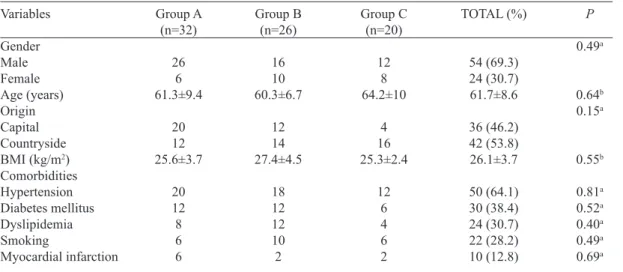
sample included 78 patients, who were predominantly male (69.3%) and from the countryside (53.8%), with a mean age
of 61.7±8.6 years and bodymass index of 26.1±3.7 kg/m2.
Groups did not differ signiicantly with regard to demograph -ic, clinical, or surgical variables, as seen in Tables 1 and 2.
The mean mechanical ventilation duration was 12.8±6.9
h. Patients in Group A (PEEP 5 cm H2O) were ventilated for
13.6±8.1 h, whereas those in Group B (PEEP 8 cm H2O) were
ventilated for 11.7±6 h and those in Group C (PEEP 10 cm
H2O) were ventilated for 13.2±4.8 h (P=0.69). There were
no differences in mean gas exchange values (PaO2/FiO2)
Fig. 1 - Consort low diagram.
Table 1. Demographic and clinical data for patients undergoing CABG.
Variables
Gender Male Female Age (years) Origin Capital Countryside BMI (kg/m2) Comorbidities Hypertension Diabetes mellitus Dyslipidemia Smoking
Myocardial infarction
Group A (n=32)
26 6 61.3±9.4
20 12 25.6±3.7
20 12 8 6 6
Group B (n=26)
16 10 60.3±6.7
12 14 27.4±4.5
18 12 12 10 2
Group C (n=20)
12 8 64.2±10
4 16 25.3±2.4
12 6 4 6 2
TOTAL (%)
54 (69.3) 24 (30.7) 61.7±8.6
36 (46.2) 42 (53.8) 26.1±3.7
50 (64.1) 30 (38.4) 24 (30.7) 22 (28.2) 10 (12.8)
P
0.49a
0.64b 0.15a
0.55b
0.81a 0.52a 0.40a 0.49a 0.69a
BMI=body mass index. aG test. bKruskal-Wallis test
Table 2. Surgical data for patients undergoing CABG.
Variables
Number of bypasses Number of drainage tubes Pump time (min) Aortic clamp time (min) Surgery time (min)
Group A (n = 32) 3 (2.75;3)
2 (2;2) 83.4 ± 21.7 60.9 ± 19.2 220.9 ± 25.3
Group B (n = 26) 2 (2;3) 2 (2;2) 67.3 ± 25.3 48.4 ± 20.2 230.5 ± 62.1
Group C (n = 20) 3 (2;3) 2 (2;2) 89.4 ± 23.5
61.5 ± 17 257.6 ± 64.9
P
0.22a 0.56a 0.08b 0.15b 0.22a
Table 5. Comparison of inspired oxygen fraction (%) applied after extubation between the three groups of patients undergoing CABG.
Groups/times Group A Group B Group C P
1st hour 26±5 27±5 27±5 0.61
3rd hour 27±6 28±7 27±6 0.70
6th hour 27±5 28±8 27±7 0.77 Data shown as the mean ± standard deviation. Kruskal-Wallis test Table 3. Comparison of gas exchange mean (mmHg) between the three groups of patients undergoing CABG.
Groups/times Group A Group B Group C P
1st hour 320.5±65 326.9±84.1 308.3±49.9
0.92
3rd hour 347.7±75.9 332.5±97.3 313.3±56.9
0.64
6th hour 333.1±67.9 343.5±118.5
311.5±80.3 0.77 Data shown as the mean±standard deviation. Kruskal-Wallis test.
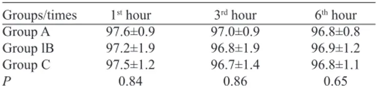
Table 4. Comparison of arterial oxygen saturation (%) between the three groups of patients undergoing CABG.
Groups/times Group A Group lB Group C P
1st hour 97.6±0.9 97.2±1.9 97.5±1.2 0.84
3rd hour 97.0±0.9 96.8±1.9 96.7±1.4 0.86
6th hour 96.8±0.8 96.9±1.2 96.8±1.1 0.65 Data shown as the mean±standard deviation. Kruskal-Wallis test.
Mean arterial oxygen saturation and inspired oxygen fraction did not differ between groups at 1, 3, and 6 h after extubation (Tables 4 and 5).
DISCUSSION
Gas exchange impairment is a signiicant complication
during the CABG postoperative period[10]. In thoracic
sur-geries, these changes may be related to intraoperative proce-dures, such as mechanical ventilation with low volumes and PEEP, pain, and thoracotomy (which alters chest wall
com-pliance)[11,12]. Therefore, we chose to evaluate oxygenation
indices after extubation, because they properly relect chang
-es in pulmonary function following on-pump surgery[13].
To reopen collapsed lung units and improve arterial ox-ygenation following thoracic surgery, different PEEP levels
have been proposed[14]. Dongelmans et al.[15], who compared
high versus physiological PEEP (10 vs. 5 cm H2O) after
CABG, showed that the highest PEEP levels improve ox-ygenation and lung compliance but are associated with in-creased mechanical ventilation duration. In their randomized clinical trial of 136 patients undergoing CABG who were
mechanically ventilated at 5, 8, or 10 cm H2O of PEEP,
Borg-es et al.[16] showed that the highest PEEP levels may increase
respiratory mechanics and provide better oxygenation indi-ces in the immediate postoperative period.
Our hypothesis that application of higher PEEP levels throughout SBT would improve oxygenation after extubation
was not supported by our measurements during the irst 6 h
after extubation. The results were consistent with those
mea-sured in the randomized clinical trial by Marvel et al.[17], in
which patients undergoing CABG and ventilation with PEEP
of 0, 5, or 10 cm H2O did not experience a sustained arterial
oxygenation beneit from higher PEEP levels.
A question that arose during our research was what PEEP level would be considered physiological to avoid alveolar collapse while performing SBT, given that the “expiratory delay function” of the glottis (which serves as an organic PEEP mechanism to prevent or minimize alveolar collapse)
is removed during artiicial ventilation[18]? During
mechani-cal ventilation of adult patients, PEEP is generally set to 3–5
cm H2O, as this is considered physiological[19]. However, our
study provided some evidence that levels between 5 and 8
cm H2O, possibly up to 10 cm H2O, may more closely mimic
normal respiratory physiology for such patients.
The knowledge of physical therapy was found to be
generally applied across the entire treatment process[20].
Physical therapists play an important role in conducting pa-tient-screening protocols for mechanical ventilation wean-ing[21,22]. Our research emphasizes identiication of optimal
variables during weaning as fundamental to this process so as to minimize patient complications.
CONCLUSION
In this sample of patients undergoing CABG, the use of different PEEP levels before extubation did not affect gas
exchange or oxygen therapy utilization in the irst 6 h after
endotracheal tube removal.
Authors’ roles & responsibilities
ROL Analysis and/or interpretation of data; study design; imple-mentation of projects and/or experiments; manuscript writ-ing or critical review of its content
DLB Analysis and/or interpretation of data; statistical analysis; inal approval of the manuscript; study design; implemen-tation of projects and/or experiments; manuscript writing or critical review of its content
MAGC Conduct of operations and/or experiments TEPB Conduct of operations and/or experiments MGBS Conduct of operations and/or experiments FASS Conduct of operations and/or experiments MOS Conduct of operations and/or experiments
REFERENCES
1. Cutlip D, Levin T, Aroesty J. Bypass surgery versus percutaneous intervention in the management of stable angina pectoris: clinical trials [Cited 2015 Jul 23]. Available from: http://www.uptodate. com/contents/bypass-surgery-versus-percutaneous-intervention-in-the-management-of-stable-angina-pectoris-clinical-studies
2. Rocha LA, Maia TF, Silva LF. Diagnóstico de enfermagem em pacientes submetidos à cirurgia cardíaca. Rev Bras Enferm. 2006:59(3);321-6.
3. Zocrato LBR, Machado MGR. Fisioterapia respiratória no pré e pós-operatório de cirurgia cardíaca. In: Machado MGR. Bases da isioterapia respiratória: terapia intensiva e reabilitação. Rio de Janeiro: Guanabara Koogan; 2008. p.338-62.
4. Nardi C, Otranto CPM, Piaia IM, Forti EMP, Fantini B. Avaliação da força muscular, capacidades pulmonares e função pulmonar respiratória de pacientes submetidos à cirurgia cardíaca. In: 4ª Mostra Acadêmica e Congresso de Pesquisa da UNIMEP [on line]: 2006. Out, 24-26. Piracicaba. Anais eletrônicos [Cited 2015 Jul 23]. Available from: http//www.unimep.br/phpg/ mostraacademica/anais/4mostra/pdfs/171pdf
5. Lourenço IS, Franco AM, Bassetto S, Rodrigues AJ. Pressure support-ventilation versus spontaneous breathing with “T-Tube” for interrupting the ventilation after cardiac operations. Rev Bras Cir Cardiovasc. 2013;28(4):455-61.
6. Umeda IIK. Manual de isioterapia na cirurgia cardíaca: guia prático. Barueri: Manole; 2004.
7. Piotto RF, Maia LN, Machado MN, Orrico SP. Effects of the use of mechanical ventilation weaning protocol in the Coronary Care Unit: randomized study. Rev Bras Cir Cardiovasc. 2011;26(2):213-21.
8. Goldwasser R, Farias A, Freitas EE, Saddy F, Amado V, Okamoto V. III Consenso Brasileiro de Ventilação Mecânica. Desmame e interrupção da ventilação mecânica. J Bras Pneum. 2007;33 (Suppl 2S):S128-S136.
9. Diniz GCLM, Machado MGR. Oxigenoterapia. In: Machado MGR. Bases da isioterapia respiratória: terapia intensiva e reabilitação. Rio de Janeiro: Guanabara Koogan; 2008. p.182-97.
10. Singh NP, Vargas FS, Cukier A, Terra-Filho M, Teixeira LR, Light RW. Arterial blood gases after coronary artery bypass surgery. Chest. 1992:102(5);1337-41.
11. Ambrozin ARP, Cataneo AJM. Pulmonary function aspects after myocardial revascularization related to preoperative risk. Rev Bras Cir Cardiovasc. 2005;20(4):408-15.
12. Park DJ, Jeong JH, Lee HO. The effects of a self-training physiotherapy program on pulmonary functions, postoperative pulmonary complications and post-thoracotomy pain after lobectomy of patients with lung cancer. J Phys Ther Sci. 2013;25(3):253-5.
13. Cui H, Zhang M, Xiao F, Li Y, Wang J, Chen H. Comparison and correlative analysis of pulmonary function markers after extracorporeal circulation. Beijing Da Xue Xue Bao. 2013;35(2):195-9.
14. Dyhr T, Nygård E, Laursen N, Larsson A. Both lung recruitment maneuver and PEEP are needed to increase oxygenation and lung volume after cardiac surgery. Acta Anaesthesiol Scand. 2004;48(2):187-97.
15. Dongelmans DA, Hemmes SN, Kudoga AC, Veelo DP, Binnekade JM, Schultz MJ. Positive end-expiratory pressure following coronary artery bypass grafting. Minerva Anestesiol. 2012;78(7):790-800.
16. Borges DL, Nina VJS, Costa MAG, Baldez TEP, Santos NP, Lima IM, et al. Effects of different PEEP levels on respiratory mechanics and oxygenation after coronary artery bypass grafting. Rev Bras Cir Cardiovasc. 2013;28(3):380-5.
17. Marvel SL, Elliott CG, Tocino I, Greenway LW, Metcalf SM, Chapman RH. Positive end-expiratory pressure following coronary artery bypass grafting. Chest. 1986;90(4):537-41.
18. Annest SJ, Gottlieb M, Paloski WH, Stratton H, Newell JC, Dutton R, et al. Detrimental effects of removing end-expiratory pressure prior to endotracheal extubation. Ann Surg. 1980;191(5):539-45.
19. David CM. Ventilação Mecânica: Da isiologia à prática clínica. 2ª ed. Rio de Janeiro: Revinter; 2011.
20. Ryu YU, Park J. Medical and narrative use of physical therapy knowledge in clinical reasoning by Korean physical therapists. J Phys Ther Sci. 2011;23(2):251-4.
21. Ely EW, Meade MO, Haponik EF, Kollef MH, Cook DJ, Guyatt GH, et al. Mechanical ventilator weaning protocols driven by nonphysician health-care professionals: evidence-based clinical practice guidelines. Chest. 2001;120(6 Suppl):454S-63S.