This clinical study aimed to evaluate the relationship of the delay between dental trauma and the initial attendance to the development of external inflammatory root resorption in permanent teeth affected by severe luxation. Sixty-seven patients, aged between 11 and 56 years, presenting 133 injured teeth with closed apex (56 extrusive luxation, 69 lateral luxation and 8 intrusive luxation) were followed up for a minimum of 24 months. The time elapsed between dental trauma and the initial attendance was recorded. The presence of necrotic pulp and external inflammatory resorption for each type of trauma was verified. Fisher’s exact test was used to verify the influence of the initial attendance date at the Dental Trauma Center and the development of inflammatory resorption (p<0.05). The odds ratio was used to check the risk of developing external inflammatory resorption due to delay in seeking treatment. Pulp necrosis was observed in 105 teeth (78.9%) and external inflammatory resorption was detected in 17.8% cases of extrusive luxation (10 teeth), 15.9% of lateral luxation (11 teeth) and 25% of intrusive luxation (2 teeth). For lateral luxation, extended delay between the date of injury and initial attendance influenced the occurrence of external inflammatory resorption (p=0.0174). Patients who began treatment 45 days after the injury were 3.4 times more likely to develop external inflammatory resorption than patients who sought treatment after the trauma. Treatment late after the occurrence of dental trauma can impair the prognosis and result in the development of external inflammatory resorption in luxated teeth.
R e l a t i o n s h i p b e t w e e n I n i t i a l
Attendance after Dental Trauma
a n d D e v e l o p m e n t o f E x t e r n a l
I n f l a m m a t o r y R o o t R e s o r p t i o n
Thiago Farias Rocha Lima1, Emmanuel João Nogueira Leal da Silva2, Brenda Paula Figueiredo de Almeida Gomes3, José Flávio Affonso de Almeida3, Alexandre Augusto Zaia3, Adriana de Jesus Soares3
1Department of Restorative Dentistry, Endodontics Area, UFPB - Universidade Federal da Paraiba, João Pessoa, PB, Brazil 2Department of Endodontics, Dental School, UERJ - Universidade do Estado do Rio de Janeiro, Rio de Janeiro, RJ, Brazil 3Department of Restorative Dentistry, Endodontics Area, Piracicaba Dental School, UNICAMP - Universidade Estadual de Campinas, Piracicaba, SP, Brazil
Correspondence: Prof. Thiago Farias Rocha Lima, Avenida Limeira, 901, 13414-018 Piracicaba, SP, Brasil. Tel: +55-19-3251-7138. e-mail: [email protected]
Key Words: dental trauma, treatment, root resorption.
Introduction
Dental trauma is considered a public health problem worldwide, affecting 4-33% of the population (1-3). It was demonstrated that dental trauma may affect children, adolescents and adults (4). Usually, the most affected are the anterior teeth (4). The main factors that predispose to trauma in young patients are the presence of a pronounced overjet and incomplete lip seal (5,6). The treatment of a traumatized tooth requires a multidisciplinary approach, as aesthetic, pulp and periodontal complications are common after a traumatic accident (7).
Luxation and avulsion are considered the most severe injuries, as they are related to higher occurrences of pulp necrosis and root resorption (8-10). Root resorption occurs as a result of clastic cells acting on the tooth surface. This process can be physiological like, for example, resorption of deciduous teeth, or pathological, especially in cases of excessive orthodontic movement, dental trauma and in the presence of periapical lesions (11-13). Among the types of pathological resorption, external inflammatory resorption
is the most commonly diagnosed and result in destruction of root cementum and dentin (13,14). Progression of these lesions may lead to tooth loss.
T
.F
.R. Lima et al.
with closed apex affected by dental luxation. The hypothesis tested is that there is a relationship between beginning of treatment and the occurrence of external inflammatory resorption in luxated teeth.
Material and Methods
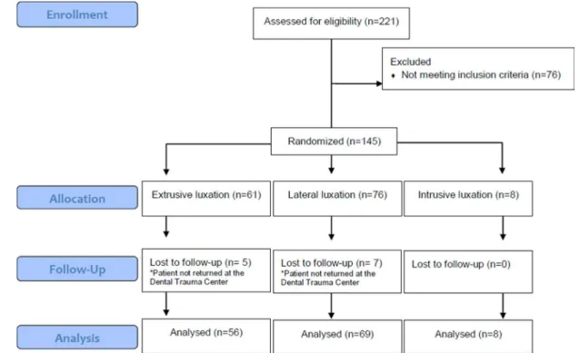
This study was approved by the Local Ethics Committee for Research with Human Beings (Protocol number 092-2011). All patients or their legal guardians were informed of the procedures protocols, risks and benefits and their right to self-determination regarding participation. A written consent was signed and a copy delivered to all volunteers. The study was conducted with reference to patients who had sought treatment for dental trauma at our Dental Trauma Center between July 2007 and July 2011. Only cases of extrusive, lateral or intrusive luxation with a minimum of 24 months follow-up were included. Patients who had coronary fractures without supporting tissue involvement, root fractures, traumatic lesions in primary teeth, teeth with cavities or restored, teeth with incomplete root formation and reimplanted teeth were excluded from this study. Furthermore, when it was found that emergency care protocol was not in accordance with the recommendations of the International Association of Dental Traumatology, the patient was excluded from this study. Figure 1 shows of the flowchart for the selection and evaluation of luxated teeth. The patient’s medical history included the following information: age, gender, type of trauma and the teeth
that were injured. The period between dental injury and initial attendance at the Dental Trauma Center was recorded during anamnesis, when the patient attended for the first time at the Dental Trauma Center. The date of the initial attendance at the Dental Trauma Center was classified as up to 15 days, between 15 and 45 days and after 45 days after dental trauma. Clinical examination was performed by three endodontists with five years clinical experience. The following parameters were recorded: dental discolouration, mobility, sensitivity to palpation and percussion, and presence of fistulae. Pulp vitality was established with the cold test using a cooling spray at −50 °C (EndoFrost; Roeko, Langenau, Germany). Periapical periodontitis and external inflammatory resorption were investigated radiographically. A diagnosis of pulp necrosis was established in cases of crown grey discolouration, loss of pulpal sensitivity to the cold test and radiographic periapical radiolucency. In the conducted study, all three criteria had to be fulfilled for a diagnosis of pulp necrosis. If there was doubt about the diagnosis (type of trauma and pulp vitality) the tooth was excluded from this study.
Ultraspeed periapical films (Kodak, São José dos Campos, SP, Brazil) were used for radiographic analysis at 55 kVp and 15 mA. All radiographs were performed using intraoral positioners and paralleling technique. The radiographs were analyzed by two of the authors, under optimal conditions, while using a white-light illuminator (Lumatron; Encor Indústria Fotográfica Ltda, Rio Claro,
Initial attendance after dental trauma SP, Brazil) and a magnifying glass large enough to allow
binocular observation. External inflammatory resorption was defined by loss of continuity of the lamina dura associated with areas of bone rarefaction. If signs of inflammatory resorption were detected, bowl-shaped cavities involving both cementum and dentine were radiographically identified. Clinical findings such as increased mobility and fistulae could also be associated with this pathological signal. If there was no consensus on the resorption, the tooth was excluded from the study.
The data were collected and statistically analyzed using SPSS for Windows (SPSS Inc., Chicago, IL, USA). All traumatized teeth were included in the statistical analysis, even when a patient had more than one luxated tooth. Fisher’s exact test was used to verify the influence of the initial attendance date and the development of inflammatory resorption for each type of trauma. The odds ratio was used to check the risk of developing external inflammatory resorption due to delay in seeking treatment. The significance level was set at p<0.05.
Results
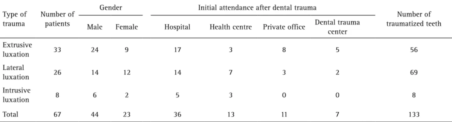
The sample in this study included 67 patients (44 males and 23 females) with 133 injured teeth with closed apex. Most of the affected teeth were maxillary central incisors (n=77), followed by maxillary lateral incisors (n=36), mandibular central incisors (n=9) and mandibular lateral incisors (n=11). The age of the patients ranged from 11 to 56 years. Injuries occurred most frequently in patients between 11 and 16 years of age (35 patients). Fifteen patients were in the 16-20 years age group and 17 patients were over 20 years old at the time of trauma. Most of the patients received emergency care in hospitals carried out by specialized dentists in maxillo-facial surgery (53.7%) (Table 1). Next, the patients were referred immediately to begin treatment at the Dental Trauma Center. Thirty-one patients (46.2%) received initial attendance at the Dental Trauma Center 15 days after the trauma, 19 (28.3%) between 15 and 45
days and 17 (25.5%) after 45 days. The prevalence of pulp necrosis and external inflammatory resorption is in Table 2. The date of initial attendance and treatment was found to influence the development of inflammatory resorption in lateral luxation (p=0.0174) (Table 3). The odds ratio showed that patients who started treatment after 45 days were 3.4 times more likely to develop external inflammatory resorption than patients who sought treatment immediately after the trauma (Table 4).
Discussion
The present study evaluated the occurrence of pulp necrosis and external inflammatory resorption in cases of dental luxation which, like avulsion, is considered a more severe type of trauma. The frequency of these consequences after dental fractures is not high, therefore fractures were not included in this study. The distribution of patients with dental luxation according to gender and age group is similar to other published studies (20-22).
Pulp necrosis occurred in 78.9% of teeth affected by dental luxation. The high frequency of necrosis may be related to the extent of root formation of the teeth included in this research. Some studies showed that the chance of necrosis is greater in teeth with a closed apex (8,15-17,20-22). Among the types of luxation, intrusion is related to the highest prevalence of pulp necrosis, because this type of
Table 1. Distribution of patients according to gender, location of initial attendance after dental trauma and number of traumatized teeth for each type of trauma
Type of trauma
Number of patients
Gender Initial attendance after dental trauma
Number of traumatized teeth Male Female Hospital Health centre Private office Dental trauma
center Extrusive
luxation 33 24 9 17 3 8 5 56
Lateral
luxation 26 14 12 14 7 3 2 69
Intrusive
luxation 8 6 2 5 3 0 0 8
Total 67 44 23 36 13 11 7 133
Table 2. Prevalence of pulp necrosis and external inflammatory resorption among luxated teeth
Type of trauma
Number of traumatized
teeth
Pulp necrosis
n(%)
External inflammatory
T
.F
.R. Lima et al.
Table 4. Odds ratios to verify the risk of development of external inflammatory resorption Initial
attendance after dental trauma
Number of traumatized
teeth
External inflammatory resorption (n)
Odds ratio
95% confidence
interval
p
Up to 15 days 54 2 1 Between 15
and 45 days 45 10 1.068 0.6590–4.1233 0.2854 After 45 days 34 11 3.4674 1.3567–8.8621 0.0094 Table 3. Relationship between initial attendance after dental trauma and development of external inflammatory resorption in luxated teeth
Type of trauma
External inflammatory resorption (n)
Up to 15 days (n)
Between 15 and 45
days (n)
After 45 days (n) p Extrusive
luxation 10 1 3 6 0.0719
Lateral
luxation 11 1 5 5 0.0174
Intrusive
luxation 2 0 2 0 1.0
trauma can crush the neurovascular bundle (8). Hecova et al. (7) found 83.3% of necrosis in cases of intrusive luxation in teeth with a closed apex.
This study demonstrated that delay in seeking treatment after trauma can impair the prognosis. Collecting these data was possible because most patients in this study were first treated in hospitals, clinics and private offices, where only emergency treatment was provided, ie, dental repositioning followed by containment. Subsequently, patients were referred for further treatment to the Dental Trauma Center. Some sought treatment a few days after emergency care, others after several weeks.
Among cases of extrusive and lateral luxation, there was inflammatory resorption in 17.8% and 15.9%, respectively. Other studies have found a lower frequency, varying from 0% to 10% (7,8,16,21,23). The values found in this study may be explained by the delay in seeking treatment. For lateral luxation, it was found that an extended delay between the date of injury and early treatment influenced the occurrence of external inflammatory resorption. For extrusive luxation, 6 of the 10 patients diagnosed with resorption were those who sought treatment only after 45 days.
The occurrence of pulp necrosis and external inflammatory resorption in intrusive luxation was the highest (100% and 25%, respectively). Hecova et al. (7) found a prevalence of 83.3% for pulp necrosis and 33.3% for external inflammatory resorption in intruded teeth with closed apex. The high frequency of these post-traumatic complications can be explained by a higher damage rate to the periodontal ligament and the pulp along with intrusion,
compared to lateral luxation or extrusion.
The odds ratio was used to assess the risk of developing inflammatory resorption due to delay for seeking treatment. In the present study, patients who began treatment 45 days after the injury were 3.4 times more likely to develop external inflammatory resorption. External inflammatory resorption may arise as a result of destruction of the protective barrier of the tooth root, associated with the presence of irritants in the root canal, like necrotic tissue and/or microorganisms. Thus, if pulp necrosis is not diagnosed early, such irritants may favour the emergence and progression of external inflammatory resorption. A recent study found that the pulp extirpation delay can interfere with the development of resorption in replanted teeth with a closed apex, and if there is a delay in the completion of the endodontic treatment, the risk of developing root resorption increases (18). In this study, replanted teeth were not included but the delay in seeking treatment causes a delay of the pulp extirpation, which can interfere with the progression of root resorption. A limitation of this study concerns the radiographic diagnosis of external inflammatory resorption performed by periapical radiographs. This method provides a two-dimensional image can hide lesions on the buccal or lingual surfaces. Furthermore, resorption in the early stages cannot be checked in periapical radiographs. Another limitation is the age range of the included patients, which was extremely wide, ranging from 11 to 56 years old. It is important to emphasize that only teeth with closed apex were included, regardless the patient age. However, further studies to assess factors related to the development and progression of external inflammatory resorption in children and adults should be encouraged.
Only external inflammatory resorption was analyzed in this study. Superficial root resorption is self-limiting and usually not related to pulp necrosis (11). Replacement resorption is often observed after severe intrusion and in replanted teeth, and its occurrence is linked to destruction of the root protective barrier, which favors the action of cells from the bone on the root surface (10,11,17).
Initial attendance after dental trauma Calcium hydroxide has a long and proven track record in
encouraging hard tissue repair and is indicated for use as part of the management of inflammatory resorption. Once there are radiographic signals of hard tissue repair, the root canal filling can be completed (3,24). According to the IADT Dental Traumatology Guidelines (25), patients affected by dental luxation must be followed up for a longer time, because if a post-traumatic complication occurs, timely treatment can be provided. The rate of pulp necrosis is high after traumatic tooth injuries. Luxated teeth are also at considerable risk for periodontal healing complications. Thus, regular controls are required for early detection of external inflammatory root resorption.
According to the results of the present study, it may be concluded that delayed treating after the occurrence of injury can impair the prognosis and promote the development of external inflammatory resorption in luxated teeth with closed apex.
Resumo
O objetivo deste estudo clínico foi avaliar a relação entre a demora na procura por tratamento e o desenvolvimento de reabsorções inflamatórias externas em dentes luxados com ápice fechado. A amostra desta pesquisa foi constituída por 67 pacientes que apresentaram 133 dentes traumatizados, dos quais 56 foram acometidos por luxação extrusiva, 69 por luxação lateral e 8 por luxação intrusiva, proservados por no mínimo 24 meses. Foi registrado o tempo decorrido entre a data do trauma e o primeiro atendimento. Verificou-se a ocorrência de necrose pulpar e reabsorções inflamatórias externas em cada tipo de traumatismo. O Teste Exato de Fisher foi aplicado para verificar a influência da data do atendimento inicial e o desenvolvimento de reabsorções inflamatórias externas em cada tipo de luxação e o teste de Odds Ratio foi aplicado para verificar o risco do desenvolvimento de reabsorções inflamatórias externas em função da demora na procura por tratamento. Os resultados revelaram que a necrose pulpar foi verificada em 105 dentes luxados (78,9%) e a reabsorção inflamatória externa foi verificada em 17.8% das luxações extrusivas (10), sendo 15,9% das luxações laterais (11) e 25% das luxações intrusivas (2). A demora na procura por tratamento influenciou o desenvolvimento de reabsorções infamatórias externas nas luxações laterais (p=0,0174). Os pacientes que procuraram tratamento após 45 dias da data do trauma apresentaram 3,4 vezes mais chance de desenvolver reabsorção inflamatória externa do que os pacientes que procuraram tratamento logo após o traumatismo. Conclui-se que o tratamento tardio após a ocorrência do traumatismo pode prejudicar o prognóstico e favorecer o desenvolvimento de reabsorções inflamatórias externas em dentes luxados com ápice fechado.
References
1. Hunter ML, Hunter B, Kingdon A, Addy M, Dummer PM, Shaw WC. Traumatic injury to maxillary incisor teeth in a group of South Wales schoolchildren. Endod Dent Traumatol 1990;6:260-264.
2. Glendor U, Marcenes W, Andreasen JO. Classification, Epidemiology and Etiology. In: Andreasen JO, Andreasen FM, Andersson L, editors. Textbook and color atlas of traumatic injuries to the teeth. 4th ed. Odder: Blackwell Munksgaard; 2007. p. 217-254.
3. Damé-Teixeira N, Alves LS, Susin C, Maltz M. Traumatic dental injury among 12-year-old South Brazilian schoolchildren: prevalence, severity and risk indicators. Dent Traumatol 2013;29:52-58.
4. Lauridsen E, Hermann NV, Gerds TA, Kreiborg S, Andreasen JO. Pattern of traumatic dental injuries in the permanent dentition among children, adolescents and adults. Dent Traumatol 2012;28:358-363.
5. Feldens CA, Kramer PF, Ferreira SH, Spiguel MH, Marquezan M. Exploring factors associated with traumatic dental injuries in preschool children: a Poisson regression analysis. Dent Traumatol 2010;26:143-148. 6. Norton E, O’Connel AC. Traumatic dental injuries and their association
with malocclusion in the primary dentition of Irish children. Dent Traumatol 2012;28:81-86.
7. Hecova H, Tzigkounakis V, Merglova V, Netolicky J. A retrospective study of 889 injured permanent teeth. Dent Traumatol 2010;26:466-475. 8. Andreasen FM, Vestergaard Pedersen B. Prognosis of luxated permanent
teeth - the development of pulp necrosis. Dent Traumatol 1985;1:207-220.
9. Andreasen FM, Andreasen JO. Luxation injuries of permanent teeth: general findings. In: Andreasen JO, Andreasen FM, Andersson L, editors. Textbook and color atlas of traumatic injuries to the teeth. 4th ed. Odder: Blackwell Munksgaard 2007. p. 372-403.
10. Soares AJ, Gomes BPFA, Zaia AA, Ferraz CCR, Souza-Filho FJ. Relationship between clinical radiographic evaluation and outcome of teeth replantation. Dental Traumatol 2008;24:183-188.
11. Fuss Z, Tsesis I, Lin S. Root resorption: diagnosis, classification and treatment choices based on stimulation factors. Dent Traumatol 2003;19:175-182.
12. Patel S, Kanagasingam S, Pitt Ford T. External cervical resorption: a review. J Endod 2009;35:616-625.
13. Patel S, Ricucci D, Durak C, Tay F. Internal root resorption: a review. J Endod. 2010;36:1107-1121.
14. Estrela C, Bueno MR, de Alencar AHG, Mattar R, Valladares Neto J, Azevedo BC, et al.. Method to evaluate inflammatory root resorption by using cone beam computed tomography. J Endod 2009;35:1491-1497. 15. Lauridsen E, Hermann NV, Gerds TA, Ahrensburg SS, Kreiborg S,
Andreasen JO. Combination injuries 3. The risk of pulp necrosis in permanent teeth with extrusion or lateral luxation and concomitant crown fractures without pulp exposure. Dent Traumatol 2012;28:379-385.
16. Ferrazzini Pozzi EC, von Arx T. Pulp and periodontal healing of laterally luxated permanent teeth: results after 4 years. Dent Traumatol2008;24:658-662.
17. Wigen TI, Agnalt R, Jacobsen I. Intrusive luxation of permanent incisors in Norwegians aged 6-17 years: a retrospective study of treatment and outcome. Dent Traumatol 2008;24:612-618.
18. Bastos JV, Ilma de Souza Côrtes M, Andrade Goulart EM, Colosimo EA, Gomez RS, Dutra WO. Age and timing of pulp extirpation as major factors associated with inflammatory root resorption in replanted permanent teeth. J Endod 2014;40:366-371.
19. Petrovic B, Marković D, Peric T, Blagojevic D. Factors related to treatment and outcomes of avulsed teeth. Dent Traumatol 2010;26:52-59. 20. Nikoui M, Kenny DJ, Barrett EJ. Clinical outcomes for permanent incisor
luxations in a pediatric population. III. Lateral luxations. Dent Traumatol 2003;19:280-285.
21. Lee R, Barrett EJ, Kenny DJ. Clinical outcomes for permanent incisor luxations in a pediatric population. II. Extrusions. Dent Traumatol 2003;19:274-279.
22. Humphrey JM, Kenny DJ, Barrett EJ. Clinical outcomes for permanent incisor luxations in a pediatric population. I. Intrusions. Dent Traumatol 2003;19:266-273.
23. Oikarinen K, Kassila O. Causes and types of traumatic tooth injuries treated in a public dental health clinic. Endod Dent Traumatol 1987;3:172-177.
24. Abott PV. Prevention and management of external inflammatory root resorption following trauma to teeth. Aust Dent J 2016:61:82-94 25. Diangelis AJ, Andreasen JO, Ebeleseder KA, Kenny DJ, Trope M, Sigurdsson
A, et al.. International Association of Dental Traumatology. International Association of Dental Traumatology: Guidelines for the management of traumatic dental injuries: 1. Fractures and luxations of permanent teeth. Dent Traumatol 2012;28:2-12.