UNIVERSIDADE TÉCNICA DE LISBOA
Faculdade de Medicina Veterinária
RASTREIO VIROLÓGICO DE CARNÍVOROS ERRANTES E
CARACTERIZAÇÃO GENÉTICA VIRAL
RICARDO CONSTANTE ROSADO
CONSTITUIÇÃO DO JÚRI ORIENTADORA
Doutor Luís Manuel Morgado Tavares Doutora Ana Isabel Simões Pereira Duarte Doutor Virgílio da Silva Almeida
Doutora Ana Isabel Simões Pereira Duarte
UNIVERSIDADE TÉCNICA DE LISBOA
Faculdade de Medicina Veterinária
RASTREIO VIROLÓGICO DE CARNÍVOROS ERRANTES E
CARACTERIZAÇÃO GENÉTICA VIRAL
RICARDO CONSTANTE ROSADO
DISSERTAÇÃO DE MESTRADO INTEGRADO EM MEDICINA VETERINÁRIA
CONSTITUIÇÃO DO JÚRI ORIENTADORA
Doutor Luís Manuel Morgado Tavares Doutora Ana Isabel Simões Pereira Duarte Doutor Virgílio da Silva Almeida
Doutora Ana Isabel Simões Pereira Duarte
ACKNOWLEDGEMENTS
I realize upon writing this, the first and last page of my dissertation, that I'm looking back at six years of my life at the Faculty of Veterinary Medicine in Lisbon. The first thing that comes to my mind is that I should have saved more time to write these acknowledgements, and yet the simple truth is that no matter how much time I could have saved for this page, the people to whom I am thankful would always be innumerable.
I won't dedicate this dissertation to anyone, but I would like to acknowledge that I wouldn't be here if it weren't for my dog, Juca, who passed away during the final month of my curricular training. He was the first to teach me the joys of early rising, lazy Sundays, running on the beach, and unconditional love. He chose this profession for me, always tried to show me how to communicate with animals, mostly in vain, and taught me that family doesn't have only two legs. I wouldn't dedicate this dissertation to him because he'd probably just eat it. I would like to thank the rest of my two-legged family, my parents, my brother and sister and our recent “acquisitions” for their support and patience. I am in your debt.
None of this would have been possible if Professor Luís Tavares hadn't given me the opportunity to “help out” after classes at the Virology Laboratory three years ago, much to the dismay of my colleagues in the laboratory and especially my current supervisor, Professor Ana Duarte, who has had to put up with me for the better part of those three years, and has taught me so much more than I could have hoped. Together they have shown me that there is a world within these walls, just as exciting and entertaining as working in the jungle, be it the concrete or the real one. The experience has been life-changing and they are now a part of me.
Throughout these past six years I've built relationships which are stronger than I could have dreamed and they have helped shape who I am today. I am proud to have been a part of the class of 2003-2008 and proud to be a member of the VETuna. I will always cherish the lessons I learned as part of the Students Association and the Pedagogical Council, hard as they may have been, and I would never have been able to accomplish as much as I did without the help of so many friends, teachers and colleagues. I'm especially thankful for the support of my closest friends, with whom I've traveled across oceans and shared both joys and sorrows. I will always carry you with me and be within your reach, wherever I go. Special thanks, to four dogs, Pilhas, Balú, Joca and Lady, and two cats, Bagerah and Pekenita. Together with their “person” they brought magic into my life and taught me lessons I will never forget.
Lastly, we are grateful to our colleagues and all the employees of the Lisbon Municipal Kennel for their collaboration and assistance. This work was sponsored by CIISA-FMV as part of the Integrated Masters in Veterinary Medicine.
ABSTRACT
Virological survey in stray carnivores and viral genetic characterisation
Free-roaming stray or feral dogs and cats living in urban areas can be responsible for the spread and maintenance of several infectious diseases. To investigate the presence of viral agents and the genomic diversity of canine and feline coronavirus (CCoV, FCoV) in Lisbon’s Municipal kennel, a virological survey was conducted which included canine distemper virus (CDV), canine and feline parvovirus (CPV,FPV), CCoV and FCoV, feline immunodeficiency virus (FIV) and feline leukaemia virus (FeLV).
Blood samples and faecal swabs were collected from 50 dogs and 50 cats at the time of euthanasia, and 24 environmental swabs were collected at a later date. Samples were either tested using a commercial ELISA kit, or amplified by PCR and RT-PCR. All coronavirus positive samples were further characterized by RT-PCR to assess the presence of different FCoV and CCoV genotypes. All PCR products were observed on 1,5%.agarose gel.
Antibodies against FIV were found in 18% of the samples, while FeLV antigen was found in 10%. Viral nucleic acid was detected in 8.2% samples for CDV, 32.7% for CCoV, 59.6% for FPV/CPV, and 70% for FCoV. Seven (43.8%) samples were positive for CCoV type I, 9 (56.2%) for CCoV type II, and as for FCoV, 9 (25,7%), 6 (17%) and 12 (34.3%) samples were positive for FCoV type I, type II and both types, respectively. No differentiation was possible in 8 (23%) of the FCoV samples. There were positive environmental samples for CDV (50%), FPV/CPV (62.5%) and FCoV (87.5%).
The results found on this study, particularly on parvovirus and FCoV brought to our attention the need for a continued and more precise evaluation of the health status of free-roaming stray or feral animals in the municipal kennel, to correctly evaluate their role as viral reservoirs within and without the kennel premises. The high prevalence of coronavirus infection found in both dogs and cats in the Lisbon Municipal Kennel allowed the viral genetic characterization, showing a high rate of co-infection with both genotypes of FCoV and absence of co-infected animals with CCoV I and II. However, further investigation is needed in order to maintain a molecular epidemiological surveillance and help identify further CoV strains, as well as understand the pathogenic potential of these viruses.
Keywords: virological survey, molecular epidemiology, small animal viruses, coronavirus,
RESUMO
Rastreio virológico de carnívoros errantes e caracterização genética viral
Os animais errantes ou assilvestrados que habitam áreas urbanas podem ser responsáveis pela distribuição e manutenção de diversas doenças infecciosas. Com o objectivo de investigar a presença de agentes virais e avaliar a diversidade genética de coronavirus canino e felino (CCoV, FCoV) no Canil Municipal de Lisboa, realizámos um rastreio virológico em que foram avaliados o vírus da esgana canina (CDV), parvovirus canino e felino (CPV, FPV), CCoV e FCoV, o vírus da imunodeficiência felina (FIV) e o vírus da leucemia felina (FeLV).
Foram colhidas amostras de sangue e zaragatoas rectais de 50 cães e 50 gatos na altura da eutanásia, e 24 zaragatoas ambientais numa data posterior. Avaliámos as amostras utilizando um teste comercial de ELISA ou amplificando as amostras por PCR e RT-PCR. Todas as amostras positivas para coronavirus foram caracterizadas por RT-PCR para avaliar a presença de genótipos diferentes de FCoV e CCoV. Todos os produtos de PCR foram visualizados num gel de agarose a 1,5%.
Foram encontrados anticorpos contra FIV em 18% das amostras e antigénio de FeLV em 10%. Detectámos ácido nucléico viral em 8.2% das amostras para CDV, 32.7% para CCoV, 59.6% para FPV/CPV a 70% para FCoV. Sete (43.8%) amostras foram positivas para CCoV tipo I, 9 (56.2%) para CCoV tipo II, e em relação ao FCoV, 9 (25,7%), 6 (17%) e 12 (34.3%) amostras foram positivas para FCoV tipo I, tipo II, e para ambos os tipos, respectivamente. Não foi possível obter diferenciação em 8 (23%) das amostras para FCoV. Houve amostras ambientais positivas a CDV (50%), FPV/CPV (62.5%) e FCoV (87.5%).
Os resultados deste estudo demonstraram a importância de uma avaliação contínua e mais precisa do estatuto sanitário dos animais errantes ou assilvestrados no canil municipal, principalmente em relação ao parvovirus e ao FCoV, de modo a avaliar correctamente o seu papel de reservatórios de doenças, tanto dentro como fora do canil. A alta prevalência de infecções por coronavirus em cães e gatos no Canil Municipal de Lisboa permitiu a caracterização genética viral, demonstrando uma percentagem elevada de co-infecções com os dois genótipos de FCoV, mas uma ausência de animais co-infectados com CCoV I e II. No entanto, é necessário mais investigação para manter uma vigilância epidemiológica ao nível molecular, de forma a ajudar na identificação de possíveis novas estirpes de coronavirus, assim como compreender o potencial patogénico destes vírus.
Palavras-chave: rastreio virológico, epidemiologia molecular, vírus de animais de
LIST OF SCIENTIFIC COMMUNICATIONS
The work related to this dissertation resulted in the presentation of three scientific posters at two different international meetings:
• Fernandes, T., Bernardino, R., Duarte, M., Fevereiro, M., Correia, J., Duarte, A., Rosado, R., Tavares, L.. “Fatal Panleukopenia virus infection in a Linx linx”. International Conference on Diseases of Zoo and Wild Animals 2009, 20th to the 24th of May. Hilvarenbeek, The
Netherlands.
• Ricardo C. Rosado, Ana Duarte, Augusto Baptista, Filomena Oliveira, Ana Machado, Leonel Fernandes, Luís Tavares. “Virological survey in shelter dogs and cats in Lisbon”. 8th
International Congress of Veterinary Virology, 23rd to the 26th of August 2009, Budapest,
Hungary.
• Ricardo C. Rosado, Ana Duarte, Augusto Baptista, Filomena Oliveira, Ana Machado, Leonel Fernandes, Luís Tavares. “Genetic characterization of coronaviruses in shelter dogs and cats in Lisbon”. 8th International Congress of Veterinary Virology, 23rd to the 26th of
TABLE OF CONTENTS
Acknowledgements...i
Abstract...ii
Resumo...iii
List of scientific communications...iv
1. Introduction...1 1.1 Laboratory work...1 1.2 Research project...3 1.3 Objectives...5 1.4 DNA Virus...6 1.4.1 Parvovirus...6 1.5 RNA Virus...10
1.5.1 Canine Distemper Virus...10
1.5.2 Coronavirus...14
1.5.3 Retroviruses...19
2. Materials and methods...25
2.1 Study population...25
2.2 Sample collection...25
2.3 Sample processing...25
2.4 Antibody detection...25
2.5 Antigen detection...26
2.6 Nucleic Acid Extraction...26
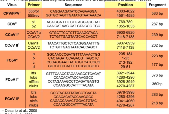
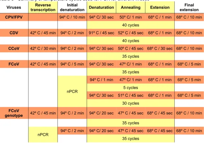
2.7 PCR and RT-PCR...26
3. Results...29
3.1 Kennel records...29
3.2 Laboratory results...29
3.2.1 Serological results...29
3.2.2 Nucleic acid amplification results...30
3.2.2.1 Dog samples...30
3.2.2.2 Cat Samples...31
3.2.2.3 Environmental samples...32
3.2.3 Genetic characterisation of coronaviruses...33
4. Discussion and conclusions...34
Illustration Index
Figure 1 - Map of the municipal kennel highlighting the rooms sampled...4
Figure 2 - Parvovirus structure (a) and genome (b)...6
Figure 3 - Structure of canine distemper virus (a) and its genome (b)...11
Figure 4 - Coronavirus structure (a) and comparison of CCoV and FCoV genome (b)...14
Figure 5 - FeLV (a) and FIV (b) structure and genome...21
Figure 6 - ELISA assay for FeLV with positive (+) and negative (-) controls marked...29
Figure 7 - PCR and RT-PCR results of Hexadog vaccine dilutions used to test sensibility of parvovirus and CDV detection...30
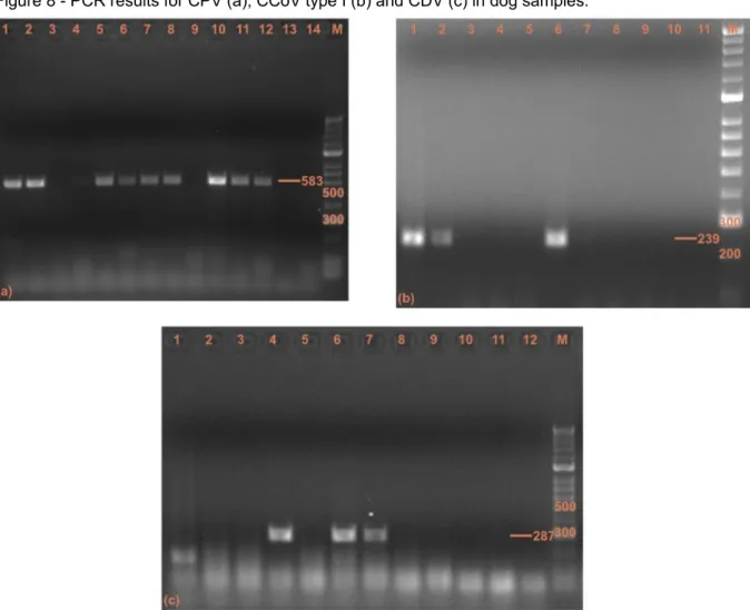
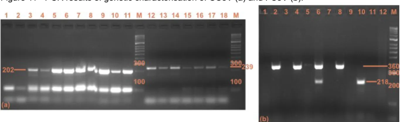
Figure 8 - PCR results for CPV (a), CCoV type I (b) and CDV (c) in dog samples...31
Figure 9 - PCR results for FPV (a) and FCoV (b) in cat samples...32
Figure 10 - PCR results for CPV and FCoV in environmental samples...32
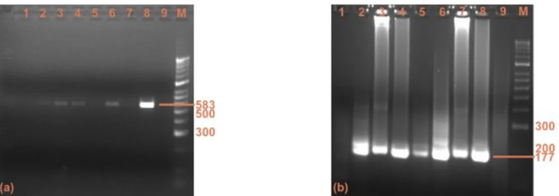
Figure 11 - PCR results of genetic characterisation of CCoV (a) and FCoV (b)...33
Index of Tables
Table 1 - Summary of agents, materials and test types performed at the Virology and Molecular Biology Laboratory at the FMV...1Table 2 - Number of samples analysed at the Virology and Molecular Biology Laboratory during the period of 15th October 2008 and 15th April 2009...2
Table 3 - Number of samples of different biological materials received at the Virology and Molecular Biology Laboratory during the period of 15th October 2008 and 15th April 2009....2
Table 4 - Primers, position and fragment sizes used for PCR amplification of different viruses ...27
Table 5 - Summary of temperature cycles used for PCR for different viruses...28
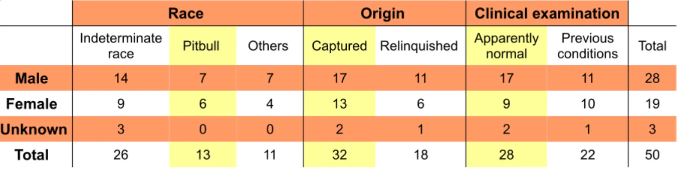
Table 6 - Details from kennel records and distribution of race, origin and clinical examination per gender...29
1. Introduction
The present study was part of the curricular training for the Integrated Masters in Veterinary Medicine from the Faculty of Veterinary Medicine (FMV), Universidade Técnica de Lisboa. It consisted of a period of six months between 15th October 2008 and 15th April 2009 in the Virology and Molecular Biology Laboratory of the FMV under the supervision of Prof. Ana Isabel Pereira Duarte. During this time we conducted a virological survey of free-roaming carnivores at the municipal kennel in Lisbon, while participating in the diagnostic procedures at the virology laboratory.
1.1 Laboratory work
The Virology and Molecular Biology Laboratory provides diagnostic services for several clinics, including the Teaching Hospital of the FMV and the Pathology Laboratory. Samples submitted by veterinary clinicians are tested for different viral diseases using molecular biology or serological techniques. Among the different agents diagnosed in the laboratory, the most common are feline coronavirus (FCoV), feline leukaemia virus (FeLV), feline immunodeficiency virus (FIV) , feline/canine parvovirus (FPV/CPV) and canine distemper virus (CDV). Other agents are tested less frequently, like feline herpesvirus (FHV), and canine coronavirus (CCoV).
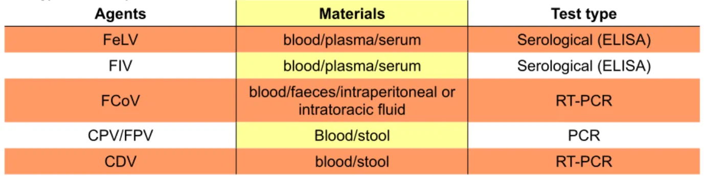
Sample materials received by the laboratory include whole blood (plasma/serum), stool, intraperitoneal/intratoracic fluid and organ samples. Different materials are required and are processed accordingly to the agent to test for and the technique used (Table 1). FeLV and FIV are tested using serological techniques, namely an enzyme-linked immunosorbent assay (ELISA) for detection of Ag and Ab, respectively. Blood samples are previously centrifuged because the assay has better results with plasma/serum than whole blood.
Table 1 - Summary of agents, materials and test types performed at the Virology and Molecular Biology Laboratory at the FMV
Agents Materials Test type
FeLV blood/plasma/serum Serological (ELISA) FIV blood/plasma/serum Serological (ELISA) FCoV blood/faeces/intraperitoneal or intratoracic fluid RT-PCR
CPV/FPV Blood/stool PCR
CDV blood/stool RT-PCR
Samples tested for other viruses use molecular biology techniques to assess the presence of viral nucleic acid, by polymerase chain reaction (PCR) for DNA viruses and reverse transcriptase-PCR for RNA viruses. Liquid samples (blood, body fluids) are centrifuged and
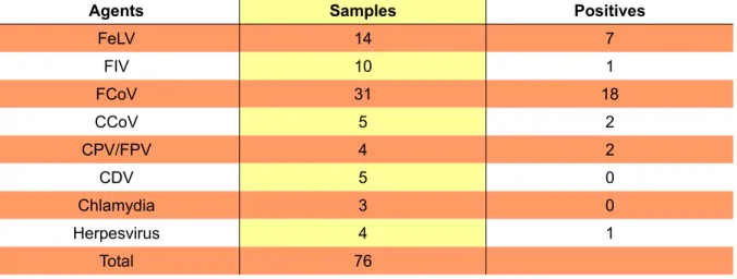
the supernatant is processed for nucleic acid extraction using an appropriate commercial kit. Solid samples (stool, organs) are homogenized with Phosphate buffered Saline (PBS), centrifuged and processed as referred for liquid samples. A summary of the samples analysed at the laboratory during the course of this study is presented in tables 2 and 3.
Table 2 - Number of samples analysed at the Virology and Molecular Biology Laboratory during the period of 15th October 2008 and 15th April 2009
Agents Samples Positives
FeLV 14 7 FIV 10 1 FCoV 31 18 CCoV 5 2 CPV/FPV 4 2 CDV 5 0 Chlamydia 3 0 Herpesvirus 4 1 Total 76
Table 3 - Number of samples of different biological materials received at the Virology and Molecular Biology Laboratory during the period of 15th October 2008 and 15th April 2009
Materials Samples
blood 35
stool 30
ocular swab 3
oral swab 4
1.2 Research project
Free-roaming stray or feral dogs and cats living in urban areas can be responsible for the spread and maintenance of several infectious diseases. These animals aren't confined or owned by anyone and both their number as well as their health status is mostly unknown to veterinarians and public health services.
There is a lot of information throughout the scientific literature about virological surveys. These surveys can vary greatly in terms of methodology and procedures, can be very specific or very general, have different sample sizes, focus on several or only one aetiological agent and some are conducted in collaboration with TNR programs (trap, neuter, release), usually oriented to control cat population. All of them help us gain greater insight into an existing disease or its agent and the way it interacts with its host. These surveys help us paint the picture of prevalence of disease in a population, but it is a picture which is never finished, because for every question we answer, new questions arise..
In this particular case, the original idea of a survey which would help us characterise the presence of specific agents in the environment and the risk to both wild and domestic animals, stumbled upon the difficult scenery of municipal kennels. Regulations conflict with compassion and the darkest of human nature as we try to understand the reasons why owners and citizens allow animals who share the same environment as them be relinquished to a kennel and ultimately euthanized after all other options are spent. In the process, we came across problems with which our colleagues at the municipal kennel have had to deal for a long time.
For the purpose of this study, “free-roaming” describes homeless animals, including socialized strays and unsocialized feral. In the case of the municipal kennel, most of the dogs were socialized, while practically all of the cats caught by the kennel employees were feral or barely socialized.
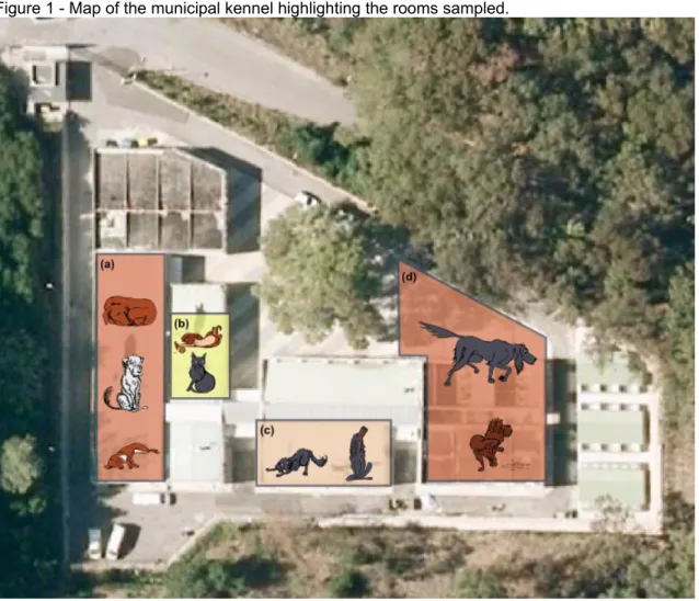
The kennel is placed in the middle of the Monsanto Natural Park, yet the facilities soon proved to be too small to contain the growing pet overpopulation in the Lisbon urban area, in spite of the abundant space surrounding it. Dogs are housed in three kennels: the main one (Figure 1-a) where most of the dogs are kept in open cells, alone, but with some contact with each others; another smaller room where aggressive animals are kept in containment cages (Figure 1-c); and an exterior kennel where more social dogs are housed in pairs, or in small groups (Figure 1-d). All cats are housed together with other animals captured in the same day, in a single room with a number of different cages (Figure 1-b). The same room also has cages for smaller dogs and young puppies. Cats considered fit for adoption, are also housed in groups in an exterior kennel.
Animals are delivered at any time of the day or night (when delivered by police forces), or captured by kennel employees and taken directly to one of the kennels after the entry record has been made. Owners who relinquish their animals sign a statement and provide identification. Veterinarians fill in details of the clinical examination and tend to any animals which require medical treatments. Although surgeries are performed, most of the treatments are supportive. When possible, animals are photographed to be advertised on the kennel website for adoption. Euthanasia is usually on a weekly basis for the simple reason of keeping the kennel population at an acceptable number. During this study an average of 26 animals per week were euthanized.
Among the problems identified at the beginning of the study by the veterinarians and employees of the kennel were the lack of proper ventilation in the interior kennels, the high number of animals present at all times, which prevented any sanitary break from taking place and the difficulty in providing better health care to the animals who needed it, due to lack of financing support.
Figure 1 - Map of the municipal kennel highlighting the rooms sampled.
Both veterinarians and employees mentioned the presence of infectious diseases among the kennel population, specially canine distemper and parvovirus among the dogs and panleukopenia among the cats.
1.3 Objectives
In order to conduct a virological survey of both dogs and cats 100 samples were collected from animals euthanized at the Lisbon municipal kennel. For the purpose of this study, canine distemper virus (CDV), canine parvovirus (CPV), canine coronavirus (CCoV), feline immunodeficiency virus (FIV), feline leukaemia virus (FeLV), feline coronavirus (FCoV), and feline parvovirus (FPV) were selected. All coronavirus positive samples were further characterized to assess the presence of different FCoV and CCoV genotypes within the animal population.
Most of these animals were captured in urban areas in and around Lisbon and a few were delivered by their owners to be euthanized for health or other issues. Since background information on the health status of these animals was lacking, antibody detection was only performed for FIV, because of its specific pathogenesis which produces persistent infection, but low level virus replication, and the fact that it has no commercial vaccine in Portugal. All other viruses were tested for the presence of antigen by enzyme-linked immunosorbent assay (ELISA) or nucleic acid by polymerase chain reaction (PCR).
This information was used to estimate the prevalence of these diseases in dogs and cats at the Lisbon Municipal Kennel and evaluate the sanitary conditions in the kennel. As a secondary objective, we tried to estimate the role of stray and feral animals in the spread and maintenance of infectious disease in both domestic populations and wild animals.
1.4 DNA Virus 1.4.1 Parvovirus
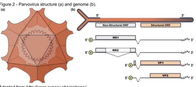
Viruses from the family Parvoviridae infect a wide variety of hosts, ranging from insects to primates (Hueffer and Parrish, 2003). The current classification of parvoviruses is based primarily on their host range and their dependence on help from other viruses for replication, traditionally separating them into autonomous viruses of vertebrates, helper-dependent viruses of vertebrates, and autonomous viruses of insects. Viruses from insects and other arthropods are included in the subfamily Densovirinae, while viruses from vertebrates are contained in the subfamily Parvovirinae (Lukashov & Goudsmit, 2001). Among these, members of the genus Parvovirus are among the smallest DNA virus to include important pathogens of both dogs and cats (Patel & Heldens., 2009). The viruses are all classified as members of the feline parvovirus subgroup of the family Parvoviridae and are named for the host from which they are isolated—hence CPV, FPV, raccoon parvovirus, mink enteritis virus (MEV), as well as bluefox parvovirus (BFPV) from Arctic foxes (Parrish, 1995). Feline panleukopenia virus (FPV) is considered the prototype parvovirus of carnivores (Truyen et al., 2009) and Canine parvovirus (CPV) is a host range variant which acquired the ability to infect dogs. While FPV infects all felids, as well as raccoons, minks and foxes, CPV infects members of the Canidae family and recently regained the ability to infect cats (Truyen, Evermann, Vieler & Parrish, 1996). Current taxonomy defines canine parvovirus and feline panleukopenia virus as one single taxonomic entity (Tattersall, 2006 cited by Truyen et al., 2009), but in the present study we refer to CPV as parvovirus in dogs and FPV parvovirus in cats.
Figure 2 - Parvovirus structure (a) and genome (b).
Parvoriruses are small and spherical, roughly 25 nm in diameter, without an envelope (Parrish, 1995) (Figure 2-a). FPV and CPV isolates may differ by as little as 0.5% in their DNA sequence (Hueffer et al., 2003). Their genome is a single-stranded DNA, roughly 5 kb long with hairpin structures at both ends and encodes two genes (Figure 2-b), which result in the expression of two non-structural proteins, NS1 and NS2, coded by the 3' end open reading frame. The capsid proteins are coded by an open reading frame towards the 5' end of the (negative sense) genomic DNA. The structural proteins VP1 and VP2 are formed by alternate splicing of the messenger RNA from the viral DNA, so that the complete sequence of VP2 is present in VP1 (Agbandje, Parrish and Rossman, 1995; Parrish, 1999). The number of capsid protein species per virion varies among parvoviruses. There are three types of polypeptides in CPV, VP1, VP2 and VP3. VP3 is formed by cleavage of 15 to 20 amino acids from the amino terminus of VP2 after virion assembly, and is not present in empty particles (Agbandje et al., 1995). The full capsid contains 60 copies of a combination of VP1, VP2 and VP3, with VP2 being the dominant protein (54-55 copies) (Parrish, 1999). The structure of the capsid defines three separate regions surrounding the threefold axes of symmetry, termed threefold spike, which are determinant in the host range differences between CPV and FPV by allowing it to bind to different transferrin receptors in the host cell (Hueffer and Parrish, 2003). The carnivore group of parvoviruses shows high conservation of DNA sequences, with less than 1.3% sequence variation between diverse isolates collected 30 years apart (Truyen et al., 1995). However, several sites in VP2 of CPV-2 display a nucleotide substitution rate similar to that of RNA viruses, which caused them to undergo a complex series of host range changes in the past three decades, and determined the appearance of diverse antigenic epitopes in naturally occuring variants of the virus (CPV-2a, 2b, 2c), (Truyen et al., 1995; Truyen et al., 1996; Shackelton, Parrish, Truyen & Holmes 2005).
All parvoviruses are extremely stable and resistant to adverse environmental influences, being able to persist on inanimate objects for 5 months or longer (McCaw & Hoskins, 2006). FPV resists heating at 56º C for 30 minutes and most common detergents and disinfectants, such as 70% alcohol, organic iodines, phenolics and quaternary ammonium compounds. One noteworthy exception is inactivation of both CPV and FPV by 6% sodium hypochlorite for 10 minutes at room temperature, which still has to be thorough, (Greene & Addie, 2006). Feline panleukopenia virus (FPV) has been known to cause disease in cats, raccoons and some related carnivores for more than a hundred years (Lamm & Rezabek, 2008), while canine parvovirus (CPV) was first isolated in 1978 (Carmicheal & Binn, 19811). The new virus
spread globally in a pandemic of disease during that same year and has since remained endemic in dogs throughout the world. There is no serological or other evidence for infection
of dogs by a related virus prior to the mid-1970s (Parrish, 19902) and its exact evolution and
origin remains elusive to date. One of the hypothesis about its sudden emergence suggested that CPV arose as a host range mutant directly from FPV in the dog or cat populations, similar to the scenario proposed for the emergence of MEV in the 1940s. Another hypothesis was that CPV emerged from a FPV vaccine virus after propagation in tissue culture and was initially spread in vaccine, which would explain the almost simultaneous appearance of CPV in the dog populations worldwide. Various isolates from wild carnivores support the hypothesis that CPV arose in a different host from the cat or dog, and that another carnivore may have harboured its immediate ancestor. DNA and amino acid analysis from one Arctic fox from Finland and red foxes from Germany revealed intermediate sequences between the FPV and CPV viruses, providing evidence of interspecies transmission between domestic and wild carnivores for the first time (Truyen, Müller, Heidrich, Tackmann & Carmichael, 1998; Truyen 1999).
Since the emergence of CPV it has continued to grow at an exponential rate, contrary to FPV, which maintains a population growth rate compatible with its endemic nature. CPV growth is characteristic of an epidemic, as it adapted to its canine host in a matter of years, spread across the world in a couple of months and subsequently generated a number of antigenic and host-range variants (Shackelton et al., 2005). CPV-2 was replaced by CPV-2a in 1981 and again by CPV-2b between 1984 and 1990 (Patel & Heldens, 2009). Interestingly, these variants show only a few amino acid substitutions in its genomic sequence. According to Truyen (2006) the adaptation to a new host was most likely due to the virus gaining the ability to bind the canine transferrin receptor, which is used to enter and infect canine cells. A new strain, emerged in the end of the 1990s, was characterised by a mutation in a major antigenic site of the viral capsid (position 426 Asp to Glu) and was named CPV-2c (Buonavoglia et al., 2001; Decaro et al., 2007a). This strain has already spread worldwide, yet it is still more prevalent in Europe (Nakamura et al., 2004; Decaro et al., 2007a; Hong et al., 2007; Kapil et al., 2007; Peréz et al., 2007). While the virulence and pathogenicity of CPV-2c were first deemed to be low, more recent reports are conflicting (Decaro et al., 2005; Decaro et al., 2008), just like reports on the efficacy of current vaccines (Decaro et al., 2008; Spibey, Greenwood, Sutton, Chalmers & Tarpey, 2008; Calderon et al., 2009).
Both CPV and FPV are highly contagious and most infections are a result of contact with contaminated faeces, but also fomites, insects and rodents. Animals may carry the virus in their hair coat for extended periods (McCaw & Hoskins, 2006). Infection in domestic dogs doesn't always result in apparent disease, as many dogs which are naturally infected never develop overt clinical signs (McCaw & Hoskins, 2006). Clinical illness is usually most severe in young puppies, especially when they harbour other pathogens such as parasites or enteric
bacteria. Incubation periods can vary between 7-14 days for CPV-2 and as brief as 4-6 days for newer variants (McCaw & Hoskins, 2006) and infected carnivores shed virus at high titres in their faeces (Truyen et al., 2009).
The pathogenesis of parvovirus infections is influenced primarily by the requirement of DNA replication for mitotic cells. Following oronasal exposure, the virus first replicates in the mucosa and lymphoid tissue of the buccal cavity and spreads next via cell-free viraemia to other internal organs. In vivo tissue tropism of FPV and CPV varies, but after progeny virus spread it can be found in virtually all organ tissues in both dogs and cats (Decaro et al., 2007c; Truyen et al., 2009). In puppies secondary sites of infection are typically the liver and heart, and animals often succumb to heart failure associated with pulmonary oedema, hepatomegaly and ascites. Older dogs commonly present with vomiting, diarrhoea and leucopoenia with a high morbidity, but a much lower mortality than in puppies. The disease induced by the Glu-426 mutant CPV-2c has been described as mild in pups, with mucoid diarrhoea and relative leucopoenia and lymphopenia (Decaro et al., 2005), but a more severe clinical course with higher mortality rates was also described in a breeding kennel in Italy (Decaro et al., 2008).
Pathogenesis of FPV infection in cats is similar and the infection is systemic. Replication causes shortening of the intestinal villi due to a sometimes complete loss of the rapidly dividing epithelial cells in the gut. Thus, the most common clinical sign is diarrhoea followed by leucopoenia and anaemia caused by replication of the virus in early progenitor cells of the bone marrow. Kittens infected during pregnancy may present cerebellar ataxia and intention tremor due to impaired development of the cerebellum caused by lytic virus replication in the Purkinje cells (Truyen et al., 2009).
The clinical diagnosis of CPV-2 infection is indecisive, since several other pathogens may cause diarrhoea in dogs. Several methods are available for the laboratory diagnosis of CPV infections. Virus isolation from blood or faeces in cell cultures and the demonstration of haemagglutination of porcine erythrocytes have been extensively used for diagnosis, but these methods are too labour-intensive and time-consuming for routine diagnostic testing (Desario et al., 2005). In practice, antigen detection in faeces is usually carried out using commercially available latex agglutination or immunochromatographic tests (Truyen et al., 2009), and recent advances in molecular technique have led to the development of highly sensitive and specific PCR and real-time PCR assays for the detection and quantisation of CPV-2 DNA in the faeces of diarrhoeic dogs (Desario et al., 2005). However, the period of faecal viral shedding is brief and virus is seldom detected by 10 to 12 days after natural infection. Furthermore, vaccine virus can yield false-positive results up to 12 days after vaccination (McCaw & Hoskins, 2006). On the other hand, the use of an antibody test such
as ELISA or indirect immunofluorescence is of limited value, because serological tests do not differentiate between infection and vaccination induced antibodies (Truyen et al., 2009). Both CPV and FPV vaccines are considered core vaccines by the Vaccinations Guidelines Group (VGG) of the World Small Animal Veterinary Association (WSAVA). Modified live vaccines have been available for some time and are recommended because of the more rapid and effective immunity they produce in comparison to inactivated virus vaccines. These are only recommended in the case of pregnant queens and kittens younger than 4 weeks, because no danger exists of post-vaccinal virus spread or clinical illness as a result of reversion to virulence (Greene & Addie, 2006; Day, Horzinek & Schultz, 2007). The recent emergence of CPV variants which infect even animals which have been repeatedly vaccinated has raised concerns that the antigenic differences between the original type 2 and its variants may decrease the effectiveness of the CPV-2 based vaccines and it would be useful to prepare vaccines using the CPV variants circulating in the field (Decaro et al., 2008).
1.5 RNA Virus
1.5.1 Canine Distemper Virus
Canine distemper (CD) is considered by many the most important worldwide infectious disease of domestic dogs, and its fatality rate is second only to that of rabies. This morbillivirus infection of dogs and other carnivores, has been recognized for at least 250 years and the first report of CD was from South America by Ulloa in 1746. Heusinger was convinced that the disease was introduced in 1760 from Peru to Spain, from where it spread to other parts of Europe and Russia within a few years. The epidemic spread of CD through Europe started around the 1760s, but the disease may have occurred earlier and was possibly confused with rabies. In 1815 Jenner observed that CD among dogs is as contagious as smallpox, measles and scarlet fever among humans and Karle succeeded in experimentally transmitting CD in 1844, by brushing the lips of young dogs with discharge from diseased dogs. The aetiology of the disease remained controversial until 1905, when Henri Carré demonstrated it was caused by a filterable virus (Carré, 19053; Blancou, 2004).
Clinical distemper has been largely controlled by routine vaccination in domestic dogs since the 1950s, but evidence of vaccinal failure caused by the emergence of viral variants in the recent years has raised concerns and renewed investigation efforts. Vaccine-induced infections have occurred in a variety of species, as have large-scale epidemics in felids and CDV may have the most far reaching implications of any infectious agent for susceptible free-living and captive carnivores. The discoveries of related viruses, such as phocine and
delphine morbilliviruses, and CDV’s similarity to the measles virus suggest viral mutability and a zoonotic potential for CDV (Deem, Spelman, Yates & Montali, 2000).
The genus morbillivirus within the family Paramyxoviridae includes important, highly contagious pathogens of animals and man. Until recently the genus comprised four distinct members: measles virus (MV) of man, canine distemper virus (CDV) of canines and their relatives, rinderpest virus (RPV) of cloven-hoofed animals, and peste-des-petits-ruminants virus (PPRV) mainly of small ruminants. All of these may cause severe disease in their respective host and MV is considered the prototype virus for the genus. Knowledge of the natural host range is a major factor in differentiating between these closely related morbillivirus species (Blixenkrone-Möller et al., 1992), and the natural host range for CDV has recently been proven to comprise all families of the order Carnivora (Deem et al., 2000).
CDV has a variable diameter between 150-250 nm, with negative-sense, single-stranded RNA, containing six non-overlaping genes encoding six structural proteins and enclosed in a nucleocapsid of helical symmetry (Greene & Appel, 2006) (Figure 3). It is surrounded by a
Figure 3 - Structure of canine distemper virus (a) and its genome (b).
N – nucleocapsid protein; P – phosphoprotein; C, V – transcriptional units; M – matrix protein; F – fusion protein; H – hemagglutinin; L – large protein. Adapted from http://www.expasy.ch/viralzone/.
lipoprotein envelope derived from the host cell membrane, where the virus incorporates the matrix protein (M) and two glycoprotein spikes, the hemagglutinin (H) and fusion protein (F). The nucleocapsid protein (N) encapsulates and protects the RNA and is associated with the phosphoprotein (P) and the large protein (L) which assists in transcription and replication (Blixenkrone-Möller et al., 1992; Barrett, 1999; Martella, Elia & Buonavoglia, 2008). Two additional proteins, C and V, are found as transcriptional units within the P gene (Beineke, Puff, Seehusen & Baumgärtner, 2009). The viral protein M is responsible for connecting the nucleocapsid and the surface glycoproteins H and F during viral maturation (Beineke et al., 2009), while the H protein is responsible for attachment to cell receptors and the F protein promotes fusion of the cell membrane with the viral envelope, and also between host cells (Martella et al., 2008). While CDV is considered to have one antigenic type, CDV strains can be divided into distinct lineages, based on phylogenetic analysis of subgenomic F, P, and complete H gene sequences, which are mainly associated with the geographical area from which the strain is isolated (Lednicky et al., 2004, Lan et al., 2005, Martella et al., 2006). CDV is extremely susceptible to heat and drying and is destroyed by temperatures greater than 50º C for 30 minutes. It may survive in excised tissues and secretions for up to 3 hours, but does not usually persist in kennels after infected dogs have been removed, especially in warm climates. The virus remains viable between pH 4.5 and 9.0 and is inactivated by ultraviolet light. As an enveloped virus, it is susceptible to ether and chloroform, dilute formalin solution, phenol and quaternary ammonium disinfectant. Routine disinfection procedures are usually effective in destroying the virus in kennel, clinic or hospital environments (Greene & Appel, 2006).
Transmission occurs mainly by direct animal-to-animal contact and the virus can be detected from most body tissues, including urine (Elia et al., 2006; Greene & Appel, 2006) However, it is most abundant in respiratory exudates and commonly spread by exposure to infectious aerosol or droplets (Green and Appel, 2006). Viral shedding occurs by 7 days postinfection and can last 60 to 90 days, although shorter periods are more typical. Transplacental infection can occur and the virus is maintained in a population by contact among infected animals and a constant supply of susceptible puppies for infection. Although immunity after exposure to the wild-type virus is prolonged or life-long, it is not as absolute after vaccination and there are estimates that 25-75% of susceptible animals become subclinically infected, but clear the virus. However, some dogs aren't able to clear the virus completely and may harbour it in their central nervous system. Spontaneous distemper is correlated in most cases with the loss of maternal antibodies, affecting dogs between 3 and 6 months the most, but disease can be severe and widespread, affecting all ages, in susceptible isolated populations and different strains may have different pathogenicity and clinical evolution (Green and Appel, 2006).
Great variations in duration, severity and clinical presentation of distemper have been found in experimentally infected dogs as well as in animals suffering from natural disease. The incubation period may vary from 1 to 4 weeks and depends on viral strain, age of the animal at the time of infection and immune status of the host. Disease manifestation ranges from virtually no clinical signs to severe disease with approximately 50% mortality. Tissue macrophages and monocytes located in or along the respiratory epithelium and tonsils represent the first cell type to pick up and propagate the virus after natural exposure by aerosol droplets. Following this local burst of virus replication the pathogen is then disseminated by lymphatics and blood to distant hematopoietic tissues during the first viraemic phase (Beineke et al., 2009). The virus multiplies from 4-6 days PI in the lymphoid follicles of the spleen, in the lamina propria of the stomach and small intestine, and in the Kupffer’s cells in the liver, which is accompanied by an initial fever 3-6 days PI. Further spread of CDV to epithelial and central nervous system (CNS) tissues 8-9 days PI depends on the immune status of the dog, and most likely takes place both as a cell-associated and plasma-phase viraemia (Green and Appel, 2006). The clinical picture in all susceptible species manifests most frequently in respiratory, gastro-intestinal, integumentary, and CNS systemic signs, which may emerge several months later, and without any preceding systemic signs. Biphasic fever and general malaise are often associated with viraemia (Deem et al., 2000).
Practical diagnosis of canine distemper is primarily based on clinical suspicion, but there are a number of specific confirmatory laboratory tests available which detect either CDV or a specific immune response in samples from the affected animal. Virus isolation is still considered the gold standard, but it's not straight forward since it requires that virulent CDV is adapted before it grows in routinely-used epithelial or fibroblast cell lines. The best results are achieved by direct cultivation of buffy coat cells or other target tissues from the infected host together with mitogen-stimulated dog lymphocytes (Greene & Appel 2006). RT-PCR can demonstrate CDV from buffy coat cells of dogs with acute infection and from serum, whole blood, cerebrospinal fluid (CSF) or urine of dogs with systemic or neurological CD (Shin et al., 1995; Frisk, Konig, Moritz & Baumgartner, 1999, Saito et al., 2006). RT-PCR can be applied for the detection of CDV from smears of epithelial cells and from other tissue samples. A positive RT-PCR result is indicative of CD infection, whereas a negative one can result from various reasons. Other confirmatory test available for antemortem diagnostic are immunofluorescence assays, ELISA and immunohistochemistry performed on blood, CSF and tissue samples (Greene & Appel 2006).
The first vaccine against CD was made in 1923 by Puntoni from the formalin-inactivated brain tissue of a dog suffering from CD encephalitis (Appel, 19994). The protection obtained
with inactivated vaccines was limited, and they are no longer used. However, active immunization was not successful before MLVs became available in the 1950s. All commercial CD vaccines available for dogs are multivalent vaccines and are considered core vaccines for both domestic dogs and animals in shelter environments (Day et al., 2007). Vaccinal virus is shed by animals for a little over a week after vaccination (Kim, Cho, Youn, Yoo & Han, 2001). Not all vaccine produce the same level of protection and increased potency of protection is often associated with induced illness, especially in wild or immunocompromised domestic carnivores (Greene & Appel 2006).
1.5.2 Coronavirus
This virus genus belongs to the family Coronaviridae, within the order Nidovirales and can be grouped into three clusters, as defined by their antigenic and genomic properties (Addie et al., 2009). Of interest to this study, feline coronavirus (FCoV) and canine coronavirus (CCoV), belong to subgroup 1a coronaviruses (Addie et al., 2009; Decaro and Buonavoglia, 2008; Pratelli et al., 2002; Lorusso et al., 2008), together with transmissible gastroenteritis virus (TGEV) of swine, porcine respiratory coronavirus (PRCoV) and porcine epidemic diarrhoea virus (PEDV). Group 2 is subdivided into groups 2a and 2b, depending whether they are bovine-like coronaviruses or severe acute respiratory syndrome (SARS)-like. The recent canine respiratory coronavirus (CRCoV) is included in the latter. The third coronavirus (CoV) group comprises CoVs of avian origin, such as the avian bronchitis virus.
Coronaviruses are the largest RNA viruses known thus far (Gorbalenya, Enjuanes, Ziebuhr & Snijder, 2006), with genome sizes ranging between 27 and 31 kb. They are roughly spherical enveloped viruses 100-200 nm in diameter, with a fringe of circa 20 nm long petal-shaped
Figure 4 - Coronavirus structure (a) and comparison of CCoV and FCoV genome (b).
S – spike protein; E – envelope protein; M – membrane protein; N – nucleocapsid protein; RNA – genomic RNA. Virion structure dapted from http://www.expasy.ch/viralzone/. Genome structure adapted from Decaro and Buonavoglia, 2008.
spikes with single-stranded positive sense RNA (Figure 4) and different CoVs have a similar genome organization, with a set of five genes in conserved order (de Vries, Horzinek, Rottier & de Groot, 1997). More than 70% of the genome in the 5' end is taken up by two overlapping open reading frames (ORFs) encoding two replicase polyproteins from which up to 16 mature products are derived. These proteins are required for RNA synthesis and other aspects of viral replication (de Vries et al., 1997; Lorusso et al., 2008). Four structural proteins are encoded by genes in a 3'-coterminal nested set of subgenomic mRNAs, namely the spike, envelope, membrane and nucleocapsid proteins (de Vries et al., 1997). The spike (S) protein forms the characteristic viral peplomers which mediate viral attachment to specific cell receptors and fusion between the envelope and the plasma membrane (Enjuanes et al., 20005). The envelope protein (E) is an integral membrane protein and thought to be
important for viral envelope assembling (Vennema et al., 1996). The membrane protein (M), a type III glycoprotein, is the most abundant structural protein. The nucleocapsid protein (N) is a highly basic phosphoprotein which modulates viral RNA synthesis, binding to it and forming a helical nucleocapsid. In addition to these structural proteins, CoVs may have a varying number of ORFs encoding non-structural proteins also expressed from subgenomic mRNAs (Lorusso et al., 2008). Most of these proteins aren't essential for replication and their exact function is unknown, but may be connected to virulence and host range (Yamanaka, Crisp, Brown & Dale, 1998; Haijema, Volders & Rottier, 2004).
CoVs lose infectivity after approximately 40 hours at room temperature or 60 hours when refrigerated (Tennant, Gaskell & Gaskell, 1994), but virus has been shown to survive outside cats for up to 7 weeks in dry conditions (Addie and Jarret, 2006). They are readily inactivated by most commercial detergents and disinfectants (McCaw & Hoskins, 2006).
Coronaviruses are important pathogens of mammals and birds. Clinical signs are usually enteric or respiratory, but can also be systemic. Feline infectious peritonitis (FIP) was first described as an important disorder of cats in 1963, and though the disease was thought to be infectious, no specific aetiologic agent was identified until 1970, when the close similarities of FIP virus (FIPV) in tissues to members of the family Coronaviridae was recognized. The close genetic relationship of FIPV to coronaviruses of dogs and swine was first reported by Pedersen et al. in 1978 and the existence of two serotypes, feline coronavirus (FCoV)-like and canine coronavirus (CCoV)-like, of feline coronaviruses was first reported in 1984. The incidence of the disease increased in the 1960s onward, and it is currently one of the leading infectious causes of death among young cats from shelters and catteries. The reason for its sudden emergence is not known, but some suggest the virus may have speciated into cats within the last half century. TGEV was first described in North America a decade earlier and at least one strain of CCoV can induce mild enteritis in cats
and enhance a subsequent infection with FIPV (Pedersen, Allen & Lyons, 2008). Recombinants between these three viruses are known to occur, favoured by the ease with which transcription units (RNAs) can be gained or lost during the divergent evolution of coronaviruses. (de Groot, Ter Haar, Horzinek & Van Der Zeijst, 1987). Another hypothesis suggests that the FIP mutation may be selective to a variant FCoV that appeared in the 1950s, which could have arisen because of the intra- and inter-species mutability of coronaviruses (Pedersen et al., 2008).
The first report on CCoV infection is dated 1971, when the virus was first isolated a coronavirus from faeces of dogs with suspected infectious enteritis in a canine military unit in Germany (McCaw & Hoskins, 2006). Since then, several CCoV outbreaks have been reported worldwide, although its true importance as a cause of infectious enteritis in the dog population is still unknown. Serological and virological investigations have demonstrated that CCoV is widespread in dog population, mainly in kennels and animal shelters (Decaro and Buonavoglia, 2008) and serological evidence suggest CCoV has been present indefinitely in the dog population and is an infrequent cause of infectious enteritis (McCaw & Hoskins, 2006).
Feline coronavirus may be subdivided serologically and by nucleotide sequencing into two types. Type I virus is the most prevalent (Hohdatsu, Okada, Ishizuka, Yamada & Koyama, 1992; Addie, Schaap, Nicolson, & Jarrett, 2003; Vennema, 1999; Kummrow et al., 2005; Shiba, Maeda, Kato, Mochizuki & Iwata, 2007). Type II virus is less common, resulting from recombination between type I feline coronavirus and canine coronavirus involving the spike gene (Herrewegh, Smeenk, Horzinek, Rottier & de Groot, 1998), and can be readily propagated in cell cultures, unlike type I virus (Pedersen et al., 1984). Both types of virus can induce FIP and feline coronavirus strains have also been subdivided into two distinct “biotypes”: Feline Enteric Coronavirus (FECV) and Feline Infectious Peritonitis Virus (FIPV). Technically, FCoV includes all strains (numerous), serotypes (types I and II) and biotypes (enteric or infectious peritonitis viruses) of the genus (Pedersen et al., 2008), but since all FCoV may induce systemic infection, such descriptions are perhaps best avoided (Addie et al., 2009).
Genetic analysis of several CCoVs detected in pups with diarrhoea in Italy revealed a number of point mutations affecting a fragment of the M gene, which led to the designation of these atypical CCoVs as FCoV-like CCoVs (Pratelli et al., 2002). Phylogenetic analysis showed that this new strain segregates with FCoVs type I rather than reference CCoVs and FCoVs type II and it has since been designated as the prototype of the newly recognised CCoV type I, whereas reference CCoVs have been referred to as CCoV type II. Unlike group 1 CoVs, CCoV type I shares with members of groups 2 and 3 a potential cleavage site in the
S protein, although the potential implications in viral pathobiology have not been determined (Pratelli et al., 2003a).
Coronaviruses are highly contagious and spread rapidly through groups of susceptible animals. Faeces are the major source of both FCoV and CCoV and the major mode of transmission is believed to be the faecal-oral route, though contamination via saliva and transplacental transmission have been described. Susceptible cats are most likely to be infected with FCoV from asymptomatic cats and litter boxes represent the main source of infection in groups of cats (Addie et al., 2009). Following natural infection with FCoV cats begin to shed virus in the faeces within one week (Pedersen, Sato, Foley & Poland, 2004) and shedding continues for weeks to months, while a small proportion of carrier cats may shed virus for life (Addie et al., 2009). Young animals are at greater risk of developing clinical disease and FIP (Hartmann 2005; McCaw & Hoskins, 2006). Dogs shed the virus starting at 1 to 4 days post-infection and it can be isolated in faeces of infected dogs for weeks to months (Tennant et al., 1994; McCaw & Hoskins, 2006; Decaro and Buonavoglia, 2008). Most FCoV infections proceed asymptomatically or only with minor signs of enteritis, but about 12% of infected cats (Addie et al., 2003), 70% of them less than a year old (Hartmann, 2005), develop a lethal systemic pyogranulomatous disease called FIP (Addie et al., 2009). Its precise cause is still unclear, but could either be a mutation favouring viral replication in monocytes and macrophages (Haijema et al., 2004; Rottier, Nakamura, Schellen, Volders & Haijema, 2005; Cornelissen, Dewerchin, Van Hamme & Nauwynck, 2007), or the interaction between viral load and the cat’s immune response (Meli et al., 2004; Dewerchin, Cornelissen & Nauwynck, 2005; Rottier et al., 2005; Kipar et al., 2006; Dye & Siddell, 2007) and it is likely that both viral genetics and host immunity play a role in the development of FIP. The two forms of the disease described are called “wet”, or effusive, and “dry”, or non-effusive, and whether a cat develops the wet or dry form is thought to depend on strength of the T-cell-mediated immune response, which is probably the only efficient immune response against disease progression (Cornelissen et al., 2007). While virus replicates to high titres in monocytes and can be found in many organs in cats with FIP, it is mainly confined to the intestine in asymptomatic cats, even though a low-level monocyte-associated viraemia can still be detected by RT-PCR (Herrewegh et al., 1995; Meli et al., 2004; Addie et al., 2009). Activation of monocytes and perivascular macrophages may lead to the development of typical widespread pyogranulomatous and vasculitis/perivasculitis lesions in various tissues and organs, including lung, liver, spleen, omentum, and brain of cats with FIP (Addie et al., 2009).
CCoV infections are usually also restricted to the alimentary tract, leading to the onset of clinical signs typical of the gastroenteric involvement including loss of appetite, vomiting, fluid diarrhoea, dehydration and, only occasionally, death. Usually, systemic disease is not
observed during CCoV infection, although the virus has been isolated from several tissues (tonsils, lungs and liver) of pups infected experimentally (Tennant, Gaskell, Kelly, Carter & Gaskell, 1991) and a highly virulent strain has been reported in Italy, where it caused a systemic disease followed by a fatal outcome in pups (Buonavoglia et al., 2006; Decaro et al., 2007b). Clinical signs consisted of fever, lethargy, loss of appetite, vomiting, haemorrhagic diarrhoea, severe leucopoenia and neurological signs (ataxia, seizures) followed by death within 2 days after the onset of the symptoms. Fatal disease commonly occurs as a consequence of mixed infections with CCoV together with CPV-2, canine adenovirus type 1 or CDV (Decaro and Buonavoglia, 2008).
Diagnosis of these diseases was difficult before the development of sensitive molecular techniques and diagnosis of FIP intra vitam is still extremely challenging in the absence of effusion (McCaw & Hoskins, 2006; Addie et al., 2009). Coronavirus particles can be observed in electron microscopy, but false-negatives are possible. The original virus strains do not grow well in tissue or cell culture systems, which renders virus isolation difficult. Positive serum titres detected by ELISA or serum neutralization only confirm exposure to the viruses, though the presence of IgM antibodies against CCoV is indicative of recent infection (Naylor, Monckton, Lehrbach & Deane, 2001; McCaw & Hoskins, 2006). Viral detection in blood, faeces, or effusions using highly sensitive RT-PCR assays are becoming more common, though the test is not a useful prognostic indicator for the development of FIP, and false-negatives in FIP-positive cats are possible (Hartmann et al., 2003; McCaw & Hoskins, 2006; Addie et al., 2009).
CCoV vaccination is not recommended at the moment due to the questionable efficiency of available vaccines and the frequency of adverse reactions (Pratelli et al., 2004a). Vaccine development has been especially aimed at preventing FIP, but the phenomenon of antibody-dependent enhancement has foiled many attempts so far, because cats with pre-existing antibodies, especially those directed against the spike protein, developed an enhanced form of disease in experimental infections, typified by an earlier development and a shortened disease course leading to a more rapid death (Addie et al., 2009). However, the role of ADE in natural infection is not clear since in field studies cats were most likely to develop FIP on first exposure to FCoV (Addie et al., 2003). There is one commercial vaccine available for cats which contains a temperature sensitive mutant of the type 2 FCoV and is designed to be administered intranasally, thus inducing local mucosal immune responses through the induction of IgA and cell mediated immunity. Unfortunately there is considerable controversy regarding the efficacy and safety of this vaccine (Addie et al., 2009). Recent efforts oriented towards the development of a vaccine which protects by cellular mediated immunity have been successful in creating a live attenuated vaccine by deletion of genes implicated in viral virulence which is ready to be tested in the field and prepared for the market if successful,
even if no explanation for this success has been found (Haijema, Schellen, Egberink & Rottier, 2009).
1.5.3 Retroviruses
Among the three genera to cause common infection in pets, feline foamy virus is a transmissible retrovirus considered non-pathogenic, but feline leukaemia virus (FeLV) and feline immunodeficiency virus (FIV) can cause a variety of life-threatening conditions in both domestic as well as wild felines, unless properly managed (Levy et al., 2008b). Dogs, on the contrary, have no well-characterized retroviral infections, in spite of periodic reports compatible with this type of infections (Dunham & Graham, 2008). Additionally in the cat, a number of endogenous, nonpathogenic retroviruses (enFeLV and RD-114) are reported, derived from ancient retroviral infections of their ancestors, which cannot be induced to produce infectious virus particles and are inherited by transmission through germ line. Retroviruses are enveloped viruses with single-stranded RNA genome which is reverse transcribed by a specialized enzyme into dsDNA and integrated into the host cell genome. The virus depends on the enzymatic machinery of the host cell for replication and so infection doesn't normally lead to cell death (Lutz et al., 2009).
Feline leukaemia virus was first described in 1964 when virus particles were observed budding from the membrane of malignant lymphoblasts from a cat with naturally occurring lymphoma. The virus was proven to be capable of transmiting lymphocytic neoplasia when experimentally injected into healthy cats and was thought to account for most disease-related deaths in pet cats for many years (Hartmann 2006). The isolation of a T-lymphotropic virus possessing the characteristics of a lentivirus from pet cats in Davis, California was reported in 1987 and it was evident that in causing an acquired immunodeficiency syndrome in cats, FIV was of substantial veterinary importance. The virus has remained the object of intense investigation as a model of lentiviral pathogenesis and prevention (Hartmann, 1998; Sellon & Hartmann, 2006). Both are among the most common infectious diseases of cats (Levy et al., 2008b).
FeLV is a γ-retrovirus of the subfamily Orthoretrovirinae typically associated with the long-latency induction of leukaemia and lymphoma in cats. It occurs in nature not as a single genomic species, but as genetically complex families of closely related viruses (Levy, 2008). FIV is part of the same subfamily as FeLV, Orthoretrovirinae, but belongs to the genus Lentivirus, which shares many properties with human immunodeficiency virus (HIV). For this reason it has become a model of lentiviral pathogenesis and prevention and remains until today the object of intense investigation (Sellon & Hartmann, 2006).
The diameter of FeLV virus is ~127 nm (DeBlois & Wesley, 1977) and the extracellular virion shows no symmetry of the nucleocapsid (Sarkar, Nowinski & Moore, 1971). Its genome is
similar to other retroviruses in the family (Figure 5-a). The single-stranded RNA transcribed into a ~8.5kb dsDNA (Soe, Devi, Mullins & Roy-Burman, 1983) by the viral reverse transcriptase (RT) is called provirus, and is integrated into the host cell genome. The gene sequence includes the group specific antigen group gene (gag), the polymerase gene (pol) and the envelope gene (env). These genes are flanked on both sides by long terminal repeat sequences (LTRs), with a regulatory function of the viral expression and replication (Hartmann, 2006). The complete FIV virion is spherical to ellipsoid in shape, 105-125 nm in diameter with short, poorly defined envelope projections (Pedersen et al., 19876). Each virion
contains two copies of the positive-sense, polyadenylated RNA genome and a number of molecules of viral proteins (Bendinelli et al., 1995). Its genome organization is similar to other retro viruses and lentiviruses, with ~9400 base pairs (Hartmann, 1998) and additional ORFs encoding accessory proteins allowing the virus to regulate its life cycle more tightly or productively (Dunham & Graham, 2008) (Figure 5-b). The gag gene codes internal virion proteins, such as the matrix, capsid and nucleocapsid protein. The capsid protein p27 of FeLV and p24 of FIV are both important for diagnosis purposes (Hosie et al., 2009) and p27 is produced in infected cells abundantly, which means it can be detected in the cytoplasm of infected cells, as well as in the plasma of infected cats (Hartmann, 2006). The pol gene codes the viral enzymatic proteins namely, the protease determinant for the virion maturation, the reverse transcriptase responsible for copying the viral genomic RNA into a complementary dsDNA molecule and the integrase that modulate the viral integration into the cell genome as provirus. The FIV pol gene encodes an additional protein, dUTPase (DU), which is a characteristic enzyme of non-primate lentiviruses (Elder et al., 1992) and minimizes the incorporation of uracil into viral DNA, proving essential for an efficient viral replication with stable and correct DNA synthesis (Inoshima, Miyazawa & Mikami, 1998). The env gene codes the surface and transmembrane glycoprotein, which are responsible for defining the virus subgroup in FeLV, and subtypes in FIV (Hartmann, 2006; Hosie et al., 2009). The genetic variation for FIV is greater than that seen for FeLV (Dunham & Graham, 2008), and the clinical relevance of this variability is that different FIV env sequences may confer different cell tropism and have different pathogenic consequences (Verschoor et al., 1995; Pancino, Castelot & Sonigo, 1995). Based on these sequence variability within the env gene five subtypes, or clades, have been recognized – A, B, C, D and E (Sodora et al., 1994; Pecoraro et al., 1996; Nishimura et al., 1998). A sixth subtype – F, has been proposed, but not yet fully characterised (Duarte & Tavares, 2006) Among the auxiliary genes of FIV, three have been identified and partially characterized. The vif gene is essential for productive FIV infection in vitro (Lockridge, Himathongkham, Sawai, Chienand & Sparger, 1999) and required for efficient viral replication in vivo during the early stage of infection in cats
(Inoshima et al., 1996). ORF-A is involved in multiple steps of the FIV cycle including both virion formation and infectivity (Gemeniano, Sawai, Leutenegger & Sparger, 2003) and the rev gene encodes a protein which controls the cytoplasmic expression of other FIV viral proteins essential for productive infection (Phillips et al., 1992; Tomonaga et al., 1993).
FeLV occurs in nature in four subgroups, designated A, B, C and T, classified first on the basis of superinfection interference testing, and later distinguished genetically. Superinfection interference is the ability retroviruses have to downmodulate the presence of a particular membrane receptor on the infected host cell, thus preventing successive rounds of infection of the same cell. (Mendoza, Anderson & Overbaugh, 2006). This interference reflects differences in the genetic sequence of the env gene, more specifically, in the surface protein, which binds to different host cell receptors for entry. Only FeLV-A is contagious and represents the predominant agent horizontally transmitted from cat to cat (Hartmann, 2006; Levy, 2008). Amino acid sequence in the surface proteins of known isolates of FeLV-A has been reported to be ~97% identical, in spite of isolation over more than a decade from widely different geographic locations (Donahue et al., 1988; Levy, 2008). Subtype B originates from
Figure 5 - FeLV (a) and FIV (b) structure and genome.
Major genes: gag – group specific antigen; pol – polymerase; env – envelope. Major viral proteins: MA – matrix; CA – capsid; NC – nucleocapsid; RT – reverse-transcriptase; PR – protease; IN – integrase; SU – surface protein; TM – transmembrane protein. Virion structure adapted from http://www.expasy.ch/viralzone/. Genome structure adapted from Dunham and Graham, 2008.
recombination of FeLV-A with the endogenous retrovirus enFeLV and subtype C is the result of mutations within the env gene (Lutz et al., 2009). Subtype T is a highly cytopathic form which evolves from FeLV-A as a result of multiple mutations on the env gene and is characterised by its tropism for T lymphocytes (Hartmann, 2006).
None of these retroviruses survive more than a few minutes outside the host and both are readily destroyed by disinfectants, soap, heating and drying. Transmission via fomites is unlikely, but there is potential for iatrogenic transmission to occur via contaminated needles, surgical instruments or blood transfusions (Hosie et al., 2009; Lutz et al., 2009).
FeLV and FIV infections are life-threatening infections which occur globally. Risk factors for infection include male gender, adulthood, and outdoor access, whereas indoor lifestyle and sterilization are associated with reduced infection rates (Levy, Scott, Lachtara & Crawford, 2006). FeLV prevalence may be influenced by the density of cats and there may be noticeable geographical and local variation. In some European countries, the USA and Canada, the prevalence of FeLV infection in individually kept cats seems to be very low, usually less than 1 %, but prevalence may be greater than 20 % in large multi-cat households without specific preventive measures for introduction of FeLV. Over the last 25 years, the prevalence and importance of FeLV infection in Europe has greatly diminished due to the availability of reliable tests, the test and removal programmes initiated, improved understanding of the pathogenesis and the introduction of highly efficacious FeLV vaccines (Lutz et al., 2009). In contrast, the prevalence of FIV has not changed since the virus was discovered, in 1986, and is highly variable between regions, with estimates of 1 to 14% in cats with no clinical signs and up to 44% in sick cats (Hartmann 1998).
Cats with FeLV viraemia act as a source of infection. Virus is shed from an infected cat in saliva, nasal secretions, faeces or milk and infection is transmitted mainly by mutual grooming, but also through bites. In viraemic queens, pregnancy usually results in embryonic death, stillbirth or in viraemic kittens which fade away rapidly. In latently infected queens, transmission does not usually take place during pregnancy. Young kittens are especially susceptible to FeLV infection while with age, cats become increasingly resistant to infection, although they can still be infected provided the challenge is sufficiently severe (Lutz et al., 2009).
The majority of natural FIV infections are acquired by biting, presumably through the inoculation of virus, or virus-infected cells, from the saliva of persistently infected cats. Transmission from mother to kittens may occur but only a proportion of the offspring become persistently infected, depending on the viral load of the queen during pregnancy and birth. Cats can be infected by experimental inoculation of virus into the nose, mouth, vagina and rectum and virus can be recovered from semen following natural or experimental infection,