DISCOVERY OF NOVEL BIOMARKERS IN GASTRIC
CANCER BASED ON POST-TRANSLATIONAL
MODIFICATIONS OF GLYCOPROTEINS
DESCOBERTA DE NOVOS BIOMARCADORES DE CANCRO
GÁSTRICO BASEADA EM MODIFICAÇÕES PÓS-TRADUÇÃO DE
GLICOPROTEÍNAS
CATARINA DE SENA BASTOS GOMES
PhD Thesis in Biomedicine
"Winning means being unafraid to lose" Fran Tarkenton
Artigo 48º, § 3º - A Faculdade não responde pelas doutrinas expendidas na Dissertação. (Regulamento da Faculdade de Medicina do Porto – Decreto-Lei nº 19337, de 29 de Janeiro de 1931).
DISCOVERY OF NOVEL BIOMARKERS IN GASTRIC
CANCER BASED ON POST-TRANSLATIONAL
MODIFICATIONS OF GLYCOPROTEINS
Orientador: Professor Doutor Celso Albuquerque Reis Categoria: Professor Afiliado
Afiliação: IPATIMUP - Instituto de Patologia e Imunologia Molecular da Universidade do Porto; Faculdade de Medicina da Universidade do Porto; Instituto de Ciências Biomédicas Abel Salazar da Universidade do Porto
Coorientador: Doutor Hugo Alexandre Carvalho Pinheiro Osório Categoria: Investigador
Afiliação: IPATIMUP - Instituto de Patologia e Imunologia Molecular da Universidade do Porto; Faculdade de Medicina da Universidade do Porto
Dissertação de candidatura ao grau de Doutor em Biomedicina submetida à Faculdade de Medicina da Universidade do Porto
Presidente – Doutor José Agostinho Marques Lopes
Diretor da Faculdade de Medicina da Universidade do Porto (por delegação reitoral)
Vogais – Doutor Hans H. Wandall
Professor Associado da Faculty of Health and Medical Sciences of Univdersity of Copenhagen
Doutora Paula Alexandra Quintela Videira
Professora Auxiliar Convidada da Faculdade de Ciências da Universidade Nova de Lisboa
Doutor Manuel Alberto Coimbra Sobrinho Simões
Professor Catedrático da Faculdade de Medicina da Universidade do Porto Doutor João António Pinto de Sousa
Professor Associado Convidado da Faculdade de Medicina da Universidade do Porto
Doutor Mário Jorge Dinis Ribeiro
Professor Associado Convidado da Faculdade de Medicina da Universidade do Porto
Doutor Celso Albuquerque Reis
Investigador do IPATIMUP - Instituto de Patologia e Imunologia Molecular da Universidade do Porto
The results included in this thesis constitute research work of scientific articles and conference proceedings published in international journals.
Ao abrigo do Art. 8º do Decreto-Lei nº 388/70 fazem parte integrante desta Dissertação os seguintes trabalhos publicados e submetidos em revistas internacionais. Em cumprimento com o disposto no referido Decreto-Lei, declaro que participei ativamente na recolha e estudo do material incluído em todos os trabalhos.
Scientific papers
Catarina Gomes; Andreia Almeida; José Alexandre Ferreira; Luísa Silva; Hugo Santos-Sousa; João Pinto-de-Sousa; Lúcio L. Santos; Francisco Amado; Tilo Schwientek; Steven B. Levery; Ulla Mandel; Henrik Clausen; Leonor David; Celso A. Reis; Hugo Osório. Glycoproteomic analysis of serum from patients with gastric precancerous lesions. J. Proteome Res., 2013, 12 (3), pp 1454–1466
Catarina Gomes; Hugo Osório; Marta Teixeira Pinto; Maria José Oliveira; Celso A. Reis. Expression of ST3GAL4 leads to SLeX expression and induces c-Met activation and an invasive
phenotype in gastric carcinoma cells. PLoS One, 2013, 8 (6):e66737.
Conference proceedings
Catarina Gomes, Hugo Santos-Sousa, Tilo Schwientek, João Pinto-De-Sousa, Hugo Osório, Celso A. Reis. Serum Glycoprotein Biomarkers in Gastric Carcinoma Patients. abstract presented at annual conference of the Society for Glycobiology, St. Pete Beach, FL, USA, November 7 – 10, 2010. Glycobiology (2010) 20(11): 1488
Catarina Gomes, Maria Luísa Silva, João Pinto-de-Sousa, Hugo Santos- Sousa, Tilo Schwientek, Leonor David, Celso Albuquerque Reis, Hugo Osório. Glycan biomarkers in gastric lesions: tissue and serum characterization. Abstracted presented at GLYCO 21:XXI International Symposium on Glycoconjugate, Vienna, Austria, August 21-26, 2011. Glycoconj J (2011) 28:264
Catarina Gomes, Hugo Osório, Marta T. Pinto, Celso A. Reis. Role of SLea and SLeX in gastric
cancer cells. Abstract presented at GLYCOT Hannover 2012: 8th International Symposium on GLycosyltransfrases, Hannover, Germany, June 5-9th 2012. www.GLYCOT2012.org [8th International Symposium on Glycosyltransferases, Hannover, 5.6.-9.6.2012]
Catarina Gomes, Hugo Osório, Marta T. Pinto, Celso A. Reis. Overexpression of ST3Gal-IV induces activation of cell signaling pathways and alteration in gastric cancer cell line phenotype. Abstract presented at joint meeting of the Society for Glycobiology and American Society for Matrix Biology San Diego, CA, USA, November 11–14, 2012. Glycobiology (2012) 22 (11): 1645
FINANCIAMENTO
Bolsa Individual de Doutoramento (SFRH/BD/44236/2008) da Fundação para a Ciência e Tecnologia (FCT)
To the Faculty of Medicine of University of Porto for accepted me and to giving me the opportunity of making part of a high quality educational training.
This work was supported by the Portuguese foundation for science and technology FCT (grant SFRH/BD/44236/2008).
Agradecimentos
Ao Professor Doutor Celso Reis, pela presença e apoio na orientação deste trabalho, pela pertinência nas sugestões e incentivos manifestados durante o decurso dos trabalhos. Agradeço, em grande, toda a liberdade científica e intelectual que me facultou, demonstrando plena confiança no trabalho por mim desenvolvido.Ao Doutor Hugo Osório, pelo apoio ao longo destes anos, assim como a preciosa ajuda em muitos dos trabalhos desenvolvidos aliados às indispensáveis discussões, sugestões e incentivos.
Ao Professor Doutor Sobrinho Simões, pelas excelentes condições de acolhimento concedidas e por ser um grande exemplo de devoção científica.
À Professora Doutora Leonor David, por ter proporcionado a inclusão num grupo de investigadores que não vão ser, de todo, esquecidos. Por toda a sincera amizade e incansável disponibilidade que sempre manifestou. Foi e será sempre um enorme prazer trabalhar e comunicar consigo.
Ao Doutor João Pinto de Sousa por toda a disponibilidade e por tornar possível a recolha e coleção de todas as amostras para este estudo.
To Professor Tilo Schwientek for the hospitality, scientific collaboration and interesting discussions during my training visit to his lab in Cologne.
To all the co-authors of the publications included in this thesis for their important contributions and suggestions.
À Luísa, uma amizade que começou bem no início do meu percurso e que vai ficar para sempre. A tua ajuda preciosa ajudou a tornar fáceis momentos que poderiam ser bem difíceis. A todos os grandes amigos que fiz no instituto e que me acompanharam nesta viagem. Aos colegas de grupo Glycobiology in Cancer pela amizade, apoio e discussão dos trabalhos. Ao
é possível e nunca me deixaram ir abaixo. Nunca vos esquecerei…
Aos meus pais, irmão e restante família, pelo amor sem limites e apoio incondicional. Por acreditarem, sempre, que o sucesso é algo que me vai acompanhar ao longo da vida e por me desejarem nada menos que a felicidade.
Ao Humberto, por ser a pessoa fantástica que é...
The present thesis has been divided into five different chapters.
The Chapter 1 and Chapter 2 consist of the scientific background and motivation of the work performed in this thesis regarding the discovery of cancer biomarkers based on alterations of glycosylation.
The Chapter 1 is a general introduction to the fields of gastric cancer, glycobiology and biomarkers in cancer.
The Chapter 2 summarizes the main objectives of the thesis.
The Chapter 3 reports the main results obtained during the work project. It consists of three main studies related to altered glycosylation observed in gastric cancer, and were subdivided in three parts:
The first study, Chapter 3.1, focuses on the identification of glycoproteins expressing truncated O-glycans in serum of patients as source for biomarker discovery. "Glycoproteomic analysis of serum from patients with gastric precancerous lesions. Published in J. Proteome Res., 2013, 12 (3), pp 1454–1466."
The second study, Chapter 3.2, focuses on the biological role of SLeX in gastric cancer cell
behavior using a cell line model overexpressing ST3Gal IV. "Expression of ST3GAL4 leads to SLeX expression and induces c-Met activation and an invasive phenotype in gastric carcinoma
cells. Published in PLoS One, 2013, 8 (6):e66737."
The third study, Chapter 3.3, focuses on the identification of the SLeX expressing
glycoproteins using a gastric cancer cell line model overexpressing ST3Gal IV, and validation in gastric carcinoma tissues as putative new biomarker in gastric cancer. "CEACAM5 carcinoembryonic antigen carries SLeX in gastric carcinoma cells - implications for diagnosis
improvement. Manuscript in preparation."
The Chapter 4 consists on a general discussion of the main findings and future perspectives, finishing with final conclusions.
All the cells that constitute epithelial surfaces are decorated by a large diversity of glycoconjugates that have been involved in numerous cellular processes, including cancer cell transformation. During malignant cancer cell transformation, cells express on their surfaces different glycan structures when compared with their normal counterpart. The expression of these altered glycans occurs mainly in glycoproteins and glycolipids and are characterized by the expression of abnormal truncated glycans (e.g. Tn, STn and T antigens) and increased sialylation in complex glycan structures (e.g. SLea and SLeX antigens). The study of these glycan
alterations common in cancer as well as the identification of proteins carriers of the altered glycans will contribute to the understanding of the carcinogenesis process and help in the finding of new cancer biomarkers.
The main objectives of this work were: to identify new serological biomarkers in gastric carcinogenesis, by the evaluation of T and STn expression in gastric carcinoma precursor lesions (gastritis and intestinal metaplasia) and in gastric carcinoma; to characterize the biological behavior of gastric carcinoma cells expressing SLeX, to evaluate the molecular
modulation of SLeX expressing cells by tyrosine kinase receptors activation; and to identify
protein carriers of SLeX.
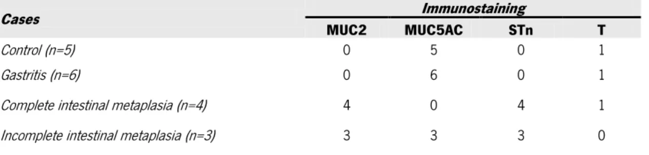
STn and T antigens were shown to be expressed in biopsies of individuals with gastritis, intestinal metaplasia and tissues from gastric carcinoma patients. The proteomic study of serum from individuals with gastritis, intestinal metaplasia and gastric carcinoma revealed the presence of these truncated glycans and lead us to identify some proteins such as acute phase proteins (mainly complement proteins), vitronectin and plasminogen. The detailed mass spectrometry analysis of glycan structures in serum plasminogen from intestinal metaplasia individuals allowed the validation of the STn structure. The overexpression of ST3Gal IV in gastric carcinoma cell line MKN45, induced the expression of type 2 sialylated Lewis structure, SLeX, in cellular and secreted proteins. The SLeX expression lead to a more aggressive in vitro
phenotype of gastric cells, characterized by an increased cellular invasion with increased adhesion capacity to extracellular matrix proteins such as collagen IV and vitronectin. Using the chicken chorioallantoic membrane (CAM) as in vivo model for the evaluation of cancer cell invasion capacity we demonstrated that cells expressing SLeX show increased capacity to
activation we observed increased phosphorylation of Src, FAK and activation of RAC1, RhoA and Cdc42 GTPases contributing for the modulation of cellular biological behavior. The identification of SLeX protein carriers, by a proteomic approach, lead us to identify the carcinoembryonic
antigen (CEA). This result was further confirmed in cells by proximity ligation assay (PLA) and validated by immunoprecipitation. The expression of CEA and SLeX and the presence of
CEA/SLeX was evaluated by immunohistochemistry and PLA in gastric carcinoma tissues, and
revealed that 80.6% of the cases show CEA/SLeX. This result was associated with the presence
of venous invasion of the carcinomas.
Overall, our results give new insights for the application of both plasminogen STn and CEA SLeX glycosylation pattern as serum biomarkers of gastric pathologies, and open new avenues
for future targeted evaluation of these specific glycobiomarkers in additional immunoassay-based approaches. Moreover, we showed that SLeX expression on the surface of malignant cells
Todas as células que constituem as superfícies epiteliais são decoradas por diversos glicoconjugados que estão envolvidos em inúmeros processos celulares, inclusive na progressão para cancro. Aquando da transformação maligna, estas células passam a expressar, à sua superfície, diferentes estruturas de glicanos. A expressão desses glicanos alterados acontece maioritariamente em glicoproteínas e glicolípidos e é caracterizada pela expressão de formas truncadas anormais (antigénios Tn, STn e T), assim como aumento da sialilação de glicanos complexos (antigénio SLea, SLeX). O estudo destas alterações comuns em
cancro, assim como a identificação de proteínas portadoras destes glicanos alterados pode contribuir significativamente para a compreensão do processo de carcinogénese e da progressão tumoral assim como ajudar na descoberta de novos biomarcadores de cancro.
Este estudo teve como objetivos: identificar novos biomarcadores serológicos envolvidos na carcinogénese gástrica através do estudo da expressão de glicanos truncados como STn e T em lesões precursoras de carcinoma gástrico (gastrite e metaplasia intestinal) e em carcinoma gástrico; caracterizar o comportamento biológico de células de carcinoma gástrico que expressam SLeX, avaliar a modelação molecular por ativação de recetores de tirosina cinase em
células que expressam SLeX; e identificar proteínas portadoras de SLeX.
A expressão de estruturas STn e T foi observada em tecidos de biopsias de indivíduos com gastrite e metaplasia intestinal, assim como em casos de carcinoma gástrico. A análise proteómica do soro de indivíduos com carcinoma gástrico e lesões precursoras de carcinoma gástrico (gastrite e metaplasia intestinal) revelou a presença dessas mesmas estruturas, e levou à identificação de proteínas de resposta de fase aguda (maioritariamente proteínas do sistema de complemento), de vitronectina e plasminogénio. O estudo detalhado, por espectrometria de massa, da estrutura de glicanos do plasminogénio sérico levou à validação da presença da estrutura STn em indivíduos com metaplasia intestinal. A sobre-expressão, in vitro, da sialiltransferase ST3Gal IV numa linha celular de carcinoma gástrico, MKN45, induziu a expressão de estruturas Lewis sialiladas do tipo 2, SLeX, em proteínas celulares e secretadas. A
expressão de SLeX levou a um comportamento mais agressivo das células in vitro, caracterizado
pelo aumento da invasão celular assim como maior capacidade de adesão celular a proteínas de matriz como colagénio IV e vitronectina. A aplicação, como modelo in vivo, da membrana corioalantóica (CAM) de embrião de galinha para a avaliação da capacidade invasiva de células
observamos aumento de ativação de c-Met em células que expressam SLeX, um recetor já
descrito como estado envolvido no processo de invasão e metastização celular. Como efeito subsequente à ativação do c-Met, um aumento de fosforilação de Src, FAK e ativação de RAC1, RhoA e Cdc42 GTPases foi observado como participando na modulação do comportamento biológico celular. O estudo proteómico de identificação das proteínas transportadoras de SLeX
identificou o antigénio carcinoembrionico (CEA), e este resultado foi posteriormente confirmado por PLA em células e validado por imunoprecipitação. A presença da co-expressão de CEA e SLeX foi também avaliada por imunohistoquímica e PLA em tecidos de carcinoma gástrico, e
revelou que 80.6% dos carcinomas gástricos apresentam co-expressão de CEA com SLeX. A
co-expressão de CEA com SLeX apresentou associação com invasão venosa dos carcinomas.
Os resultados obtidos poderão contribuir significativamente para a aplicação de alterações de glicosilação de proteínas, como plasminogénio com STn e CEA com SLeX, como possíveis
novos biomarcadores de patologias gástricas, e abrem novas portas para o desenvolvimento de novos testes sorológicos que avaliem estas alterações. Adicionalmente, este trabalho permitiu demonstrar que a expressão de SLeX em células tumorais está associada com a invasão
CHAPTER 1
General Introduction General View Of Cancer Incidence And Mortality
Gastric Cancer - General Aspects Epidemiology Perspective Gastric Carcinogenesis Host Genetic Susceptibility
Helicobacter pylori Virulence Factors Gastric Cancer Classification Glycans: Widespread Molecules That Decorate Cell Surfaces
Glycosylation
Glycosylation The Most Diverse Post-Translational Modification Of Proteins The Biosynthetic Pathway of N-Glycosylation
Biosynthesis of Mucin Type O-Glycosylation Aberrant Glycosylation In Cancer
Aberrant Glycosylation in Gastric Cancer Biomarkers For Cancer Detection
Glycoproteomic Advances in Gastric Cancer Biomarker Discovery Glycan-Based Serological Assays in Cancer
References
CHAPTER 2
Aims and Objectives
CHAPTER 3
Results
3.1 Glycoproteomic Analysis of Serum From Patients With Gastric Precancerous Lesions 3.2 Expression of ST3GAL4 Leads to SLeX Expression and Induces c-Met Activation and
an Invasive Phenotype in Gastric Carcinoma Cells
3.3 CEACAM5 Carcinoembryonic Antigen Carries SLeX in Gastric Carcinoma
Cells - Implications for Diagnosis Improvement
CHAPTER 4
General Discussion
Glycoproteomics For Discovery Of New Cancer Biomarkers
Serum Glycoproteomics for Biomarker Finding in Gastric Lesions Serum Plasminogen Glycan Characterization in Gastric Lesions
Modulation Of Gastric Cellular Glycophenotype By Sialyltransferases Overexpression: Biological Behavior And Biomarker Identification
Biological Role of SLeX Expression, due to Overexpression of ST3Gal IV, in Gastric Cancer Cells
References Summary and Conclusions
CHAPTER 5
Other Contributions
5.1 Alterations in Glycosylation as Biomarkers for Cancer Detection 5.2 ST6GalNAc-I Controls Expression of Sialyl-Tn Antigen in Gastrointestinal Tissues 5.3 Challenging the Limits of Detection of Sialylated Thomsen-Friedenreich Antigens By In-Gel Deglycosylation and Nano-LC-MALDI-TOF-MS
APPENDIX
Chapter 1
General Introduction
Content
General View Of Cancer Incidence And Mortality Gastric Cancer - General Aspects
Epidemiological Perspective Gastric Carcinogenesis Host Genetic Susceptibility
Helicobacter pylori Virulence Factors Gastric Cancer Classification
Glycans: A Widespread Molecules That Decorates Cell Surfaces
Glycosylation
Glycosylation The Most Diverse Post-Translational Modification Of Proteins
The Biosynthetic Pathway of N-glycosylation Biosynthesis of Mucin Type O-glycosylation
Aberrant Glycosylation In Cancer
Aberrant Glycosylation in Gastric Cancer
Biomarkers For Cancer Detection
Glycoproteomic Advances in Gastric Cancer Biomarker Discovery Glycan-Based Serological Assays in Cancer
GENERAL VIEW OF CANCER INCIDENCE AND MORTALITY
Cancer is a leading cause of death worldwide (Twombly 2005) and a tremendous effort has been made to improve health by developing new approaches for early diagnosis, treatment and prevention.
In 2008, it was estimated an overall incidence of more than 12 million new cancer cases resulting in approximately 8 million cancer deaths (Ferlay et al., 2010). Lung cancer remains the most frequent cancer worldwide, presenting both higher incidence and mortality rates. The ranking, in terms of incidence, is followed by breast cancer, the second most common cancer overall, colorectal cancer, stomach cancer, prostate cancer and liver cancer. Regarding mortality, gastric cancer is the second cause of cancer death worldwide followed by liver cancer, colorectal cancer and breast cancer.
GASTRIC CANCER - GENERAL ASPECTS
Epidemiological Perspective
The International Agency for Research on Cancer (IARC), a part of the World Health Organization (WHO), often estimates the cancer incidence and mortality in broad areas of the world and more recently it provides a more detail estimate comprising the country level through the GLOBOCAN series.
According to the last estimate, gastric cancer is one of the main cause of cancer death worldwide, especially in developing countries (Ferlay et al., 2010). About one million new cases were estimated to have occurred (989,000 cases, 7.8% of the total), making it currently the fourth most common cancer malignancy in the world. This estimative differs significantly from the first in 1975, when stomach cancer was globally the most common neoplasm (Parkin et al., 1984; La Vecchia et al., 2010). Incidence of gastric cancer in developing countries represents more than 70% of total cases, and half of the world total cases occurs in Eastern Asia, mainly in China (Ferlay et al., 2010). In addition, gastric cancer is the second leading cause of cancer death in both sexes worldwide and the highest mortality rates are estimated in Eastern Asia with Central and Eastern Europe, and Central and South America also present high mortality rates. Nonetheless, the lowest mortality rate is observed in Northern America.
Besides these GLOBOCAN perspectives, there is also estimates of cancer burden in Europe. Overall, it was estimated about 3 million new cancer cases diagnosed and 1.7 million cancer
deaths in Europe in 2006 (Ferlay et al., 2007). This study estimated that overall, the most frequent cancer types were breast cancer, followed by colorectal cancers, lung cancer, prostate cancer and gastric cancer. In terms of mortality, lung cancer was the most common cause of death from cancer, followed by colorectal cancer, breast cancer, gastric cancer and prostate cancer. Higher incidence and mortality rates are still recorded in Eastern European countries. This European estimative differs from the GLOBOCAN worldwide estimative and fortunately, gastric cancer incidence and mortality are declining throughout Europe, in both men and women (Boyle et al., 2005; Ferlay et al., 2007).
The incidence of gastric cancer varies from country to country, probably as a result of genetic, epigenetic, and environmental factors. Gastric cancer has been associated with many factors known to contribute to the development and progression of the disease (Crew et al., 2006). Some of the factors thought to be involved in the development of gastric cancer are sex, where gastric cancer occurs twice as often in men; age, where gastric cancer is more common in people over the age of 55 (Yamaoka et al., 2009); race, in the United States of America some studies point for differences in incidence among the different populations (Schlansky et al., 2011) and dietary factors in particular a high salt intake along with the use of nitrate for food preservation (Ramon et al., 1993; Tsugane 2005; Peleteiro et al., 2011). Other related risk factors are fruit and vegetables intake (Riboli et al., 2003; Soerjomataram et al., 2010), smoke habit (La Torre et al., 2009), alcohol consumption (Duell et al., 2011) and high body mass index (Hampel et al., 2005; Kubo et al., 2006). In addition to these environmental factors, Helicobacter pylori, a gram-negative bacterium that is thought to be present in the stomach of half the global population, has also been implicated in gastric carcinogenesis (Peek et al., 2002; Lochhead et al., 2007; Lochhead et al., 2008).
In this regard, the decline in incidence and mortality rates observed in gastric cancer throughout Europe is generally attributed to better food preservation, enhanced nutrition and improved control of Helicobacter pylori infection.
Gastric Carcinogenesis
Based on epidemiologic evidence, the IARC classified in 1994 H. pylori as a class I carcinogen (IARC 1994). Since then, continuous increasing data supports the role of H. pylori in gastric carcinogenesis that was firstly established with a very elegant study with the Mongolian gerbil animal model demonstrating gastric cancer induction by H. pylori infection (Watanabe et al., 1998).
H. pylori colonization is generally acquired during childhood (Malaty et al., 2002; Rowland et al., 2006) but if not treated the infection persists lifelong (Vincent 1995; Blaser et al., 2004; Lehours et al., 2007). The transmission infection mode is reported to be human to human (Graham et al., 1991; Neale et al., 1995), most probably by gastric-oral transmissions associated with gastroenteritis and vomiting and oral-oral or fecal-oral transmission (Leung et al., 1999; Parsonnet et al., 1999; De Schryver et al., 2006; Perry et al., 2006; Solnick et al., 2006).
As referred above, H. pylori infection is considered a major risk factor that together with other environmental factors triggers a cascade of gastric lesions that cause alterations in the gastric mucosa resulting in atrophy of the mucosal barrier which may increase the risk of carcinogenesis in the underlying epithelial layer leading ultimately to gastric cancer (Uemura et al., 2001; Correa et al., 2007). In 1992, Pelayo Correa first described the gastric carcinogenesis pathway characterized by a multiple step process started by H. pylori infection (Correa 1992) (Figure 1).
Figure 1: Gastric carcinogenesis pathway proposed by Pelayo Correa in 1992, characterized by a multi-step process
started by H. pylori infection leading to gastric tissue inflammation that can evolve to atrophy, metaplasia, dysplasia and ultimately gastric cancer.
This multistep model propose that H. pylori infection, in association with other environmental factors, triggers a cascade of events that starts with a chronic inflammation or a chronic gastritis that can evolve to atrophic gastritis with focal loss of glands. As consequence of this atrophy, several changes can occur and the local gastric cells can be replaced by intestinal type cells conferring a tissue intestinal phenotype named intestinal metaplasia. The next step in the cascade is characterized by atypical changes in nuclear morphology and irregular tissue architecture and is called dysplasia (reviewed in Correa et al., 2007). High-grade dysplasia commonly progress to gastric cancer of the more common intestinal subtype, the final step of the cascade (de Vries et al., 2007).
Regarding this well established model, gastric mucosal atrophy is a major risk factor to develop gastric cancer. In addition, different patterns of atrophic gastritis are associated with
different risk of developing gastric cancer. A recent system of gastritis classification, the OLGA (Operative Link for Gastritis Assessment)-staging system, consider both the topography (antral and corpus mucosa) and the extent of gastric mucosa atrophy (score) to determine gastritis-associated gastric cancer risk (Rugge et al., 2008). Taking in consideration this classification, several studies consistently associated increased risk of developing gastric cancer with OLGA stages III/IV (Rugge et al., 2007; Rugge et al., 2010; Rugge et al., 2011; Marcos-Pinto et al., 2013). More recently, a modification in the OLGA system of classification, OLGIM (Operative Link on Intestinal Metaplasia Assessment), was proposed to consider the assessment of intestinal metaplasia instead of atrophy and this new classification has been shown to be more accurate in determine gastric cancer risk (Capelle et al., 2010). These classifications show to be important for the application of follow-up strategies for patient-specific clinico-pathological future evaluations (Dinis-Ribeiro et al., 2012; Marcos-Pinto et al., 2012).
All H. pylori strains have the capacity of promoting gastric inflammation, however most of the infected individuals show few or no symptoms and a very small percentage of H. pylori infected individuals will develop gastric carcinoma (Dooley et al., 1989; Suerbaum et al., 2002; Amieva et al., 2008). Considering this reality, the outcome of H. pylori infection for each individual is difficult to predict since it depends on different aspects such as host genetic susceptibility, strain virulence characteristics and environmental factors.
Host Genetic Susceptibility
It is recognized the importance of host genetics in gastric cancer development and the association with H. pylori infection. The prevalence of a number of genetic polymorphisms in some inflammation-related genes have been demonstrated to be associated with risk of developing gastric lesions, mainly in H. pylori infected individuals.
It has been shown that human genetic pro-inflammatory polymorphisms within the genes interleukin (IL)-1B and IL-1RN are associated with risk for gastric cancer development (El-Omar et al., 2000; Machado et al., 2001; Figueiredo et al., 2002). IL-1B gene encodes for a pro-inflammatory cytokine IL-1β that plays an important role in initiating and amplifying the inflammatory response upon H. pylori infection (Noach et al., 1994). On the other hand, IL-1RN gene encodes for an interleukin 1 receptor antagonist IL-1ra, an anti-inflammatory cytokine, that competitively binds IL-1β receptors modulating the potentially damage effects of IL-1β (Arend et al., 1998). The expression of IL-1B-511*T, IL-1B-31*C and IL-1RN*2 alleles have been associated
with increased IL-1β production (Pociot et al., 1992; Danis et al., 1995; Santtila et al., 1998; Hwang et al., 2002) and have been found to confer an increased risk for the development of gastric cancer (Machado et al., 2001) chronic atrophic gastritis (Furuta et al., 2002) and intestinal metaplasia (Figueiredo et al., 2002; Zambon et al., 2002).
Furthermore, polymorphism in the tumor necrosis factor α (TNF-α) gene and interferon gamma receptor 1 (IFNGR1) has also been associated with increased gastric cancer risk (El-Omar 2001; Machado et al., 2003; Canedo et al., 2008). The pro-inflammatory TNF-α cytokine, encoded by TNF-α gene, has been shown to be increased in gastric mucosa of patients infected by H. pylori (Noach et al., 1994; Arend et al., 1998), and the expression of TNF-α-308*A allele has been associated with increased risk for chronic atrophic gastritis and gastric cancer development (Machado et al., 2003). IFNGR1 is a cytokine receptor for IFNϒ, a cytokine that has been described to play a pivotal role in promoting H. pylori induced mucosal inflammation when upregulated (Smythies et al., 2000). The presence of IFNGR1-56*T polymorphism has been reported as a relevant host susceptibility factor for gastric cancer development (Canedo et al., 2008).
In addition, other findings claimed that MUC1 polymorphism present different susceptibility for the development of gastric lesions (Silva et al., 2001) and gastric carcinoma (Carvalho et al., 1997). It was also reported that H. pylori adhesion to gastric cells depends on the size of the MUC1 variable number tandem repeats (VNTR) domain (Costa et al., 2008).
Helicobacter pylori Virulence Factors
Although the final clinical outcome is dependent also on host and environmental factors, it was also observed that not every H. pylori strain resulted in similar damage for the host, leading to the distinction between high and low pathogenic strains. Thus, it was possible to identify bacterial features that confer increased risk to develop gastric disease (van Doorn et al., 1998; Figueiredo et al., 2005).
The presence of cytotoxin-associated gene pathogenicity island (cagPAI) was associated with more severe disease outcomes and was considered to be involved in the development of atrophic gastritis, peptic ulcer disease and gastric carcinoma (Blaser et al., 1995; Kuipers et al., 1995; Figueiredo et al., 2002; Nomura et al., 2002), a feature that has been well established in animal models (Ogura et al., 2000; Wiedemann et al., 2009). Bacterial strains that are cagPAI positive (cagPAI+, more virulent strains), are characterized by containing genes that encode a type IV
secretion system, which functions as a needle that permits the insertion of bacterial products into the host cells (Censini et al., 1996; Tanaka et al., 2003) as well as the gene encoding the CagA protein, one of the proteins delivered to epithelial cells through this system (Backert et al., 2000). Once inserted in epithelial cells, CagA protein associates with proteins from the tight junction complexes, leading to loss of epithelial integrity (Amieva et al., 2003) and its phosphorylation on tyrosine residues by Src family kinases promotes the interaction with several cell signal-transduction pathways resulting in cytoskeleton rearrangement, motility, proliferation and apoptosis (Segal et al., 1999; Asahi et al., 2000; Odenbreit et al., 2000; Stein et al., 2000). The presence of an intact type IV secretion system as well as CagA protein phosphorylation is also associated with the production of interleukin (IL)-8 in gastric cells (Brandt et al., 2005; Figueiredo et al., 2005; Lai et al., 2011).
Several other virulent factors have been characterized and described such as the vacuolating cytotoxin gene (vacA) that encodes a toxin that induce epithelial cell damage by cell vacuolization, membrane channel formation, disruption of endosomal/lysosomal function, apoptosis, and immunomodulation (Cover et al., 1992; Cover 1996; reviewed in Cover et al., 2005).
Additional, H. pylori virulence factors can comprise genes that encodes for blood group antigen binding adhesin (BabA) (Ilver et al., 1998) and sialic acid-binding adhesin (SabA) (Mahdavi et al., 2002). The expression of these adhesin proteins in the surface of H. pylori strains are involved with the capacity of the bacteria to colonize gastric mucosa by adhering to epithelial cells through host antigens receptors. H. pylori adhesion to the gastric epithelial cells constitutes a crucial step for gastric mucosa colonization and establishment of a successful infection, because it provides protection from clearance mechanisms such as liquid flow, peristaltic movements or renewal of the mucous layer. The host antigens receptors involved in bacteria colonization are glycan structures present on proteins or glycolipids in the surface of the host gastric epithelial cells namely H-type 1 and Lewis b (Leb) for Bab A adhesin (Boren et al., 1993;
Ilver et al., 1998) and Sialyl Lewis X (SLeX) and Sialyl Lewis A (SLea) for SabA adhesin (Johansson
et al., 2005; Walz et al., 2005; Aspholm et al., 2006). H. pylori has the capacity of modulate the expression of these two adhesins to achieve a long-term colonization, according to the host antigen receptors expression. For this reason, H. pylori take advantage of expressing SabA adhesin in inflamed gastric tissues that is characterized by increased expression of sialylated structures (Ota et al., 1998; Mahdavi et al., 2002). Infection with H. pylori that expresses BabA is associated with increased epithelial proliferation and inflammation, increased risk for duodenal
ulcer, gastric atrophy, intestinal metaplasia, and gastric adenocarcinoma (Gerhard et al., 1999; Prinz et al., 2001; Yu et al., 2002).
Gastric Cancer Classification
Most of gastric cancers are adenocarcinomas that occur in the lining of the stomach and account for nearly 90% of the total cases. About 2-7% of the gastric malignancies are lymphoma of mucosa-associated lymphoid tissue (MALT) (Wotherspoon 1998), and the remainder percentage includes very rare cases of gastric stromal tumors (sarcomas) developed from the muscle or connective tissue of the stomach wall (Duffaud et al., 2003), and carcinoid tumors (Zhang et al., 2011).
Different stages of gastric adenocarcinoma are characterized by the capacity of invasion deeply into the stomach wall extending to nearby organs such as lymph nodes, abdominal cavity, liver, pancreas, esophagus or intestine (Washington 2010). Furthermore, gastric carcinoma cells released by the primary tumor into lymphatic and/or blood circulation can spread into distant target organs (lungs, brain, ovaries, bones) forming secondary tumors (a process also called metastization) (Kemp et al., 2010; Ahn et al., 2011; Yamanishi et al., 2011).
Several classification systems have been proposed to aid in the description of gastric cancer based on microscopic pattern (Ming, Carneiro, and Goseki). The two most commonly used are the World Health and Organization (WHO) Lauren systems. According to WHO, gastric carcinomas are classified in adenocarcinoma, papillary carcinoma, tubular carcinoma, mucinous carcinoma and signet ring-cell carcinoma. However Lauren's classification is the predominant histological classification worldwide that divides gastric carcinoma in two major subtypes - intestinal type and diffuse type carcinomas (Lauren 1965) although considering an third subtype designated unclassified or mixed type carcinoma. Nevertheless, intestinal type and diffuse type carcinomas don't differ only by histological appearance, but also by epidemiology, pathogenesis, and genetic profiles differences (Shah et al., 2011). Morphologically, these two types of gastric carcinoma differ in the intercellular adhesion molecules that are well preserved in intestinal-type tumors and defective in diffuse carcinomas. In intestinal-type carcinoma, cells are still attached in tubular or glandular formations (similar to adenocarcinomas arising in the intestinal tract - hence their designation as intestinal-type); in contrast to the lack of adhesion molecules in diffuse carcinomas allows individual tumor cells to grow and invade neighboring tissues without the formation of tubules or glands. Moreover, the molecular basis of these two carcinoma types is also different. In the diffuse-type carcinoma the main carcinogenic event is the loss of function of E-cadherin, a key
cell surface protein for intercellular adhesion and maintenance of the organization of epithelial tissues. Impairment of E-cadherin function occurs through biallelic inactivation of its related gene CDH1 via germline or somatic mutation, allelic imbalance events (loss of heterozygosity), epigenetic silencing of gene transcription through aberrant methylation of the CDH1 promoter (Oliveira et al., 2006; Carneiro et al., 2008; Yamamoto et al., 2011; Corso et al., 2012), or by modulation of E-cadherin N-glycosylation (Pinho et al., 2011). In the intestinal-type carcinoma the main carcinogenic event is H. pylori infection followed by a cascade of events where several molecular changes occur leading to transformation of gastric cells into intestinal type cells (as mentioned above).
Although H. pylori associated preneoplastic lesions are a feature of intestinal-type gastric cancer, some evidences claimed that there is an association between H. pylori infection and diffuse-type gastric cancer (Handa et al., 1996; Uemura et al., 2001). The association of H. pylori infection and diffuse-type gastric cancer is observed during the progression of atrophic gastritis in individuals presenting active gastritis (Sipponen et al., 1992; Solcia et al., 1996). Conversely, in hereditary diffuse gastric cancer this association is not observed (Carneiro et al., 2004).
Besides gastric carcinoma, H. pylori infection in gastric mucosa is also associated with the development of mucosa-associated lymphoid tissue (MALT), increasing the risk of gastric MALT lymphoma (Parsonnet et al., 1994; Sagaert et al., 2010). This involvement is support by the fact that most of the patients with low-grade MALT-lymphoma are infected with H. pylori, and that, in most of these cases, eradication of H. pylori infection results in tumor regression (Thiede et al., 1997; Zullo et al., 2010; Kuo et al., 2012).
Briefly, there are several factors that contribute for gastric disease development. As mentioned above, also glycan antigen receptors expressed in the surface of gastric epithelial cells contribute for the development of gastric diseases.
GLYCANS: WIDESPREAD MOLECULES THAT DECORATE CELL
SURFACES
It is amazing to observe that all living cells present a vast collection of glycan structures that decorate cell surfaces (Varki et al., 2009a; Varki 2011). Glycan expression on intracellular and cell surfaces proteins and lipids are implicated in numerous biological functions (Varki 1993). Oligosaccharide moieties are involved in cell-cell and cell-matrix interactions and recognition, intra-
and intercellular protein trafficking, pathogen infection, immuno responses and recognition of self, modulation of protein function and signaling, as well as altering the dynamics of glycoprotein endocytosis and regulation of proper maturation and folding of newly synthesized proteins (Varki 1993; Moremen et al., 2012)
Glycosylation
Glycosylation is characterized by the enzymatic covalent attachment of a carbohydrate to a polypeptide, lipid, polynucleotide, carbohydrate, or other organic compound catalyzed by glycosyltransferases using specific sugar nucleotide donor substrates; in opposition to the non-enzymatic chemical reaction of glycation.
The synthesis of a glycan structure is a very complex and organized process that involves a huge number of genes including those that code for glycosyltransferases, glycosidases and chemical chaperons, as well as the enzymes responsible for nucleotide sugars synthesis and transport (Rini et al., 2009). Furthermore, the availability and localization of the nucleotide sugar donors can compromise glycosyltransferases activity in the synthesis of a glycan structure.
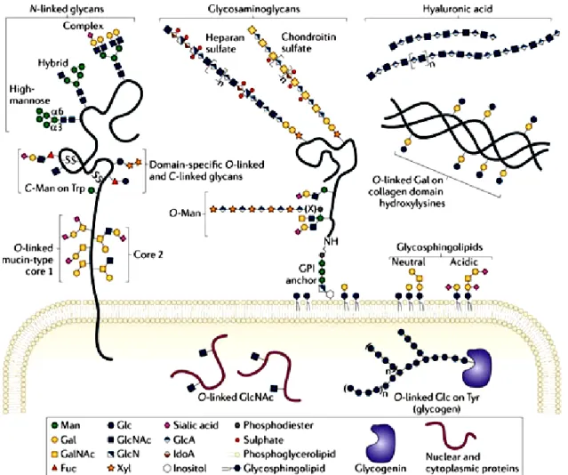
The human genome contains about 250 to 500 genes that are involved in the glycan assembly, like those that code for proteins involved in the synthesis and degradation of glycans or transport of sugar donors, accounting for approximately 2% of the total human genome (Schachter et al., 2009). Generally the enzymatic attachment of glycans occurs in non-carbohydrate molecules forming the glycoconjugates. Glycoconjugates comprise a different class of molecules such as glycosphingolipids, glycosaminoglycans present as free polysaccharides or as part of proteoglycans, glycoproteins and glycosylphosphatidylinositol-linked proteins (Figure 2).
Glycan structures can be attached to a lipid structure forming glycolipids. Almost all glycolipids in vertebrates are glycosphingolipids (GSLs) that belongs to the sphingolipid family (Schnaar et al., 2009). GSLs are characterized by the linkage of a glycan structure to an sphingolipid core structure called ceramide. The biosynthesis of GSLs occurs by a stepwise addition of sugars first to ceramide, typically in a β-linked galactose (galactosylceramide-GalCer) or glucose (glucosylceramide-GlcCer), and then to the arising glycan. GlcCer synthesis starts in the cytoplasmic face of the endoplasmic reticulum (ER) and early Golgi apparatus and is further elongated by a series of glycosyltransferases in the Golgi lumen. Conversely, GalCer synthesis occur on the ER lumen and then go through the Golgi, where it may be sulfated to form sulfatide. The elongation process leads to a variety of different combinations that subclassify GSLs as
neutral (no charged sugars or ionic groups), sialylated (having one or more sialic acid residues) generally known as gangliosides, or sulfated. GSLs are found embedding in the cell membranes, due to hydrophobic properties of the lipid tail, contributing for the maintenance of cell membranes integrity and clustered in lipid rafts contributing for cell signaling processes (Schnaar et al., 2009).
Figure 2: Common classes of animal glycan structures.The major classes of animal glycans are shown, with an emphasis on typical vertebrate sugar chains. Most glycans on membrane and secreted proteins are found in N-linkage to Asn or in O-linkage to Ser/Thr.O-linked glycans are classified by their initiating monosaccharide. Addition of GalNAc initiates mucin-type O-linked glycans and extension with Gal, GlcNAc or GalNAc produces eight different core structures. Man linked initiates another class of O-linked glycan (O-Man glycans), as well as Fuc residues (O-Fuc glycans). O-linked GlcNAc is found on the extracellular domains of some proteins and on numerous cytosolic and nuclear proteins. Furthermore, O-linked glycan structures can be found attached to other amino acids, including Glc residue to Ser and Tyr residues of glycogenin, and Gal on hydroxylysine of collagen domains. Also glycosaminoglycans (GAGs) are O-linked glycans initiated by a conserved tetrasaccharide (GlcA-β1,3-Gal-β1,3-Gal-β1,4 Xyl-β) and classified by the composition of their disaccharide repeat that are usually found attached to proteins forming proteoglycans. A GAG-like polymer that forms hyaluronic acid is the only glycan that is not linked to a protein or lipid. In addition to proteins, sphingolipids can be modified by glycosylation, which are ceramide-linked glycans.The monosaccharide abbreviations are Man, Mannose; Gal, Galactose; GalNAc, N-acetylgalactosamine; Fuc, Fucose; Glc, Glucose; GlcNAc, N-acetylglucosamine; GlcN, Glucosamine; Xyl, Xylose; GlcA, Glucuronic acid; and IdoA, Iduronic acid. Adapted from (Moremen et al., 2012).
Glycosaminoglycans (GAGs) are larger linear polysaccharides, formed by repeated disaccharide building blocks composed of an amino sugar (N-acetylglucosamine-GlcNAc, glucosamine-GlcN that is variously N-substituted, or N-acetylgalactosamine-GalNac) and either an uronic acid (glucuronic acid-GlcA or iduronic acid-IdoA) or galactose (Gal). GAGs can be found as free polysaccharides, such as hyaluronic acid, or as part of proteoglycans. Proteoglycans are characterized by one or more GAGs covalently attached to a protein core. GAGs are attached to proteins by a conserved tetrasaccharide (GlcA-β1,3-Gal-β1,3-Gal-β1,4 Xyl-β) to Ser residues in an O-linkage that will be further elongated with disaccharide repeats giving rise to two different classes: chondroitin sulfate and heparan sulfate (Esko et al., 2009). Besides proteoglycans, GAGs can also be found as a free polysaccharide such as hyaluronic acid. Hyaluronic acid is the only glycosaminoglycan synthesized in the plasma membrane, with the growing polymer being secreted into the extracellular environment. The synthesis of hyaluronic acid is catalyzed by hyaluronan synthases (HAS) and consists of repeating disaccharides composed of GlcNAc and GlcA. Hyaluronic acid is the only glycan that is not linked to a protein or lipid (Hascall et al., 2009). Proteoglycans and glycosaminoglycans can be found on the cell surface, inside the cell, and in the extracellular matrix (ECM) and account for many functions like promoting cell adhesion to ECM; binding of cytokines, chemokines and growth factors; acting as receptors for proteases and as coreceptors for various and tyrosine kinase growth factors receptors (Esko et al., 2009).
As mentioned above, proteins can also be glycosylated giving rise to glycoproteins. Glycoproteins show a huge heterogeneity in its glycan structure that can be usually attributed to a non template driven biosynthetic process in endoplasmic reticulum and Golgi compartment, and a lack of any proofreading machinery. In addition, the composition of the final glycan structure relies on the polypeptide backbone as well as a number of variable factors such as the expression levels of glycosidases and glycosyltransferases and the availability of substrates, which fluctuate during cell growth, differentiation and development (Schwientek et al., 2002; Varki et al., 2009b; Du et al., 2010; Fernandez-Valdivia et al., 2011). There are several types of glycan linkage on proteins that present different glycan biosynthesis and composition wich will be discussed with detail in the next section.
GLYCOSYLATION THE MOST DIVERSE POST-TRANSLATIONAL
MODIFICATION OF PROTEINS
Newly formed proteins synthesized in the ER, can be further decorated with one or more biochemical moieties, a process so called post-translational modification (PTM), giving rise to a huge protein heterogeneity. Several types of modifications can occur in proteins and the understanding of the extent and pattern of these PTMs gives insight into the function and dynamics of the proteome.
PTM can be classified in two main categories: the first include the covalent addition of chemical groups by enzymatic catalysis, the second comprises the cleavage of peptide backbones by the action of proteases or autocatalysis. Many modifications resulting in the addition of chemical group to an amino acid residue can be found in proteins, and the major types of protein covalent modifications are phosphorylation, acetylation, glycosylation, methylation, and ubiquitylation. These covalent addition are classified according to the type of amino acid side residue involved, the class of the enzyme implicated in the process and the degree of reversibility (Walsh et al., 2005). Among these major types of PTMs, glycosylation is the most diverse and complex modification that occurs in proteins, with at least one half of the known proteins estimated to be glycosylated (Apweiler et al., 1999). This type of modifications strongly influences many of the protein functional aspects, including cellular localization, turnover and protein quality control (Fukuda et al., 1989; Parodi 1999; Arnold et al., 2007). Protein glycosylation can be classified into several types according to the glycan linkage site: N-linked glycosylation, O-linked glycosylation, C-linked glycosylation (C-mannosylation), Phospho-linked glycosylation or phosphoglycosylation and glycophosphatidylinositol (GPI)-anchored glycosylation or glypiation (Spiro 2002; Moremen et al., 2012) (Figure 2).
Proteins at the cell surface can be integrated in the cell membrane by a GPI-anchored moiety, called GPI-anchored proteins firstly described in 1985 (Ferguson et al., 1985). Proteins are attached to GPI via their carboxyl termini through a phosphodiester linkage of phosphoethanolamine to a trimannosyl-non acetylated glucosamine (Man3-GlcN) core (Man(α1– 2)Man(α1–6)Man(α1–4)GlcN), and can be found in the outer leaflet of the lipid bilayer facing the extracellular environment. The reducing end of GlcN is linked to phosphatidylinositol (PI) which is then anchored by another phosphodiester linkage to the cell membrane through its hydrophobic region and the distal, nonreducing mannose residue is attached to the protein via an
ethanolamine phosphate (EtNP) bridge between the C-6 hydroxyl group of mannose and the α-carboxyl group of the carboxy-terminal amino acid. The Man3-GlcN core oligosaccharide core can suffer various modifications during all the process of secretion from the cell (Ferguson et al., 2009). GPI-anchored proteins have been described as having a critical role in a variety of receptor mediated signal transduction pathways, adhesion, and antigenicity (Maeda et al., 2011).
Another type of protein glycosylation is the phosphoglycosylation, the most abundant protein glycosylation in parasites. This type of glycosylation is characterized by the enzymatic addition of sugar residues to serine (Ser) residues in the polypeptide chain, through a phosphodiester linkage. The presence of phosphoglycans in proteins, more specifically GlcNAcα-1-P, was firstly described in 1980 by Gustafson and Milner in endopeptidase Proteinase I isolated from Dictyostelium discoideum (Gustafson et al., 1980), but it was only in 1995 that this linkage was demonstrated to be in Ser residues, catalyzed by UDP-GlcNAc:Ser protein N-acetylglucosamine-1-phosphotransferase (Ser:GlcNAc phosphotransferase) and the process called phosphoglycosylation (Freeze et al., 1995). Since that, other sugar residues were found to be linked to Ser residues by a phosphodiester linkage such as Mannose (Man) residues (Ilg et al., 1994) or Xylose (Xyl) (Haynes et al., 1996). A specific role for phosphoglycoproteins is not yet elucidated, however some studies associate phosphoglycoproteins with the immunogenicity of the parasites (Cooper et al., 1993; Ilg et al., 1993).
Moreover, mannose (Man) carbon-carbon (C-C) linkages to the C2 position of tryptophan (Trp) residues were also described and named C-mannosylation (Hofsteenge et al., 1994; de Beer et al., 1995; Loffler et al., 1996). This type of protein glycosylation can be found in cells from a variety of mammals (Krieg et al., 1997), and involves a protein O-mannosyltransferase (POMT) that uses dolichyl-phosphate-mannose as a precursor (Doucey et al., 1998), know to occur in a consensus sequence -W-x-x-W/F- (x could be any amino acid) in which the first Trp residue becomes mannosylated. Sequences presenting alternative phenylalanine (Phe) to Trp residue show a reduced efficiency of 3.5-fold in C-mannosylation (Krieg et al., 1998).
In addition to the above described types of glycosylation, there are two main types of protein glycosylation in eukaryotes, the N- and the O-linked glycans. N- glycosylation is described by the addition of a sugar precursor to a nitrogen group of an asparagine (Asn) amino acid. In contrast, O-glycosylation is characterized by the addition of monosaccharides to a hydroxyl group of Ser, threonine (Thr) and tyrosine (Tyr) residues; and, to a lesser extent, to hydroxyproline and hydroxylysine (Spiro 2002). The different classes of O-glycans are known to be dependent on
the first added sugar (Brockhausen et al., 2009), including α- or β-linked O-Gal (Seyer et al., 1977), β-linked O-GlcNAc (Hart 1997), α-linked O-Man (Endo 1999), α-linked O-Fuc (Harris et al., 1993; Hofsteenge et al., 2001), α- or β-linked O-Glc (Harris et al., 1993), β-linked O-Xyl (GAGs) (Kresse et al., 1994; Lin 2004) and the most abundant form of O-linked glycosylation in higher eukaryotes α-linked O-GalNAc also known as mucin type O-glycans (Van den Steen et al., 1998). The structural complexity of the chains initiated by O-linked GalNAc is very high, exceeding that of other O-linked and N-linked chains. In addition, there is the O-GlcNac glycosylation that is the only type of protein glycosylation, mainly found on intracellular nucleocytosolic proteins. This type of glycosylation is characterized by a reversible process comprising a single GlcNAc residue added to Ser and/or Thr, and is important in the modulation of the biological activity of intracellular proteins (Holt et al., 1986), often competing with phosphorylation (Wang et al., 2008).
The Biosynthetic Pathway of N-glycosylation
In all eukaryotes, the core pathway for the establishment of N-linked glycosylation is well conserved (Stanley et al., 2009b). This process mainly takes place in the endoplasmic reticulum (ER) and further processing and rebuilding of the N-glycans occurs in the Golgi compartment generating a large diversity of possible structural outcomes (Dennis et al., 2009; Varki et al., 2009c). N-glycosylation is considered an important protein posttranslational modification in eukaryotic cells given the high occurrence of N-glycans in glycoproteins. It is estimated that about 90 % of all glycoproteins carry N-linked glycans with an average of 1.9 N-linked glycans per polypeptide chain (Apweiler et al., 1999).
Sugars that constitute N-glycan structures are covalently attached to the proteins at asparagine (Asn) residues by an N-glycosidic bond, and five different N-glycan linkages have been reported, of which N-acetylglucosamine to asparagine (GlcNAcβ1-Asn) is the most common (Stanley et al., 2009b). This N-glycosylation process starts in the ER and is characterized by a linkage to Asn residue, via a common trimannosyl chitobiosyl pre-assembled core sugar structure. The assembled of this core structure can occur whenever a consensus sequence Asn-X-Ser/Thr is present in the nascent protein, where X can be any amino acid with the exception of proline. Less commonly, the sequence Asn-X-Cys can also be used to construct N-glycan structures (Gavel et al., 1990; Sato et al., 2000).
The biosynthesis of the core trimannosyl chitobiosyl structure, starts in the cytosolic surface of the ER membrane, and is characterized by a stepwise addition of two N-acetylglucosamines (2
GlcNAc) and five mannoses (5xMan) to dolichylphosphate (Dol-P) giving rise to Dol-P-P-GlcNAc2Man5 (Helenius et al., 2001; Helenius et al., 2004). Thereafter, the Dol-P-P-GlcNAc2Man5
structure is "flipped" to the luminal side of ER membrane where four mannose and three glucose additional residues are added forming the final core structure Dol-P-P-GlcNAc2Man9Gluc3. The substrate donors for the addition of the last four Man and three Glc are the Man and Dol-P-Glc, also made on the cytoplasmic face of the ER and “flipped” onto the luminal face. Each of the sugar additions is catalyzed by a specific glycosyltransferase located on both sides of the ER membrane (Helenius et al., 2001; for a review see Stanley et al., 2009b).
The transfer of the complex core structure from the Dol-P-P-GlcNAc2Man9Gluc3 donor to the
nascent polypeptide backbone takes place in the ER lumen and is catalyzed by the oligosaccharyltransferase (OST) enzyme complex (Helenius et al., 2001; Helenius et al., 2004; Stanley et al., 2009b). Following the covalent attachment of the core oligosaccharide to Asn residues, a series of processing reactions trims the N-glycan in the ER. First, the three Glc residues (the terminal α1–2Glc and the two inner α1–3Glc) are removed sequentially by α-glucosidases I and II, followed by one mannose removal by ER α-mannosidade which specifically removes the terminal α1–2Man from the central arm of Man9GlcNAc2. These initial steps are
known to be important in regulating glycoprotein folding, a process that is mediated by the interaction between enzymes and chemical chaperons in the ER that recognize specific features of the trimmed glycan. At this point, misfolded proteins can be recognized and targeted for ER degradation by two different quality control processes, the calnexin/calreticulin system and the ER degradation-enhancing α-mannosidase I–like protein (EDEM). Calnexin (membrane-bound) and calreticulin (soluble) sequester the newly synthesized glycoprotein and act as molecular chaperones by a deglucosylation reglucosylation cycle promoting correct folding, preventing aggregation of folding intermediates, blocking premature oligomerization, and by facilitating formation of native disulfide bonds (Stanley et al., 2009b). Another group of proteins that are believed to interact with the deglucosylated chain and help in the correct folding of glycoproteins are EDEMs. Little is known about EDEMs functions, however evidence suggests that EDEMs have catalytic activity and that overexpression enhances misfolded glycoprotein degradation (Freeze et al., 2009). During this quality control process, if glycoproteins fail to fold or oligomerize properly they are eventually retrotranslocated to the cytoplasm and destroyed by N-deglycosylation and proteasomal degradation, a process called ER-associated degradation (ERAD).
When correctly folded, glycoproteins leave the ER and travel through the Golgi compartment where several steps of removal and addition of sugars occur by the action of several glycosidases and glycosyltransferases, creating a variety of glycan structures. In the majority of the multicellular organisms, glycoproteins entered in the cis-Golgi compartment and trimming of α1–2Man residues is carry on by the action of α1–2 mannosidases, originating the Man5GlcNAc2
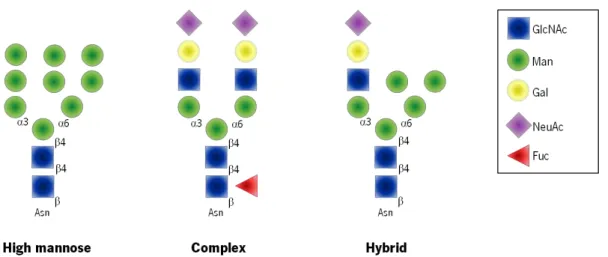
intermediate glycan structure for the synthesis of hybrid and complex N-glycans. Further processing reactions for the biosynthesis of hybrid and complex N-glycans are initiated in the medial-Golgi and are pursued in the trans-Golgi (Helenius et al., 2001). In the end, N-glycan structures share a common pentasaccharide core region (Manα1–6(Manα1-3)Manβ1– 4GlcNAcβ1–4GlcNAcβ1-Asn) that can be classified into three main classes: high-mannose type, complex type and hybrid type (Figure 3).
The biosynthesis of these three different glycan structures occurs in a stepwise manner by the action of several enzymes such as N-acetylglucosaminyltransferases, galactosyltransferases, sialyltransferases and fucosyltransferases, in a very well-orchestrated fashion (Stanley et al., 2009b). In brief, the high mannose type structure is characterized by the presence of only mannose residues attached to the pentasaccharide core structure; complex type has two antennae or branches initiated by the addition of two GlcNAc residues catalyzed by N-acetylglucosaminyltransferases (GnTs); and hybrid type in which mannose residues are attached to the Manα1–6 arm and a GlcNAc residue to the Manα1–3 arm of the core structure (Helenius et al., 2001; Helenius et al., 2004; Stanley et al., 2009b).
Figure 3: Types of N-glycans. N-glycans added to protein at Asn-X-Ser/Thr sequons, that shear a common core Man3GlcNAc2Asn, are of three general types in a mature glycoprotein: High mannose, Complex, and Hybrid.
The enzymes responsible for the synthesis of complex structures in N-glycans are common to the elongation process of the mucin type O-glycosylation, and occur both in the Golgi compartment. For this reason, the terminal protein glycosylation can be very similar between N- and O-glycans.
Biosynthesis of Mucin Type O-glycosylation
In contrast to N-glycosylation, the O-glycosylation is a stepwise process with monosaccharide added incrementally, beginning with the addition of a single sugar residue to either a Ser or Thr amino acids in the protein backbone and then to the nascent sugars in a process called elongation. O-linked glycosylation is a relatively late-stage event in the protein maturation, and involves a large set of enzymes localized at the Golgi compartment (Hanisch 2001).
The most ubiquitous type of initial O-glycosylation is the one formed by the addition of GalNAc (O-GalNAc) to Ser or Thr, also described as mucin type glycosylation. The addition of the first monosaccharide residue on protein backbones is mediated by a family of UDP-GalNAc:polypeptide GalNAc-transferases (ppGalNAc-Ts) and occurs in the Golgi compartment (Roth et al., 1994; Clausen et al., 1996; Rottger et al., 1998; Bennett et al., 2012; Gerken et al., 2013). Although most protein glycosylation events are controlled by one or two genes encoding the enzymes responsible for the initiation step of glycosylation, human mucin-type O-glycosylation is controlled by a large family of up to 20 homologous genes encoding ppGalNAc-Ts, fifteen of which were confirmed to be expressed and functionally active (Clausen et al., 1996; Ten Hagen et al., 2003; Tian et al., 2009; Bennett et al., 2012). The addition of GalNac residues by ppGalNAc-Ts is not dependent on a defined consensus recognition sequence and in theory, any Ser or Thr residues can be O-glycosylated. However, some aspects have been identified that allow an improved prediction of the polypeptide O-glycosylation sites (Hansen et al., 1995; Julenius et al., 2005; Gerken et al., 2011). Some authors claimed that the initiation of GalNAc glycosylation by ppGalNAc-Ts is ruled by the sequence context of putative O-glycosylation sites. Each ppGalNAc T isoform may be uniquely sensitive to peptide sequence and overall charge, which together dictates the substrate sites that will be glycosylated (Clausen et al., 1996; Kato et al., 2001; Gerken et al., 2006; Wandall et al., 2007; Gerken et al., 2011). Moreover, there are evidences for a dynamic regulatory mechanism of the initial GalNAc addition, either by competition for different substrate sites or by competition of ppGalNAc-Ts with the glycosyltransferases responsible for the formation of core glycans (Hanisch et al., 1999; Gill et al., 2011). Also very interesting is that tissues show specific ppGalNAc-Ts enzyme expression
(Bennett et al., 1998; Bennett et al., 1999; Mandel et al., 1999; Gomes et al., 2009). Overall, the cellular repertoire of glycosyltransferases, with their distinct donor and acceptor sugar specificities, their localizations in sub-compartments of the Golgi and their sequential action will dictate the cell's specific O-glycosylation profile.
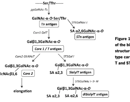
In mucin type glycosylation the addition of the first monossacharide residue by ppGalNAc-Ts, which transfer a GalNAc residue from a sugar donor UDP-GalNAc to Ser/Thr residues of the acceptor protein, gives rise to the formation of the Tn antigen (GalNAc-Ser/Thr) (Figure 4). A two-step model of O-GalNAc biosynthesis is proposed, where a subset of GalNAc-Ts add GalNAc at low density to the polypeptide chain, preferentially with unglycosylated Ser/Thr residues or those containing GalNAc-Ser/Thr flanking the active site; and another subset of GalNAc-Ts that catalyze the addition of GalNAc residues to Ser/Thr adjacent to the existing GalNAc-Ser/Thr sites (reviewed in Gill et al., 2011). This initial step regulates the site and level of occupancy of total O-glycan modification in the target proteins, which will be then further elongated giving rise to the global final glycan chain structure. Extension of the GalNAc residue can generate eight different cores (Figure 4) and cores 1 to 4 are the most common in humans. Elongation of the GalNAc residue can be employed by C1GalT enzyme or T synthase, originating the core 1 structure, also known as T antigen (Galβ1-3GalNAc-Ser/Thr); or by C3GnT making the core 3 structure (GlcNAcβ1-3GalNAc-Ser/Thr) (Figure 4). Further elongation of these core 1 and core 3 structures is performed by C2GnT family of enzymes that catalyze the specific addition of a β1,6GlcNAc originating the corresponding core 2 and core 4 structures (Figure 4). Therefore, the C2GnT1 and C2GnT3 function to synthesize the core 2 structure, whereas C2GnT2 catalyzes the core 4 synthesis (reviewed in Gill et al., 2011).
Additional core extension can occur in the GlcNAc moiety of the core 2 and core 4 O-glycans in two different ways: addition of a simple Gal alone by β3/4GalT enzymes or by addition of poly-lactosamine repeats (Gal-GlcNAc) through the concerted action of β3/4GalT and β3/4GnT enzymes. Conversely, the core 3 GlcNAc moiety is only extended through the addition of Gal by β4GalT4 or β3GalT5. The extension of core structures by the action of β3/4 Gal-Ts and β3/4 Gn-Ts leads to type 1 and type 2 chains formation, where type 1 chains are characterized by the β1,3 linkage of the Gal residue to the GalNAc, while in type 2 chains this linkage is β1,4. Both chains are frequently terminated by Lewis-type blood group-related antigens. Finally, all O-glycan are capped to terminate O-glycosylation through addition of sialic acid by ST3Gal- and ST6Gal-sialyltransferases.
Figure 4: Schematic representation of mucin type O-glycosylation biosynthesis pathway. The image
depicts the biosynthetic pathway of mucin type glycosylation that can generate eight different cores structures (with cores 1 to 4 the most common in humans), and are extended giving rise to a vast diversity of structures. For image simplicity only representative enzymes were included. Gray boxes correspond to cancer-associated antigens.
As mentioned above, there are four additional core O-glycans structures (core 5–8) that have been biochemically characterized in tissues. They are thought to be generated through direct