ISSN 0100-879X
BIOMEDICAL SCIENCES
AND
CLINICAL INVESTIGATION
www.bjournal.com.br
www.bjournal.com.br
Volume 45 (1) 1-92 January 2012
Braz J Med Biol Res, January 2012, Volume 45(1) 25-32
doi:10.1590/S0100-879X2011007500159
Effect of thymosin alpha-1 on subpopulations of Th1, Th2, Th17,
and regulatory T cells (Tregs)
in vitro
Xia Yang, Feng Qian, Hai-Yang He, Kai-Jun Liu, Yuan-Zhi Lan, Bing Ni, Yi Tian, Xiao-Lan Fu, Ji
Zhang, Zi-Gang Shen, Jian Li, Yi Yin, Jin-Tao Li and Yu-Zhang Wu
Institutional Sponsors
The Brazilian Journal of Medical and Biological Research is partially financed by
Faculdade de Medicina de Ribeirão Preto Campus
Ribeirão Preto
Ex plor e H igh - Pe r for m a n ce M S Or bit r a p Te ch n ology I n Pr ot e om ics & M e t a bolom ics
Effect of thymosin alpha-1 on
subpopulations of Th1, Th2, Th17, and
regulatory T cells (Tregs)
in vitro
Xia Yang
1*, Feng Qian
2*, Hai-Yang He
1, Kai-Jun Liu
1, Yuan-Zhi Lan
2,
Bing Ni
1, Yi Tian
1, Xiao-Lan Fu
1, Ji Zhang
1, Zi-Gang Shen
1,
Jian Li
1, Yi Yin
1, Jin-Tao Li
1and Yu-Zhang Wu
11Institute of Immunology, Third Military Medical University, Chongqing, PR China 2Department of General Surgery, Southwest Hospital, Third Military Medical University, Chongqing, PR China
Abstract
Thymosin alpha 1 (Tα1) has been shown to have beneficial effects on numerous immune system parameters, but little is known about the effects of Tα1 on patients with gastric carcinoma. The objective of this study was to determine the effect of Tα1 on subpopulations of Th1, Th2, Th17, and regulatory T cells (Tregs) in vitro, and to evaluate its efficacy as an immunoregulatory
factor in patients with gastric carcinoma. We compared the effect of Tα1 on the frequency of CD4+ and CD8+ T cells,
espe-cially the CD4+CD25+Foxp3+ Tregs in peripheral blood mononuclear cells (PBMCs) from gastric carcinoma patients (N = 35) and healthy donors (N = 22). We also analyzed the changes in the proliferation of PBMCs in response to treatment with Tα1, and examined the production of Th1, Th2, and Th17 cytokines by PBMCs and tumor-infiltrating lymphocytes. The treatment of PBMCs from gastric cancer patients, with Tα1 (50 µg/mL) alone increased the percentage of CD4+CD25+Foxp3+ (suppressive antitumor-specific Tregs) from 1.68 ± 0.697 to 2.19 ± 0.795% (P < 0.05). Our results indicate that Tα1 increases the percentage of Tregs and IL-1β, TNF-α, and IL-6 in vitro.
Key words: Tα1; Gastric carcinoma; Tregs; Th1; Th2; Th17
Introduction
Correspondence: Jin-Tao Li and/or Yu-Zhang Wu, Institute of Immunology, Third Military Medical University, 30 Gaotanyan Street, Shapingba District, Chongqing 400038, China. E-mail: ljtqms@yahoo.com.cn and/or wuyuzhang20006@sohu.com
*These authors contributed equally to this study.
Received June 21, 2011. Accepted November 16, 2011. Available online December 2, 2011. Published January 16, 2012.
Gastric cancer is the most lethal among malignant
diseases worldwide. Over half of gastric cancer cases are
reported to occur in developing countries, especially in China (1). Gastric cancers are highly resistant to chemotherapy
and radiotherapy; the most common intervention is surgical
resection of the tumor with concomitant lymphadenectomy. Regrettably, many clinical studies have shown that surgical
resection is associated with an increased risk of recurrence,
postoperative complications, and mortality (2). As such, the outcome of surgery for gastric cancer remains very poor, and clinical research has focused on developing more effective adjunct therapies. Some efforts have been made to identify enhanced chemotherapeutic agents to improve the patient’s quality of life, to extend survival, and to decrease the duration of illness while the patient reaches remission. Recently, a promising new series of chemotherapeutic agents, including
various platinum compounds, fluoropyrimidine derivatives,
and S-1 (tegafur, 5-chloro-2,4-dihydropyrimidine, and potas-sium oxonate) (3), have been described. However, these remain to be fully characterized and clinically validated.
There remains a need to seek innovative therapies to treat
and resolve gastric cancer.
The cytotoxic action of host T and natural killer (NK)
cells suggests that these cells may inherently harbor ef-fective antitumor properties. However, tumors employ a
wide variety of evasive mechanisms to fight the antitumor immune responses mounted by the host (4-6), so that an
effective immune response is down-regulated and tolerance to the tumor is established. Among the mechanisms used by tumors to escape host immunity are the production of
cytokines such as TGF-β (7), IL-10 (6,8), IL-17 (9,10), and
perturbation or destruction of immune cells. In addition, recent study has implicated an immunopathological role of
tumor-medi-26 Xia Yang et al.
ated anti-host defense (11,12), leading to tumor tolerance. Therefore, the objective of most cancer immunotherapies is to provide essential immunogenic elements to boost
tumor-specific immunity and to eliminate those elements
that lead to tumor tolerance.
Certain biological response modifiers (BRMs) are
thought to be essential for the induction of protective and
therapeutic antitumor immunity. Thymosin alpha 1 (Tα1)
was described in the 1970s as a clinically applicable BRM
for the treatment of immunodeficiencies and cancers. Tα1
is a polypeptide hormone with immunoregulatory proper-ties that is expressed by various tissues and organs of mammals. Consisting of 28-amino acid residues that are
N-terminally acetylated, Tα1 is proteolytically processed from prothymosin α (13). Tα1 has been shown to have beneficial effects on numerous immune system parameters
(14), including increasing the antitumor activity of dendritic
cells (15), and macrophages (16). Today, Tα1 is being tested
in late-stage clinical trials for the treatment of melanoma (17). Although the preliminary results from the clinical trials
appear to be promising, little is known about the effects of Tα1 on the proliferation of suppressive antitumor-specific
Tregs in patients with gastric carcinoma.
In this study, we investigated the ability of Tα1 to
modify various immune cell subpopulations in peripheral
blood mononuclear cells (PBMCs) and tumor-infiltrating
lymphocytes (TILs) from patients with gastric carcinoma.
We compared the effect of Tα1 on the frequency of CD4+
and CD8+ T cells, especially the CD4+CD25+Foxp3+ Tregs
found in PBMCs from gastric carcinoma patients and healthy donors. Moreover, we analyzed the changes in the
proliferation of PBMCs in response to treatment with Tα1.
Finally, we examined the production of Th1, Th2, and Th17
cytokines by PBMCs and TILs.
Material and Methods
Patients
PBMCs or TILs were studied in a group of 35 adult
patients (19 females and 16 males) aged 22-70 years (av
-erage age: 47.6 ± 11.2) diagnosed with gastric carcinoma.
A donor control group consisted of 22 healthy subjects (12
females and 10 males) aged 23-68 years (average age: 41.5 ± 10.4). The pathological characteristics of all the
patients were analyzed by the Department of Pathology (Southwest Hospital, Chongqing, China) between 2008 and 2010, which included classifying their histological sub-type and staging their tumor-node-metastasis (TNM). The protocol was approved by the Institutional Ethics Board of Southwest Hospital, Third Military Medical University, and written informed consent was obtained from all patients prior to enrollment.
PBMC isolation
PBMCs were isolated according to manufacturer
instructions by centrifugation at 1500 g for 20 min on a
Ficoll-Hypaque gradient (Sigma, USA). The phase contain-ing the white blood cells was retained and washed three
times with RPMI 1640 medium supplemented with 10%
heat-inactivated fetal calf serum (Gibco Invitrogen, Italy).
The PBMCs were diluted to 1 x 10⁶ cells/mL for analysis.
TIL isolation
TILs were isolated from tumor tissues by enzymatic digestion as previously described (18). In brief, after surgical resection, the tissues maintained under sterile conditions
were finely minced and enzymatically digested in RPMI 1640 U/mL hyaluronidase V, 30 U/mL DNase I, 300 U/mL collagenase type IV (all from Sigma), and antibiotics (100 U/mL penicillin, 100 µg/mL streptomycin, and 100 µg/mL
gentamycin, all from Life Technologies, USA). After digestion
for 3-4 h at room temperature, the suspension was filtered through a 25-µm nylon filter. The filtrate was separated on
a Ficoll-Hypaque gradient to isolate the TILs and tumor cells, after which the cells were washed three times with
PBS supplemented with 5% FBS (19).
Cell culture
The PBMCs and TILs were treated with various
con-centrations of Tα1 as described (20). Briefly, the cells were incubated with RPMI 1640 medium supplemented with 10% heat-inactivated fetal calf serum and recombinant human IL-2 (100 U/mL) in 96-well, round-bottom plates at 37°C in an atmosphere of 5% CO2 for 72 h. The PBMCs
obtained from patients were cultured with or without Tα1
as detailed below. Cell yield and viability were evaluated using the Trypan blue dye exclusion method. Cells were then harvested and the supernatants were collected and
stored at -80°C for cytokine analysis.
Flow cytometry
All antibodies, the fixation/permeabilization concentrate
and diluent, and the permeabilization buffer were purchased from eBiosciences (eBioscience, Inc., USA). After culturing for 72 h, the cells were collected and washed twice with PBS. The cells were stained with PE-Cy5.5-conjugated anti-human CD4 monoclonal antibody (mAb), FITC-conjugated anti-human CD8 mAb, and PE-conjugated anti-human CD25 mAb or the appropriate isotype control mAbs in 100
µL PBS for 45 min at 4°C. These antibodies were used at a concentration of 20 μL (0.125 µg) per 106 cells in a total staining volume of 100 µL. Positive controls for APC,
FITC, PE-Cy5.5, and PE staining were also included in the analysis. The cells were then washed twice with PBS, and subjected to intracellular staining using APC-conjugated anti-human Foxp3 mAb according to manufacturer
instruc-tions. The antibody was used at a concentration of 20 µL (0.125 µg) per 106 cells in a 100-µL total staining volume. The
cells were then resuspended in permeabilization buffer and
(BD Biosciences, USA) using the Cell Quest software.
Cytokine secretion in the culture supernatants was assessed with a human Th1/Th2 11plex multiplex kit and a human IL-17A simplex kit (both from Bender MedSys -tems, Austria) according to manufacturer instructions. The
concentrations of IL-1β, IL-2, IL-4, IL-5, IL-6, IL-10, IL-12, IFN-γ, TNF-α, TNF-β, and IL-17A were determined by flow cytometry. Briefly, 25 µL culture supernatants or cytokine
standards was aliquoted into tubes, and multiplex antibody beads were added to each sample. For detection, biotin-conjugated multiplex antibodies were added to the samples, and these were incubated at room temperature and in the
dark for 2 h and then washed to remove unbound proteins.
The samples were incubated at room temperature in the
dark for 1 h with streptavidin-PE solution. After washing, the
samples were resuspended in PBS and 10,000 events were
counted per sample for measurement by flow cytometry on a BD FACSAria instrument (Becton, Dickinson and Company, USA). The data for each cytokine were acquired using the
FlowCytomix Software (Bender MedSystems).
[3H]-thymidine analysis
Sixteen hours before collecting the cells, 1 µCi [3
H]-thymidine was added to each well and adequately mixed. The radioactivity emitted by the cells in the individual wells
was measured using an LS 6500 liquid scintillation counter (Beckman) and was reported as counts per min (cpm).
Statistical analysis
Data are reported as means ± SD and were analyzed sta
-tistically by the Student t-test (SPSS 13.0, SPSS Inc., USA).
P < 0.05 was considered to be statistically significant.
Results
Tα1 promotes the generation of CD4+CD25+Foxp3+
(Tregs) from PBMCs. We further investigated the effect of
Tα1 on Tregs, which prevent host antitumor immune re -sponses. The PBMCs from patients with gastric cancer and
healthy donors were co-cultured in Tα1-containing medium.
The frequencies of Tregs were determined by measurement
of CD4+, CD25+, and Foxp3+ molecular markers, which are
most commonly associated with Treg phenotypes. There
was no significant change in the percentage of CD4 in
PBMCs isolated from healthy people and cancer patients
in the presence or absence of Tα1 (P > 0.05; data not
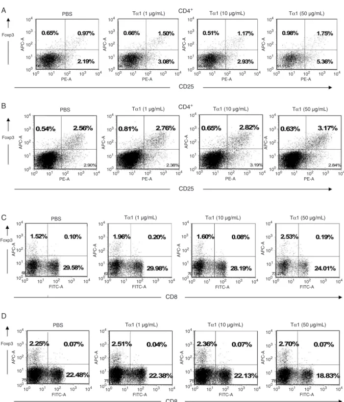
shown). As shown in Figure 1A, CD4+CD25+Foxp3+ levels
in Tα1-treated PBMCs from healthy donors were higher than that in Tα1-untreated PBMCs. The mean percentage
of CD4+CD25+Foxp3+ Tregs in PBMCs from healthy donors
was found to be increased from 1.45 ± 0.638% under PBS treatment to 2.22 ± 0.401% under Tα1 (50 µg/mL) treatment (P < 0.05). As shown in Figure 1B, the mean percentage
of CD4+CD25+Foxp3+ Tregs in PBMCs from patients with
gastric cancer was statistically increased from 1.68 ± 0.697%
under PBS treatment to 2.19 ± 0.795% under Tα1 (50 µg/ mL) treatment (P < 0.05). No significant differences were observed either at baseline or in the changes of HIV pa
-tients induced by Tα1 treatment, in terms of CD4 and CD8 lymphocyte subset changes at week 12 (21). These results indicate that Tα1 treatment can induce CD4+CD25+Foxp3+
lymphocyte expansion, suggesting that the Tα1-induced
Treg expression subsequently could hinder the antitumor action mediated by the host immune response. The effect
of Tα1 on the expression of CD4+CD25+Foxp3+ Tregs has
not been reported.
Moreover, the CD8+ and Foxp3+ markers are known
to be involved in another Treg (CD8+Foxp3+)
subpopula-tion. In our analysis, CD8+Foxp3+ T cells did not exhibit
any statistically significant changes in the presence or absence of Tα1 on PBMCs from healthy donors (Figure 1C) and patients (Figure 1D; P > 0.05). Therefore, these results showed that Tα1 treatment was able to increase the
CD4+CD25+Foxp3+ Treg, but not the CD8+Foxp3+ Treg
differentiation. The effect of Tα1 in terms of CD8+ Foxp3+
Treg expression has not been reported.
Tα1 elevates the levels of the inflammatory cytokines IL-1β and TNF-α in the PBMCs from gastric cancer patients. We used cytokine-FACS analysis to detect the effects of Tα1 on Th1 cytokine production. PBMCs and TILs were stimulated with Tα1 at various concentrations, and the se
-cretion of IL-1β, IL-2, IL-12 (p70), IFN-γ, TNF-α, and TNF-β
was subsequently measured. The data in Figure 2, shows
that the production of IFN-γ in PBMCs from healthy donors
was higher than in PBMCs from gastric cancer patients.
Treatment with Tα1 at various concentrations resulted in no significant changes of IFN-γ production in PBMCs from either donors or patients (P > 0.05). No IFN-γ secretion was detected in TILs from patients. Production of IL-1β in
PBMCs from healthy donors was lower than in PBMCs from
patients. Treatment with 1 µg/mL Tα1 caused an increase to 178% in the secretion of IL-1β by PBMCs from patients compared to those untreated (100%; P < 0.05). In contrast, 1 µg/mL Tα1 induced secretion of TNF-α to more than 500%
in PBMCs from patients compared to other various
treat-ment conditions. However, Tα1 decreased TNF-α levels
in PBMCs from healthy donors under various treatment
conditions (P < 0.05). The IL-2, IL-12 (p70), and TNF-β secretion profiles in PBMCs from patients and donors or TILs from patients were not significantly different under any
condition (data not shown).
Tα1 elevates the levels of IL-6 Th2-cytokines in PBMCs. Cytokine profiles were assessed after stimulation with Tα1 and Th2 cytokines (including IL-4, IL-5, IL-6, and IL-10). As shown in Figure 3, Tα1 treatment caused no significant
changes in IL-10 secretion by PBMCs from patients and
healthy donors (P > 0.05). The level of IL-10 was close to 0 pg/mL in TILs from patients. Tα1 showed no significant
changes in secretion of IL-10 in TILs from gastric cancer
28 Xia Yang et al.
10 µg/mL increased the secretion of IL-6 to over 140% in PBMCs from both healthy donors and patients (P < 0.05).
The IL-4 and IL-5 secretion levels were undetectable in PBMCs from patients and donors or TILs from patients under the various treatment conditions (data not shown).
Tα1 seems to be inconsistent in inducing the secretion
of IL-17A. To further confirm the role of Tα1 in Th17 cytokine secretion in PBMCs, IL-17A was examined by flow cytom
-etry analysis. As shown in Figure 4, we found that Tα1 at concentrations of 10 µg/mL decreased IL-17A secretion to 42% in PBMCs from healthy donors (P < 0.05), but there were no significant changes detected in the secretion of
IL-17A in PBMCs from patients. IL-17A in TILs from patients
was increased by 60% with 10 µg/mL Tα1, compared to PBS-treated cells (100%; P < 0.05). These data suggest that Tα1 might reduce the secretion of IL-17A in cells from healthy subjects. However, Tα1 did not decrease the se -cretion of IL-17A in PBMCs from gastric cancer patients.
Tα1, therefore, has an inconsistent function in inducing the
secretion of IL-17A.
Figure 2. Effect of Tα1 on Th1 cytokine secretion in PBMCs from healthy donors (HD) and gastric cancer patients (GCa) and in TILs from gastric cancer patients. PBMCs and TILs were cultured with PBS or 1, 10, or 50 µg/mL Tα1 for 72 h. The levels of Th1 cytokines were determined using the Bio-Plex cytokine assay. PBMCs = peripheral blood mononuclear cells; TILs = tumor-infiltrating lymphocytes. Data are reported as means ± SD for 3 separate experiments (P < 0.05, t-test).
Figure 3. Effect of Tα1 on IL-6 cytokine secretion in PBMCs from healthy donors (HD) and gastric cancer patients (GCa), and in TILs from gastric cancer patients. PBMCs and TILs were cultured with PBS or 1, 10, or 50 µg/mL Tα1 for 72 h. The levels of Th2 cytokines were determined using the Bio-Plex cytokine assay. PBMCs = peripheral blood mononuclear cells; TILs = tumor-infiltrating lymphocytes. Data are reported as means ± SD for 3 separate experiments (P < 0.05, t-test).
30 Xia Yang et al.
Tα1 had no significant effect on T lymphocyte prolifera
-tion. To investigate the impact of Tα1 on PBMC proliferative
capacity, PBMCs from healthy individuals and patients with
gastric cancer were incubated with Tα1 at various concentra -tions. Cell proliferation was measured by the incorporation
of [3H]-thymidine. Tα1 caused no significant change in the
proliferative capacity of PBMCs from healthy donors and gastric cancer patients when compared to PBS-treated
cells (P > 0.05).
Discussion
Recent studies have demonstrated that some immune
cells such as T and NK cells produce specific cytokines,
which normally function as suppressors of tumors. Similarly,
one of the key elements sought to develop effective tumor
immunotherapies is the means by which to induce effective host-derived immune effectors in the tumor
microenviron-ment. These immune cells and cytokines influence tum
-origenesis, including growth and metastasis. One of the
main mechanisms for immune elimination of tumor cells is the release (exocytosis) of cytolytic granules by cytotoxic
CD8+ T lymphocytes (CTLs) and NK cells, a process that
is mediated by adequate contact and antigenic recognition of tumor cells. Still another pathway requires the interaction of Fas ligands on the surface of CTLs with Fas molecules present on target tumor cells. However, it has been dem-onstrated that the interaction between target and NK cells can be prevented by negative signaling, causing NK cells to lose their cytotoxic capacity and to become functionally
“exhausted”. Moreover, IL-15, IL-2, IFN-α, and IL-21 (22)
have been demonstrated to promote NK- and CD8+ T
cell-mediated cytotoxicity, to sustain function, and to increase
antitumor immunity. Animal studies have shown that Tα1
had no effect on T cell numbers (both CD4+ and CD8+),
and therefore, had no effect on T lymphocyte proliferation
(23). Therefore, our in vitro data are consistent with this
finding.
Control of tumor growth depends on the clearance of the tumor by antitumor immune responses, and on effectively
abrogating immunological unresponsiveness. CD4+CD25+
Tregs have been shown to functionally contribute to the maintenance of immune tolerance and prevention of automunity. However, Tregs can also prevent host antitumor
im-mune responses.Removal of CD4+CD25+ Tregs has been
shown to be effective for boosting tumor-specific immunity
in vivo in a mouse model (24). In humans, tumor Tregs appear to exert immunosuppressive effects on the prolifera-tion and activaprolifera-tion of immune effector cells, increasing the
growth of tumors in vivo and reducing the survival rate of
individuals with ovarian cancer (12). CD4+CD25+ Tregs are
one of the active weapons utilized by tumors to circumvent host immunity. When Tregs were incubated together with
CD56+ NK cells, the NK cells lost their cytotoxic function
(5). Depletion of Tregs in tumors can evoke specific host
immunity, representing an attractive anticancer strategy.
Conversely, it could be beneficial for the control of autoim
-mune disorders. In a CLP septic mouse model, Tα1 was
found to decrease the percentage of CD4+CD25+Foxp3+
T lymphocytes (25). The present study revealed that Tα1
can increase levels of Tregs in PBMCs from patients with
gastric cancer, suggesting that Tα1 could increase the risk
of patients to develop tumor immunotolerance.
Other immune effectors, such as cytokines and chemok
-ines, are known to play either positive or negative roles
in cancer immunity in the tumor microenvironment. Much
attention has been given recently to defining the cytokine secretion profiles of immune cells since they are involved in antitumor immunity. IL-6 has been documented as a pleiotropic cytokine, a potent pro-angiogenic molecule that
stimulates T lymphocyte proliferation and differentiation.
Furthermore, IL-6 is also thought to be a tumor cell growth
factor for tumor cell proliferation, which contributes to drug
resistance, cachexia, and other problems (26).
In addition, IL-10 can act as an inhibitory cytokine, play -ing an important role in the regulation of T cell responses and
tumor-immune escape effects (27). IFN-α and its receptor, IFNR-α, are involved in inhibiting angiogenesis in tumor
rejection mediated by CD4+ or CD8+ T cells (28,29). It has
been reported that IL-4 levels were significantly higher in HBV patients, while IL-10 levels were significantly lower with Tα1 treatment compared to controls (20). Tα1 was documented to increase the secretion of IL-10 by 53% in Panc-1 pancreatic cells. However, Tα1 did not cause an
increase of IL-10 in BxPC-3 cells, another pancreatic cancer
cell line (30). These data suggest that the Tα1-stimulating
effect on the secretion of IL-10 is variable.
Previous reports have indicated that Tα1 did not result in any significant differences in IFN-γ concentrations be
-tween HBV patients and controls at baseline conditions. However, Tα1 significantly increased the concentration of IL-2 of HBV-infected patients compared with healthy donors (20). Some reports have indicated that treatment of Tα1 increased serum IL-1β levels, but not IL-2 levels, in rats (31). Our findings suggest that Tα1 at some concentration can elevate levels of the inflammatory cytokines IL-1β and TNF-α in the PBMCs from patients. Previous research has suggested that the elevation of serum IL-6 and TNF-α in
patients with prostate cancer correlated with poor survival
(32). We conclude that Tα1 can elevate the levels of IL-1β and TNF-α in the PBMCs from patients, providing preclinical evidence supporting the efficacy of a Tα1 blockade against
malignant gastric carcinoma.
Cytokine IL-17 has been shown to have a potent effect
on tumor cell proliferation and angiogenesis (33). Similarly,
treatment with Tα1 was also found to stimulate secretion
of IL-17 in Panc-1 and BxPC-3 cells (30).
Our data show that Tα1 at different concentrations
can increase the levels of Tregs and the production of
PBMCs, suggesting that different qualitative effects of Tα1
may depend on the concentration. In addition, we found
that Tα1 at a concentration of 10 µg/mL decreased the
secretion of IL-17A in PBMCs from healthy donors.
How-ever, Tα1 did not modify the secretion of IL-17A in PBMCs
from gastric cancer patients. These data indicate that the
effect of Tα1 on the regulation of IL-17A secretion is also inconsistent. Taken together, our results suggest that the effect of Tα1 on regulation of Th17 cytokine secretion is inconsistent. Our data suggest that the effect of Tα1 on the
up-regulation of Tregs in PBMCs from gastric patients is
confirmedwith the changes of secretion of TNF-α, IL-1β, and IL-6. While the data are suggestive, we recognize that
some observed changes are small, and sometimes not
statistically significant. Therefore, these results need to be confirmed in larger studies.
Acknowledgments
Research supported by the National Natural Science
Foundation of China (#30972716).
References
1. Parkin DM, Bray F, Ferlay J, Pisani P. Global cancer statis -tics, 2002. CA Cancer J Clin 2005; 55: 74-108.
2. Sah BK, Chen MM, Yan M, Zhu ZG. Reoperation for early postoperative complications after gastric cancer surgery in a Chinese hospital. World J Gastroenterol 2010; 16: 98-103.
3. Boku N, Yamamoto S, Fukuda H, Shirao K, Doi T, Sawaki A, et al. Fluorouracil versus combination of irinotecan plus cis-platin versus S-1 in metastatic gastric cancer: a randomised phase 3 study. Lancet Oncol 2009; 10: 1063-1069. 4. Curiel TJ, Wei S, Dong H, Alvarez X, Cheng P, Mottram P,
et al. Blockade of B7-H1 improves myeloid dendritic cell-mediated antitumor immunity. Nat Med 2003; 9: 562-567.
5. Wolf AM, Wolf D, Steurer M, Gastl G, Gunsilius E, Grubeck-Loebenstein B. Increase of regulatory T cells in the periph-eral blood of cancer patients. Clin Cancer Res 2003; 9:
606-612.
6. Bhairavabhotla RK, Verm V, Tongaonkar H, Shastri S, Din -shaw K, Chiplunkar S. Role of IL-10 in immune suppression in cervical cancer. Indian J Biochem Biophys 2007; 44:
350-356.
7. Toutirais O, Chartier P, Dubois D, Bouet F, Leveque J, Catros-Quemener V, et al. Constitutive expression of TGF-beta1, interleukin-6 and interleukin-8 by tumor cells as a major component of immune escape in human ovarian carcinoma. Eur Cytokine Netw 2003; 14: 246-255.
8. Singh H, Jain M, Sachan R, Mittal B. Association of TNFA (-308G>A) and IL-10 (-819C>T) promoter polymorphisms with risk of cervical cancer. Int J Gynecol Cancer 2009; 19: 1190-1194.
9. Wang L, Yi T, Kortylewski M, Pardoll DM, Zeng D, Yu H. IL-17 can promote tumor growth through an IL-6-Stat3 signaling pathway. J Exp Med 2009; 206: 1457-1464.
10. He D, Li H, Yusuf N, Elmets CA, Li J, Mountz JD, et al. IL-17 promotes tumor development through the induction of tumor promoting microenvironments at tumor sites and myeloid-derived suppressor cells. J Immunol 2010; 184: 2281-2288.
11. Khazaie K, von Boehmer H. The impact of CD4+CD25+ Treg on tumor specific CD8+ T cell cytotoxicity and cancer. Semin Cancer Biol 2006; 16: 124-136.
12. Curiel TJ, Coukos G, Zou L, Alvarez X, Cheng P, Mottram P, et al. Specific recruitment of regulatory T cells in ovarian carcinoma fosters immune privilege and predicts reduced survival. Nat Med 2004; 10: 942-949.
13. Haritos AA, Goodall GJ, Horecker BL. Prothymosin alpha: isolation and properties of the major immunoreactive form of thymosin alpha 1 in rat thymus. Proc Natl Acad Sci U S A
1984; 81: 1008-1011.
14. Hsia J, Sarin N, Oliver JH, Goldstein AL. Aspirin and thy -mosin increase interleukin-2 and interferon-gamma produc -tion by human peripheral blood lymphocytes. Immunophar-macology 1989; 17: 167-173.
15. Shrivastava P, Singh SM, Singh N. Effect of thymosin alpha 1 on the antitumor activity of tumor-associated macrophage-derived dendritic cells. J Biomed Sci 2004; 11: 623-630.
16. Shrivastava P, Singh SM, Singh N. Antitumor activation of peritoneal macrophages by thymosin alpha-1. Cancer Invest
2005; 23: 316-322.
17. Goldstein AL, Goldstein AL. From lab to bedside: emerging clinical applications of thymosin alpha 1. Expert Opin Biol Ther 2009; 9: 593-608.
18. Roman S, Petrusca D, Moldovan I, Paraoan M, Petrescu A, Damian D, et al. Evaluation of apoptosis of tumor and of apparently normal cells in human renal carcinoma. Immunol Lett 1999; 67: 15-22.
19. Shimizu Y, Weidmann E, Iwatsuki S, Herberman RB, Whi -teside TL. Characterization of human autotumor-reactive T-cell clones obtained from tumor-infiltrating lymphocytes in liver metastasis of gastric carcinoma. Cancer Res 1991; 51:
6153-6162.
20. Loggi E, Gramenzi A, Margotti M, Cursaro C, Galli S, Vitale G, et al. In vitro effect of thymosin-alpha1 and interferon-alpha on Th1 and Th2 cytokine synthesis in patients with eAg-negative chronic hepatitis B. J Viral Hepat 2008; 15: 442-448.
21. Chadwick D, Pido-Lopez J, Pires A, Imami N, Gotch F, Villacian JS, et al. A pilot study of the safety and efficacy of thymosin alpha 1 in augmenting immune reconstitution in HIV-infected patients with low CD4 counts taking highly active antiretroviral therapy. Clin Exp Immunol 2003; 134: 477-481.
22. Yi JS, Du M, Zajac AJ. A vital role for interleukin-21 in the control of a chronic viral infection. Science 2009; 324: 1572-1576.
23. Hadden JW, Verastegui E, Hadden E. IRX-2 and thymosin alpha1 (Zadaxin) increase T lymphocytes in T lymphocy-topenic mice and humans. Ann N Y Acad Sci 2007; 1112: 245-255.
32 Xia Yang et al.
immunity by removing CD25+CD4+ T cells: a common ba-sis between tumor immunity and autoimmunity. J Immunol
1999; 163: 5211-5218.
25. Wan J, Shan Y, Shan H, Li G, Wang T, Guan J, et al. Thymosin-alpha1 promotes the apoptosis of regulatory T cells and survival rate in septic mice. Front Biosci 2011; 17: 3004-3013.
26. Zaki MH, Nemeth JA, Trikha M. CNTO 328, a monoclonal antibody to IL-6, inhibits human tumor-induced cachexia in nude mice. Int J Cancer 2004; 111: 592-595.
27. Urosevic M, Dummer R. HLA-G and IL-10 expression in human cancer - different stories with the same message. Semin Cancer Biol 2003; 13: 337-342.
28. Qin Z, Blankenstein T. CD4+ T cell-mediated tumor rejection involves inhibition of angiogenesis that is dependent on IFN gamma receptor expression by nonhematopoietic cells. Im-munity 2000; 12: 677-686.
29. Qin Z, Schwartzkopff J, Pradera F, Kammertoens T, Se
-liger B, Pircher H, et al. A critical requirement of interferon gamma-mediated angiostasis for tumor rejection by CD8+ T cells. Cancer Res 2003; 63: 4095-4100.
30. Li M, Feurino LW, Li F, Wang H, Zhai Q, Fisher WE, et al. Thymosinalpha1 stimulates cell proliferation by activating ERK1/2, JNK, and increasing cytokine secretion in human pancreatic cancer cells. Cancer Lett 2007; 248: 58-67. 31. Yao W, Zhu Q, Yuan Y, Qiao M, Zhang Y, Zhai Z. Thymosin
alpha 1 improves severe acute pancreatitis in rats via regula-tion of peripheral T cell number and cytokine serum level. J Gastroenterol Hepatol 2007; 22: 1866-1871.
32. Michalaki V, Syrigos K, Charles P, Waxman J. Serum levels of IL-6 and TNF-alpha correlate with clinicopathological fea -tures and patient survival in patients with prostate cancer. Br J Cancer 2004; 90: 2312-2316.