UNIVERSIDADE DE LISBOA FACULDADE DE CIÊNCIAS
Socially driven changes in neural and behavioural plasticity in zebrafish
Doutoramento em Biologia (Etologia)
Magda Cristina Teles
Tese orientada por: Prof. Doutor Rui F. Oliveira Prof. Doutor Paulo Jorge Fonseca
Documento especialmente elaborado para a obtenção do grau de doutor
NOTA PRÉVIA
Para a elaboração da presente dissertação, e nos termos do nº1 Artigo 41, do regulamento de Estudos Pós-Graduados da Universidade de Lisboa, publicado no Diário da República nº 209, II Série de 30 de Outubro de 2006, foram usados integralmente dois (2) artigos científicos publicado, três (3) submetidos para publicação em revistas internacionais indexadas e um capítulo de um livro. Tendo os trabalhos mencionados sido efectuados em colaboração, o autor da dissertação esclarece que em todos os trabalhos participou na recolha e análise de dados, discussão dos resultados e redação dos manuscritos.
_______________________________
This dissertation was developed with the support of the Fundação para a Ciência e a Tecnologia, PhD fellowship (SFRH/BD/44848/2008) and EXCL/BIA-ANM/0549/2012 project, and by the EU “Copewell” project (Grant no: 265957).
Agradecimentos
O presente trabalho não teria sido possível sem a ajuda de muitas pessoas que passaram pela minha vida ao longo destes anos, e a todas elas gostaria de expressão a minha gratidão.
Ao meu orientador, Professor Doutor Rui F. Oliveira que me tem guiado nesta longa caminhada e inspirado com a sua determinação, clareza de ideias e principalmente com a sua paixão pela ciência. É uma pessoa incansável que eu admiro muito (de coração).
Ao Professor Doutor Paulo Fonseca, que aceitou a co-orientação deste projeto mostrando-se sempre disponível para ajudar.
Ao Professor Doutor David Gonçalves, pelas discussões na elaboração dos trabalhos mas principalmente por me ter apresentado o mundo dos peixes, vais ser sempre o meu chefinho!
Ao Professor Doutor Daniel Peterson e Letia Peterson, pela sua hospitalidade durante a minha permanência no seu laboratório e à Sarah e Laura pela companhia e programas organizados.
A todos os colegas que fizeram ou fazem parte deste grupo, obrigada pela amizade e simpatia de todos durante estes anos.
À Olinda, uma das melhores pessoas que já cruzaram a minha vida, obrigada pelas discussões, troca de ideias e pelo teu entusiasmo todas as vezes que falávamos das Tilápias (não foram poucas).
Ao Miguel (o meu Mi), que me acompanhou sempre até mesmo à distância, obrigada por seres sempre optimista e estares sempre presente.
À Sara... nem tenho palavras pela ajuda e força que me tem dado nesta fase final (terrível), não é uma amiga de sempre mas será para sempre de certeza! Estou cá para ti!
À Leonor, que é realmente uma fonte de inspiração e de força.
Ao meu núcleo de amigos pelo apoio e amizade ao longo destes anos, obrigada por compreenderem que eu nunca podia ir, era mesmo verdade.
À minha família, especialmente aos meus pais e minha irmã pelo vosso amor e apoio incondicional a toda a hora e em qualquer situação.
Ao David por estar sempre ao meu lado, por toda a paciência que tem tido comigo mesmo quando eu não tenho a mesma com ele, por estar sempre disponível para ouvir, por ser o meu melhor amigo e principalmente por me fazer tão feliz.
Ao Vasquinho quero agradecer apenas por existir.
Finalmente à Fundação para a Ciência e Tecnologia (FCT) pelo financiamento que permitiu a minha dedicação total a este projeto, assim como a participação em várias conferências internacionais ( FCT, SFRH/BD/44848/2008 e FCT, EXCL/BIA-ANM/0549/2012), assim como ao projecto Europeu “Copewell” (Projecto número: 265957).
Para os meus Pais e para o meu filho
Resumo
Cada espécie apresenta uma variedade de comportamentos específicos (repertório comportamental) que evoluíram de maneira adaptativa de forma a integrar o comportamento com o meio ambiente onde os animais estão inseridos. De um modo geral, a maioria dos animais vive em ambientes sociais, sendo que as suas ações afectam e são afectadas pela atividade de outros. Mesmo em ambientes sociais muito simples com um número de indivíduos reduzido, o comportamento exibido por um indivíduo vai induzir uma resposta comportamental noutro. Dependendo do número de indivíduos que constituem o grupo, graus de complexidade vão sendo acrescentados à interação entre os mesmos. Assim sendo, grupos com maiores dimensões representam tipicamente ambientes socais mais complexos, uma vez que mais interações com parceiros sociais diferentes são mais prováveis de acontecer.
À capacidade que os indivíduos têm para alterarem o seu comportamento de modo a optimizarem as suas relações sociais nestes grupos chamamos de competência social, e esta implica a capacidade de identificarem pistas sociais no ambiente e produzirem uma resposta comportamental apropriada (Plasticidade social). Assim sendo, a plasticidade comportamental assenta na flexibilidade do comportamento, que é caracterizada por uma variação da resposta comportamental ao mesmo estímulo, e encontra-se dependente de processos cognitivos como a aquisição, retenção e uso de informação pública disponível.
Vários exemplos de competência social, onde os animais extraem informação do ambiente e alteraram a sua resposta comportamental com base na informação adquirida foram descritos na natureza: (1) os animais podem observar interações entre terceiros, e recolher informação que usam posteriormente em encontros futuros (social
eavesdropping); (2) ou servir-se da informação recolhida sobre relações conhecidas para
inferir relações desconhecidas (inferência transitiva); (3) a presença destes observadores, pode também por sua vez influenciar a interação observada num fenómeno designado por efeito de audiência; (4) a familiaridade vs não familiaridade entre os animais também influencia as interações sociais num fenómeno designado por “efeito querido inimigo” onde a familiaridade promove uma redução da agressão contra vizinhos, redução esta que não é observada contra estranhos; (5) por último, um efeito de experiência social, onde eventos passados influenciam acontecimentos futuros num efeito de vencedor e derrotado.
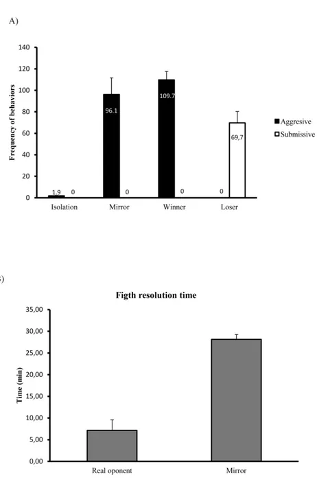
Após uma interação agonística normalmente surge um vencedor e um derrotado e estes estatutos sociais vão influenciar interações futuras de maneira que o vencedor da primeira interação tem maior probabilidade de vencer uma segunda, enquanto que o derrotado tem maior probabilidade de a perder. Curiosamente, em interações sociais onde a informação sobre o estatuto não é definida, como por exemplo, interações não resolvidas quando os animais lutam contra a sua própria imagem no espelho, a mudança de estatuto social (para vencedor ou derrotado) não ocorre, demonstrando a relevância que a informação social tem nas decisões de alteração de comportamento.
De acordo com o que foi descrito anteriormente, um único genótipo pode então ser modulado socialmente dando origem a múltiplos fenótipos comportamentais. Esta plasticidade comportamental, depende naturalmente de uma plasticidade a nível dos circuitos neurais subjacentes ao comportamento social. Os mecanismos neurais implícitos à plasticidade comportamental podem atuar de duas formas: (1) provocando alterações estruturais nos circuitos, que conduzem a mudanças comportamentais que ocorrem lentamente e são duradouras; (2) ou modulando bioquimicamente a atividade neural, provocando alterações comportamentais significativamente mais rápidas, mas contudo transientes. Recentemente foi proposto que esta modulação ocorre a nível da
social decison-making (SDM) network que integra um conjunto de núcleos neurais
responsáveis pela regulação de comportamentos sociais (agressão, corte ou comportamento parental) com núcleos do sistema mesolímbico de recompensa que avaliam a saliência do estímulo através de uma via de sinalização dopaminérgica. Todas estas áreas estão reciprocamente ligadas, contêm receptores para hormonas esteróides e neuromoduladores, e todas elas respondem individualmente à exibição de comportamentos sociais. A hipótese subjacente a esta rede neural é que a informação é codificada de forma dinâmica, de tal forma que determinado perfil comportamental parece ser melhor explicado pelo perfil de ativação da rede na sua globalidade, do que pela atividade individual de cada nódulo. Conceptualmente, a pluralidade de combinações possíveis de ativação dos diferentes nódulos, poderá explicar a diversidade de comportamentos exibida entre espécies, entre indivíduos da mesma espécie e dentro do mesmo individuo, uma vez que esta rede é evolutivamente conservada.
Esta tese tem como principais objectivos o estudo e a possível identificação dos mecanismos proximais subjacentes à flexibilidade comportamental numa perspectiva integrativa. Para tal usámos como espécie modelo o peixe-zebra, que são animais altamente sociais, que vivem em cardumes com relações sociais bem estruturadas, tais como hierarquias de dominância e territorialidade.
No paradigma comportamental utilizado os animais foram expostos a diferentes tipos de experiências sociais: 1) interações com oponentes reais que deram origem a vencedores e derrotados, que consequentemente ajustaram o seu reportório comportamental ao novo estatuto social, e 2) lutas com espelhos, interações não resolvidas onde a expressão do comportamentos agressivos foi dissociada da experiência de vitória ou derrota. Como os peixes não reconhecem a sua própria imagem no espelho, atacam-na como se esta fosse um intruso. No entanto, como o
comportamento de submissão nunca é expresso pelo oponente, ou seja, a imagem do espelho reproduz o comportando agressivo do animal focal, o indivíduo focal nunca experiencia uma vitória ou derrota. Animais que não interagiam socialmente, i.e., que se encontravam em isolamento visual mas não químico, foram utilizados como grupo controlo.
Deste modo, o paradigma experimental utilizado para além de permitir o estudo da flexibilidade comportamental, ou seja, de diferenças específicas observadas em vencedores e derrotados aquando da comparação com o grupo controlo, permite ainda estudar o tipo de informação utilizada para esta alteração de comportamento, comparando os animais que lutam com o espelho com vencedores e derrotados, ou seja,
self- assessment, opponent-only assessment ou mutual assessment, caso o mecanismo
utilizado seja uma integração entre os dois modelos, num balanço entre comportamento exibido pelo próprio e pelo oponente.
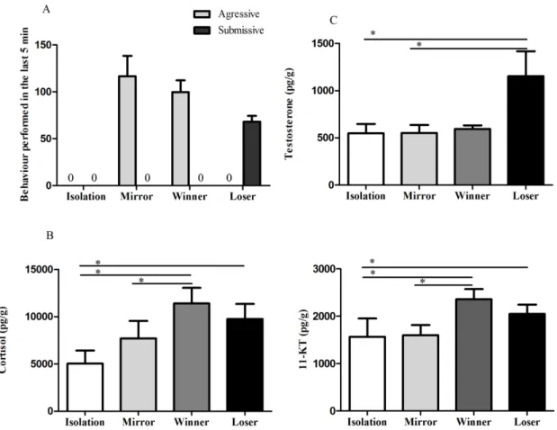
No primeiro conjunto de experiências estudámos a influência dos neuromoduladores nos mecanismos de plasticidade, e para tal caraterizámos a resposta endócrina a desafios sociais (Capítulo II) e a modulação social de monoaminas (Capítulo III) e de nonapéptidos (Capítulo IV) no cérebro. Este conjunto de trabalhos identificou uma resposta dos androgénios nas lutas com oponentes reais, tanto em vencedores como em derrotados, assim como uma ativação na produção dos glucocorticóides (cortisol). No Capítulo III referente ao estudo de monoaminas, associámos o estatuto de vencedor a um aumento de atividade serotonérgica e dopaminérgica no Telencéfalo, sugerindo que o mecanismo de recompensa poderá estar envolvido na alteração de estatuto social em vencedores. No estudo do Capítulo IV, referente à variação nonapeptídica após interações sociais verificámos que a arginina-vasotocina respondeu de forma mais generalizada no grupo dos derrotados do que a
isotocina, apontando para a relevância deste péptido na regulação do comportamento agressivo em peixe-zebra, como já tinha sido descrito para outras espécies. Curiosamente, nenhum destes sistemas foi ativado em resposta à luta com o espelho, apesar deste animais apresentarem níveis de agressão semelhantes àqueles expressos pelos vencedores, apontado para o papel fundamental que a percepção social tem em transições entre diferentes estados comportamentais.
A nível neuromolecular testámos a hipótese da social decision-making network, contrastando alterações entre funcionalidade localizada e conectividade dentro desta rede, em resposta a alterações do estatuto social (Capítulo V) e terminámos com a caraterização de genes-chave envolvidos nos diferentes mecanismos de plasticidade (Capítulo VI).
Os dados apresentados no Capítulo V, sustentam a hipótese da social
decision-making network, dando um suporte funcional para uma atividade em rede, ao invés da
atividade específica de nodos individuais. Tendo em consideração os padrões de actividade neural conseguimos distinguir vencedores, derrotados e animais que lutaram contra a sua própria imagem no espelho.
Os resultados obtidos no Capítulo VI mostram que cada estado comportamental é caracterizado por um padrão neuromolecular de expressão de genes associados a diferentes mecanismos de plasticidade neural. Tal como ocorreu a nível fisiológico, também a nível neuromolecular os animais que lutam com o espelho apresentam padrões neurais distintos de vencedores e derrotados, indicando uma vez mais a importância a importância da percepção social na alteração de fenótipos comportamentais.
Palavras-chave: Comportamento Social, Plasticidade Social, Hormonas Esteróides, Monoaminas, Nonapéptidos, Neurogenómica, Peixe-zebra
Abstract
Social competence, the ability of individuals to regulate the expression of their social behaviour in order to optimize their social relationships in a group, is especially benefic for individuals living in complex social environments, and implies the ability to perceive social cues and produce appropriate behavioural output responses (Social Plasticity). Numerous examples of social competence can be found in nature, where individuals extract social information from the environment, and change their behavioural response based on the collected information. At the neuronal level, two major plasticity mechanisms have been proposed to underlie social plasticity, structural reorganization and biochemical switching of the neuronal networks underlying behaviour. The neural substrate for behavioural plasticity has been identified as the social decision-making (SDM) network, such that the same neural circuitry may underlie the expression of different behaviours depending on social context. The goal of this work is to study the proximate mechanism underlying behavioural flexibility in the context of experience-dependent behavioural shifts, in an integrative framework. For this purpose we exposed male zebrafish to two types of social interactions: (1) real-opponent interactions, from which a Winner and Loser emerged; and (2) Mirror-elicited interactions, that produced individuals that did not experience a change in social status, despite expressing similar levels of aggressive behaviour to those participating in real-opponent fights. In a first set of experiments, we studied the influence of neuromodulators on social plasticity mechanisms, by characterizing the endocrine response to social challenges, as well as the social modulation of brain monoamines and nonapeptides. Next we tested the SDM network hypothesis by contrasting changes in functional localization vs. connectivity across this network. Finally we characterized changes in expression of key genes for different neuroplasticity mechanisms in response
to changes in social status. Our research suggests different social plasticity mechanisms underlying Winners and Losers both at physiological and molecular levels, for Mirror-fighters, where the experience of winning or losing was decoupled for the fighting experience, few changes were detected. This, by itself suggests a pivotal role of social perception in triggering shifts between socially driven behavioural states.
Keywords: Social plasticity, Social decision-making network, steroid hormones, Monoamines, Nonapeptides, Neurogenomic, Zebrafish
Contents
Agradecimentos ... iii
Resumo ... vii
Abstract ... xiii
Contents ... xv
Chapter I. General Introduction ... 1
1.1.The social brain ... 3
1.2.Behavioural plasticity mechanisms ... 6
1.3.Neuroanatomy of the social brain ... 7
1.4.Neuromodulators contribution to plastic responses ... 9
1.5.Neurogenomics of social plasticity ... 16
1.6.Zebrafish as an experimental model in social neurosciences ... 19
1.7.Aims and hypothesis ... 20
1.8.References ... 23
Chapter II. Androgen response to social competition in a shoaling fish ... 33
Abstract ... 35
1. Introduction ... 37
2. Material and methods ... 39
2.1. Animal housing ... 39
2.2. Social challenge tests ... 39
2.3. Hormone assays ... 40 2.4. Data analysis ... 41 2.5. Ethics statement ... 41 3. Results ... 42 4. Discussion ... 43 References ... 45
Chapter III. Social modulation of brain monoamine levels in zebrafish ... 49
Abstract ... 51
1. Introduction ... 53
2. Materials and Methods ... 55
2.1. Animals and housing ... 55
2.2. Experimental design ... 56
2.3. Sampling ... 57
2.4. Analysis of brain monoamines and metabolites... 57
2.5. Behavioral observations ... 59 2.6. Statistical analysis ... 59 2.7. Ethics statement ... 60 3. Results ... 61 3.1. Behavior ... 61 3.2. Brain monoamines ... 63
3.3. Relationship between monoamines and behavior ... 67
4. Discussion ... 67
Bibliography ... 74
Chapter IV. Social modulation of nonapeptides in the zebrafish brain ... 79
Abstract ... 81 1. Introduction ... 83 2. Methods ... 86 2.1. Animals ... 86 2.2. Behavioural paradigm ... 86 2.3. Brain collection ... 87
2.4. Quantification of nonapeptides by high performance liquid chromatography with fluorescence detection (HPLC-FL) ... 87
2.5. Behavioural analysis ... 88
2.6. Statistical analysis ... 89
3. Results ... 90
3.1. Behaviour ... 90
3.2. Nonapeptide levels in the brain ... 91
3.3. Relationship between nonapeptides and behaviour ... 94
4. Discussion ... 95
Bibliography ... 100
Chapter V. Social interactions elicit rapid shifts in functional connectivity in the social decision-making network of zebrafish ... 105
Abstract ... 107
1. Introduction ... 109
2. Material and methods ... 112
(a) Fish housing ... 112
(b) Social treatments ... 112
(c) Microdissection of regions of interest in the brain ... 113
(d) Gene expression analysis ... 114
(e) Behavioural observations ... 115
(f) Statistical analysis ... 115
3. Results ... 117
(a) Social behaviour states ... 117
(b) Effect of social behaviour state and brain region on immediate early gene expression ... 118
(c) Differences in functional localization among social behaviour states across the social decision-making network ... 118
(d) Differences in functional connectivity among social behaviour states across the social decision-making network ... 121
(e) Association between immediate early gene expression and behaviour .... 124
4. Discussion ... 124
(a) Functional localization ... 125 xvii
(b) Functional connectivity ... 127
5. Conclusion ... 129
References ... 131
Supplementary Material ... 134
Chapter VI. Social plasticity relies on different neuroplasticity mechanisms across the brain social decision-making network in zebrafish ... 143
Abstract ... 145
1. Introduction ... 147
2. Material and methods ... 151
2.1. Animals ... 151
2.2. Experimental procedure ... 152
2.3. Blood collection and hormone analysis ... 153
2.4. Brain microdissection ... 153 2.5. Gene expression ... 154 2.6. Behavioural analysis ... 155 2.7. Statistical analysis ... 155 2.8. Ethics Statement ... 156 3. Results ... 157 3.1. Behaviour ... 157
3.2. Expression of bdnf across the SDMN ... 158
3.4. Expression of neuroligin genes across the SDMN ... 161
3.5. Expression of neurogenesis genes across the SDMN ... 162
3.6. Circulating cortisol levels ... 162
3.7. Association patterns between behaviour, gene expression, and cortisol levels... 163
4. Discussion ... 165
4.1. Socially triggered neuroplasticity profiles for each social phenotype .... 165
4.2. Status-specific and fighting triggered neuromolecular states of the SDMN
... 169
4.2. Brain region specific neuroplasticity ... 170
4.3. Role of cortisol on socially-driven neuroplasticity ... 171
Funding statement ... 173
References ... 173
Supplementary Material ... 179
Chapter VII. General Discussion ... 183
7.1.Overview of results ... 185
7.2.Behavioural and physiological modulators of social plasticity ... 187
7.2.1. When behavioural repertoire speaks about social status ... 187
7.2.2. Physiological modulation of social plasticity... 189
7.3.Neurogenomic shifts: the first line of response ... 198
7.4.Concluding remarks and future perspectives ... 201
7.5.References ... 203
Supplement I - Quantifying aggressive behaviour in zebrafish ... 207
Summary ... 209 1. Introduction ... 210 2. Materials ... 214 3. Methods ... 215 3.1. Animal housing ... 215 3.2. Individual tagging ... 215 3.3. Behavioural recording ... 218 4. Notes ... 223 References ... 226 xix
Chapter I. General Introduction
Chapter I
Chapter I. General Introduction
1.1. The social brain
Animal species present a variety of species-specific behaviours (behavioural repertoires) that evolved in an adaptive way in order to integrate behaviour with the natural environment, and that will ultimately dictate the Darwinian fitness of individuals.
In broad terms, the vast majority of animals live in social environments, and their lives are affected by the presence and activity of others around them. In the most simplified environment, the behaviour expressed by an individual will induce a response in another, and depending on group size, layers of complexity will be added to the relationship between individuals and third parties. This way, larger groups will typically represent a more complex social environment than smaller groups, since more interactions with different social partners will be more likely to occur in the later one (Taborsky & Oliveira 2012).
The social brain hypothesis (SBH) proposed by Dunbar (1998) posits that the complexity of the social environment is one of the main driving forces for brain evolution. This hypothesis is supported by comparative data in primates and ungulates where relative brain size covaries with group size (Pérez-Barbería & Gordon 2005; Dunbar & Shultz 2007) linking brain size to high cognitive demands of more complex social interactions. There has been an attempt to generalize this hypothesis to all vertebrate taxa, however the relationship for other species was qualitative rather than quantitative, and exclusively associated with monogamy (Dunbar & Shultz 2007), one of the dimensions of sociality.
In a cooperative breeding cichlid, Neolamprologus pulcher, it was recently showed that the relative brain size (specific macroareas) was affected by group-size rearing, for instance, hypothalamus and cerebellum increased in fish reread in large groups, whereas
the optic tectum was bigger in fish reared in small groups, which may indicate some degree of specialization for each brain area (Fischer et al. 2015). It is also important to stress out that total brain size was unaffected, presenting the first experimental evidence for “mosaic evolution” where selective pressures act on individual brain parts (de Winter & Oxnard 2001) over “concerted evolution” where the overall brain size is selected (Finlay & Darlington 1995). At the behavioural levels, group complexity also affected the establishment of social hierarchies, where fish reared in large groups showed more submissive and less aggressive behaviour towards larger conspecifics. This behavioural response increases the chances of being tolerated in the territory of a larger dominant conspecific, which greatly enhances the survival chances under natural conditions, suggesting a better ability to cope with social challenges (Fischer et al. 2015). On the other hand, in guppies artificially selected for brain size it affected survival rate under a predation threat. Large-brained females had higher survival compared to small-brained females (Kotrschal et al. 2015), and in a predator inspection task, large-brained fish spend less time performing inspections, and group size affected the distance kept from the predator which may indicate a cognitive advantage for larger brains (van der Bijl et al. 2015).
According to this framework, the complexity of the social environment drives brain evolution and consequently cognitive abilities, which ultimately enhance fitness.
Social competence, the ability of individuals to regulate the expression of their social behaviour in order to optimize their social relationships in a group (Taborsky & Oliveira 2012) will be especially beneficial for individuals living in complex social environments, and implies the ability to perceive social cues, and produce the appropriate behaviour output response (social plasticity). Therefore, social plasticity relies on behavioural flexibility, that is variation in the behavioural response to the same
Chapter I. General Introduction stimulus that will often depend on cognitive skills (acquisition, retention and use of information) (Taborsky & Oliveira 2012). This type of plasticity is reversible, and occurs within an individual’s lifetime.
Numerous examples of social competence can be found in nature, where individuals extract social information from the environment, and change their behavioural response based on the collected information. Animals may eavesdrop interactions between third parties and collect information from the observed individuals to use in subsequent encounters [“social eavesdropping” (Oliveira et al. 1998; Earley 2010; Abril-de-Abreu et al. 2015b)], or use the collected information of known relationships to deduce unknown ones [“transitive inference” (Grosenick et al. 2007)]. The presence of bystanders may also influence the behaviour of interacting conspecifics in an “audience effect” (Doutrelant et al. 2001; Pinto et al. 2011; Cruz & Oliveira 2015)]. Familiarity with an opponent is also known to reduce aggression from a territory owner towards a neighbour rather than towards a stranger depending on the relative threat that both represent [“dear enemy effect” (Temeles 1994; Aires et al. 2015)]. Previous social experience can affect subsequent behaviour as in the case of “winner–loser effects” demonstrated across different animal taxa (Hsu et al. 2006; Rutte et al. 2006), where previous winners are more likely to win successive contests, and losers will be more likely to lose even against different opponents. Interestingly, social interactions where no information can be extracted, as is the case of unsolved fights when animals are fighting their own mirror image, behavioural flexibility (i.e. shifts between behavioural states) does not occur (Teles et al. 2013), demonstrating the relevance of social information for behavioural decisions. Thus, social competence is a key factor in the generation of different behavioural states that encompasses neural, genomic and physiological information (Cardoso et al. 2015).
1.2. Behavioural plasticity mechanisms
At the neuronal level, two major plasticity mechanisms have been proposed and both may underlie behavioural plasticity: structural reorganization and biochemical switching of the neuronal networks underlying behaviour (Zupanc & Lamprecht 2000). These mechanisms are expected to operate at different time scales. Structural reorganization is expected to underlie long-lasting behavioural changes, and may include core structural modifications through processes such as adding or removing cells from the circuit (neurogenesis and apoptosis), changes on the connectivity or de
novo formation of synaptic connections (synaptogenesis), or by altering the
responsiveness of the circuit (i.e. balance of neurotransmitter and/or neuromodulator receptors in specific neurons) (Zupanc & Lamprecht 2000; Oliveira 2009; Cardoso et al. 2015). On the other hand, biochemical switching, is expected to underlie short-term reversible transitions between behavioural states, and involves the modulation of synaptic transmission by changing the release of neurotransmitter molecules at the presynaptic level, changing the number, type, or properties of neurotransmitter receptors postsynaptically, or by altering the dynamic of neuromodulatory molecules (i.e. monoamines, nonapeptides or hormones) in a socially dependent fashion (Oliveira 2009).
Interestingly, these two different neural plasticity mechanisms have a parallel with two different levels of endocrine regulation. Hormones can have different effects depending on their activation time, and are expected to be involved in different types of plasticity. Organizational effects occur early in development, typically within a critical or sensitive window during which the exposure to the active molecule induces a long-lasting and irreversible differentiation of a behavioural state and implies structural changes in the brain. On the other hand, activational effects typically occur later in time,
Chapter I. General Introduction typically in adulthood, and are reversible allowing behavioural shifts (Arnold & Breedlove 1985). Thus, the integration of endocrine parameters in social decisions is expected to be mediated by activational effects in the case of behavioural flexibility (Cardoso et al. 2015), and by organizational effects early in developmental stages, by shaping fixed behavioural patterns that can also be influenced by environmental triggers. For instance, rats exposed to prenatal stress showed increased anxiety and depression related behaviours when tested for novelty in adulthood (Vallée et al. 1997). Nevertheless, at the molecular level, both mechanisms rely on the social regulation of gene expression, so that different neurogenomic states will ultimately induce different behavioural responses (Aubin-Horth & Renn 2009; Oliveira 2012).
1.3. Neuroanatomy of the social brain
According to this framework a single genotype can be socially modulated, resulting in particular phenotypes. But how does a single genome orchestrate multiple complex forms of behaviour? The neural substrate for behavioural plasticity has been identified as the social brain network (SBN) originally proposed by Newman (1999) in mammals, and confirmed to be evolutionary conserved across different vertebrate taxa by Goodson (2005). This social behaviour network is composed by six nodes located in the forebrain and midbrain areas [i.e. bed nucleus of the stria terminalis / extended medial amygdala (BNST/meAMY), lateral septum (LS), preoptic area (POA), anterior hypothalamus (AH), ventromedial hypothalamus, and the periaqueductal gray (PAG)], that are reciprocally connected and that together regulate several dimensions of sociality including, sexual behaviour, parental behaviour, and different forms of aggressive behaviour (Newman 1999; Goodson 2005).
As mentioned earlier, the behavioural actions expressed in response to a specific context must be adaptive to the animal, and therefore the stimuli must be properly evaluated, in terms of their valence and salience, in order to produce an appropriate response (Mendl et al. 2010). More recently, O’Connell and Hoffman (2012) proposed the inclusion of the mesolimbic reward system, which is generally assumed to evaluate stimulus salience via dopaminergic signalling (Wickens et al. 2007) and shares overlapping nodes with the SBN (lateral septum and bed nucleus of the stria terminalis), as an integrated evolutionarily ancient social decision-making (SDM) network. This network is highly conserved across vertebrates pointing to the fundamental role of the involved brain areas in vertebrate social evolution (O’Connell & Hofmann 2011, 2012), Figure 1.
(Adapted from: O’Connell & Hofmann 2011) Figure 1 – Social decision-making (SDM) network. a) Schematic representation of the interactive nodes of the networks, brain nuclei in the social behaviour network (left) and mesolimbic reward system (right), as well as brain regions involved in both systems (centre), b) Sagittal view of a mammalian and teleost brain highlighting the connectivity between nodes of the social decision-making circuit.
Chapter I. General Introduction According to the SDM network hypothesis the information is encoded in a dynamic fashion, and each behavioural state is better represented by the overall profile of activation across the network, rather than by the activity of single individual nodes. Different combinations of activation across nodes, and variation in the strength of the connections among them, will allow the same genotype to generate an almost infinite variation in neural states that would produce equivalent behavioural states (Goodson 2005; Cardoso et al. 2015). Given that most nodes of the SDM network widely express receptors for neuromodulators and steroid hormones the state of this network can be also co-regulated by these molecules (Newman 1999; Goodson 2005).
1.4. Neuromodulators contribution to plastic responses
Monoamines and neuropeptides are considered the two major classes of neuromodulators, and the action of both on social behaviour as well as their sensitivity to environmental factors, have been extensively documented (Libersat & Pflueger 2004; Goodson & Thompson 2010), which makes them major candidates to mediate changes in brain states, underlying socially driven behavioural flexibility in the SDM network.
Neuromodulators are released into broader areas than neurotransmitters, bind to receptors in the cell membrane, that are generally linked to G proteins, and subsequently activate intracellular signalling cascades with effects on the electrical activity of neurons that could last seconds, minutes, hours, days, or even weeks. At the molecular levels, neuromodulators have a wide variety of effects including: (a) modulation of ion channels and receptors, and (b) changes in protein synthesis, enzyme activity, and gene transcription (Libersat & Pflueger 2004).
a) Monoamines
Among the major representatives of the monoamines that are known to modulate well-defined behaviours are serotonin and dopamine. Serotonin (5-hydroxytryptamine, 5-HT) distribution in the CNS has been studied in different species, and found to be located exclusively in the brainstem (Takahashi et al. 1986; Ishimura et al. 1988; Johnston et al. 1990), where the majority of the serotonergic cell bodies reside in the dorsal and median raphe nuclei. These neurons project axons almost to the entire brain, including cortical, limbic, midbrain, and hindbrain regions (Charnay & Léger 2010), and due to this wide projection pattern, 5-HT is involved in many biological processes, such as learning and memory, mood, food intake, sleep, reproduction, circadian rhythm, thermo-regulation, pain, and social behaviour (Kiser et al. 2012). This molecule is catabolized from the amino acid tryptophan via 5-hydroxytryptophan and the rate-limiting factor in serotonin synthesis is the enzyme tryptophan hydroxylase (TPH1 and TPH2), whose activity depends on the availability of tryptophan (Ruddick & Evans 2006). Following release into the synapse, serotonin is either recycled back into the presynaptic neuron by the serotonin transporter (SERT), or degraded to 5-hydroxyindoleacetic acid (5-HIAA) by the enzyme MAO. The ratio 5-HIAA/5-HT can though be used as a measurement of serotonergic activity (Shannon 1986).
Dopamine (DA) on the other hand, is a catecholamine synthesized from tyrosine via 3,4-dihydroxyphenylalanin (L-DOPA). The first step is carried out by tyrosine hydroxylase and is the rate-limiting step. In mammals, dopamine is degraded by the enzyme MAO, like in serotonin, and produce three metabolites: homovanillic acid (HVA), 3,4-dihydroxyphenylacetic acid (DOPAC), and the 3-methoxy- tyramine (Daubner et al. 2011). The ratio between its metabolites and DA is also used as an index of activity.
Chapter I. General Introduction The influence of monoamines on plastic responses, have been widely documented in the context of social status hierarchy and unpredictability, ubiquitous features in social groups.
5-HT appears to play a central role in aggressive interactions but its effects are to some extent paradoxical. While several studies have pointed out that pharmacological manipulations that increase 5-HT inhibit aggression in a wide range of vertebrates, from fish to humans (Summers et al. 2005), other studies, in contrast, have showed increased serotonergic activity in specific brain regions during the expression of aggressive behaviour (Winberg & Nilsson 1993; Overli et al. 1999; Summers et al. 2005). In early stages of hierarchy formation the serotonergic system appears to be activated in both dominants and subordinates, in the hippocampus, nucleus accumbens and amygdala of lizards, and in the telencephalon of fish [Anolis carolinensis, (Summers et al. 2003); rainbow trout (Oncorhynchus mykiss, (Overli et al. 1999)]. Similarly, after a chronic (5 days) agonistic interaction both dominants and subordinates showed higher levels of 5-HT activity in the telencephalon in the bicolour damselfish (Stegastes partitus, (Winberg et al. 1996), whereas in zebrafish only subordinate males increased serotonergic activity (measured as the 5-HIAA/5-HT ratios) in the hindbrain (raphe nucleus) (Dahlbom et al. 2012). In rats single or repeated social defeat increases serotonergic neuronal activity within the dorsal raphe nucleus in losers as evidenced by an increase in extracellular serotonin (Amat et al. 2010) and c-fos expression in serotonergic neurons (Paul et al. 2011). Finally, a cross-strain comparison of male mice obtained through different artificial selection breeding programs for aggression that studied the differential role of the 5-HT1A receptor in aggressive and non-aggressive
mice, found that highly aggressive mice had lower serotonin levels in the prefrontal cortex, and that two out of three aggressive strains had higher 5-HT1A receptor
sensitivity (Caramaschi et al. 2007). Together, these results indicate that in addition to the activational effects on aggressive behaviour, these neuromodulator can also have organizational effects early in development.
Dopaminergic system has been classically associated with reward and motivation, with an increase in dopamine transmission leading to an increased feeling of reward. In Long-Evans rats, in a resident-intruder paradigm dopamine and serotonin levels in medial prefrontal cortex (PFC) changed in the opposite direction in dominate animals, with a sustained decrease in serotonin during and after the confrontation and an increase in dopamine after the fight in PFC and in the nucleus accumbens (NAC) (van Erp & Miczek 2000). A similar pattern has been previously observed in salmonids where dominant individuals showed higher DA activity in telencephalon than subordinate fish (Winberg et al. 1991).
DA is also involved in signalling unpredictability in the environment. Reversal learning is a behavioural task that requires that the animal responds to changes in reward contingencies, that is the animal has to adapt previously learned behaviours to changes in the environment. This type of learning is highly linked to both 5-HT and DA modulation. Rats treated with parachlorophenylalanine (PCPA), a drug that depletes 5-HT by the inhibition of tryptophan hydroxylase, were impaired at approaching the stimulus associated with the reward, after the reversal and this was related with lower levels of 5-HT in the ventromedial frontal cortex (Izquierdo et al. 2012). Recently, it was shown the pivotal role of the striatal dopamine (DA) in this process. The authors monitor the DA release in the ventromedial frontal cortex using a fast-scan cyclic voltammetry. During discrimination performance (pre-reversal), cue presentation induced phasic DA release, whereas reward delivery did not. The opposite pattern was observed in the post-reversal, that is striatal DA release occurred after reward delivery,
Chapter I. General Introduction while cue-induced release diminished. Trial-by-trial analysis showed rapid replacement of cue-induced DA release on trials immediately following initial correct responses. This effect of positive feedback was observed in animals that learned the reversal, but not in “non-learners” (Klanker et al. 2015). Also in the Atlantic Salmon (Salmo salar
L.), in an omission of the expected reward (OER) paradigm, increased DOPAC and
DOPAC/DA ratios were found in the brain stem in the OER group under stress conditions (acute confinement) (Vindas et al. 2014).
b) Nonapeptides
The nonapeptide family includes the mammalian forms arginine vasopressin (AVP) and oxytocin (OT), as well as the nonmammalian homologues, arginine-vasotocin (AVT), and isotocin (IT) in bony fish, respectively, and mesotocin (MT) as an homologue of OT in lungfish, amphibians, birds, reptiles and some marsupials (Goodson 2008; Donaldson & Young 2008) (Thompson & Walton 2009).
All vertebrate species exhibit magnocellular and parvocellular nonapeptide cell groups in the preoptic area and hypothalamus, including the supraoptic and paraventricular nuclei in amniotes (Goodson 2008). Vasopressin and oxytocin neurons produce and pack nonapeptides into large dense-core vesicles that will be transported along the axons and can terminate in the neurohypophysis, where they are released in the bloodstream in the eminence of the anterior pituitary, and stimulate the adrenocorticotropin (ACTH) release (De Vries 2008), or project to the hindbrain, to influence autonomic functions (Thompson & Walton 2009), or other forebrain regions (Saito et al. 2004; Biran et al. 2015). In mammals these vesicles can also be released locally at the dendrites (Moos & Freund-Mercier 1984; Landgraf & Neumann 2004; Ludwig & Leng 2006) modifying their own electrical activity (Moos & Freund-Mercier
1984; Morris & Ludwig 2004). In non-mammalian species there is no direct evidence of dendritic release, although the anatomical features of both neuronal types, such as the close proximity and direct membrane appositions (Saito et al. 2004) may indicate a similar mechanism.
The nonapeptide system has been implicated in a variety of social behaviours, such as parental behaviour, sexual behaviour, pair-bond, mate choice, aggression and social recognition (Goodson & Bass 2001; Insel 2010), as well as in plasticity in behavioural responses in several species.
The plasticity underlying aggression, that is the ability to express aggressive behaviour under the right circumstances, may depend on previous experience and social status. In mammals this social status plasticity is linked to changes in AVP and AVP receptor (Avpr) distribution, with different effects being observed in dominants and subordinates. In mice, administration of lysine-vasopressin after a social defeat increases submissive behaviour in subsequent encounters compared to saline-treated animals (Roche & Leshner 1979). In the Syrian hamsters, AVP or AVP antagonist injections in the anterior hypothalamus-medial preoptic area (AH-MPOA) result in transient reversals of dominant/subordinate relationships. Subordinate animals treated with AVP display increased flank-mark behaviour (the way they communicate dominance status) while dominates treated with Avpr1 antagonist decreased its expression (Ferris et al. 1986). Consistently with these results, subordinate hamsters present fewer Avp-ir neurons in the magnocellular nucleus circularis (a cell group between supraoptic nucleus and paraventricular nucleus) than dominants (Ferris 1989), and following repeated agonistic encounters dominant hamsters have higher levels of
Avpr1a binding in the ventromedial hypothalamus (VMH) when compared to their
subordinate opponents (Cooper et al. 2005). In socially isolated hamsters, the increase
Chapter I. General Introduction in aggression was correlated with the increase of Avpr1a binding in the anterior hypothalamus (AH), the paraventricular nucleus (PVN) and lateral hypothalamus, whereas socially experienced hamsters (i.e. allowed to interact with a conspecific 3 times a week) present a significant binding increase only in the amygdala (Albers et al. 2006). Taken together these results indicate that the AVP system, especially through the
Avpr1a can be altered by social experience. Nevertheless, while treatment with Avpr1a
antagonist reduced aggression in golden hamsters (Ferris et al. 2006), contrary to what was expected Avpr1a knockout (KO) mice did not show reduced aggression (Wersinger
et al. 2008), and it was the isoform Avpr1b that proved to be critical for proper
expression of aggression, as Avpr1b KO mice showed significant impairments in aggressive displays compared to wild-type controls (Wersinger & Ginns 2002).
In fish, in addition to the magnocellular and parvocellular cell groups, there is a third cellular type, the gigantocellular, and these three types of cells are distributed along the ventral portion of the preoptic area (POA) [reviewed in (Urano & Ando 2011)]. In fish the modulation of aggression is also ambiguous. In some teleost species, the expression of social dominance has been associated with higher number or size of AVT-ir cells in magnocellular (mPOA) or gigantocellular (gPOA) neuronal population, whereas social submission is associated with the number or size of pPOA AVT-ir cells [e.g. zebrafish, Danio rerio (Larson et al. 2006); African cichlid, Astatotilapia burtoni (Greenwood et al. 2008); butterfly fishes (Dewan et al. 2011)]. In other fish species the reversal is also true: social submission is associated with changes in the mPOA and gPOA populations (Almeida & Oliveira 2015), and aggressive behaviour with variations in size of the pPOA AVT-ir (Lema 2006). AVT manipulation can also increase or decrease aggression depending on the species (Godwin & Thompson 2012), and the expression of nonapeptide receptors can also vary depending on social status.
Evidence from zebrafish indicates arginine vasotocin-like receptor 1b (V1b) as one of the highest differentially expressed gene in the hypothalamus of dominant animals (Filby et al. 2010), whereas in the pupfish transcripts encoding the isoform V1a1 were expressed at higher levels in the telencephalon and hypothalamus of subordinate males, and it was the variant V1a2 that was more abundant in dominants telencephalon (Lema
et al. 2015), similarly to what has been previously described in mice.
The oxytocin role on aggression has been scarcely investigated and, only one study in female Syrian hamsters (Mesocricetus auratus) clearly demonstrated that endogenous OT modulates resident-intruder aggression in adults. This experiment showed that OT infusions into the preoptic area-anterior hypothalamus (POA-AH) decreased resident-intruder aggression, and that OTR antagonist facilitated aggression (Harmon et al. 2002). In territorial finches (violet-eared waxbill) peripheral injections of an OTR antagonist reduced aggressive behaviour in females, and colocalization of OT-Fos found in the preoptic area and hypothalamus, was correlated negatively with aggression (Goodson et al. 2015), suggesting that OT may be mediating the stress response and not the aggression.
Finally the involvement of these peptides in gregariousness, a key dimension of sociality that implies behavioural flexibility, is very well described in birds. In different species of finches, mesotocin receptor distribution in the lateral septum correlates with flock size, and administration of mesotocin increases while a mesotocin antagonist reduces social behaviour, such as flock formation (Goodson et al. 2009).
1.5. Neurogenomics of social plasticity
Behaviour traits exhibit a great deal of plasticity and their modulation requires the integration of multiple systems as we previously stated. At the proximate level, the
Chapter I. General Introduction direct consequence of the activation of any specific neural circuits underlying behaviour is a burst in gene expression. A neurogenomic state corresponds to distinct transcriptome profiles across the SDM network, (Robinson et al. 2008; Zayed & Robinson 2012) corresponding to different behavioural states. Switches between states (behavioural flexibility) are orchestrated by signalling pathways that interface the social environment and the genotype (Aubin-Horth & Renn 2009; Oliveira 2012).
At least three different neuronal activity-dependent molecular mechanisms can be proposed to translate social information into a neurogenomic state (Wolf & Linden 2012): protein phosphorylation; immediate early genes activation (IEGs), and microRNAs.
a) Activation (e.g. phosphorylation) of the intracellular signalling pathway mitogen-activated protein kinase (MAPK/ERK) is involved in the transduction of signals through a cascade of protein kinases in response to stimuli. Once this pathway is activated, ERK phosphorylates a variety of target proteins, including other protein kinases and transcription factors, for example CREB that is phosphorylated by ERK and other kinases such as protein kinase C (Roberson et
al. 1999). This transcription factor (Brindle et al. 1993) binds to the CRE site
present in IEG promoters, and acts as a key regulator of IEG expression activation. Several different protein kinases possess the capability of driving the phosphorylation of CREB, making it a point of potential convergence for multiple intracellular signalling cascades (Wu et al. 2001).
b) IEGs are the first genomic response upon cell depolarization, whose transcription can be induced without requiring de novo protein synthesis or
previous activation of any other responsive genes (Clayton 2000). It has been shown recently that several IEGs are poised for near-instantaneous transcription by stalling the DNA polymerase II (Pol II) in the vicinity of the promoter (Saha
et al. 2011). In line with this, IEGs have been classified into different groups
depending on the presence or absence of the DNA polymerase II (Pol II) stalling. For rapid IEGs, that are expressed within a few minutes after stimulation, DNA polymerase II (Pol II) stalling is in the promoter region, whereas in delayed IEGs, that are expressed later (ca. 1 hour post-stimulation), largely lacked this poised Pol II (Saha et al. 2011; Saha & Dudek 2013). This mechanism of stalling has been shown to be relevant in regulating timing and dynamics of gene responses, but not for steady-state accumulation of mRNA over time (Saha et al. 2011). Depending on their function, IEG proteins can act themselves as transcription factors (e.g. c-fos and egr-1), or as effector proteins (e.g. arc and homer1a), regulating synaptic function (Clayton 2000).
c) The transcription of microRNAs (miRNA), which are non-coding RNAs, function as post-transcriptional repressors of gene expression. These RNA molecules can control specific biological processes by switching off a few target genes at particular time (temporal switches) or places (during development) (Lai 2005). An example of this mechanism is the brain-expressed miR-133, recently found to play an important role in controlling behavioural shifts in migratory locusts (Locusta migratoria) (Yang et al. 2014). miR-133 controls dopamine production by targeting the genes henna and pale, which are involved dopamine synthesis and release, and related to the behavioural phase transitions (from gregarious phase to the solitary) of the migratory locust (Ma et al. 2011).
Chapter I. General Introduction 133 sense oligonucleotides (agomir) delivery suppressed henna and pale expression, which consequently decreased dopamine production, resulting in the behavioural shift from the gregarious phase to the solitary phase, while miR-133 inhibition, promoted gregarious-like behaviour of solitary locusts. Thus, microRNA plays an important role as an activational switch in this species acting a key mediator of a transition between behavioural states.
1.6. Zebrafish as an experimental model in social neurosciences
Zebrafish (Danio rerio) have already proven to be a powerful model organism for the study of behavioural neuroscience including complex cognitive disorders like depression, autism spectrum disorder (ASD), drug abuse, cognitive deficits and psychoses (Kalueff et al. 2014). Several behavioural paradigms used in rodents to study these disorders have already been successfully adapted to zebrafish, such as exploration (open field), anxiety-like (light-dark and alarm substance), locomotion (novel tank), and social and cognitive (shoaling, social preference, predator avoidance and T-maze) tests (Kalueff et al. 2014). Zebrafish are also highly social animals that live in groups with structured social relationships including shoaling, dominance hierarchies, and territoriality (Spence et al. 2008). The utility of this species in behavioural neuroscience has grown markedly because of: its available molecular [forward and reverse genetic methods (Sivasubbu et al. 2007; Bill et al. 2009)], electrophysiological (Higashijima et
al. 2003) and optogenetic (Douglass et al. 2008) tools; the variety of wild-type lines
with distinct behavioural phenotypes (Kalueff et al. 2014), conditional transgenic lines (Kawakami et al. 2010); the similarity its genome presents with the human genome, where approximately 70% of the genes have human orthologues (Howe et al. 2013); and the conserved regulatory mechanisms with mammals, including shared modulatory
neurotransmitter systems (Panula et al. 2010) and homologous brain areas (Wullimann & Mueller 2004). Moreover, their small size (adults 3–4 cm long), short inter-generation time (3 months), and the large number of eggs per spawn, makes this species suitable for large-scale behavioural screens. All these features allow the study of the mechanisms underlying relevant behavioural traits.
1.7. Aims and hypothesis
The goal of this work is to study and potentially identify the proximate mechanism underlying behavioural flexibility in the context of experience dependent behavioural shifts, in an integrative framework.
Teleost fish are a group with unparalleled diversity among vertebrates in social organization. There are solitary species and species where individuals form massive schools with hundreds of others, species where no parental care is provide and species that provide parental care, either maternal, paternal or biparental (Kornfield & Smith 2000).There are also monogamous and polygynous species, species where males mimic female’s behaviour (Godwin 2010), and sex change in adult animals depending on particular social conditions (Kuwamura et al. 2002). Thus, this group offers a unique opportunity to study how animals have adapted to social selective pressures.
To accomplish our goals we chose zebrafish which are highly social animals that live in groups with structured social relationships including shoaling, dominance hierarchies, and territoriality (Spence & Simth 2005; Spence et al. 2008). Social behaviour in zebrafish is flexible, as recently shown by the occurrence of acute winner and loser effects (Oliveira et al. 2011), where short-term social interactions induce changes in social behaviour. This plasticity predicts some social cognitive skills such as social preference (Engeszer et al. 2007), social recognition [Kin recognition (Gerlach et
Chapter I. General Introduction
al. 2008) and individual recognition (N. Madeira and R.F. Oliveira, unpublished data)]
social attention [(Abril-de-Abreu et al. 2015a)], social learning [stimulus enhancement (Lindeyer & Reader 2010), observational conditioning (Suboski et al. 1990)], and social inference (eavesdropping (Abril-de-Abreu et al. 2015b); audience effects (Cruz & Oliveira 2015), skills already described for this species.
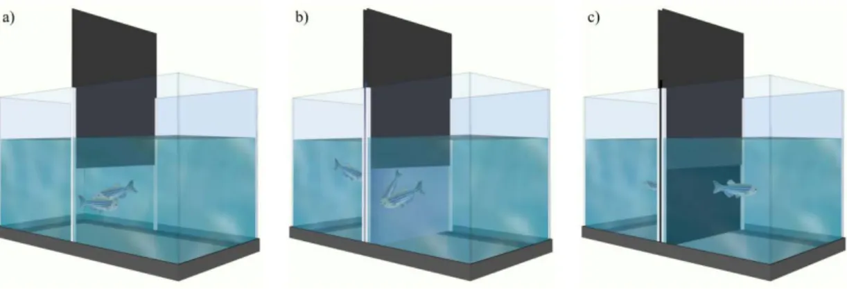
Here we used a behavioural paradigm where animals could experience winning or losing a social interaction and consequently adjust their behaviour to a new social status (behavioural shift). The specific cues that trigger changes in social status were also investigated. There are at least two potential sources of social status information available in a social interaction: the own aggressive behaviour expressed by the individual; and the behaviour expressed by the opponent. Theoretically, animals can use just one of these two types of information (self-assessment or opponent only assessment, respectively) or combine both in mutual assessment, which assumes that contestants know their own competitive abilities, gather information about the opponent, and integrate both into a behavioural adaptive response (Elwood & Arnott 2012). Therefore, the perception that the individual has of the interaction is a key feature in the modulation of the behavioural response. In order to assess the type of assessment zebrafish uses to trigger a status-dependent behavioural shift, three social treatments were used (Figure 2):
1) Staged fights between pairs of real-opponent conspecifics, which resulted in a winner and a loser (Figure 2a);
2) Mirror-fights, which resulted in unsolved interactions and the expression of aggressiveness is decoupled from the experience of winning or losing (Figure 2b); fish do not recognize themselves on a mirror, and attack their own image as if it is an intruder (Oliveira et al. 2005), however since submissive behaviour is never expressed
by the opponents (i.e. the mirror image replicates the aggressive behaviour of the focal fish) focal fish never experiences a victory;
3) No agonistic interaction, which were used as a reference group (Figure 2c).
Figure 2 – Experimental apparatus: a) Real opponent interaction, fish fought with a conspecific, and a Winner and a Loser emerge; b) Mirror interaction, fish fought with their own image on the mirror but did not experience a change in social status; c) Control group, no agonistic interaction or mirror stimulation.
These social treatments generated four social behaviour states: Winners and Losers of the real opponent interaction; Mirror-fighters; and fish with no social interaction.
Our hypothesis is that winners and losers will have different neuronal states that are experience dependent, and for mirror fighters the following premises can be generated:
1) if only the individuals own behavioural expression would be relevant for the individual’s assessment of fight outcome, then mirror-fighters should have a neuromolecular profile similar to that of winners;
2) if only the behavioural feedback from opponent would be relevant, then mirror-fighters should have a neuromolecular profile similar to that of losers;
3) if the comparison between perceived behaviour of the opponent and the own expressed behaviour is needed, then mirror-fighters should not activate a response because in mirror interactions they equal each other, and therefore no change in social
Chapter I. General Introduction status would be experienced by the subject, leading to a neuromolecular profile different from those of both winners and losers.
All tests were done in pairs in order to give individuals the access to conspecific odours, which would otherwise only be present in real opponent dyads, therefore avoiding confounding effects of putative chemical cues in the comparisons between treatments.
In a first set of experiments we studied the influence of neuromodulators on social plasticity mechanisms. For this purpose we characterized the endocrine response to social challenges (Chapter II), and the social modulation of brain monoamines (Chapter III) and nonapeptides (Chapter IV). Next we tested the SDM network hypothesis by contrasting changes in functional localization vs. connectivity across this network in response to changes in social status (Chapter V). Finally, we characterized the changes in expression of key genes for different neuroplasticity mechanisms (e.g. neurogenesis, synaptogenesis, changes in synaptic strength) in response to changes social status (Chapter VI).
1.8. References
Abril-de-Abreu R, Cruz J, Oliveira RF (2015a) Social Eavesdropping in Zebrafish: Tuning of Attention to Social Interactions. Scientific Reports, 5, 12678.
Abril-de-Abreu R, Cruz AS, Oliveira RF (2015b) Social dominance modulates eavesdropping in zebrafish. Royal Society Open Science, 2, 150220.
Aires RF, Oliveira GA, Oliveira TF, Ros AFH, Oliveira RF (2015) Dear Enemies Elicit Lower Androgen Responses to Territorial Challenges than Unfamiliar Intruders in a Cichlid Fish. PloS One, 10, e0137705.
Albers HE, Dean A, Karom MC, Smith D, Huhman KL (2006) Role of V1a vasopressin receptors in the control of aggression in Syrian hamsters. Brain Research, 1073-1074, 425–430.
Almeida O, Oliveira RF (2015) Social Status and Arginine Vasotocin Neuronal Phenotypes in a Cichlid Fish. Brain, Behavior and Evolution, 85, 203–213.
Amat J, Aleksejev R, Paul E, Watkins L, Maier S (2010) Behavioral control over shock blocks behavioral and neurochemical effects of later social defeat. Neuroscience, 165, 1031– 1038.
Arnold AP, Breedlove SM (1985) Organizational and activational effects of sex steroids on brain and behavior: a reanalysis. Hormones and Behavior, 19, 469–498.
Aubin-Horth N, Renn SCP (2009) Genomic reaction norms: using integrative biology to understand molecular mechanisms of phenotypic plasticity. Molecular Ecology, 18, 3763–3780.
Van der Bijl W, Thyselius M, Kotrschal A, Kolm N (2015) Brain size affects the behavioural response to predators in female guppies (Poecilia reticulata). Proceedings of the Royal
Society B: Biological Sciences, 282, 20151132.
Bill BR, Petzold AM, Clark KJ, Schimmenti LA, Ekker SC (2009) A Primer for Morpholino Use in Zebrafish. Zebrafish, 6, 69–77.
Biran J, Tahor M, Wircer E, Levkowitz G (2015) Role of developmental factors in hypothalamic function. Frontiers in Neuroanatomy, 9, 47.
Brindle P, Linke S, Montminy M (1993) Protein-kinase-A-dependent activator in transcription factor CREB reveals new role for CREM repressors. Nature, 364, 821–824.
Caramaschi D, Boer S de, Koolhaas J (2007) Differential role of the 5-HT 1A receptor in aggressive and non-aggressive mice: An across-strain comparison. Physiology &
Behavior, 90, 590–601.
Cardoso SD, Teles MC, Oliveira RF (2015) Neurogenomic mechanisms of social plasticity. The
Journal of Experimental Biology, 218, 140–149.
Charnay Y, Léger L (2010) Brain serotonergic circuitries. Dialogues in Clinical Neuroscience, 12, 471–487.
Clayton DF (2000) The genomic action potential. Neurobiology of Learning and Memory, 74, 185–216.
Cooper MA, Karom M, Huhman KL, Albers HE (2005) Repeated agonistic encounters in hamsters modulate AVP V1a receptor binding. Hormones and Behavior, 48, 545–551. Cruz A, Oliveira R (2015) Audience effects and aggressive priming in agonistic behaviour of
male zebrafish, Danio rerio. Animal Behaviour, 107, 269–279.
Dahlbom SJ, Backström T, Lundstedt-Enkel K, Winberg S (2012) Aggression and monoamines: effects of sex and social rank in zebrafish (Danio rerio). Behavioural Brain Research, 228, 333–338.
Daubner SC, Le T, Wang S (2011) Tyrosine hydroxylase and regulation of dopamine synthesis.
Archives of Biochemistry and Biophysics, 508, 1–12.
Chapter I. General Introduction
Dewan AK, Ramey ML, Tricas TC (2011) Arginine vasotocin neuronal phenotypes, telencephalic fiber varicosities, and social behavior in butterflyfishes (Chaetodontidae): potential similarities to birds and mammals. Hormones and Behavior, 59, 56–66.
Donaldson ZR, Young LJ (2008) Oxytocin, vasopressin, and the neurogenetics of sociality.
Science, 322, 900–904.
Douglass AD, Kraves S, Deisseroth K, Schier AF, Engert F (2008) Escape behavior elicited by single, channelrhodopsin-2-evoked spikes in zebrafish somatosensory neurons. Current
Biology, 18, 1133–1137.
Doutrelant C, McGregor P, Oliveira R (2001) The effect of an audience on intrasexual communication in male Siamese fighting fish, Betta splendens. Behavioral Ecology, 12, 283–286.
Dunbar RIM (1998) The social brain hypothesis. Evolutionary Anthropology: Issues, News, and
Reviews, 6, 178–190.
Dunbar RIM, Shultz S (2007) Evolution in the social brain. Science, 317, 1344–1247.
Earley RL (2010) Social eavesdropping and the evolution of conditional cooperation and cheating strategies. Philosophical transactions of the Royal Society of London. Series B,
Biological Sciences, 365, 2675–2686.
Elwood RW, Arnott G (2012) Understanding how animals fight with Lloyd Morgan’s canon.
Animal Behaviour, 84, 1095–1102.
Engeszer RE, Barbiano LA DA, Ryan MJ, Parichy DM (2007) Timing and plasticity of shoaling behaviour in the zebrafish, Danio rerio. Animal Behaviour, 74, 1269–1275.
Van Erp AMM, Miczek KA (2000) Aggressive Behavior, Increased Accumbal Dopamine, and Decreased Cortical Serotonin in Rats. The Journal of Neuroscience, 20, 9320–9325. Ferris CF, Lu S-F, Messenger T et al. (2006) Orally active vasopressin V1a receptor antagonist,
SRX251, selectively blocks aggressive behavior. Pharmacology, Biochemistry, and
Behavior, 83, 169–174.
Ferris C, Meenan D, Axelson J, Albers H (1986) A vasopressin antagonist can reverse dominant/subordinate behavior in hamsters. Physiology & Behavior, 38.
Filby A, Paull G, Hickmore T, Tyler C (2010) Unravelling the neurophysiological basis of aggression in a fish model. BMC Genomics, 11, 498.
Finlay BL, Darlington RB (1995) Linked regularities in the development and evolution of mammalian brains. Science, 268, 1578–1584.
Fischer S, Bessert-Nettelbeck M, Kotrschal A, Taborsky B (2015) Rearing-Group Size Determines Social Competence and Brain Structure in a Cooperatively Breeding Cichlid.
The American Naturalist, 186, 123–140.
Gerlach G, Hodgins-Davis A, Avolio C, Schunter C (2008) Kin recognition in zebrafish: a 24-hour window for olfactory imprinting. Proceedings of the Royal Society B: Biological
Sciences, 275, 2165–2170.
Godwin J (2010) Neuroendocrinology of sexual plasticity in teleost fishes. Frontiers in
Neuroendocrinology, 31, 203–216.
Godwin J, Thompson R (2012) Nonapeptides and social behavior in fishes. Hormones and
Behavior, 61, 230–238.
Goodson JL (2005) The vertebrate social behavior network: evolutionary themes and variations.
Hormones and Behavior, 48, 11–22.
Goodson JL (2008) Nonapeptides and the evolutionary patterning of sociality. Progress in
Brain Research, 170, 3–15.
Goodson JL, Bass AH (2001) Social behavior functions and related anatomical characteristics of vasotocin/vasopressin systems in vertebrates. Brain Research Reviews, 35, 246–265. Goodson JL, Schrock SE, Kingsbury M a (2015) Oxytocin mechanisms of stress response and
aggression in a territorial finch. Physiology & Behavior, 141, 154–163.
Goodson J, Schrock S, Klatt J (2009) Mesotocin and nonapeptide receptors promote estrildid flocking behavior. Science, 325, 862–866.
Goodson JL, Thompson RR (2010) Nonapeptide mechanisms of social cognition, behavior and species-specific social systems. Current Opinion in Neurobiology, 20, 784–794.
Greenwood AK, Wark AR, Fernald RD, Hofmann HA (2008) Expression of arginine vasotocin in distinct preoptic regions is associated with dominant and subordinate behaviour in an African cichlid fish. Proceedings of the Royal Society B: Biological Sciences, 275, 2393– 2402.
Grosenick L, Clement TS, Fernald RD (2007) Fish can infer social rank by observation alone.
Nature, 445, 429–432.
Harmon AC, Huhman KL, Moore TO, Albers HE (2002) Oxytocin inhibits aggression in female Syrian hamsters. Journal of Neuroendocrinology, 14, 963–969.
Higashijima S, Masino MA, Mandel G, Fetcho JR (2003) Imaging neuronal activity during zebrafish behavior with a genetically encoded calcium indicator. Journal of
Neurophysiology, 90, 3986–3997.
Howe K, Clark MD, Torroja CF et al. (2013) The zebrafish reference genome sequence and its relationship to the human genome. Nature, 496, 498–503.
Hsu Y, Earley R, Wolf L (2006) Modulation of aggressive behaviour by fighting experience: mechanisms and contest outcomes. Biological Reviews, 81, 33–74.
Insel TR (2010) The challenge of translation in social neuroscience: a review of oxytocin, vasopressin, and affiliative behavior. Neuron, 65, 768–779.