The article was published by Academy of Chemistry of Globe Publications www.acgpubs.org/RNP © Published 05/01/2014 EISSN:1307-6167
Rec. Nat. Prod. 8:3 (2014) 228-233
Saponins from Cephalaria aristata C. Koch
Derya Gülcemal
*1, Milena Masullo
2, Serdar Gökhan
Ş
enol
3,
Özgen Alanku
ş
-Çalı
ş
kan
1, and Sonia Piacente
*21
Ege University, Faculty of Science, Department of Chemistry, Bornova, 35100, İzmir, Türkiye 2
Salerno University, Department of Pharmacia, 84084 Fisciano (Salerno), Italy
3
Department of Biology, Faculty of Science, Ege University, Bornova, 35100 İzmir, Türkiye
(Received June 11, 2013; Revised September 15, 2013; Accepted October 24, 2013)
Abstract: One new oleanane-type saponin, 3-O-β-D-glucopyranosyl-(1→4)-β-D-xylopyranosyl-(1→3)-α -L-rhamnopyranosyl-(1→2)-α-L-arabinopyranosylhederagenin 28-O-β-D-glucopyranosyl-(1→6)-β -D-gluco-pyranosyl ester (1) was isolated from the MeOH extract of whole plant parts of Cephalaria aristata C. Koch along with three known oleanane-type saponins, 3-O-α-L-rhamnopyranosyl-(1→2)-α-L-arabinopyranosyl hederagenin 28-O-(β-D-glucopyranosyl-(1→6)-β-D-glucopyranosyl) ester (2), 3-O-β-D-glucopyranosyl-(1→
4)-β-D-xylopyranosyl-(1→3)-α-L-rhamnopyranosyl-(1→2)-α-L-arabinopyranosyl hederagenin (3) and 3-O-α -L-rhamnopyranosyl-(1→2)-α-L-arabinopyranosylhederagenin (4). Also three triterpenoids and a steroid glucoside oleanolic acid (5), β-amyrin (6), 20-β-hydroxyursolic acid (7) and 29-hydroxystigmast-5-en-3-O-β -D-glucopyranoside (8). Their structures were established by the extensive use of 1D- and 2D-NMR experiments along with ESIMS and HRMS analysis.
Keywords: Cephalaria aristata; Dipsacaceae; Oleanane-type saponins. © 2014 ACG Publications. All rights reserved.
1. Introduction
The genus Cephalaria (Dipsacaceae) consists of 94 species distributed mainly in the Mediterranean region and the Middle East. In Turkey, there are 40 Cephalaria taxa and 24 of them are endemic [1-3].
Cephalaria aristata is an annual herb which mainly grows at inner and east Anatolia, also Armenia
[2]. Several Cephalaria species have been used as folk medicine for their hypothermal, alleviative, relaxant and anti-infective activities [4,5]. Flavonoids [6,7], iridoids [8] and triterpene saponins with antimicrobial [9-11], antifungal [9], molluscicidal [10] and cytotoxic activities [10,12] have been previously reported from this genus. As a part of our ongoing research of new compounds from Turkish Dipsacaceae species [A-D] [13-16] we carried out a study on Cephalaria aristata C. Koch. This paper reports the isolation of one new oleanane-type triterpene glycoside (1) from the methanol extract of the whole plant of C. aristata along with three known oleanane-type glycosides (2-4), three aglycones (5-7) and one sterol glucoside (8). Their structures were elucidated by extensive
*
spectroscopic methods including 1D- (1H, 13C and TOCSY) and 2D-NMR (DQF-COSY, HSQC, HMBC and ROESY) experiments as well as ESIMS and HRMS analysis.
2. Materials and Methods
2.1. General procedures
Optical rotations were measured on a JASCO DIP 1000 polarimeter. IR measurements were obtained on a Bruker IFS-48 spectrometer. NMR experiments were performed on a Bruker DRX-600 spectrometer (Bruker BioSpinGmBH, Rheinstetten, Germany) equipped with a Bruker 5 mm TCI CryoProbeat 300 K. All 2D-NMR spectra were acquired in CD3OD (99.95%, SigmaAldrich) and
standard pulse sequences and phase cycling were used for DQF-COSY, HSQC, and HMBC spectra. The NMR data were processed using UXNMR software. Exact masses were measured by a Voyager DE mass spectrometer. Samples were analyzed by matrix-assisted laser desorption ionization time-of-flight (MALDITOF) mass spectrometry. A mixture of analyte solution and a-cyano-4-hydroxycinnamic acid (Sigma) was applied to the metallic sample plate and dried. Mass calibration was performed with the ions from ACTH (fragment 18-39) at 2465.1989 Da and angiotensin III at 931.5154 Da as internal standard. ESIMS analyses were performed using a ThermoFinnigan LCQ Deca XP Max iontrap mass spectrometer equipped with Xcalibur software.
2.2. Plant Material
Cephalaria aristata C. Koch was collected from Darende-Malatya, Turkey in June 2010 and
identified by one of the co-author, Serdar G. Şenol (Deparment of Biology, Faculty of Sciences, Ege University, İzmir, Turkey). A voucher specimen (EGE 40857) is deposited in the Ege University Botanical Garden & Herbarium Research and The Application Center, İzmir, Turkey.
2.3. Extraction and Isolation
Air-dried and grinded plant material (900 g) were extracted with MeOH (4x5 L) at room temperature. After filtration, the solvent was removed by rotary evaporation yielding (60 g) of MeOH extract. The MeOH extract was dissolved in H2O (350 ml), and successively partitioned with n-hexane
(3x150 mL), CH2Cl2 (4x150 mL), and n-BuOH saturated with H2O (4x150 mL). The n-BuOH phase
(10 g) was chromatographed over Sephadex column chromatography to give ten main fractions (A-J). Fraction A (2.5 g) was subjected to vacuum liquid chromatography (VLC) using reversed-phase material (RP-18) employing H2O (600 mL), H2O-MeOH (8:2, 1000 mL; 6:4, 1000 mL; 4:6, 1500 mL;
3:7, 1000 mL; 2:8, 1200 mL) and MeOH (800 mL) to give 6 main fractions. Fraction 2 (50 mg) was subjected to RP-HPLC column chromatography with the solvent system MeOH:H2O (50:50, 250 ml)
to yield compound 1 (3 mg). Fraction 3 (50 mg) was submitted to RP-HPLC column chromatography with the solvent system MeOH:H2O (60:40, 250 ml) to yield compounds 2 (3 mg) and 3 (2 mg).
Fraction 4 (60 mg) was subjected to RP-HPLC column chromatography with the solvent system MeOH:H2O (70:30, 250 ml) to yield compound 4 (3 mg). Fraction 5 (50 mg) was subjected to
RP-HPLC column chromatography with the solvent system MeOH:H2O (90:10, 200 ml) to yield
compounds 5 (3 mg), 6 (4 mg) and 7 (3 mg). Fraction 6 (40 mg) was submitted to RP-HPLC column chromatography with the solvent system MeOH:H2O (95:15, 250 ml) to yield compound 8 (2 mg).
2.4. Acid hydrolysis
The crude saponin mixture (300 mg) was heated at 60 °C with 1:1 0.5 N HCl-dioxane (100 ml) for 2 h, and the mixture was then evaporated in vacuo. The residue was partitioned with CH2Cl2-H2O, and
the H2O layer was neutralized with Amberlite MB-3. The H2O layer was then concentrated and passed
through a silica gel column, using CHCl3-MeOH-H2O (7:1:1.2, lower layer) as eluting solvent to
values with those reported in the literature [17] (Eskander et al., 2006). The optical rotations were determined after dissolving the sugars in H2O and allowing them to stand for 24 h; D-xylose [α]
22 D
+10 (c 0.1), L-arabinose [α]22D +80.5 (c 0.1), D-glucose [α] 22
D +51.0 (c 0.1), L-rhamnose [α] 22
D +13.8
(c 0.1).
O HOH2C
C O OH HO O O OH O HO α-L-ara α-L-rha O HO HO OH OH
β-D-glcI β -D-xyl O HO HO OH O O HO HO OH OH β-D-glcIII β-D-glcII O O O O HO OH
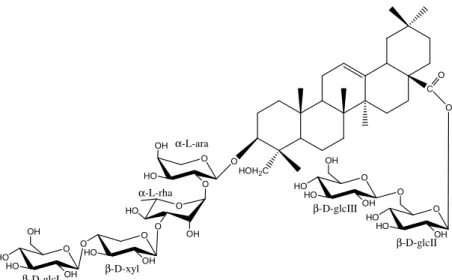
Figure 1.Structure of compound 1
O HOH2C
C O OH HO O O OH O HO α-L-ara α-L-rha O HO HO OH OH
β-D-glcI β-D-xyl
O HO HO OH O O HO HO OH OH β-D-glcIII β-D-glcII O O O O HO OH
Figure 2.Key HMBC of compound 1
3. Results and Discussion
3.1. Structure elucidation
The HRMALDITOF mass spectrum of 1 (m/z 1391.6465 [M+Na]+, calcd for C64H104O31Na,
1391.6459) supported a molecular Formula of C64H104O31. The ESIMS spectrum showed a major ion
[M+Na-162-162]+, corresponding to the loss of a hexose unit, 1023 [M+Na-162-162-44]+, due to consecutive losses of carbon dioxide, 861 [M+Na-162-162-44-162]+, corresponding to the loss of a hexose unit, 729 [M+Na-162-162-44-162-132]+, corresponding to the loss of a pentose unit, 583 [M+Na-162-162-44-162-132-146]+, corresponding to the loss of a deoxy-hexose unit, 451 [M+Na-162-162-44-162-132-146-132]+, corresponding to the loss of a pentose unit were observed.
The 1H NMR spectrum of 1 displayed signals for six tertiary methyl groups at δ 0.74, 0.83, 0.94, 0.97, 1.00 and 1.21, for an olefinic proton at δ 5.27 (t, J=3.5 Hz), one oxygen-bearing methine proton at δ 3.64 (dd, J=11.5, 4.2 Hz, H-3) and one primary alcoholic function at δ 3.36, 3.61 (H-23) (Table 1). These signals along with the carbon resonances in the 13C NMR spectrum for the methyl groups at δ 13.8, 17.7, 33.5, 23.8, 16.6, 26.3 and the two olefinic carbons at δ 123.5, 146.3 suggested that compound 1 possessed hederagenin as aglycon [18]. The downfield shifts of C-3 (δ 82.2) and C-28 (177.7) of the aglycon suggested that compound 1 was a bidesmosidic glycoside. The 13C NMR spectrum showed 64 carbon signals, of which 30 were assigned to the aglycon moiety and 34 to a sugar portion made up of six sugar units. The 1H NMR spectrum displayed in the sugar region signals corresponding to six anomeric protons at δ 4.37 (d, J=7.5 Hz), 4.37 (d, J=7.5 Hz), 4.54 (d, J=7.5 Hz), 4.55 (d, J=3.7Hz), 5.26 (d, J=1.2 Hz) and 5.38 (d, J=7.5 Hz) which were unambiguously correlated by HSQC experiment to the corresponding carbon resonances at δ 103.2, 104.5, 105.8, 104.5, 100.9 and 95.3, respectively. The chemical shifts of all the individual protons of the six sugar units were ascertained from a combination of 1D-TOCSY and DQF-COSY spectral analysis, and the 13C chemical shifts of their relative attached carbons were assigned unambiguously from the HSQC spectrum (Table 1). These data showed the presence of three β-glucopyranosyl units (δ 4.37, 2H and 5.38), one α-rhamnopyranosyl unit (δ 5.26), one β-xylopyranosyl unit (δ 4.54) and one α -arabinopyranosyl unit (δ 4.55). An unambiguous determination of the sequence and linkage sites was obtained from the HMBC spectrum, which showed key correlation peaks between the proton signal of
δ H-1ara (δ 4.55) and the carbon resonance of C-3 (δ 82.2), H-1rha (δ 5.26) and C-2ara (δ 75.9), H-1xyl (δ
4.54) and C-3rha (δ 82.2), H-1glcI (δ 4.37) and C-4xyl (δ 78.1), H-1glcII (δ 5.38) and C-28 (δ 177.7) and
H-1glcIII (δ 4.37) and the carbon resonance of C-6glcII (δ 69.2). The absolute configurations of the sugar
units was determined by acid hydrolysis of the crude saponin mixture as D-glucose, D-xylose, L-rhamnose and L-arabinose and were assigned on the basis of their optical rotation values [17].
On the basis of all these evidence, the structure of the new compound 1 was established as 3-O-β -D-glucopyranosyl-(1→4)-β-D-xylopyranosyl-(1→3)-α-L-rhamnopyranosyl-(1→2)-α
-L-arabinopyranosylhederagenin 28-O-β-D-glucopyranosyl-(1→6)-β-D-glucopyranosyl ester.
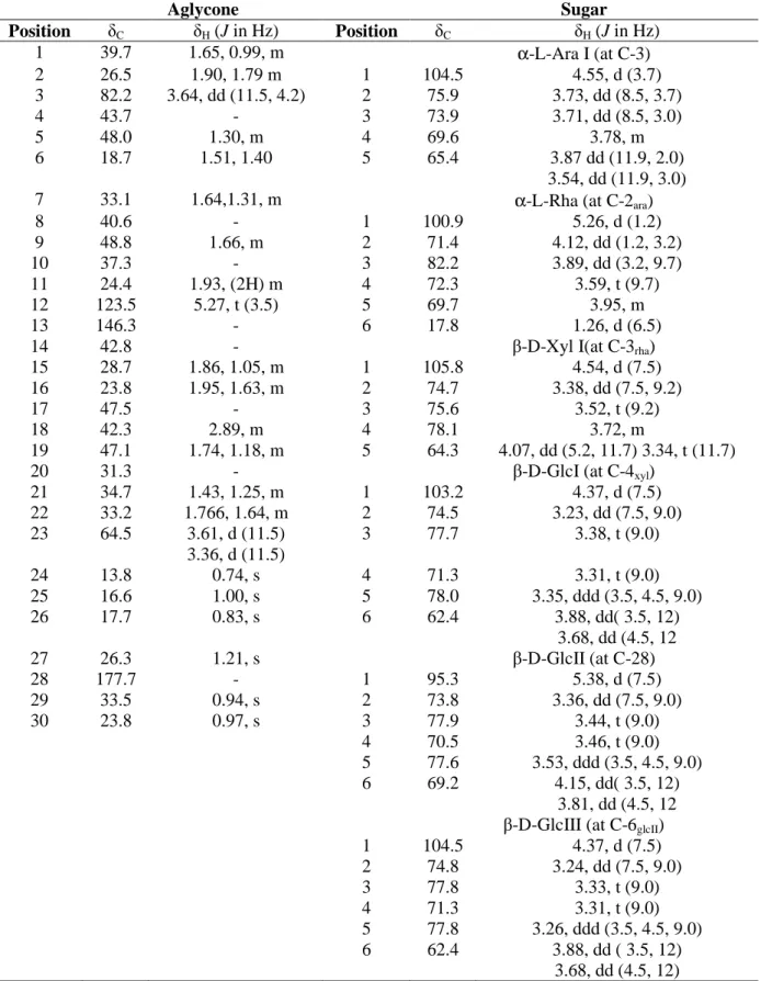
Table 1. 13C and 1H NMR data (J in Hz) of the aglycon and sugar moieties of compound 1 (600Mz, δ ppm, in CD3OD).
References
[1] V. A. Matthews (1972). Cephalaria Schrader ex Roemer & Schultes, In: Flora of Turkey and the East Aegean Islands, ed: Davis, P.H., Edinburgh University Press, Edinburgh, vol. 4, pp. 585-597.
Aglycone Sugar
Position δC δH (J in Hz) Position δC δH (J in Hz)
1 39.7 1.65, 0.99, m α-L-Ara I (at C-3)
2 26.5 1.90, 1.79 m 1 104.5 4.55, d (3.7)
3 82.2 3.64, dd (11.5, 4.2) 2 75.9 3.73, dd (8.5, 3.7)
4 43.7 - 3 73.9 3.71, dd (8.5, 3.0)
5 48.0 1.30, m 4 69.6 3.78, m
6 18.7 1.51, 1.40 5 65.4 3.87 dd (11.9, 2.0)
3.54, dd (11.9, 3.0)
7 33.1 1.64,1.31, m α-L-Rha (at C-2ara)
8 40.6 - 1 100.9 5.26, d (1.2)
9 48.8 1.66, m 2 71.4 4.12, dd (1.2, 3.2)
10 37.3 - 3 82.2 3.89, dd (3.2, 9.7)
11 24.4 1.93, (2H) m 4 72.3 3.59, t (9.7)
12 123.5 5.27, t (3.5) 5 69.7 3.95, m
13 146.3 - 6 17.8 1.26, d (6.5)
14 42.8 - β-D-Xyl I(at C-3rha)
15 28.7 1.86, 1.05, m 1 105.8 4.54, d (7.5)
16 23.8 1.95, 1.63, m 2 74.7 3.38, dd (7.5, 9.2)
17 47.5 - 3 75.6 3.52, t (9.2)
18 42.3 2.89, m 4 78.1 3.72, m
19 47.1 1.74, 1.18, m 5 64.3 4.07, dd (5.2, 11.7) 3.34, t (11.7)
20 31.3 - β-D-GlcI (at C-4xyl)
21 34.7 1.43, 1.25, m 1 103.2 4.37, d (7.5)
22 33.2 1.766, 1.64, m 2 74.5 3.23, dd (7.5, 9.0)
23 64.5 3.61, d (11.5) 3.36, d (11.5)
3 77.7 3.38, t (9.0)
24 13.8 0.74, s 4 71.3 3.31, t (9.0)
25 16.6 1.00, s 5 78.0 3.35, ddd (3.5, 4.5, 9.0)
26 17.7 0.83, s 6 62.4 3.88, dd( 3.5, 12)
3.68, dd (4.5, 12
27 26.3 1.21, s β-D-GlcII (at C-28)
28 177.7 - 1 95.3 5.38, d (7.5)
29 33.5 0.94, s 2 73.8 3.36, dd (7.5, 9.0)
30 23.8 0.97, s 3 77.9 3.44, t (9.0)
4 70.5 3.46, t (9.0)
5 77.6 3.53, ddd (3.5, 4.5, 9.0)
6 69.2 4.15, dd( 3.5, 12)
3.81, dd (4.5, 12
β-D-GlcIII (at C-6glcII)
1 104.5 4.37, d (7.5)
2 74.8 3.24, dd (7.5, 9.0)
3 77.8 3.33, t (9.0)
4 71.3 3.31, t (9.0)
5 77.8 3.26, ddd (3.5, 4.5, 9.0)
6 62.4 3.88, dd ( 3.5, 12)
[2] P. H. Davis, R. R. Mill, and K. Tan (1988). Cephalaria Schrader ex Roemer & Schultes, In: Flora of Turkey and the East Aegean Islands, eds: P. H. Davis, R. R. Mill and K. Tan, Edinburgh University Press, Edinburgh, vol. 10, p. 156.
[3] H. Duman (2000). Cephalaria Schrader ex Roemer & Schultes, In: Flora of Turkey and the East Aegean Islands, eds: A. Güner, N. Ozhatay, T. Ekim and K. H. C. Başer, Edinburgh University Press, Edinburgh, vol 11, pp. 147-149.
[4] L. D. Zvidadze, G. E. Dekanosidze, O. D. Dzhikiya, E. P. Kemertelidze and A. S. Shashkov (1981). Triterpene glycosides of Cephalaria gigantea. II. Structure of giganteasides D and G, Bioorg. Khim., 7, 736-740.
[5] D. Godevac, N. Menkovic, L. Vujisic, V. Tesevic, V. Vajs and S. Milosavljevic (2010). A new triterpenoid saponin from the aerial parts of Cephalaria ambrosioides, Nat. Prod. Res., 24, 1307–1312. [6] I. S. Movsumov, E. A. Garaev and M. I. Isaev (2006). Flavonoids from Cephalaria gigantea flowers,
Chem. Nat. Compd., 42, 677-680.
[7] I. S. Movsumov, E. A. Garaev and M. I. Isaev (2009). Flavonoids from Cephalaria grossheimii, Chem. Nat. Compd., 45, 422-423.
[8] D. Godevac, B. Mandic, V. Vajs, V. Tesevic, N. Menkovic, P. Janackovic and S. Milosavljevic (2006). Triterpenoid saponins and iridoid glycosides from the aerial parts of Cephalaria pastricensis, Biochem. Syst. and Ecol., 34, 890-893.
[9] S. Kirmizigul, H. Anil, F. Ucar and K. Akdemir (1996). Antimicrobial and antifungal activities of three new triterpenoid glycosides, Phytother. Res., 10, 274-276.
[10] S. Pasi, N. Aligiannis, H. Pratsinis, A. L. Skaltsounis and I. B., Chinou (2009). Biologically active triterpenoids from Cephalaria ambrosioides, Planta Med., 75, 163-167.
[11] N. B. Sarıkahya and S. Kırmızıgül (2012). Antimicrobially active hederagenin glycosides from Cephalaria elmaliensis. Planta Med., 78 (8): 828-833.
[12] N. Tabatadze, R. Elias, R. Faure, P. Gerkens, M. C. Pauw-Gillet, E Kemertelidze, A. Chea and E. Ollivier (2007). Cytotoxic triterpenoid saponins from the roots of Cephalaria gigantea, Chem. Pharm. Bull., 55, 102-105.
[13] O. Alankus-Caliskan and H. Anil (1995). A bidesmosidic triterpene saponin from Cephalaria transsylvanica, Phytochemistry, 38, 1493-1495.
[14] O. Alankus-Caliskan, S. Emirdag, E. Bedir, S. Avunduk and H. Anil (2004). Triterpene saponins from Knautia integrifolia var. bidens, Z. Naturforsch B., 59, 821-824.
[15] E. Polat, O. Alankus-Caliskan, T. Karayildirim and E. Bedir (2010). Iridoids from Scabiosa atropurpurea L. subsp maritima Arc. (L.), Biochem. Syst. and Ecol., 38, 253-255.
[16] D. Gulcemal, M. Masullo, O. Alankus-Caliskan, T. Karayildirim, S. G. Senol, S. Piacente and E. Bedir (2010). Monoterpenoid glucoindole alkaloids and iridoids from Pterocephalus pinardii, Magn. Reson. Chem., 48, 239-243.
[17] J. Eskander, C. Lavaud, I. Pouny, H. S. M. Soliman, S. M. Abdel-Khalik and I. I. Mahmoud (2006). Saponins from the seeds of Mimusops laurifolia, Phytochemistry, 67, 1793–1799.
[18] K. Tori, S. Sera, A. Shimaoka and Y. Tomita (1974). Carbon-13 NMR spectra of olean-12-enes. Full signal assignments including quaternary carbon signals assigned by use of indirect 13C, 1H spin couplings, Tetrahedron Lett., 15, 4227–4230.
[19] X. C. Li, C. R. Yang, Y. Q. Liu, R. Kasai, K. Ohtani, K. Yamashaki, K. Miyahara and K. Shingu (1995). Triterpenoid glycosides from Anemoclema glaucifolium, Phytochemistry, 39, 1175–1179. [20] D. A. Panov, V. I. Grishkovets, V. V. Kachala and A. S. Shashkov (2006). Triterpene glycosides from
Kalopanax septemlobum. VI. Glycosides from leaves of Kalopanax septemlobum var. typicum introduced to Crimea, Chem. Nat. Compd., 42, 49-54.
[21] J. Kong, X. G. Li, B. Y. Wei and C. R. Yang (1993). Triterpenoid glycosides from Decaisnea fargesii, Phytochemistry, 33, 427–30.
[22] S. B. Mahato and A. P. Kundu (1994). 13C NMR spectra of pentacyclic triterpenoidssa compilation and some salient features, Phytochemistry, 37, 1517-1575.
[23] O. V. Makarova and M. I. Isaev (1997). Isoprenoids of Sambucus nigra, Chem. Nat. Compd., 33, 702-703.
[24] A. Rashid, S. Firdous, V. U. Ahmad, K .M. Khan, S. B. Usmani and M. Ahmed, (1999). A new glycoside from Polianthes tuberosa Linn., Pakistan J. Sci. Ind. R., 42, 26.