Protective Macroautophagy Is Involved in
Vitamin E Succinate Effects on Human
Gastric Carcinoma Cell Line SGC-7901 by
Inhibiting mTOR Axis Phosphorylation
Liying Hou1, Yuze Li1,2, Huacui Song1, Zhihong Zhang1,3, Yanpei Sun1, Xuguang Zhang1,4, Kun Wu1*
1Department of Nutrition and Food Hygiene, School of Public Health, Harbin Medical University, Harbin, China,2Department of the Fourth Internal Medicine, The Fourth Hospital of Heilongjiang Province, Harbin, China,3Food Processing Institute, Heilongjiang Academy of Agricultural Sciences, Harbin, China,
4Department of Internal Medicine, Hematology and Oncology, Harbin Children’s Hospital, Harbin, China
*wukun_15000@126.com
Abstract
Vitamin E succinate (VES), a potential cancer therapeutic agent, potently induces apoptosis and inhibits the growth of various cancer cells. Autophagy has been supposed to promote can-cer cell survival or trigger cell death, depending on particular cancan-cer types and tumor microen-vironments. The role of autophagy in the growth suppressive effect of VES on gastric cancer cell is basically unknown. We aimed to determine whether and how autophagy affected the VES-induced inhibition of SGC-7901 human gastric carcinoma cell growth. SGC-7901 cells were treated with VES or pre-treated with autophagy inhibitor, chloroquine (CQ) and 3-methy-ladenine (3-MA). Electron microscopy, fluorescence microscopy and Western blot were used to study whether VES induced autophagy reaction in SGC-7901 cells. Western blot evaluated the activities of the mammalian target of rapamycin (mTOR) axis. Then we used 3-(4,5-dimethylthiazol-2-yl)-2,5-diphenyltetrazolium bromide (MTT) and flow cytometry to detect the level of cell viability and apoptosis. Collectively, our data indeed strongly support our hypothe-sis that VES treatment produced cytological variations that depict autophagy, increased the amount of intracellular green fluorescent protein—microtubule associated protein 1 light chain 3 (GFP-LC3) punctate fluorescence and the number of autophagic vacuoles. It altered the expression of endogenous autophagy marker LC3. VES activated the suppression of mTOR through inhibiting upstream regulators p38 MAPK and Akt. mTOR suppression consequently inhibited the activation of mTOR downstream targets p70S6K and 4E-BP-1. The activation of the upstream mTOR inhibitor AMPK had been up-regulated by VES. The results showed that pre-treatment SGC-7901 with autophagy inhibitors before VES treatment could increase the capacity of VES to reduce cell viability and to provoke apoptosis. In conclusion, VES-induced autophagy participates in SGC-7901 cell protection by inhibiting mTOR axis phosphorylation. Our findings not only strengthen our understanding of the roles of autophagy in cancer biology, but may also be useful for developing new treatments for gastric cancer patients.
OPEN ACCESS
Citation:Hou L, Li Y, Song H, Zhang Z, Sun Y, Zhang X, et al. (2015) Protective Macroautophagy Is Involved in Vitamin E Succinate Effects on Human Gastric Carcinoma Cell Line SGC-7901 by Inhibiting mTOR Axis Phosphorylation. PLoS ONE 10(7): e0132829. doi:10.1371/journal.pone.0132829
Editor:Ying-Jan Wang, National Cheng Kung University, TAIWAN
Received:February 5, 2015
Accepted:June 19, 2015
Published:July 13, 2015
Copyright:© 2015 Hou et al. This is an open access article distributed under the terms of theCreative Commons Attribution License, which permits unrestricted use, distribution, and reproduction in any medium, provided the original author and source are credited.
Data Availability Statement:All relevant data are within the paper and its Supporting Information files.
Funding:This work was supported by grants from the National Natural Science Foundation of China (no. 81172651) to KW.
Introduction
Gastric carcinoma is among the most commonly diagnosed cancers in the world and is the second most frequent cause of cancer-associated mortality[1]. The incidence of gastric carci-noma and mortality from this disease have drastically decreased in most countries over the past 70 years, but gastric carcinoma is still the fourth most common cancer[2]. Gastric carci-noma is the third most common malignancy in China[3]. The major gastric carcicarci-noma treat-ment modalities include surgery and chemotherapy, but survival among patients is low. The failure of chemotherapy is due to the development of drug resistance and toxicity. New strate-gies that overcome the abovementioned difficulties are required for treating gastric carcinoma.
Vitamin E succinate (VES;α-tocopheryl succinate) is a natural vitamin E (VE) derivative
that shows potent anticancer effects on various cancers, including gastric carcinoma; VES is
not toxic to normal tissues and cells in vitro and in vivo[4–10]. VES induces SGC-7901 human
gastric carcinoma cell apoptosis by multiple signaling pathways, such as extrinsic Fas,
mito-gen-activated protein kinase (MAPK), and endoplasmic reticulum stress pathways[11–13].
Autophagy involves the degradation of dysfunctional and unnecessary cellular components and is related to various human diseases, especially cancer[14]. Autophagy, also known as macroautophagy, involves the transport of cytosolic components into the lysosomal lumen for degradation. Autophagy is important in preventing cellular damage and maintaining cellular
homeostasis. Autophagy is involved in the suppression of human tumors[15–19]. Under
meta-bolic stress, autophagy promotes cancer cell survival, but also triggers cell death[20,21]. Thus,
the effects of autophagy are contradictory; pathways involved in cell survival and death are pro-moted by autophagy[22]. Tumor cell lines treated with various chemotherapeutic drugs exhibit
autophagy. Autophagy is upregulated in gastric cancer, as shown in previous studies[19,23,
24]. Tumor cells are protected from the cytotoxic effects of cancer therapy by autophagy,
which functions as the cell’s survival mechanism[25]. Autophagy serves an important function
in stress response and cellular homeostasis maintenance and is regulated by a number of cross-talking signaling pathways[26]. Mammalian target of rapamycin (mTOR) is involved in autop-hagy and growth regulation; mTOR coordinates the balance regulation between cell develop-ment and autophagy under different cellular physiological conditions and environdevelop-mental stress [27]. mTOR is a conserved serine/threonine kinase that is involved in the regulation of carcino-genic and metabolic events, such as autophagy, at certain key points[28]. mTOR stimulates protein synthesis by phosphorylating key translation regulators, such as ribosomal protein S6 kinase (p70S6K) and eukaryotic initiation factor 4E binding protein 1 (4E-BP-1)[29]. mTOR also prevents mammalian cell autophagy. Cell development suppression and autophagy are catabolic processes that are induced by mTOR inhibition[30]. Mammalian mTOR activation is controlled by the kinase cascade of the PI3K/ protein kinase B (Akt) signaling pathway[31] or by the reduction of phosphorylation of various protein kinases, such as p38 mitogen-activated protein kinase (p38 MAPK)[32].
We aimed to determine the occurrence of VES-induced autophagy in SGC-7901 cells and whether or not VES-induced autophagy could prevent cell death. The regulatory effects of the mTOR axis on autophagy in VES-treated cells were investigated.
Materials and Methods
Antibodies and reagents
Signaling, 9272), rabbit anti-p-Akt (S473) (Cell Signaling, 9271S), rabbit anti-mTOR (Cell Sig-naling, 2972S), rabbit anti-p-mTOR (Ser2448) (Cell SigSig-naling,5536S), rabbit anti-p70S6K (Cell Signaling,5707S), rabbit anti-p-p70S6K (T389) (Cell Signaling, 9205S), rabbit anti-4E-BP-1 (Cell Signaling, 8594S), rabbit anti-p-4E-BP-1 (Thr37/46) (Cell Signaling, 9459S), rabbit antip38 MAPK (Cell Signaling,,8690S), rabbit anti-p-p38 MAPK (The180/Tyr1820) (Cell
Sig-naling, 4092), rabbit anti-5'Adenosine monophosphate-activated protein kinaseα(AMPKα)
(Cell Signaling, 5831S), rabbit anti-p-AMPKα(Thr172) (Cell Signaling, 2535S) and rabbit
Actin (Santa Cruz Biotechnology, sc-1616). Alkaline phosphatase conjugated second anti-body used for western blotting was anti-rabbit IgG (promega, S373B). Green fluorescent
pro-tein—microtubule associated protein 1 light chain 3 (GFP-LC3) plasmid was obtained from
YRGene, China (Yrbio, VXY0542). All cell culture solutions were obtained from Thermo and all plastic-ware was obtained from Nunc, unless otherwise stated.
Cell culture
Human SGC-7901 gastric carcinoma cells were obtained from Chinese academy of sciences cell resource center. Human SGC-7901 gastric carcinoma cells were cultured in a monolayer in Roswell Park Memorial Institute (RPMI)-1640 medium containing 10% fetal bovine serum
(FBS), 1% penicillin (10,000 IU) and 1% streptomycin (10,000μg/ml) in a humidified 5% CO2
incubator at 37°C. For experiments, the level of fetal bovine serum was reduced to 2% in order to maintain the cell survival and inhibit the cell proliferation to the maximum extent. VES was dissolved in sterile absolute ethanol to produce a 10 mg/mL stock solution, which was subse-quently diluted in RPMI-1640 complete condition media to yield different concentrations. An equal amount of ethanol was used as solvent control.
Cell viability assay
Cell viability was measured by MTT assay. Cells were plated at 8,000 cells per well in 96-well plates. The MTT solution was dissolved in a culture medium to yield a final concentration of 5 mg/mL and added to each well at the end of VES treatment. The plates were incubated for 4 h. Dimethyl sulfoxide (Beyotime, China) was added to the plates to solubilize the MTT tetra-zolium crystals. The optical density was determined at 570 nm using a Benchmark Plus micro-plate reader (Bio-Rad, USA).
Cell cycle analysis
Cells were fixed using ice-cold 75% ethanol in phosphate buffered saline (PBS) and were
incu-bated with 50μg/mL propidium iodide (PI), 3.8 mmol/L sodium citrate, and 0.5μg/mL RNase
A (The CycleTEST PLUS DNA Reagent Kit, BD, USA) at 4°C for 3 h. The sample was then analyzed by flow cytometry for 488 nm excitation and 590~630 nm emission (Becton Dickin-son, USA). Cell cycle phase was analyzed with the CellQuest software.
Transmission electron microscopic analysis
acetate and lead citrate. The cell ultrastructure was observed by transmission electron micro-scopic (H-7650, HITACHI, Japan).
Autophagy analysis by GFP-cytosolic microtubule associated with
protein LC3 distribution
The SGC-7901 cells were transiently transfected using the Lipofectamine 2000 reagent
(Invi-trogen, 11668–027), according to the manufacturer's recommendations. The cells were
trans-fected with a control vehicle or a GFP–LC3 fusion protein expression vector (pEGFP-C1-LC3).
The transfected cells were harvested after VES treatment, and LC3 localization and autophago-some formation were analyzed by fluorescence microscopy (Nikon, Japan).
Western blot analyses
The cells were lysed in the lysis buffer (150 mM NaCl, 0.1% NP-40, 0.5% sodium deoxycholate,
0.1% SDS, 50 mM-Tris, 1mM-dithiothreitol, 5 mM-Na3VO4, 1mM-phenylmethylsulfonyl
fluoride, 10μg/ml trypsin, 10μg/ml aprotinin, 5μg/ml leupeptin; pH 7.4). After incubation
for 30 min at 4°C, the sample was centrifuged at 15,000gfor 8 min at 4°C, and the supernatant
was collected as whole cell lysate and stored at -80°C until use. Equivalent amounts of proteins
were separated on 10% SDS–PAGE gels and transferred to a nitrocellulose membrane. Western
blot analyses were performed using LC3-II, AMPK, p-AMPK, p38 MAPK, p-p38 MAPK,
Akt, p-Akt, mTOR, p-mTOR, p70S6K, p-p70S6K, 4E-BP-1, p-4E-BP-1, andβ-actin antibodies.
The membrane was incubated with the secondary alkaline phosphatase-conjugated IgG and detected using the Western Blue Stabilized Substrate for alkaline phosphatase (Promega, USA). The bands were analyzed using ChemiImager 4000 (Alpha Innotech, USA).
Apoptosis assessment
The combined effects of VES and autophagy inhibitors on the cell nuclear morphology were
analyzed by Hoechst 33342 (Beyotime, C1022) staining. SGC-7901 cells (1 × 10⁵cells/well;
24 wells) were pretreated with autophagy inhibitors chloroquine (CQ) and 3-methyladenine
(3-MA). Cells were incubated with VES (20μg/mL) in growth medium at 37°C for 24 h. The
cells were stained with Hoechst 33342 (1μg/mL) for 15 min after incubation and washed twice
with PBS. The cells were observed under a confocal microscope (Nikon, Japan).
Apoptotic cell death was observed using Annexin V-FITC Apoptosis Detection kit (BD, USA) according to the manufacturer's recommendation. The cells were washed twice with cold PBS after the treatment and collected by centrifugation. The pellet was resuspended in 1×
binding buffer and stained with 5μL fluorescein isothiocyanate-labeled Annexin V at 4°C for
15 min in the dark. PI (10μL) was added at 4°C for 5 min in the dark. Cells were analyzed by
flow cytometry (Becton Dickinson, USA) with the CellQuest software. The levels of apoptosis were determined using fluorescence detector. Fluorescence of FITC was detected at Ex = 488 nm and Em = 525 nm. Fluorescence of PI was detected at Ex = 488 nm and Em = 615 nm.
Statistical analyses
Statistical analyses were conducted by one-way analysis of variance. The data are presented as mean ± standard error. Experiments were performed at least three times. Results were
Results
VES inhibited gastric carcinoma cell SGC-7901 viability
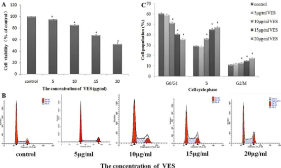
The changes in 3-(4,5-dimethylthiazol-2-yl)-2,5-diphenyltetrazolium bromide (MTT) tetrazo-lium salt formation in VES-treated SGC-7901 cells were examined to study the effect of VES on gastric carcinoma cell viability. VES significantly reduced MTT tetrazolium salt formation in cells in a dose-dependent manner (Fig 1A). SGC-7901 cell viability was inhibited by approxi-mately 5.3%, 14.7%, 34.4%, and 47.8% compared with the vehicle control (0.01% v/v
Anhy-drous ethanol) under 5, 10, 15, and 20μg/mL VES treatments, respectively, for 24 h. Flow
cytometry-based cell cycle analysis was performed to analyze the anti-mitogenic activity under
VES treatment. VES at 20μg/mL increased the number of SGC-7901 cells arrested at the S and
G2/M phases, and this effect was dose dependent. (Fig 1B and 1C).
VES induced an autophagy in SGC-7901 cells
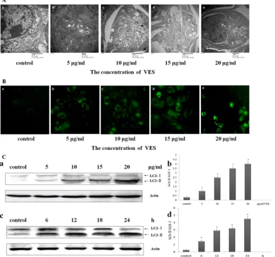
Whether or not VES induced autophagy in SGC-7901 cells was investigated. Autophagic bod-ies or autophagosomes comprising double- or single-membrane structures are located in the cytoplasm. Such autophagosomes were analyzed for topical autophagic changes by electron microscopy. The electron micrographs of normal SGC-7901 cells revealed intact cells, distinct nuclei and nuclear membranes, rough endoplasmic reticula, and homogeneous cytoplasms (Fig 2Aa). In VES-treated SGC-7901 cells, membrane-bound autophagosomes were observed
in the cytoplasm (Fig 2Ab to 2Ae). Electron micrographs revealed that VES at 5 and 10μg/mL
increased the number of autophagosomes containing mitochondria, endoplasmic reticulum (ER) membranes, and ribosomes in SGC-7901 cells (Fig 2Ab to 2Ac). Partial degradation of the contents of late-degradation autophagic vacuole was evident, and the engulfed rough ER
was degraded in cells treated with 10μg/mL VES (indicated by arrows). The contents of
Fig 1. Inhibition of gastric cancer cell SGC-7901 viability by VES.(A) Incubation of VES for 24 h decreased cell viability in a concentration-dependent manner as determined by MTT assay in SGC-7901. (B) Cells were treated with or without different concentrations of VES for 24 h. DNA contents were determined by flow cytometry analysis. (C) DNA histogram shows the accumulation of S and G2/M phases cells induced by VES.*p<0.05 means significantly different from the control group.
numerous small vesicles (indicated by arrows) showed the fusion between autophagic vacuole and the multivesicular endosome (Fig 1Ad and 1Ae). VES-treated cells showed more autopha-gosomes than untreated cells (Fig 2Aa).
Light chain 3 (LC3) localization in cells transfected with green fluorescent protein
(GFP)-labeled LC3 (GFP–LC3) was determined. Cytoplasm of transfected cells showed the
distribu-tion of GFP–LC3 dots (Fig 2B). The distribution of green dots in the control group was
dis-persed and indistinct. The punctate fluorescence in the VES treatment groups increasingly coagulated and intensified with increasing VES concentration. The increase in number and
Fig 2. VES induces an autophagic response in SGC-7901 cells.(A) Ultrastructural observations in SGC-7901 cells with transmission electron microscope. (a) Untreated SGC-SGC-7901 cells exhibited normal of cytoplasm, cell organelles, and nuclei morphologies. VES-treated SGC-7901 cells showed characteristic ultrastructural morphology of autophagy, (b to c) a significant number of autophagic vacuoles, (d) a giant autophagic vacuole filled with abundant degraded organelles, (e) autophagic vacuoles fused with multivesicular endosome. (a, c, & d, 20 000×; b, 30 000×; e, 12 000×). Typical changes were indicated by arrows. (B) Treatment of SGC-7901 cells with VES for 24 h prominently enhanced formation of autophagic vacuoles and fluorescent intensity, as determined by transfection of GFP-LC3 (C) Protein levels of LC3-I and LC3-II were measured using Western blot analysis. Data was representative of three individual experiments with similar results. (a) SGC-7901 cells were treated with 0, 5, 10, 15, and 20μg/mL VES for 24 h. (c)
SGC-7901 cells were treated with 20μg/mL VES for 0, 6, 12, 18, and 24 h. (b) and (d) Relative densitometry of
protein expression was determined by LC3-II protein densitometry with LC3-I. Actin was used as a loading control.*meansp<0.05 compared with control.
fluorescence intensities of the GFP-LC3 dots or vacuoles was induced by VES after 24 h of treatment (Fig 2B).
LC3 protein is an autophagy marker and its expression was analyzed to determine how VES affected autophagy in the cells. The LC3-II protein levels significantly increased with increasing VES concentration in a time-dependent manner (Fig 2C). The processed form of LC3-II also increased, thereby indicating an increase in autophagy incidence. Hence, VES could potentially induce autophagy in SGC-7901.
EFfect of VES on mTOR-related signaling pathways
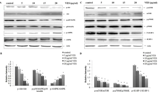
mTOR is an important component of the autophagy-related pathway. mTOR activation was analyzed to determine the molecular mechanism underlying VES-mediated autophagy induc-tion in SGC-7901 cells. VES effects on mTOR and p-mTOR (Ser2448) expressions were ana-lyzed. In particular, VES effects on phosphorylated p70S6 kinase at Thr-389 and the 4E-BP-1 at Thr37/46 were determined. mTOR, p70S6K, and 4E-BP-1 phosphorylation levels decreased with increasing VES concentration (Fig 3C and 3D).
Western blot analysis results showed that VES inhibited the phosphorylation of mTOR and its downstream targets, p70S6K and 4E-BP-1. Such inhibition triggered autophagy progression. We also investigated how VES affected the activation of the upstream signals of mTOR inhibi-tor, AMP-activated protein kinase (AMPK), and mTOR activators, namely, Akt and p38 MAPK. The levels of Akt, p38 MAPK, and AMPK in VES-treated SGC-7901 cells were ana-lyzed. Phosphorylations of Akt (p-Akt) and phosphorylation of p38 MAPK (p-p38 MAPK) decreased with increasing VES concentrations. In addition, activated p-AMPK levels were up-regulated in VES-treated cells compared with control group (Fig 3A and 3B). However, VES failed to affect total steady state protein levels.
Fig 3. Effects of VES on mTOR axis.SGC-7901 cells were treated with 0, 5, 10, 15, and 20μg/mL VES for
24 h, and activation of p38 MAPK (p-p38 MAPK), Akt (p-Akt), AMPK (p-AMPK), mTOR (p-mTOR), p70S6K (p- p70S6K), and 4E-BP-1 (p-4E-BP-1) were examined using Western blot. The data in (A) and (C) were representative of three individual experiments with similar results. The data in (B) and (D) were expressed as mean±S.D from three individual experiments. Actin was used as a loading control.*meansp<0.05 compared with control.
Blocking early and late autophagy stages enhanced the antiproliferative
effect of VES
The function of autophagy in VES-treated SGC-7901 cells was investigated by inhibiting autophagy using 3-methyladenine (3-MA) and chloroquine (CQ). VES-treated or untreated
SGC-7901 cells were exposed to 10 mM 3-MA or 20μM CQ. The changes in cell morphology
and viability and cell cycle variations were analyzed.
Untreated control cells adhered well and displayed normal SGC-7901 cell morphology. When autophagy was inhibited, the number of dead cells increased and the cells became round and detached (S1 Fig).
MTT assay and cell cycle analysis were performed to determine the function of autophagy in VES-induced inhibition of SGC-7901 cell viability. The SGC-7901 cells were subjected to the abovementioned methods. Results of the MTT assay showed that the combination of VES with either CQ or 3-MA led to a significantly higher suppression of cell viability than VES alone
(p<0.05,Fig 4A).
The early stage of cell autophagy was firstly blocked by 3-MA, and then the cell cycle was analyzed by flow cytometry. The group treated with VES and 3-MA showed a significantly higher number of cells arrested at S phase than both the control group and VES alone
(p<0.05,Fig 4B and 4C). CQ blocked late-stage autophagy and increased the cytostatic effect
of VES. The group treated with VES and CQ showed a significantly higher number of cells
arrested at S phase than both the control group and VES alone (p<0.05,Fig 4B and 4C).
The role of autophagy on VES-induced apoptosis
The biological activity of VES-induced autophagy in apoptosis was determined. The possible enhancement of VES-induced apoptosis by autophagy was determined using a confocal micro-scope and by flow cytometry after staining the cells with either Hoechst 33343 or Annexin V-FITC/PI.
The effects of the combined treatment of autophagy inhibitors and VES on cellular apopto-sis in SGC-7901 cells were analyzed. A higher number of cells in the group treated with a com-bination of CQ, 3-MA, and VES displayed apoptosis-related morphological changes, such as cell shrinking and chromatin condensation and crescent formation / margination compared with the untreated group or the group treated with CQ or 3-MA alone. DNA fragmentation and apoptotic body formation were observed in the VES + CQ and VES + 3-MA groups (Fig 5A). Flow cytometric analysis results also showed that treatment with CQ (20μM), 3-MA
(10 mM), or VES (20μg/mL) induced apoptosis in 4.31%, 3.53%, or 34.08% of the cells,
respec-tively. Combination of CQ, 3-MA, and VES at the abovementioned concentrations induced apoptosis in 57.03% or 41.06% of the cells (Fig 5B and 5C). The combination of CQ and VES or 3-MA and VES showed higher cooperative effect on the occurrence of VES-induced apopto-sis than each agent applied alone. These data confirmed that VES-induced autophagy contrib-uted to the prevention of VES-induced apoptosis in human gastric carcinoma cells.
Discussion
We focused on VES in this study. A VE analog can substantially enhance apoptosis in
malig-nant cells, as shown in vitro and in vivo studies using various species and tissues[13,33–37].
SGC-7901 cell viability was inhibited under VES treatment in a dose-dependent manner
(Fig 1A). Our results are in agreement with previously reported findings[10,13]. Flow
increased under VES treatment (Fig 1B and 1C), thereby suggesting that inhibition of cell cycle progression may a mechanism underlying the antiproliferative effect of VES.
According to a previously report researched by Karim MR[38], vitamin E act as a novel enhancer of macroautophagy. VES, as a VE analog, has been proved as a autophagy inducer in human gastric cancer by our study. Transmission electron microscopy (TEM) is a conventional method for observing autophagy; the phenomenon of autophagy was initially discovered using this method[39]. Autophagy is also detected by observing autolysosome formation under
GFP–LC3 transfection[39,40]. The microtubule-associated protein 1 (MAP1)–LC3, which is
important in autophagy, is the first mammalian protein to be detected in autophagosome
membranes[24,40,41]. A copious amount of LC3B is synthesized during autophagy. Thus,
LC3-II is an important autophagy marker.[42]. The abovementioned strategies were used to demonstrate the induction effect of VES on SGC-7901 cell autophagy (Fig 2).
mTOR is a major component of autophagy regulation and integrates numerous extracellu-lar signals, including glucose, amino acids, growth factors, and energy status[43]. The impor-tant function of mTOR as an autophagy repressor suggests that mTOR activity
downregulation efficiently triggers autophagic response[43–45]. mTOR phosphorylation
increases the levels of downstream targets, such as p70S6K and 4E-BP-1 to regulate many
dif-ferent cellular processes[46,47]. The p70S6K phosphorylation level is important in the
initia-tion of the translainitia-tion of proteins associated with cell growth and proliferainitia-tion. The p70S6K, 4E-BP-1, and mTOR phosphorylations decreased in VES-treated SGC-7901 cells, suggesting that VES inhibited mTOR activity (Fig 3C and 3D).
The serine/threonine protein kinase Akt (protein kinase B) mediates mTOR activity[48], and positively regulates mTOR. VES-induced inhibition of Akt phosphorylation was detected in a dose-dependent manner in our studies. The mTOR pathway is regulated by the MAPK pathway, as discussed in a previous study[49]. We monitored p38 MAPK phosphorylation in the MAPK signaling pathway in VES-treated SGC-7901 cells. VES markedly suppressed p38 MAPK phosphorylations, and such inhibitory effect results in autophagy. AMPK activation decreased mTOR activity[50], thereby indicating that AMPK is a negative mTOR regulator. AMPK phosphorylation was up-regulated by VES treatment (Fig 3A and 3B).
Inhibition of mTOR phosphorylation and its signal transduction by VES may partly pro-mote potent autophagy-inducing activity. The results implied that VES induced autophagy via the mTOR axis.
Autophagic cell death is an alternative cell death pathway for the chemotherapy of different cancer cells, which often develop resistance against drugs that activate the apoptotic pathway [51]. Previous studies have confirmed the dual function of autophagy in cell death. Autophagy promotes cell survival, but also induces cell death by promoting the extensive digestion of
intracellular organelles[52,53]. The function of autophagy in VES-treated SGC-7901 cells was
investigated. Autophagy was inhibited by CQ and 3-MA. CQ is an antimalarial drug that inhib-its autophagy at the late stage by blocking the fusion between autophagosome and lysosome fusion and by enzymatic autophagosomal cargo digestion[54]. Higher CQ concentrations
could reduce the viability of tumor cells[55,56], but the CQ concentration used in this study
Fig 4. Autophagy inhibition enhances VES-induced viability suppression in SGC-7901.SGC-7901 cells were exposed to VES, CQ, or 3-MA alone or in presence of 10 mM 3-MA or 20μM CQ for 24 h. (A) MTT
assay show that combined action of both VES and CQ and VES and 3-MA significantly suppress cell viability compared with VES treatment alone (*, compared with control group,p<0.05; #, compared with VES group, p<0.05). (B) and (C) DNA histogram shows that accumulation of S phase cells induced by VES was enhanced by CQ and 3-MA (*, compared with control group,p<0.05; #, compared with VES group,
p<0.05).
failed to induce tumor cell death in vitro (Figs4and5). 3-MA is a PI3K inhibitor that inhibits autophagy at the early stage and is a common inhibitor of autophagy[57]. 3-MA effectively
inhibited autophagy at the early stages[58] and also failed to induce tumor cell death (Figs4
and5). Autophagy blockage by either CQ or 3-MA enhances SGC-7901 cell apoptosis (Fig 5) and suppresses cell viability (Fig 4), respectively, under in vitro application of VES. The aug-mentation of tumor cell death by CQ and 3-MA treatments were correlated with the abilities of these compounds to block the process of autophagy. Thus, autophagy inhibition is a mecha-nism underlying the major antineoplastic effects of CQ and 3-MA at the abovementioned con-centrations. These results suggested that VES-induced autophagy is used as a means for adaptation in SGC-7901 cells. Combination of VES and autophagy inhibitors increases the effects of therapeutic methods on the induction of tumor cell apoptosis.
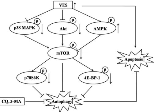
VES induces autophagy by inhibiting the mTOR axis pathway in human gastric carcinoma
SGC-7901 cells as depicted inFig 6. VES induced the suppression of both Akt and p38 MAPK,
and up-regulated AMPK, leading to the inhibition of mTOR activity. And downstream regula-tors p70S6K and 4E-BP-1 were suppressed consequently, resulting in autophagy in SGC-7901
Fig 5. Autophagy inhibition enhances VES-induced apoptosis in SGC-7901.(A) SGC-7901 cells were exposed to VES, CQ, or 3-MA alone or in presence of 10 mM 3-MA or 20μM CQ for 24 h, respectively,
stained with Hoechst 33343, and visualized under a confocal microscopy (1000×) (B) SGC-7901 human gastric cancer cells were treated as (A), stained with Annexin V-FITC/PI, and detected through flow cytometry analysis. The significance of the obtained four quadrants: FITC Annexin V-negative/PI-negative cells as viable cells, FITC Annexin negative cells as early apoptotic cells, FITC Annexin V-positive/PI-positive cells as late apoptotic cells, and FITC Annexin V-negative/PI-V-positive/PI-positive cells as necrosis cells. (C) Columns: mean of triplicate treatments; bars:±SD. (*, compared with control group,p<0.05; #, compared with VES group,p<0.05).
doi:10.1371/journal.pone.0132829.g005
Fig 6. Hypothetical model of VES autophagy mechanism in human gastric carcinoma SGC-7901 cells.
VES induced the suppression of both Akt and p38 MAPK, and up-regulated AMPK, leading to the inhibition of mTOR activity. And downstream regulators p70S6K and 4E-BP-1 were suppressed consequently, resulting in autophagy in SGC-7901 cells. VES cooperated with autophagy inhibitors to induce increased anticancer effect compared with VES alone.
cells. VES cooperated with autophagy inhibitors to induce increased anticancer effect com-pared with VES alone. Identifying the mTOR signaling transduction pathway clarified the molecular mechanisms that cause autophagy-mediated cell viability inhibition by antitumor agents and may contribute to the creation of novel therapeutic strategies for tumor growth sup-pression. Information is lacking on the detailed mechanisms that mediate the activation of kinases related to mTOR. We present an important perspective on cancer cell response to VES.
This study has several limitations. Autophagy was inhibited only using pharmacological approaches, which blocked the process of autophagy at early and late stages. Genetic investiga-tions should be conducted to further verify the results of this study. The siRNA specific to the autophagy-related gene 7 (ATG7) must be applied to inhibit ATG7 expression and block autophagy at the initial stages.
We showed for the first time that VES induce macroautophagy. Such autophagy in gastric carcinoma cells counteracted the antiproliferative effects of VES. The autophagy induction in VES-treated SGC-7901 cells was mediated by the down-regulation of mTOR pathway. The use of an autophagy inhibitor may enhance VES anti-cancer effects. We presented new perspec-tives on the mechanism underlying VES-induced cell death and discussed the potential use of combined VES and autophagy inhibitors in cancer chemotherapy.
Supporting Information
S1 Fig. Morphological observation under a phase contrast microscope The SGC-7901 cells were seeded in six-well flat bottom plates.VES-treated or untreated SGC-7901 cells were
exposed to 10 mM 3-MA or 20μM CQ. The morphologies of the SGC-7901 cells were
observed under a phase contrast microscope (Nikon, Japan) after 24 h of treatment. Bars:
100μm.
(TIF)
Author Contributions
Conceived and designed the experiments: KW LH. Performed the experiments: LH HS YS XZ. Analyzed the data: ZZ. Contributed reagents/materials/analysis tools: YL. Wrote the paper: LH.
References
1. Ferlay J, Shin HR, Bray F, Forman D, Mathers C, Parkin DM. Estimates of worldwide burden of cancer in 2008: GLOBOCAN 2008. Int J Cancer. 2010; 127(12):2893–917. Epub 2011/02/26. doi:10.1002/ijc. 25516PMID:21351269.
2. Parkin DM, Bray F, Ferlay J, Pisani P. Global cancer statistics, 2002. CA: a cancer journal for clinicians. 2005; 55(2):74–108. PMID:15761078.
3. Yang L, Parkin DM, Ferlay J, Li L, Chen Y. Estimates of cancer incidence in China for 2000 and projec-tions for 2005. Cancer Epidemiol Biomarkers Prev. 2005; 14(1):243–50. Epub 2005/01/26. doi: 14/1/ 243 [pii]. PMID:15668501.
4. Bang OS, Park JH, Kang SS. Activation of PKC but not of ERK is required for vitamin E-succinate-induced apoptosis of HL-60 cells. Biochem Biophys Res Commun. 2001; 288(4):789–97. doi:10.1006/ bbrc.2001.5839PMID:11688977.
5. Israel K, Yu W, Sanders BG, Kline K. Vitamin E succinate induces apoptosis in human prostate cancer cells: role for Fas in vitamin E succinate-triggered apoptosis. Nutr Cancer. 2000; 36(1):90–100. Epub 2000/05/08. doi:10.1207/S15327914NC3601_13PMID:10798221.
7. Sahu SN, Edwards-Prasad J, Prasad KN. Effect of alpha tocopheryl succinate on adenylate cyclase activity in murine neuroblastoma cells in culture. J Am Coll Nutr. 1988; 7(4):285–93. Epub 1988/08/01. PMID:3209780.
8. Wu K, Zhao Y, Liu BH, Li Y, Liu F, Guo J, et al. RRR-alpha-tocopheryl succinate inhibits human gastric cancer SGC-7901 cell growth by inducing apoptosis and DNA synthesis arrest. World journal of gastro-enterology: WJG. 2002; 8(1):26–30. PMID:11833065.
9. Yu W, Israel K, Liao QY, Aldaz CM, Sanders BG, Kline K. Vitamin E succinate (VES) induces Fas sen-sitivity in human breast cancer cells: role for Mr 43,000 Fas in VES-triggered apoptosis. Cancer Res. 1999; 59(4):953–61. Epub 1999/02/24. PMID:10029090.
10. Zhang X, Peng X, Yu W, Hou S, Zhao Y, Zhang Z, et al. Alpha-tocopheryl succinate enhances doxoru-bicin-induced apoptosis in human gastric cancer cells via promotion of doxorubicin influx and suppres-sion of doxorubicin efflux. Cancer Lett. 2011; 307(2):174–81. Epub 2011/05/04. doi:10.1016/j.canlet. 2011.04.001PMID:21536373.
11. Wu K, Li Y, Zhao Y, Shan YJ, Xia W, Yu WP, et al. Roles of Fas signaling pathway in vitamin E succi-nate-induced apoptosis in human gastric cancer SGC-7901 cells. World journal of gastroenterology: WJG. 2002; 8(6):982–6. PMID:12439910.
12. Wu K, Zhao Y, Li GC, Yu WP. c-Jun N-terminal kinase is required for vitamin E succinate-induced apo-ptosis in human gastric cancer cells. World journal of gastroenterology: WJG. 2004; 10(8):1110–4. PMID:15069708.
13. Huang X, Zhang Z, Jia L, Zhao Y, Zhang X, Wu K. Endoplasmic reticulum stress contributes to vitamin E succinate-induced apoptosis in human gastric cancer SGC-7901 cells. Cancer letters. 2010; 296 (1):123–31. doi:10.1016/j.canlet.2010.04.002PMID:20435408.
14. Shi Z, Li CY, Zhao S, Yu Y, An N, Liu YX, et al. A systems biology analysis of autophagy in cancer ther-apy. Cancer Lett. 2013; 337(2):149–60. Epub 2013/06/25. doi:10.1016/j.canlet.2013.06.004 S0304-3835(13)00454-0 [pii]. PMID:23791881.
15. Liang XH, Jackson S, Seaman M, Brown K, Kempkes B, Hibshoosh H, et al. Induction of autophagy and inhibition of tumorigenesis by beclin 1. Nature. 1999; 402(6762):672–6. Epub 1999/12/22. doi:10. 1038/45257PMID:10604474.
16. Lockshin RA, Zakeri Z. Apoptosis, autophagy, and more. Int J Biochem Cell Biol. 2004; 36(12):2405– 19. Epub 2004/08/25. doi:10.1016/j.biocel.2004.04.011PMID:15325581.
17. Morselli E, Galluzzi L, Kepp O, Vicencio JM, Criollo A, Maiuri MC, et al. Anti- and pro-tumor functions of autophagy. Biochim Biophys Acta. 2009; 1793(9):1524–32. Epub 2009/04/18. doi:10.1016/j.bbamcr. 2009.01.006S0167-4889(09)00024-X[pii]. PMID:19371598.
18. Qu X, Yu J, Bhagat G, Furuya N, Hibshoosh H, Troxel A, et al. Promotion of tumorigenesis by heterozy-gous disruption of the beclin 1 autophagy gene. J Clin Invest. 2003; 112(12):1809–20. Epub 2003/11/ 26. doi:10.1172/JCI20039JCI200320039 [pii]. 14638851; PubMed Central PMCID: PMC297002. PMID:14638851
19. Wu WK, Cho CH, Lee CW, Wu YC, Yu L, Li ZJ, et al. Macroautophagy and ERK phosphorylation coun-teract the antiproliferative effect of proteasome inhibitor in gastric cancer cells. Autophagy. 2010; 6 (2):228–38. Epub 2010/01/21. PMID:20087064.
20. Liang C, Jung JU. Autophagy genes as tumor suppressors. Curr Opin Cell Biol. 2010; 22(2):226–33. Epub 2009/12/01. doi:10.1016/j.ceb.2009.11.003S0955-0674(09)00196-3 [pii]. PMID:19945837; PubMed Central PMCID: PMC2854193.
21. Moreau K, Luo S, Rubinsztein DC. Cytoprotective roles for autophagy. Curr Opin Cell Biol. 2010; 22 (2):206–11. Epub 2010/01/05. doi:10.1016/j.ceb.2009.12.002S0955-0674(09)00231-2 [pii]. PMID:
20045304; PubMed Central PMCID: PMC2860226.
22. Baehrecke EH. Autophagy: dual roles in life and death? Nat Rev Mol Cell Biol. 2005; 6(6):505–10. Epub 2005/06/02. doi: nrm1666 [pii]. doi:10.1038/nrm1666PMID:15928714.
23. Ahn CH, Jeong EG, Lee JW, Kim MS, Kim SH, Kim SS, et al. Expression of beclin-1, an autophagy-related protein, in gastric and colorectal cancers. APMIS. 2007; 115(12):1344–9. Epub 2008/01/11. doi:10.1111/j.1600-0463.2007.00858.xAPMapm_858.xml [pii]. PMID:18184403.
24. Yoshioka A, Miyata H, Doki Y, Yamasaki M, Sohma I, Gotoh K, et al. LC3, an autophagosome marker, is highly expressed in gastrointestinal cancers. Int J Oncol. 2008; 33(3):461–8. Epub 2008/08/13. PMID:18695874.
26. Kondo Y, Kanzawa T, Sawaya R, Kondo S. The role of autophagy in cancer development and response to therapy. Nat Rev Cancer. 2005; 5(9):726–34. Epub 2005/09/09. doi: nrc1692 [pii]. doi:10.1038/ nrc1692PMID:16148885.
27. Jung CH, Ro SH, Cao J, Otto NM, Kim DH. mTOR regulation of autophagy. FEBS letters. 2010; 584 (7):1287–95. doi:10.1016/j.febslet.2010.01.017PMID:20083114; PubMed Central PMCID: PMC2846630.
28. Yang Z, Klionsky DJ. Mammalian autophagy: core molecular machinery and signaling regulation. Curr Opin Cell Biol. 2010; 22(2):124–31. Epub 2009/12/26. doi:10.1016/j.ceb.2009.11.014S0955-0674(09) 00228-2 [pii]. PMID:20034776; PubMed Central PMCID: PMC2854249.
29. Asnaghi L, Bruno P, Priulla M, Nicolin A. mTOR: a protein kinase switching between life and death. Pharmacological research: the official journal of the Italian Pharmacological Society. 2004; 50(6):545– 9. doi:10.1016/j.phrs.2004.03.007PMID:15501691.
30. Turcotte S, Chan DA, Sutphin PD, Hay MP, Denny WA, Giaccia AJ. A molecule targeting VHL-deficient renal cell carcinoma that induces autophagy. Cancer Cell. 2008; 14(1):90–102. Epub 2008/07/05. doi:
10.1016/j.ccr.2008.06.004S1535-6108(08)00194-3 [pii]. PMID:18598947; PubMed Central PMCID: PMC2819422.
31. Degtyarev M, De Maziere A, Orr C, Lin J, Lee BB, Tien JY, et al. Akt inhibition promotes autophagy and sensitizes PTEN-null tumors to lysosomotropic agents. J Cell Biol. 2008; 183(1):101–16. Epub 2008/ 10/08. doi:10.1083/jcb.200801099jcb.200801099 [pii]. PMID:18838554; PubMed Central PMCID: PMC2557046.
32. Tang G, Yue Z, Talloczy Z, Hagemann T, Cho W, Messing A, et al. Autophagy induced by Alexander disease-mutant GFAP accumulation is regulated by p38/MAPK and mTOR signaling pathways. Human molecular genetics. 2008; 17(11):1540–55. doi:10.1093/hmg/ddn042PMID:18276609; PubMed Central PMCID: PMC2902290.
33. Neuzil J, Tomasetti M, Zhao Y, Dong LF, Birringer M, Wang XF, et al. Vitamin E analogs, a novel group of "mitocans," as anticancer agents: the importance of being redox-silent. Mol Pharmacol. 2007; 71 (5):1185–99. Epub 2007/01/16. doi: mol.106.030122 [pii]. doi:10.1124/mol.106.030122PMID:
17220355.
34. Neuzil J, Weber T, Schroder A, Lu M, Ostermann G, Gellert N, et al. Induction of cancer cell apoptosis by alpha-tocopheryl succinate: molecular pathways and structural requirements. FASEB journal: official publication of the Federation of American Societies for Experimental Biology. 2001; 15(2):403–15. doi:
10.1096/fj.00-0251comPMID:11156956.
35. Prasad KN, Kumar B, Yan XD, Hanson AJ, Cole WC. Alpha-tocopheryl succinate, the most effective form of vitamin E for adjuvant cancer treatment: a review. J Am Coll Nutr. 2003; 22(2):108–17. Epub 2003/04/04. PMID:12672706.
36. Wang XF, Dong L, Zhao Y, Tomasetti M, Wu K, Neuzil J. Vitamin E analogues as anticancer agents: lessons from studies with alpha-tocopheryl succinate. Mol Nutr Food Res. 2006; 50(8):675–85. Epub 2006/07/13. doi:10.1002/mnfr.200500267PMID:16835868.
37. Zhang Y, Ni J, Messing EM, Chang E, Yang CR, Yeh S. Vitamin E succinate inhibits the function of androgen receptor and the expression of prostate-specific antigen in prostate cancer cells. Proceed-ings of the National Academy of Sciences of the United States of America. 2002; 99(11):7408–13. doi:
10.1073/pnas.102014399PMID:12032296; PubMed Central PMCID: PMC124244.
38. Karim MR, Fujimura S, Kadowaki M. Vitamin E as a novel enhancer of macroautophagy in rat hepato-cytes and H4-II-E cells. Biochemical and biophysical research communications. 2010; 394(4):981–7. doi:10.1016/j.bbrc.2010.03.103PMID:20307493.
39. Klionsky DJ, Abeliovich H, Agostinis P, Agrawal DK, Aliev G, Askew DS, et al. Guidelines for the use and interpretation of assays for monitoring autophagy in higher eukaryotes. Autophagy. 2008; 4 (2):151–75. Epub 2008/01/12. PMID:18188003; PubMed Central PMCID: PMC2654259.
40. Kabeya Y, Mizushima N, Ueno T, Yamamoto A, Kirisako T, Noda T, et al. LC3, a mammalian homo-logue of yeast Apg8p, is localized in autophagosome membranes after processing. EMBO J. 2000; 19 (21):5720–8. Epub 2000/11/04. doi:10.1093/emboj/19.21.5720PMID:11060023; PubMed Central PMCID: PMC305793.
41. Mann SS, Hammarback JA. Gene localization and developmental expression of light chain 3: a com-mon subunit of microtubule-associated protein 1A(MAP1A) and MAP1B. J Neurosci Res. 1996; 43 (5):535–44. Epub 1996/03/01. doi:10.1002/(SICI)1097-4547(19960301)43:5<535::AID-JNR3>3.0.
CO;2-J[pii]. 10.1002/(SICI)1097-4547(19960301)43:5<535::AID-JNR3>3.0.CO;2-J. PMID:
8833088.
cancer cells. Oncol Rep. 2012; 27(4):1079–89. Epub 2011/12/20. doi:10.3892/or.2011.1593PMID:
22179718; PubMed Central PMCID: PMC3583434.
43. Kim DH, Sarbassov DD, Ali SM, King JE, Latek RR, Erdjument-Bromage H, et al. mTOR interacts with raptor to form a nutrient-sensitive complex that signals to the cell growth machinery. Cell. 2002; 110 (2):163–75. Epub 2002/08/02. doi: S0092867402008085 [pii]. PMID:12150925.
44. Hu C, Zou MJ, Zhao L, Lu N, Sun YJ, Gou SH, et al. E Platinum, a newly synthesized platinum com-pound, induces autophagy via inhibiting phosphorylation of mTOR in gastric carcinoma BGC-823 cells. Toxicol Lett. 2012; 210(1):78–86. Epub 2012/02/11. doi:10.1016/j.toxlet.2012.01.019PMID:
22322152.
45. Iwamaru A, Kondo Y, Iwado E, Aoki H, Fujiwara K, Yokoyama T, et al. Silencing mammalian target of rapamycin signaling by small interfering RNA enhances rapamycin-induced autophagy in malignant gli-oma cells. Oncogene. 2007; 26(13):1840–51. Epub 2006/09/27. doi: 1209992 [pii]doi:10.1038/sj.onc. 1209992PMID:17001313.
46. Bonapace L, Bornhauser BC, Schmitz M, Cario G, Ziegler U, Niggli FK, et al. Induction of autophagy-dependent necroptosis is required for childhood acute lymphoblastic leukemia cells to overcome gluco-corticoid resistance. J Clin Invest. 2010; 120(4):1310–23. doi:10.1172/JCI39987PMID:20200450; PubMed Central PMCID: PMC2846044.
47. Feng Z, Zhang H, Levine AJ, Jin S. The coordinate regulation of the p53 and mTOR pathways in cells. Proc Natl Acad Sci U S A. 2005; 102(23):8204–9. doi:10.1073/pnas.0502857102PMID:15928081; PubMed Central PMCID: PMC1142118.
48. Hay N, Sonenberg N. Upstream and downstream of mTOR. Genes Dev. 2004; 18(16):1926–45. Epub 2004/08/18. doi:10.1101/gad.121270418/16/1926 [pii]. PMID:15314020.
49. Carracedo A, Ma L, Teruya-Feldstein J, Rojo F, Salmena L, Alimonti A, et al. Inhibition of mTORC1 leads to MAPK pathway activation through a PI3K-dependent feedback loop in human cancer. J Clin Invest. 2008; 118(9):3065–74. doi:10.1172/JCI34739PMID:18725988; PubMed Central PMCID: PMC2518073.
50. Kimura N, Tokunaga C, Dalal S, Richardson C, Yoshino K, Hara K, et al. A possible linkage between AMP-activated protein kinase (AMPK) and mammalian target of rapamycin (mTOR) signalling pathway. Genes to cells: devoted to molecular & cellular mechanisms. 2003; 8(1):65–79. PMID:12558800.
51. Beljanski V, Knaak C, Smith CD. A novel sphingosine kinase inhibitor induces autophagy in tumor cells. J Pharmacol Exp Ther. 2010; 333(2):454–64. Epub 2010/02/25. doi:10.1124/jpet.109.163337
jpet.109.163337 [pii]. PMID:20179157; PubMed Central PMCID: PMC2872961.
52. Brech A, Ahlquist T, Lothe RA, Stenmark H. Autophagy in tumour suppression and promotion. Mol Oncol. 2009; 3(4):366–75. Epub 2009/06/30. doi:10.1016/j.molonc.2009.05.007PMID:19559660.
53. Bursch W, Ellinger A, Gerner C, Frohwein U, Schulte-Hermann R. Programmed cell death (PCD). Apo-ptosis, autophagic PCD, or others? Ann N Y Acad Sci. 2000; 926:1–12. Epub 2001/02/24. PMID:
11193023.
54. Mahoney E, Byrd JC, Johnson AJ. Autophagy and ER stress play an essential role in the mechanism of action and drug resistance of the cyclin-dependent kinase inhibitor flavopiridol. Autophagy. 2013; 9 (3):434–5. Epub 2013/02/21. doi:10.4161/auto.23027PMID:23422081; PubMed Central PMCID: PMC3590270.
55. Prasad RN, Mahajan RC, Ganguly NK. Effect of chloroquine on cellular immune responses of normal and P. knowlesi-infected rhesus monkeys. Immunol Cell Biol. 1987; 65 (Pt 3):211–6. Epub 1987/06/01. doi:10.1038/icb.1987.23PMID:3623607.
56. Yin F, Guo M, Yao S. Kinetics of DNA binding with chloroquine phosphate using capacitive sensing method. Biosens Bioelectron. 2003; 19(4):297–304. Epub 2003/11/15. doi: S0956566303001970 [pii]. PMID:14615086.
57. Zhang X, Wei H, Liu Z, Yuan Q, Wei A, Shi D, et al. A novel protoapigenone analog RY10-4 induces breast cancer MCF-7 cell death through autophagy via the Akt/mTOR pathway. Toxicol Appl Pharma-col. 2013; 270(2):122–8. Epub 2013/04/30. doi:10.1016/j.taap.2013.04.011S0041-008X(13)00158-0 [pii]. PMID:23624174.