Diana Catarina José Pinheiro de Castro
Licenciada em Biologia Humana
Characterization of autophagy induced by linoleic acid
Dissertação para obtenção do Grau de Mestre em Genética Molecular e Biomedicina
Orientador: Doutor José Manuel Fuentes Rodríguez
Co-orientador: Doutora Mireia Niso Santano
Centro de Investigación Biomédica en Red de Enfermedades Neurodegenerativas
(CIBERNED),
Universidad de Extremadura, Espanha
III
Diana Catarina José Pinheiro de Castro
Licenciada em Biologia Humana
Characterization of autophagy induced by linoleic acid
Dissertação para obtenção do Grau de Mestre em Genética Molecular e Biomedicina
Dissertação apresentada na Faculdade de Ciências e Tecnologia da Universidade Nova de Lisboa para obtenção do Grau de Mestre em Genética Molecular e Biomedicina. A presente dissertação foi desenvolvida em colaboração
com o Centro de Investigación Biomédica en Red de Enfermedades Neurodegenerativas (CIBERNED),
Universidad de Extremadura, Espanha
Orientador: Doutor José Manuel Fuentes Rodríguez
Co-orientador: Doutora Mireia Niso Santano
V Characterization of autophagy induced by linoleic acid
Copyright Diana Catarina José Pinheiro de Castro, FCT/UNL, UNL
VII
Agradecimentos
Ao longo de todos os caminhos é muito importante o apoio dos que nos são chegados, pois não se chega a lado nenhum sozinho.
Em primeiro lugar, quero agradecer aos meus avós Jorge e Mimi por todo o apoio que me deram e continuam a dar. Por estarem sempre presentes em todos os momentos e pelo ser humano fantástico que são. Espero que vos orgulhe, como vocês me orgulham a mim.
Aos meus pais por me fazerem acreditar que eu posso ser o que eu quiser e nunca me deixarem desistir. Por me aturarem mesmo quando sou insuportável. Um muito obrigado!!
Ao meu Zé, por todas as provas de carinho e de paciência, pelas quais tem passado. Por seres aquela pessoa que nem é preciso abrir a boca, para saberes qual o meu estado de espírito. Por estares sempre perto, mesmo estando longe.
Ao Jorginho, peço desculpas pelas ausências demoradas e agradeço-te por seres o meu eterno companheiro das jogatanas.
À Vitória, pelos seus mimos e ron-rons super reconfortantes.
Ao Professor Doutor José Manuel Fuentes, pela sua simpatía e disponibilidade imediata. Por ter sido sempre tao rápido nas respostas e esclarecimentos de dúvidas que tive ao longo do ano.
À Doutora Mireia Niso Santano, por ser a melhor co-orientadora que podia ter tido. A forma como ensina é fenomenal, porque consegue explicar coisas complexas, como se fossem muito simples, e isso é algo que nem todos somos capazes de fazer.
À Sokhna por me obrigar a desenvolver o castelhano à força, quer nos almoços, quer nos experiências. Muito obrigado por partilhares o teu conhecimento (fosse em que língua fosse!), e me ajudares a desenvolver espirito crítico em relação a tudo. És uma excelente pessoa e mereces tudo de bom pela tua vida fora. Boa sorte!
Ao Mário, por ser a pessoa mais prestável que conheço, sempre pronto a ajudar os colegas, mesmo que isso lhe atrasasse o seu próprio trabalho. Sucesso em Nova York!
À Rosana, pela sua boa disposição e estar sempre preocupada pelos outros.
À Guadalupe por me ensinar todas as técnicas, no inicio, e pela paciência com o meu “portunhol”.
VIII
À Filipa, palavras não chegam para te agradecer todos os momentos que passámos desde a entrada na Universidade até aqui. És a minha companheira dos estudos e das festas. Desejo-te toda a sorte e sei que vais chegar até onde queres.
À Rosélia, que é sem dúvida das melhores pessoas que conheci. Obrigado pelas nossas conversas, pelos devaneios, pelos trabalhos e pelo convivio. Que saudades tuas, riquinha!
À Catarina e à Margarida, pela excelente companhia que são.
À Joana, pelas palavras de apoio e positivismo que sempre que falamos me dás, és a melhor madrinha académica!
Ao Andrey, pela convivência e amizade que ficam depois de alguns anos, agora já estamos mais perto de abrir o meu laboratório, com os teus sistemas operativos!
IX
Abstract
Parkinson's disease is one of the most common neurodegenerative disorder that is slowly progressive and manifested by muscle rigidity, tremor, decreased mobility and postural instability. The disease is caused by a combination of genetic and environmental factors.
The most prominent pathological features are the severe loss of dopaminergic neurons in the substantia nigra pars compacta and the presence of cytoplasmic protein inclusions called Lewy bodies,
primarily composed of fibrillar α-synuclein and ubiquitinated proteins within some remaining nigral neurons.
Autophagy is a catabolic process that maintain cellular homeostasis, through the selection of misfolded proteins, damaged organelles, and even pathogenic organisms to be degraded by lysosomes. Autophagy can mediate cytoprotection (for instance neuroprotection and cardioprotection in the context of ischemic preconditioning) and delay the pathogenic manifestations of aging.
Dysregulation of autophagy has been observed in the brain tissues from Parkinson’s disease patients and animal models. In recent years, some reports have shown a new relationship between macroautophagy and lipid metabolism.
In this work, we used the most consumed polyunsaturated fatty acid in our diet, linoleic acid, to evaluate if it induces autophagy and if there is a possible relationship between linoleic acid-induced autophagy and the neuroprotective/toxic mechanisms triggered by this compound. We found that linoleic acid induces autophagy at concentrations equal or higher than 200 μM, and we describe its activation pathway, using Western blotting and immunofluorescence assays. Our results suggest that linoleic acid-activated autophagy process is mammalian target of rapamycin-independent, class III phosphatidylinositol 3-kinase/Beclin1-independent and AMP-activated protein kinase-dependent. As for the neuroprotective capacity of linoleic acid, we observed that alone it shows some toxicity. However, if co-administered with an inducer of reactive oxygen species (such as paraquat), linoleic acid does not increase paraquat toxicity. On the other hand, when linoleic acid is co-administered with puromycin (protein aggregates generator) it has a neuroprotective effect.
Keywords: Parkinson’s disease (PD), autophagy, polyunsaturated fatty acid (PUFA), linoleic acid (LA), microtubule-associated protein 1 light chain 3 (LC3)
XI
Resumo
A doença de Parkinson é uma das doenças neurodegenerativas mais comuns, progressiva e que se manifesta por rigidez muscular, tremores, instabilidade postural e diminuição da mobilidade. A doença é causada por uma combinação de fatores genéticos e ambientais.
Os fatores patológicos mais proeminentes são a perda severa de neurónios dopaminérgicos, na região da substância nigra pars compacta, e a presença de agregados proteicos denominados Corpos de
Lewis, compostos por α-sinucleina e proteinas ubiquítinadas, com diminuição dos neurónios nigroestriatais.
A autofagia é um processo essencial das nossas células para manter a homeostase celular, selecionando proteínas alteradas, organelos, ou até mesmo organismos patogénicos para serem degradados pelos lisossomas. Ao nível do organismo, a autofagia pode mediar citoprotecção (por exemplo neuroprotecção e cardioprotecção no contexto de pré-condicionamento isquémico) e retardar as manifestações do envelhecimento patogénicos.
A desregulação da autofagia tem sido observada nos tecidos de cérebro de pacientes e de modelos animais, com PD. Nos últimos anos alguns estudos têm mostrado uma nova relação entre a macroautofagia e o metabolismo lipídico.
Usando o ácido gordo poli-insaturado mais consumido na nossa dieta, o ácido linoleico, queremos saber se este induz a autofagia e se há uma relação entre a possível autofagia induzida por ácido linoleico e a neuroproteção/toxicidade. Verificamos que o ácido linoleico induz autofagia a concentrações iguais ou superiores a 200 µM, e descrevemos a sua via de activação, usando para tal, a técnica de Western blot e de Imunofluorescência. Relativamente às vias de activação de autofagia por LA, os nossos resultados sugerem que é mammalian target of rapamycin-independent, class III phosphatidylinositol 3-kinase/Beclin1-independent e AMP-activated protein kinase -dependent. Quanto à sua capacidade neuroprotectora, os resultados sugerem que apesar de o ácido linoleico sozinho apresentar alguma toxicidade, se for incubado com um indutor de espécies reactivas de oxigénio (como paraquat), não aumenta a toxidade e se for incubado com puromicina (gerador de agregados proteicos) tem efeito neuroprotector.
XII
“If you want to reach where most of people don't reach, do what most don't do.”
XIII
Table of contents
Agradecimentos ... VII Abstract ... IX Resumo ... XI Table of contents ... XIII Abreviation list ... XV List of figures ... XVII List of tables ... XIX
1. Introduction ... 1
1.1 Physiopathology of Parkinson’s Disease ... 2
1.2 Risk factors ... 4
1.2.1 Environmental factors ... 5
1.2.2 Genetic factors ... 5
1.3 Glial cells and Parkinson’s Disease ... 6
1.3.1 Microglia ... 7
1.3.2 Astrocytes ... 7
1.4 Ubiquitin-proteasome system ... 8
1.5 Autophagy ... 9
1.5.1 Signaling Pathway ... 13
1.5.2 Autophagy and Parkinson’s Disease ... 15
1.5.3 Non-canonical autophagy ... 16
1.5.4 Lipid droplets in autophagy ... 18
1.5.5 Lipid role in autophagy ... 20
1.6 Diet ... 20
1.6.1 PUFA ... 22
1.6.2 Linoleic Acid ... 23
2. Rational and aims ... 25
3. Materials and methods ... 27
3.1 Appliances ... 27
3.2 Reagents ... 28
3.3 Antibodies ... 30
3.4 Cell lines ... 31
3.5 Cell maintenance ... 31
XIV
3.7 Cell culture... 31
3.8 Treatments ... 32
3.9 Linoleic acid ... 34
3.10 Western Blot ... 34
3.11 MTT assay ... 35
3.12 Flow Cytometry ... 35
3.13 Trypan Blue ... 36
3.14 Immunofluorescence... 36
3.14.1 Lipid droplets ... 36
3.14.2 small interfering RNA (siRNA) ... 37
3.15 Plasmid transfection ... 37
3.16 Data analysis ... 38
4. Results ... 39
4.1 Effect of LA in cell viability ... 39
4.2 LA induces autophagy ... 41
4.3 LA promotes autophagy flux ... 43
4.4 LA-induced autophagy is Atg5-dependent ... 46
4.5 LA induces mTOR-independent autophagy ... 46
4.6 LA enhances the phosphorilation of AMPKα ... 47
4.7 LA -induced autophagy is BECN1-independent ... 50
4.8 Effect of LA in differents organelles ... 53
4.8.1 LA does not alter organelle structures ... 53
4.8.2 LA induces autophagy even with lysosome and mitochondria damage. ... 56
4.9 LA has neuroprotector role in neuronal cell lines ... 58
4.9.1 LA does not protect neurons against cell death ... 58
4.9.2 LA protects neurons from protein aggregates ... 59
5. Discussion ... 64
6. Conclusions ... 68
XV
Abreviation list
AMBRA1 Activating molecule in Beclin1-regulated autophagy 1
AMP Adenosin monophosphate AMPK AMP-activated protein kinase
Atg Autophagy-related genes
BCA Bicinchonic acid BFA Brefeldin A
BSA Bovine serum albumin
BECN1 Beclin 1
BECN/PI3K Beclin/phosphatidylinositol-3-phosphate
Co Control CC Compound C
CMA Chaperone-mediated autophagy CoA Coenzyme A
CQ Chloroquine Cyt C Cytochrome C
DA Dopamine
DMEM Dulbecco’s Modified Eagle Medium
DMSO Dimethyl sulfoxide DNA Desoxyribonucleic acid ER Endoplasmic reticulum
FASN Fatty acid synthase FBS Fetal bovine serum
IF Immunofluorescence LA Linoleic acid
LAP LC3-associated phagocytosis pathway LB Lewy Bodies
LC3 Microtubule-associated protein 1 light chain 3 L-DOPA L-3,4-dihydroxyphenylalanine
LP Lipid droplets
MAO-B Monoamine oxidase B
MAP4K3 Mitogen-activated protein kinase kinase kinase kinase 3 MeDi Mediterranean diet
XVI
MPTP 1-methyl-4-phenyl-1,2,3,6-tetrahydropyridine mTOR Mammalian target of rapamycin complex 1
mTORC1 Mammalian target of rapamycin complex 1 NAD+ Nicotinamide adenine dinucleotide
NADPH Nicotinamide adenine dinucleotide phosphate NP-40 Nonidet P-40
p-AMPK Phospho-AMPK PBS Phosphate buffered saline
PD Parkinson’s Disease PFA Paraformaldehyde
PI3K/AKT Phosphatidylinositol 3-kinase/serine-theronine kinase
PI(3)P Phosphatidylinositol 3-phosphate
PINK1 PTEN-induced putative kinase 1
PKA Protein kinase A p-mTOR Phospho-mTOR
PQ2+Paraquat
PUFA Polyunsaturated fatty acid
RNA Ribonucleic acid ROS Reactive oxygen species
siRNA Small interfering ribonucleic acid UPS Ubiquitin-proteasome System
TTBS Tris buffered saline with tween 20 ULK1 Uncoordinated-51-like protein kinase WB Western Blot
XVII
List of figures
Figure 1.1 A brain with parkinson’s disease with the substantia nigra and Lewy body labeled. ... 3
Figure 1.2 Biochemical senescence and pathogenetic pathways to cell death in Parkinson disease.. .... 4
Figure 1.3 The ubiquitin–proteasome pathway of protein degradation. ... 9
Figure 1.4 Autophagy and neurodegenerative disorders.. ... 10
Figure 1.5 Model of ULK1 regulation by AMPK and mTORC1 in response to glucose signals ... 14
Figure 1.6 Regulation of VPS34 complex formation in response to nutrients. ... 15
Figure 1.7 Canonical and non-canonical autophagy pathways. ... 18
Figure 1.8 Projection of a confocal stack of an abdominal adipocyte isolated from an Akt (PKB) Drosophila melanogaster mutant. ... 19
Figure 1.9 Structure of the main PUFAs present in the human diet. ... 22
Figure 1.10 Struture of linoleic acid. ... 23
Figure 4.1 Effect of LA in cell proliferation, by MTT assay.. ... 40
Figure 4.2 Effect of LA in cell viability. ... 40
Figure 4.3 LA induces autophagy. . ... 42
Figure 4.4 Autophagic flux induced by LA... 43
Figure 4.5 Determination of autophagy flux by fluorescence microscopy. ... 44
Figure 4.6 LA promotes autophagy flux. ... 45
Figure 4.7 LA-induced autophagy is Atg5-dependent. ... 46
Figure 4.8 LA-induced autophagy is mTOR-independent. ... 47
Figure 4.9 LA enhances the phosphorilation of AMPKα.. ... 48
Figure 4.10 Compound C induces autophagy. ... 49
Figure 4.11 LA-induced autophagy is AMPK-dependent. ... 50
Figure 4.12 Autophagy induced by LA is BECN1 independent. ... 51
Figure 4.13 3-MA does not inhibit the autophagy induced by LA... 51
Figure 4.14 LA-induced autophagy is BECN1-independent. ... 52
Figure 4.15 Implication of BECN1 in LA-induced autophagy. ... 52
Figure 4.16 LA does not impair lysosomes. ... 54
Figure 4.17 Effect of LA treatment in organelle structures .. ... 55
Figure 4.18 LA-induced autophagy is organelles-independent. ... 56
Figure 4.19 LA-induced autophagy is Golgi-apparatus-independent... 57
Figure 4.20 LA-induced autophagy is ER-independent ... 57
Figure 4.21 LA induces lipid droplets biogenesis. ... 58
Figure 4.22 LA does not protect cells against cell death induced by PQ. ... 59
Figure 4.23 LA does not reduce %PI+ cells induced by PQ. ... 59
Figure 4.24 LA protect cells against cell death induces by puromycin. ... 60
Figure 4.25 LA reduce %PI+ cells, with puromycin.. ... 60
XIX
List of tables
1
1.
Introduction
Parkinson disease (PD) is a neurodegenerative disorder that affects the nervous system in the area that coordinate the activity, muscle tone and movement. It results from a combination of genetic and environmental factors, and manifests with a broad range of symptoms (Appukuttan, Ali et al.
2013). There are motor and non-motor symptoms.
The first symptoms of the disease usually appear around 60 years, reaching disability in the course of 5 or 15 years. The incidence of this neurodegenerative disorder increases with age affecting more than 3% of the population over 65 years, although in the last 40 years, the age of onset has been anticipated being as frequent from 50 (Rodriguez, Rodriguez-Sabate et al. 2015).
The parkinsonian motor symptoms include bradykinesia, muscular rigidity, tremor at rest, postural and gait impairment. (Kalia and Lang 2015). Non-motor features include olfactory
dysfunction, cognitive impairment, psychiatric symptoms, sleep disorders, autonomic dysfunction, pain and fatigue. They are also frequently present before the onset of the classical motor symptoms (Kalia
and Lang 2015).
Symptoms of PD clinically appear when almost 50 % of neurons are lost. This neuronal loss is located in the area between the brain and spinal cord, brainstem, particularly those neurons that are in a black substance called core. The portion of this core is called the substancia nigra pars compacta (SN).
It is so named because some neurons in this area are pigmented in black color due to the presnce of the neuromelanin pigment. (Rodriguez, Rodriguez-Sabate et al. 2015).
As pigmented neurons of the SN disappear, they fail to produce dopamine (DA). DA is an organic chemical of the catecholamine and phenethylamine families that plays several important roles in the brain and body and acts as a neurotransmitter, and is capable of transporting information from one neuron to another group through chemical and electrical mechanisms. DA is responsible for transmitting information from the SN to other areas of the brain forming circuit connections. Because of the degeneration of dopaminergic neurons in the SN, DA levels decrease characteristic disorders of the disease appear progressively (Segura-Aguilar, Paris et al. 2014).
2
started at time of diagnosis for all patients. However, treatment should be initiated when symptoms cause the patient disability or discomfort, for improving function and quality of life.
Bradykinesia and rigidity reliably respond to early dopaminergic treatments in the disease. At best, MAO-B inhibitors are only moderately beneficial. DA agonists or L-DOPA are needed for more severe symptoms and progressive disability. In contrast to bradykinesia and rigidity, tremor is inconsistently responsive to DA replacement therapy, especially in lower doses. Anticholinergic drugs, such as trihexyphenidyl or clozapine can be effective for tremor (Appukuttan, Ali et al. 2013).
Available therapies for PD only treat symptoms of the disease. The major goal of PD research is the development of disease-modifying drugs that slow or stop the underlying neurodegenerative process. Potential pharmacological targets for disease modification in PD include neuroinflammation, mitochondrial dysfunction and oxidative stress, calcium channel activity, LRRK2 (Leucine-rich repeat kinase 2) kinase activity, as well as α-synuclein accumulation, aggregation, and cell-to-cell transmission (including immunotherapy techniques) (Luo, Hoffer et al. 2015).
1.1
Physiopathology of P
arkinson’s Disease
Besides the loss of dopaminergic neurons, there are the presence of protein inclusions called Lewy bodies (LB) primarily composed of fibrillar α-synuclein and ubiquitinated proteins within some remaining nigral neurons (Figure 1.1). The redox imbalance in DA metabolism upon aging,
inflammation, and exposure to environmental toxins that likely act in concert with genetic predisposition are supposedly the causes of PD. During these processes, different reactive oxygen or nitrogen species (ROS/RNS) are formed in excess causing damage to organelles and macromolecules (Aranda, Sequedo
3 Figure 1.1 A brain with PD with the substantia nigra and LB labeled. (A) Cross section of the midbrain showing the pigmented substantia nigra in a normal brain (bottom) and depigmented nigra in a brain with PD (upper). (B) Microscopic section of a substantia nigra pigmented neuron containing neuromelanin (white arrow) and a LB (black arrow) within the cytoplasm of the neuron. The LB has a dense core and a lighter halo.
http://medicaliaorg.ning.com/group/neurosciences/forum/topics/parkinson-s-disease-and-other-movement-disorders.
ROS represent a link between exposure to environmental factors (e.g., pesticides, herbicides, and heavy metals) and genetic risk factors of PD. It is important to note that protein aggregation may be not only an increase in ROS generation, but also can be caused by a dysfunction in degradative systems. The cells maintain a state of constant renewal through continuous synthesis and degradation of all intracellular components including soluble proteins and organelles. The two most important mechanisms regulating the cellular quality control are the ubiquitin-proteasome system (UPS) and autophagy (Shen, Tang et al. 2013).
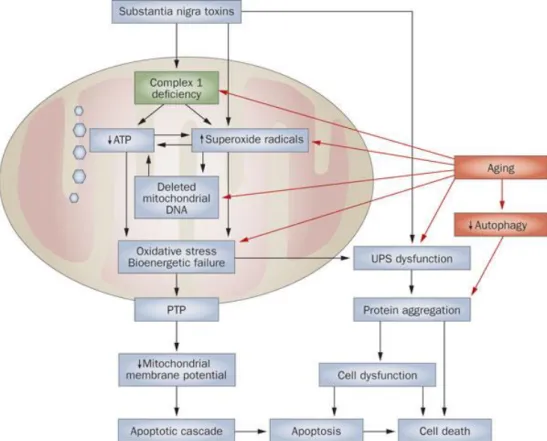
One of the most important characteristic hallmarks of PD is mitochondrial dysfunction (Figure
1.2). It has been postulated that the accumulation of mitochondrial ROS in different tissues during the
years can result in alterations in mtDNA, and subsquently cell death, leading to a decrease in tissue function associated with age (Rubinsztein, Codogno et al. 2012).
Mitochondria are the primary source of potentially damaging endogenous ROS, which have been linked to neurodegeneration too, and can induce protein carbonyls, lipid peroxidation and DNA damage. Importantly, release of cytochrome c (Cyt C) from mitochondria triggers apoptosis, and so the clearance of damaged mitochondria is vital for cell survival. ROS are also able to increase the release of Cyt C and induce the mitochondrial permeability transition pore (mPTP), both activate apoptosis
4 Figure 1.2 Biochemical senescence and pathogenetic pathways to cell death in Parkinson disease. (Schapira and Schrag 2011).
To prevent cellular damage from faulty mitochondria a number of protective mechanisms have involved; for example, mitochondria have an endogenous proteolytic mechanism to degrade misfolded proteins, proteins located on the inner and outer membranes can be degraded in the cytosol by the UPS and also specific mitochondrial components can be sequestered and directed to lysosomes for degradation by autophagy. Furthermore, electron microscopy analysis has revealed that entire mitochondria are detectable in both yeast vacuoles and mammalian lysosomes. It is the selective degradation of mitochondria by autophagy that is now termed mitophagy (Wager and Russell 2013).
1.2
Risk factors
5
age can be to increase the risk of disrupting cellular regulation, being longer exposed to harmful environmental effects and/or the combination of genetic susceptibility.
1.2.1 Environmental factors
Today a lot of pesticides, herbicides and industrial chemicals have been linked to the development of the disease. Among the most studied compounds found to participate extensively in neurodegeneration , there are rotenone (a substance of plant origin used as an insecticide and acts as a potent inhibitor of complex I of the mitochondrial respiratory chain), the 6-hydroxydopamine (6-OHDA)(a neurotoxin that induces oxidative stress), or bipiridínicos such as 1-methyl-4-phenylpyridine (MPP+)(a MPTP (1-methyl-4-phenyl-1,2,3,6-tetrahydropyridine) derivative) and the herbicide paraquat
(PQ2+).
MPTP crosses the blood brain barrier and is taken up by dopaminergic neurons through DA transporter after oxidation to MPP+ by MAO-B. This cation is very reactive and inhibits complex I
(NADH ubiquinone oxidoreductase) of the electron transport chain. That is why, today, MPTP/MPP+
and other inhibitors of this complex, including rotenone and PQ2+, are used as PD models in in vivo and
in vitro. (Padman, Bach et al. 2013)
PQ2+ (N,N′-dimethyl-4,4′-bipyridinium dichloride) is the organic compound with the chemical
formula [(C6H7N)2]Cl2. It is classified as a viologen, a family of redox-active heterocycles of similar
structure. This salt is one of the most widely used herbicides. It is quick-acting and non selective, killing green plant tissue on contact, but it is also toxic to human being and animals (Filograna, Godena et al.
2016).
1.2.2 Genetic factors
Beyond gender, race, age and environmental factors, there are familiar mutations of PD. The most convincing evidence came with the discovery of monogenic forms of PD. SNCA, which encodes
the protein α-synuclein, was the first gene to be associated with inherited PD. Mutations in LRRK2 and
parkin are the most common causes of dominantly and recessively inherited PD, respectively
(Wirdefeldt, Adami et al. 2011).
6
evidence is given by α-synuclein, the principal component of LB. Missense mutations in the gene encoding α-synuclein (PARK1) and multiplications of the α-synuclein gene locus (PARK4) lead to familial cases of PD (Gan-Or, Dion et al. 2015). Large studies determined that the hereditary
component of PD is at least 27%, and in some populations, single genetic factors are responsable for more than 33% of PD patients. Interestingly, many of these genetic factors, such as LRRK2, GBA,
SMPD1, SNCA, PARK2, PINK1, PARK7, SCARB2 and others, are involved in the autophagy-lysosome
pathway (ALP). Some of these genes encode lysosomal enzymes, whereas others correspond to proteins that are involved in transport to the lysosome, mitophagy, or other autophagic-related functions.
Table 1.1 Monogenic forms of Parkinson’s disease, by gene. Adapted from ((Kalia and Lang 2015).
Autosomal dominant
Protein
SNCA α-synuclein
LRRK2 Leucine-rich repeat kinase 2
VPS35 Vacuolar protein sorting 35
EIF4GI Eukaryotic translation initiation factor 4-ϒ 1
DNAJC13 Receptor-mediated endocytosis 8 (REM-8)
CHCH2 Coiled-coil-helix-coiler-coil-helix domain containing 2
Autosomal recessive
Parkin Parkin
PINK1 PTEN-induced putative kinase 1
DJ-1 DJ-1
1.3
Glial cells
and Parkinson’s Disease
Glial cells, sometimes called neuroglia or simply glia, are non-neuronal cells that maintain homeostasis, form myelin, and provide support and protection for neurons in the central and peripheral nervous systems. In the central nervous system, glial cells include oligodendrocytes, astrocytes, ependymal cells and microglia, and in the peripheral nervous system glial cells include Schwann cells and satellite cells. To study the mechanisms of neurodegeneration in PD, researchers have focused their attention primarily on the affected nigral dopaminergic neurons. However, it is now known that the neighboring glial cells play a significant role in the degenerescence of these neurons (Su, Zhang et al.
7
1.3.1 Microglia
Microglia is a type of glial cell located throughout the brain and spinal cord. Microglia account for 10–15% of all cells found within the brain. As the resident macrophage cells, they act as the first and main form of active immune defense in the central nervous system (CNS).
Microglia also has a role in neurodegenerative disorders. Many of the normal trophic functions of glia may be lost or overwhelmed when the cells become chronically activated in progressive neurodegenerative disorders. In such disorders, there is abundant evidence that activated glia play destructive roles by direct and indirect inflammatory attack (Sanchez-Guajardo, Tentillier et al. 2015).
Recent research suggests a complex role for microglia not only in PD but in other disorders involving α-synuclein aggregation, such as multiple system atrophy. In these neurodegenerative processes, the activation of microglia is a common pathological finding, which disturbs the homeostasis of the neuronal environment. The term activation comprises any deviation from what otherwise is considered normal microglia status, including cellular abundance, morphology or protein expression. The microglial response during disease will sustain survival or promote cell degeneration (Finkbeiner,
Cuervo et al. 2006).
1.3.2 Astrocytes
Astrocytes, the most numerous of glial cells, constitute a major class of cells in the mammalian brain and outnumber neurons by several folds in the human brain.
Astrocytes can release and supply neurons with neurotrophic factors such as nerve growth factor (NGF), neurotrophin-3, and basic fibroblast growth factor (bFGF) as well as metabolic substrates such as lactate and the antioxidant glutathione for the survival and proper functioning of neurons. On the other hand, when astrocytes undergo a state of gliosis in response to neuronal injury or toxic insults, together with microglia, they release cytokines and chemokines that are deleterious to neurons (Rappold
and Tieu 2010).
8
clearance of extracellular debris, which might aid in the survival of neurons (Janda, Lascala et al.
2015).
1.4
Ubiquitin-proteasome system
The UPS is a highly conserved and tightly regulated pathway for the coordinated degradation of a wide variety of proteins with half-lives ranging from minutes to several days. UPS curates proteome stability in various subcellular sites including the nucleus, the cytosol, the endoplasmic reticulum (ER), the mitochondria, and even the extracellular space (McKinnon and Tabrizi 2014). Therefore, UPS is
the main site of protein synthesis quality control and it is also involved in the recycling of both normal short-lived proteins and of nonrepairable misfolded or unfolded proteins (Figure 1.3). The expression
levels and activity of the UPS constituent components are tightly regulated, at both the transcriptional and posttranslational level, either under basal conditions or at conditions of increased oxidative and/or proteotoxic stress (Tsakiri and Trougakos 2015).
Impairment of the UPS has been implicated in the pathogenesis of a wide variety of neurodegenerative disorders including Alzheimer’s, Parkinson’s and Huntington’s diseases(Le 2014).
The most significant risk factor for the development of these disorders is aging, which is associated with a progressive decline in UPS activity and the accumulation of oxidatively modified proteins. Nevertheless, the gradual accumulation of stressors during aging along with the (mostly lifestyle-related) unbalanced redox homeostasis or high glucose levels eventually result in increasingly damaged proteome. This outcome may then increase genomic instability due to reduced fidelity in processes like DNA replication and repair, which then results in higher levels of proteome instability and so forth.To date, no therapies have been developed which can specifically upregulate this system (Bang, Kang et
9 .
1.5
Autophagy
Autophagy is an intracellular catabolic mechanism which is responsible for the degradation of cellular components such as proteins or organelles, by the action of lysosomal enzymes (Mizushima,
Yoshimori et al. 2010). Proteins involved in this process are called ATG proteins. There are three types
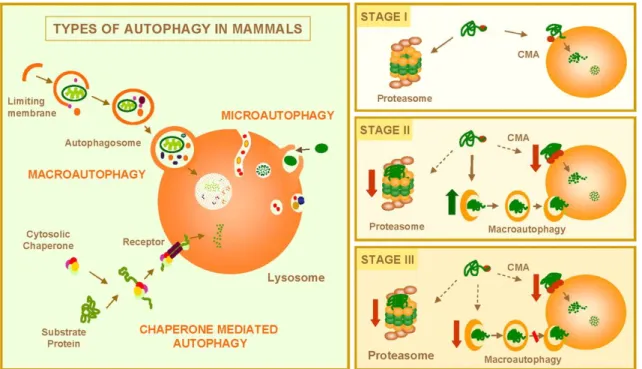
of autophagy: chaperone-mediated autophagy (CMA), microautofagia and macroautophagy (Figure
1.4) (Glick, Barth et al. 2010).
CMA involves direct translocation of unfolded proteins across the lysosome membrane. Chaperone proteins mediate this process by binding to cytosolic substrates that enter the lysosome through interaction with a receptor/channel on the lysosomal membrane. For a protein to be a CMA substrate, it must have in its amino acid sequence a pentapeptide motif biochemically related to KFERQ. This substrate protein-chaperone complex binds to lysosome-associated membrane protein type 2A (LAMP-2A), which acts as the receptor for this pathway. LAMP-2A, a single span membrane protein, is one of the three spliced variants of a single gene lamp2.
10
pinch off into the lysosomal lumen, they are rapidly degraded (Mizushima, Yoshimori et al. 2010).
Microautophagic pathway is especially important for survival of cells under starvation, nitrogen deprivation or after treatment with rapamycin.
Figure 1.4 Autophagy and neurodegenerative disorders. Left, Mammalian cells use three types of autophagy.
Microautophagy and macroautophagy involve the sequestration of complete cytosolic regions directly by the lysosomes or in an intermediate compartment, the autophagic vacuole, which is then delivered to lysosomes. In chaperone-mediated autophagy, single soluble proteins are recognized by a cytosolic chaperone and a receptor at the lysosomal membrane that mediates their translocation across the membrane into the lysosomal lumen. Right, Misfolded or altered proteins are selectively degraded by the ubiquitin/ proteasome system or by chaperone-mediated autophagy (STAGE I). However, when these altered proteins organize in toxic multimeric complexes, they often alter the proteolytic activity of these two pathways. Upregulation of macroautophagy can compensate for this deficit (STAGE II). Aggravating factors, such as oxidative stress and aging, can precipitate the failure of macroautophagy with the consequent detrimental effect on cell functioning, often resulting in cellular death (STAGE III). (Finkbeiner, Cuervo et al. 2006).
Macroautophagy (therefore refered as autophagy) involves the engulfment of the cargo into double-membraneautophagosomes, which subsequently fuse with endosomes and lysosomes for cargo degradation (Chen, Khambu et al. 2014). Autophagosome formation is a complex and highly regulated
process that requires more than 30 autophagy-related proteins (Atg) that have been identified by molecular dissection of autophagic process through genetic screening of yeast. These proteins form functional complexes Atg mediating macroautophagy individual stages: initiation or induction, nucleation, membrane elongation, load detection and autophagosomes fusion with lysosomes. Macroautophagy induction occurs after cell conditions such as, stress or nutrient deficiency.
11
III phosphatidylinositol 3-kinase (PI3K)/Vps34 complex I, and the Atg9/mATG9 transmembrane protein system.
The process starts upon activation of cell-specific signaling pathways that suppress mammalian target of rapamycin (mTOR) signaling complex (mTORC1)(Cheng, Ren et al. 2013). This is followed
by vesicle nucleation at the isolation membrane, most likely at ER level. It involves the recruitment and assembly of several proteins including Vps34, Beclin-1, and UVRAG among others. Vps34 has a PI3K activity and produces phosphatidylinositol 3-phosphate, which is needed for the targeting of Atg family proteins involved in subsequent vesicle elongation. First, ULK1/2-mAtg13-FIP200 (equivalent to Atg1-Atg13-Atg17 in yeast) complex is build and then two ubiquitin-like conjugation systems (involving Atg12-Atg5-Atg16) are recruited and mediate the lipid conjugation of Atg8/LC3, among other reactions involved in the vesicle elongation step (Rubinsztein, Codogno et al. 2012).
Among known Atg-encoded proteins, only microtubule-associated protein 1 light chain 3 (LC3), a mammalian orthologue of yeast Atg8, can localize to all types of autophagic membranes, including the phagophore (the immature autophagosome, also known as the isolation membrane), the autophagosome, and the autolysosome (a hybrid organelle formed by fusion of the autophagosome and lysosome). Nascent LC3 (pro-LC3) is processed by Atg4-family proteins, which are cysteine proteases, into LC3-I immediately after synthesis. During autophagy, cytosolic LC3-I is conjugated to phosphatidylethanolamine (PE) to become LC3-II by the activating enzyme Atg7 and the conjugating enzyme Atg3. The conjugation of LC3 to PE is also facilitated by the Atg12–Atg5 conjugate along with Atg16L1. LC3-II is then recruited to autophagosomal membranes (Ge and Schekman 2014).
Finally, LC3 is released from LC3-PE by a second Atg4-dependent cleavage, while LC3-II in the autolysosomal lumen is degraded by autophagy. Thus, LC3 conversion (LC3-I to LC3-II) and lysosomal degradation of LC3-II reflect the progression of autophagy, and detecting LC3 by immunoblot analysis is often used to monitor autophagic activity. However, the number of autophagic organelles at a given moment is regulated by both the on-rate (autophagosome formation) and off-rate (degradation upon fusion with lysosomes). Thus, although the amount of LC3-II correlates with the number of autophagosomes, its amount at a certain time point does not necessarily indicate the degree of autophagic flux, a term used to indicate overall autophagic degradation (i.e., delivery of autophagic cargo to the lysosome) rather than autophagosome formation. Furthermore, not all LC3-II is present on autophagic membranes (Gomez-Sanchez, Yakhine-Diop et al. 2016). A significant amount of LC3-II
12
in the presence and absence of bafilomycin A1 (a vacuolar H+-ATPase inhibitor), lysosomal protease
inhibitors (e.g., E64d and pepstatin A), or lysosomotropic agents (e.g., chloroquine - CQ) to inhibit lysosomal degradation of LC3-II (Hoyer-Hansen, Bastholm et al. 2007).
Another widely used autophagy marker, p62, also called sequestosome 1 (SQSTM1), binds directly to LC3 and GABARAP (Atg8 orthologues) family proteins via a short LC3 interaction region (LIR) (Ichimura and Komatsu 2010). This may serve as a mechanism to deliver selective autophagic
cargo for degradation by autophagy. The p62 protein is itself degraded by autophagy and serves as a marker to study autophagic flux. When autophagy is inhibited, p62 accumulates, while when autophagy is induced, p62 quantities decrease (Jiang and Mizushima 2015).
Increasing evidences suggest that autophagic deregulation causes accumulation of abnormal proteins or damaged organelles, which is a characteristic of chronic neurodegenerative conditions, such as PD, a multifactorial disorder, which is neuropathologically characterized by age-dependent neurodegeneration of dopaminergic neurons in the midbrain. Indeed, promoting the clearance of aggregate proteins via pharmacological induction of autophagy has proved to be an useful mechanism for protecting cells against the toxic effects of these proteins in the context of neurodegenerative diseases and protecting neurons from apoptosis (Segura-Aguilar, Paris et al. 2014).
In some cases, autophagy displays substrate specificity, even though autophagy is often considered to be a nonselective pathway for the degradation of bulk cytoplasmic components. Indeed, the unique feature of the autophagy process where the initial sequestering compartment expands into an autophagosome allows for flexible cargo selection. In addition, superfluous or damaged organelles and misfolded or aggregated proteins are selectively targeted for degradation by autophagy (Svenning and
Johansen 2013).
Selective autophagy is based on the recognition and degradation of a specific cargo, in a process depending on receptor proteins that bind Atg8/LC3 to facilitate enrichment of cargoes sequestrated for degradation (Pimentel-Muinos and Boada-Romero 2014). Among them, mitophagy has been
increasingly implicated in the pathogenesis of PD through the PINK1-PARKIN-mediated pathway. Selective autophagy can be classified according to the cargoes involved (Wager and Russell 2013). We
13
1.5.1 Signaling Pathway
o mTOR pathway
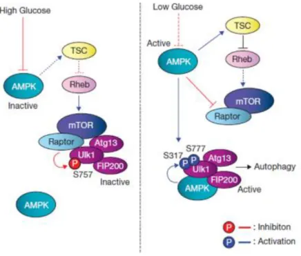
Autophagy is inhibited by the mammalian target of rapamycin (mTOR), mTOR is a highly conserved serine/threonine kinase that is capable of integrating signals from many stimuli including amino acids, energy levels, oxygen, growth factors, and stress to coordinate cell growth and maintain metabolic homeostasis. Under nutrient sufficiency, high mTOR activity prevents ULK1 activation by phosphorylating the Ser 757 residue and disrupting the interaction between ULK1 and AMPK (Figure
1.5). Activation of pathways that stimulate mTOR activity is considered to inhibit autophagy. mTOR
forms two functionally distinct complexes in mammals, mTORC1 (mTOR complex 1) and mTORC2 (mTOR complex 2) (Marlin and Li 2015). mTORC1 is sensitive to both growth factors and nutrients,
and the presence of amino acids has been shown to be essential for activation of the mTORC1 kinase. Proteins including Ste-20-related kinase (mitogen-activated protein kinase kinase kinase kinase 3) MAP4K3 and Vps34 have been described to play a role in amino acid signaling possibly through regulation of phosphatases and endocytic trafficking upstream of mTORC1 (Kim, Kundu et al. 2011).
o AMPK pathway
Autophagy is promoted by AMP activated protein kinase (AMPK), which is a key energy sensor and regulates cellular metabolism to maintain energy homeostasis (Mihaylova and Shaw 2011). Under
glucose starvation, AMPK promotes autophagy by directly activating ULK1 through phosphorylation of Ser 317 and Ser 777. Importantly, AMPK is an established negative regulator of the mTOR signaling cascade (Gwinn, Shackelford et al. 2008). This can be accomplished by AMPK-mediated
phosphorylation of the TSC complex which is a negative regulator of mTORC1 activation at the lysosome
(Chen, Zhao et al. 2015). Alternatively, AMPK can directly phosphorylate the Raptor subunit of
mTORC1, which induces 14-3-3 binding and inhibits mTORC1 target phosphorylation. Through these both mechanisms, AMPK is able to relieve mTOR-mediated autophagy repression (Choudhury, Yang
14 Figure 1.5 Model of ULK1 regulation by AMPK and mTORC1 in response to glucose signals. Left: when glucose is sufficient, AMPK is inactive and mTORC1 is active. The active mTORC1 phosphorylates ULK1 on Ser 757 to prevent ULK1 interaction with and activation by AMPK. Right: when cellular energy level is limited, AMPK is activated and mTORC1 is inhibited by AMPK through the phosphorylation of TSC2 and Raptor. Phosphorylation of Ser 757 is decreased, and subsequently ULK1 can interact with and be phosphorylated by AMPK on Ser317 and Ser 777. The AMPK-phosphorylated ULK1 is active and then initiates autophagy. (Kim, Kundu et al. 2011).
o Beclin1/PI3K
Beclin-1 (BECN1) is a mammalian ortholog of the yeast Atg6 and BEC-1. This protein interacts with either Bcl-2 or PI3K class III, playing a critical role in the regulation of both autophagy (Figure
1.6) and cell death. A BECN1 binding partner, AMBRA1 (activating molecule in Beclin1-regulated autophagy 1), has also been identified as a target for ULK1-mediated phosphorylation. Under nutrient-rich conditions, AMBRA1 binds BECN1and Vps34 at the cytoskeleton through an interaction with dynein. However, it is unclear if BECN1 binds Atg14 and AMBRA1 in the same complex at the site of the phagophore. Interestingly, AMBRA1 was shown to act in an mTORC1-sensitive positive-feedback loop to promote K63-linked ubiquitination of ULK1 through recruitment of the E3-ubiquitin ligase, TNF receptor associated factor 6 (TRAF6). This pathway of autophagy was not associated with LC3 processing but appeared to involve autophagosome formation from late endosomes and the trans-Golgi
(Kang, Zeh et al. 2011).
15 Figure 1.6 Regulation of VPS34 complex formation in response to nutrients. Starvation activates JNK1 kinase, possibly
through direct phosphorylation by AMPK. JNK1 phosphorylates Bcl-2, relieving Bcl-2-mediated repression of Beclin-1-VPS34 complexes. Bcl-2 may inhibit Beclin-1-VPS34 complexes by disrupting Beclin-1-Beclin-1-VPS34 interaction (left arrow) or by stabilizing an inactive Beclin-1 homodimeric complex (right arrow). (Russell, Yuan et al. 2014).
1.5.2 Autophagy and Parkinson’s Disease
Aggregated and ubiquitinated proteins cause synaptic impairment, damage to organelles, and cell death in the central nervous system. Many types of neurodegenerative diseases are accompanied by the accumulation of aggregated and ubiquitinated proteins. Autophagy is involved in the degradation and removal of aggregated proteins, and the inhibition of constitutive autophagy leads to neurodegeneration in the central nervous system. Autophagy helps to clear damaged organelles and protein aggregates or lipid droplets (LP), which represent unwanted and usually toxic cargo that may lead to cellular dysfunction (Lynch-Day, Mao et al. 2012).
16
Another important breakthrough has been the demonstration that Parkin, mutated in autosomal recessive forms of PD, is recruited to damaged mitochondria to facilitate the mitochondria-selective autophagy, mitophagy (Egan, Shackelford et al. 2011). Mitophagy is the process by which
mitochondria are selectively degraded by the highly conserved autophagic machinery. It occurs during developmental processes in specialized cells such as erythrocytes, while in other cell types damaged mitochondria are removed in order to maintain a functional mitochondrial population. They are also the primary source of potentially damaging endogenous ROS, which have been linked to neurodegeneration, and can induce protein carbonyls, lipid peroxidation, and DNA damage. Importantly, release of Cyt C from mitochondria triggers apoptosis.Then, the clearance of damaged mitochondria is vital for cell survival (Janda, Isidoro et al. 2012).
ROS are also able to increase the release of Cyt c and induce the mitochondrial permeability transition pore (mPTP), both of which activate apoptosis. Accumulating evidence from epidemiological studies and toxin-induced animal models of PD strongly point to environmental toxins as possible triggers of nigrostratial dopaminergic neurons degeneration. Almost all of these toxic substances have been shown to deregulate macroautophagy by pathologically enhancing or interfering with autophagic flux (Giuliano, Cormerais et al. 2015). Low levels of ROS and RNS play an increasingly recognized
role in signal transduction and regulation of physiological processes, including autophagy (Son, Shim
et al. 2012).
The parkinsonian pro-oxidants produce high levels of ROS and RNS that act both positively and negatively on signaling pathways and lipid-protein complexes regulating autophagy. The relationship between autophagy and oxidative stress in PD and other pathologies is very complex and cannot be summarized in one phrase “defective autophagy induces oxidative stress and oxidative stress induces autophagy,” mainly because the effects of excessive ROS and RNS on the autophagic machinery can be either activating or inhibiting (Tavakkoli, Miri et al. 2014). The balance between positive and
negative signals will tune the autophagic process in cell type and oxidant-specific manner and can turn on induced autophagy, as well as turn off the basal autophagy.
1.5.3 Non-canonical autophagy
17
This phosphoinositide pool also organizes the events that culminate in lipidation of the autophagy protein LC3 in preparation for fusion of the double-membrane autophagosome to lysosomes (Bankaitis
2015).
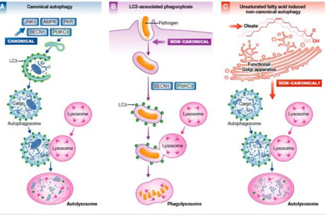
The ability of autophagy to sequester and clear large particles from the cytoplasm has broader implications as evidenced by the discovery of non-canonical autophagy pathways. This pathway counters the strategies used by intracellular pathogens to encourage their entry into cells and to subsequently inhibit maturation of conventional phagolysosomes so that these organelles are co-opted as suitable microenvironments for pathogen propagation. These immune response applications are classified as non-canonical autophagy pathways because the action is initiated at the plasma membrane, and the operant autophagosomes are systems enclosed by a single membrane (Figure 1.7).
While the pathway remains dependent on the BECN1/PI3K complex, it is neither dependent on components of the canonical autophagy pre-initiation complex nor is it subject to control by the TOR nutrient sensor kinase. Such a noncanonical LC3-associated phagocytosis pathway (LAP) more generally promotes immune responses by facilitating antigen presentation, by regulating interferon production, and by dampening inflammation potential by clearing cell corpses (Niso-Santano, Malik
et al. 2015).
The non-canonical pathway described by (Niso-Santano, Malik et al. 2015) shares some
18 Figure 1.7 Canonical and non-canonical autophagy pathways. Canonical autophagic pathway (A) involves different
components than the LC3-associated phagocytosis pathway (B) and the oleate-induced non-canonical autophagic pathway (C).
(Bankaitis 2015).
1.5.4 Lipid droplets in autophagy
LDs are storage organelles for the neutral lipids (Figure 1.8) present in most cell types. The
LDs core consists mainly of triacylglycerols (TAGs) and steryl esters (STEs). Evidence points to the ER as the site of formation of the LDs, being the main source of the autophagosomal membrane. LDs serve important functions in the cell by providing lipids and energy as well as by storing free fatty acids that may otherwise become cytotoxic. Deletion of biosynthetic enzymes of STEs and TAGs has opposite effects on the lipidation state of Atg8, suggesting novel and complementary roles for these neutral lipids in regulation of the autophagic process (Shpilka, Welter et al. 2015).
A complex relationship between autophagy and LDs is described: on one hand autophagy is implicated in lipophagy, a process of LD degradation, while on the other hand LDs are linked to autophagy regulation. When autophagy is inhibited by fatty acid synthase (FASN) there are an amount of LDs, so we believe that LDs are essential to the autophagic process.
19
Physiologically, FASN is expressed in liver cells and lipogenic tissue and is regulated by insulin, glucocorticoids, and glucagon as well as by nutrients. The NADPH-dependent process plays a central role in energy homeostasis by converting excess carbon intake into FAs for storage (Tao, He et al.
2013).
FASN is expressed at relatively low levels in normal cells (except liver, brain, lung and adipose tissue), whereas it is highly expressed in a wide variety of cancers, including cancer of the prostate, breast, brain, lung, ovary, endometrium, colon, thyroid, bladder, kidney, liver, pancreas, stomach, oesophagus, eye, mesothelium and skin (Grube, Dunisch et al. 2014).
Several natural and synthetic FASN inhibitors such as the antifungal agent cerulenin and its synthetic derivative C75, the β-lactone orlistat as well as the bactericide triclosan have been shown to inhibit cancer cell growth, by inducing cell death. Nuclear fragmentation assay and Western blotting (WB) analysis after targeting FASN with those inhibitors demonstrated autophagy and apoptosis
(Sadowski, Pouwer et al. 2014).
In a related, LDs have recently emerged as organelles that participate in aggregate clearance. The involvement of LDs in autophagosome biogenesis and the ability of autophagosomes to deliver LDs to the vacuole/lysosome may be a key mechanism by which cells eliminate aggregated proteins (Shpilka
and Elazar 2015).
Figure 1.8 Projection of a confocal stack of an abdominal adipocyte isolated from an Akt (PKB) Drosophila melanogaster mutant. The adipocyte was stained with Nile Red to image neutral lipids in order to evidence and quantify LD
20
1.5.5 Lipid role in autophagy
The macroautophagy within hepatocytes function is the degradation of intracellular lipid reserves. Although the lipolytic function of lysosomes was known previously, the mechanism of lipid delivery to the lysosome was unclear. They contain numerous lysosomal hydrolases and lipases operating in an acidic environment (pH <5.2) to degrade the charge delivered. Studies in cultured hepatocytes lacking autophagy by pharmacological inhibition with 3-methyladenine (3-MA) or using RNA interference against ATG5 and ATG7 have shown that inhibition of macroautophagy results in an increase in the accumulation of triglyceride (TG) hepatocellular, when compared to controls. Increased TG accumulation occurs under both basal conditions and when hepatocytes receive a lipogenic stimulus such as treatment with physiological concentrations of oleic or after culturing in a medium lipogenic, methionine, and choline deficient medium (Brenner, Galluzzi et al. 2013).
Electron microscopy studies demonstrate that inhibition of macroautophagy in hepatocytes and liver leads to a marked increase in the number and size of LD, showing that lipid accumulation occurs in the form of LD. Interestingly, the increase in hepatocellular reserves TG resulting from a decrease in lipolysis lipid reserves is due to a decrease in the delivery of lipid load to lysosomes, and not an increase in hepatocellular TG synthesis or reduced secretion in the form of very low density lipoproteins (VLDL). Immunofluorescence colocalization experiments between a neutral lipid dye (BODIPY 493/503) and autophagosome marker (LC3) or lysosomal marker (LAMP1) have revealed the localization of cellular lipid components autophagosome and lysosomal system under conditions which macroautophagy is activated with rapamycin (a TOR inhibitor) or a lipid providing stimulus. Additionally, pharmacological or genetic ablation of autophagy reduces bodipy-LC3 and bodipy-LAMP1 colocalization observed
(Wrighton 2015).
1.6
Diet
Nutritional epidemiological studies in PD have focused on groups of food items, macronutrients (such as protein, fat, and carbohydrates), or other specific nutrients. Several dietary habits have been shown to modify the risk of developing PD.
Intake of coffee and tea in relation to PD has been studied extensively. Caffeine acts as an adenosine receptor antagonist and experimental evidence suggests that it may exert a neuroprotective effect. Of the 7 case-control studies that investigated tea intake for a possible association with PD, three reported an inverse association, three found no association, and one reported an increased risk (Agim
21
Calcium and vitamin D intake were associated with PD risk when the source was dairy products, but not when the source was non-dairy products suggesting that a compound in dairy products other than calcium or vitamin D was responsible for the association (Agim and Cannon 2015).
The role of antioxidants in PD has been studied based on the hypothesis that oxidative stress is involved in the pathogenesis of the disease. A meta-analysis of 7 case-control and one cohort study that assessed intake of antioxidants in relation to PD risk reported an inverse association with moderately high vitamin E intake and PD, whereas high intake of vitamin E (defined as fourth quartile or fifth quintile of intake) was not associated with further reduction in PD risk (Albarracin, Stab et al. 2012).
Total dietary fat intake is supplied in three categories: saturated fatty acids, unsaturated fatty acids, and cholesterol. MUFAs (mono-unsaturated fatty acids) and PUFAs (poly-unsaturated fatty acids) have been shown to have antiinflammatory and neuroprotective properties by reducing the oxidative stress and inhibiting neuronal apoptosis. PUFAs help regulation of dopamine activity in basal ganglia, controlling movement. Supplementation or higher intake of unsaturated fatty acids was shown to alleviate neurotoxin-induced PD-like syndrome (de Lau, Bornebroek et al. 2005). As the
neuroprotective effect of PUFA has been repeatedly shown, both in vivo and in vitro, high fat diet has
been shown to be associated with increased risk of PD. In a small case-control study, higher Mediterranean diet (MeDi) score was significantly associated with lower risk of PD (Dong, Beard et
al. 2014).
The MeDi has received attention in recent years because of growing evidence associating MeDi with lower risk for AD, cardiovascular disease, several forms of cancer, and overall mortality. The MeDi is characterized by high intake of vegetables, legumes, fruits, and cereals; high intake of unsaturated fatty acids (mostly in the form of olive oil) compared to saturated fatty acids; a moderately high intake of fish; a low-to-moderate intake of dairy products, meat and poultry; and a regular but moderate consumption of ethanol, primarily in the form of wine and generally during meals (Alcalay, Gu et al.
2012).
22
1.6.1 PUFA
PUFA are basic components involved in the architecture and function of cellular membranes, being endogenous mediators for cell signaling and involved in the regulation of gene expression. They are precursors of eicosanoids, such as prostaglandins and leukotrienes, and docosanoids such as protectins or resolvins (Igarashi, Kim et al. 2012). They can act as transcription factors modulating
protein synthesis, as ligands in signal transduction, and as membrane components able to regulate the fluidity, permeability and dynamics of cell membranes (Johansson, Monsen et al. 2015). Unsaturated
FAs can induce non-canonical BECN1-independent autophagy in vitro and in vivo through a phylogenetically conserved mechanism that requires an intact Golgi apparatus (Enot, Niso-Santano et
al. 2015).
This type of fat helps to increase the rates of high-density lipoprotein cholesterol (HDL) and maintain low rates of low-density lipoprotein cholesterol (LDL). Excess involves the production of toxic compounds (Cetrullo, Tantini et al. 2012). They can be obtained in blue fish and vegetables such as
corn, soybean, sunflower, pumpkin, nuts etc.
23
In relation to their structure, PUFA are fatty acids that contain more than one double bond in their backbone and they can be divided in two types: Omega-3 (n-3) and Omega-6 (n-6) (Figure 1.9).
The nutritional dietary recommendations of n-3 PUFA are clearly defined and consensual (amounts and types), but the recommendations of the n-6 PUFA are more controversial (Chen, Zhang et al. 2014).
Existing reports suggest that omega-6 essential fatty acids are tipically proinflammatory and are linked with initiation and progression of carcinogenesis and others reports that suggest n-3 PUFA supplementation is a potential neurogenic and oligodendrogenic treatment to naturally improve post-stroke brain repair and long-term functional recovery (Rovito, Giordano et al. 2013) (Hu, Zhang et
al. 2013). Studies using animal models of PD show that, in addition to its essential role in protecting
dopaminergic neurons, maternal n-3 PUFAs supplementation played an important role in maintaining the cognitive integrity in inflammation-induced neurotoxicity (Delattre, Carabelli et al. 2016).
Further, PUFAs are essential components of neuronal and glial cell membranes. They regulate the production of pro/anti-inflammatory cytokines that may also contribute to neurodegenerative diseases such as PD (Lopez-Vicario, Alcaraz-Quiles et al. 2015).
1.6.2 Linoleic Acid
Linoleic acid (LA) is an omega-6 PUFA. It is a carboxylic acid with an 18-carbon chain and two cis double bonds, with the first double bond located at the sixth carbon from the methyl end (Figure
1.10).
LA belongs to one of the two families of essential fatty acids, which means that the human body cannot synthesize it. LA comes from other food components and itis used in the biosynthesis of arachidonic acid and thus some prostaglandins, leukotrienes, and thromboxane. It is found in the lipids of cell membranes (Choque, Catheline et al. 2014).
24
In fact, LA is abundant in nuts, fatty seeds (flax seeds, hemp seeds, poppy seeds, sesame seeds, etc.) and their derived vegetable oils: poppy seed, safflower, sunflower, corn, and soybean oils.
25
2.
Rational and aims
One of important cellular dysfunction in PD pathogenesis is autophagy desregulation. As it has been described the importance of PUFAs in autophagy modulation, it would be interesting to study their potential neuroprotective effects in PD models. Linoleic acid is a PUFA and it is the most present in our diet. If it possesses a neuroprotective effect, it would be a cheap and accessible compound..
In order to achieve our objective, we emphasize the following specific topics:
Perform a dose-response of linoleic acid to determine the non cytotoxic concentrations that induce autophagy.
Characterization of autophagy by linoleic acid to prove the implication of different signaling pathways.
Find out the relation between LA-induced autophagy and various organelles.
27
3.
Materials and methods
3.1
Appliances
Rocker - Labnet Rocker 25 model Tube shaker - Bunsen
Analog Magnetic shuffler - Bunsen serie MC-8 Autoclave - Raypa Steam Sterilizer
Analytical balance - ADAM PW 124 model Electronic balance - AND GF 300 model Thermostatic bath - Bunsen serie BA
Camera Hammamatsu Orca-ER (adapted inverted optical microscope fluorescence Olympus IX81 model)
Camera Olympus model DP70 (adapted inverted optical microscope fluorescence Chemiluminescence imager – Amersham Imagen 600, GE Healthcare
Olympus IX51 model)
Laminar flow hood - TELSTAR model AV-100 Fume hood - Flores Valles
Refrigerated centrifuge table Heraeus Megafuge 1.0R model Refrigerated centrifuge table Hermle Z 36 HK model Freezer -20oC Edesa Práctica model
Freezer -80oC Heraeus HERA freeze model
Freezer -80oC Thermo Scientific Forma 994 model
Automatic cell counter - Bio-Rad TC10™ Automated Cell Counter model Electrophoresis equipment Bio-Rad Mini-Protean® III Cell model
Transfer equipment Bio-Rad Trans-Blot SD Semi-Dry Transfer Cell model Spectrophotometer Thermo Scientific NanoDrop 2000 model
Refrigerator 4ºC and -20ºC Edesa Style model
Power Supplies - Bio-Rad POWER PAC 200 and POWER PAC 300 models Automatic homogenizern Accumet AB150 Ficher Scientific
CO2 incubator with temperature control - Thermo Scientific HEPA Class 100 model
28
Inverted optical microscope fluorescence Nikon Eclipse Ti
Inverted optical microscope fluorescence Olympus modelo IX51 and IX81 pH-metro CRISON GLP 21 model
Metal block thermostat - Bunsen serie TMR
3.2
Reagents
Ambion Laboratories
• BLOCK-iT™ Alexa Fluor® Red Fluorescent Oligo
Applied Biosystems Laboratories
• Negative control to siRNA(Silencer ® Negative Control #1 siRNA) (scrambled)
Bio-Rad Laboratories
• Coomassie® Brilliant Blue R-250 • Sodium dodecyl sulfate (SDS)
• Polyacrylamide gel 12% Mini-PROTEAN® TGX™ Precast Gel
• Molecular weight marker for protein electrophoresis (Precision Plus Protein™ Dual Color Standars)
• Buffer Tris/glycine 10X
• Buffer Tris/glycine/SDS (Laemmli) 10X
Fluka Chemika Laboratories
• Paraformaldehyde (PFA)
GIBCO Laboratories
• Medium DMEM
• Medium Opti-MEM® I Reduced Serum Medium • Trypsin EDTA 10X 2,5 %
• Versene 1X
GE Healthcare Laboratories
• PVDF Hybond-P membranes
• Photographic paper HyperFilm™ ECL
HyClone Laboratories
29
Invitrogen Laboratories
• Lipofectamine® 2000 Transfection Reagent
KODAK Laboratories
• Fixative solution for photographic film • Developing solution for photographic film
Molecular Probes Laboratories
• Alexa Fluor® 488 anti-rabbit IgG antibody • Alexa Fluor® 568 anti-mouse IgG antibody
Panreac Laboratories • Glacial acetic acid
• Hydrochloric acid (HCl) 37% • Calcium chloride (CaCl2) • Magnesium chloride (MgCl2) • Potassium chloride (KCl) • Sodium chloride (NaCl) • Absolute ethanol • sodium fluoride (NaF)
• Disodium phosphate (Na2HPO4)
• Potassium phosphate (KH2PO4)
• Glycerin • Glycine • Methanol .
Pierce Laboratories
ECL Plus Western Blotting Substrate
QIAGEN Laboratories
HiPerfect Transfection Reagent (HiP)
Roche Laboratories Nonidet (NP40)
Sigma-Aldrich Laboratories
Bicinchoninic acid (BCA) Linoleic acid (LA)
Bovine serum albumin (BSA) β – mercaptoethanol
30
2- [4- (2-hydroxyethyl) -1-piperazinyl (1)] - ethanesulfonic (HEPES) Hoechst 33342
m-chloro carbonylcyanide phenylhydrazone (CCCP) Nile red
Oxaloacetate sodium pyruvate Red ponceau Sucrose
Copper sulfate (II) Thapsigargin Triclosan Triton X-100 Trizma® (Tris) base
Trypan blue solución (0,4 %) Tween 20
Propidium Iodide (PI)
Southern Biotech Laboratories
Glue Fluoromount G
Thermofisher Laboratories
Black plate 96-well polystyrene Cat. No.:437869/437958 Silencer negative control #1 AM4611
3.3
Antibodies
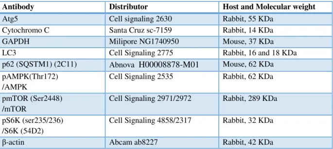
The following primary antibodies were used:
Table 3.1 Primary antibodies.
Antibody Distributor Host and Molecular weight
Atg5 Cell signaling 2630 Rabbit, 55 KDa
Cytochromo C Santa Cruz sc-7159 Rabbit, 14 KDa
GAPDH Milipore NG1740950 Mouse, 37 KDa
LC3 Cell Signaling 2775 Rabbit, 16 and 18 KDa
p62 (SQSTM1) (2C11) Abnova
H00008878-M01
Mouse, 62 KDapAMPK(Thr172) /AMPK
Cell Signaling 2535 Rabbit, 62 KDa
pmTOR (Ser2448) /mTOR
Cell Signaling 2971/2972 Rabbit, 289 KDa
pS6K (ser235/236) /S6K (54D2)
Cell Signaling 4858/2317 Rabbit, 32 KDa
31
3.4
Cell lines
SH-SY5Y (human neuroblastoma derived cell line);
H4 WT, H4 ATG5 KO (human neuroglioma derived cell line);
MEF WT and ATG5 KO (mouse embryon fibroblast);
U251 (human glioma derived cell line).
3.5
Cell maintenance
Maintenance is a vital process when working with cell cultures. It allows us throughout our experiments, thawing vials of cells to amplify, seed, treat and freeze new road that will ensure our work in the future.
3.6
Defrosting/freezing
Cells are frozen in a solution of 10% dimethylsulfoxide (DMSO) in FBS (10ml DMSO in 90 ml FBS). Although toxic at room temperature, DMSO is a cryoprotectant which protects cells from ice crystals, preventing cell death, at extreme temperatures. However, DMSO is not always appropriate because it can induce differentiation into certain cell lines, in which case it is better to go to glycerol. Generally, we freeze a cell density of approximately 1 million in 1 mL of FBS / DMSO, in a dry cryotube and store it in a freezer of -80°C. The ideal of a freeze is that it is slow and progressive.
3.7
Cell culture
Human cell lines and mouse cell lines were obtained as described: H4 cells stably expressing GFP-LC3, both WT and ATG5 and U2OS cell lines (kindly given by Prof. Junying Yuan, Harvard Medical School, USA); SH-SY5Y from ATCC (American Type Culture Collection, Manassas, VA, USA); U251 (kindly given by Prof. Nadezda Apostolova, Skopie University, Republic of Macedonia); MEF WT and MEF ATG5 (kindly given by Prof. Mizushima, University of Tokio, Japan) Cell lines were routinely maintained at 37ºC, 5% CO2, in the following media:
o H4 GFP LC3 (1 L DMEM; 10% FBS; 2 mM L-glutamine; 10 U/mL