Low Prevalence of
Chlamydia trachomatis
Infection in
Non-Urban Pregnant Women in Vellore, S. India
Navjyot K. Vidwan1*, Annie Regi2, Mark Steinhoff1, Jill S. Huppert1, Mary Allen Staat1, Caitlin Dodd1, Rida Nongrum2, Shalini Anandan2, Valsan Verghese2
1Cincinnati Children’s Hospital Medical Center, Cincinnati, Ohio, United States of America,2Christian Medical College, Vellore, Tamil Nadu, India
Abstract
Objective:To determine the prevalence and risk factors forChlamydia trachomatis(CT) infection in pregnant women and the rate of transmission of CT to infants.
Methods:Pregnant women ($28 weeks gestation) in Vellore, South India were approached for enrollment from April 2009 to January 2010. After informed consent was obtained, women completed a socio-demographic, prenatal, and sexual history questionnaire. Endocervical samples collected at delivery were examined for CT by a rapid enzyme test and nucleic acid amplification test (NAAT). Neonatal nasopharyngeal and conjunctival swabs were collected for NAAT testing.
Results:Overall, 1198 women were enrolled and 799 (67%) endocervical samples were collected at birth. Analyses were completed on 784 participants with available rapid and NAAT results. The mean age of women was 25.8 years (range 18–
39 yrs) and 22% (95% CI: 19.7–24.4%) were primigravida. All women enrolled were married; one reported.one sexual
partner; and six reported prior STI. We found 71 positive rapid CT tests and 1/784 (0.1%; 95% CI: 0–0.38%) true positive CT infection using NAAT.
Conclusions:To our knowledge, this is the largest study on CT prevalence amongst healthy pregnant mothers in southern India, and it documents a very low prevalence with NAAT. Many false positive results were noted using the rapid test. These data suggest that universal CT screening is not indicated in this population.
Citation:Vidwan NK, Regi A, Steinhoff M, Huppert JS, Staat MA, et al. (2012) Low Prevalence ofChlamydia trachomatisInfection in Non-Urban Pregnant Women in Vellore, S. India. PLoS ONE 7(5): e34794. doi:10.1371/journal.pone.0034794
Editor:Katharina Kranzer, London School of Hygiene and Tropical Medicine, United Kingdom ReceivedSeptember 14, 2011;AcceptedMarch 9, 2012;PublishedMay 2, 2012
Copyright:ß2012 Vidwan et al. This is an open-access article distributed under the terms of the Creative Commons Attribution License, which permits unrestricted use, distribution, and reproduction in any medium, provided the original author and source are credited.
Funding:This work was supported by Fogarty International Center, National Institutes of Health (R24TW007988, the Fogarty International Clinical Research Scholars Support Center), and Cincinnati Children’s Hospital Medical Center Infectious Diseases Department. The funders had no role in study design, data collection and analysis, decision to publish, or preparation of the manuscript.
Competing Interests:The authors have declared that no competing interests exist. * E-mail: nkvidwan@gmail.com
Introduction
Chlamydia trachomatis(CT) is one of the most common genital pathogens worldwide [1]. Strategies to screen, test, diagnose and treat this curable, yet prevalent and indolent disease have been adopted in many countries. Disease in women can include severe outcomes such as pelvic inflammatory disease and infertility. In addition, neonates of infected women may have increased morbidity and mortality. Newborn prematurity and low birth weight is shown to be associated with maternal chlamydia infection. Notably 50–75% of infants born to infected mothers become infected at one or more sites including the conjunctiva, nasopharynx, vagina, and rectum leading to purulent conjuncti-vitis, pneumonia, and trachoma [2]. Hammerschlag recognized that the most frequent site of perinatally acquired chlamydia infection in US neonates is the nasopharynx with rates as high as 70% and that CT pneumonia can occur in 30% of infants with nasopharyngeal infection [3]. In addition, Rosenman found that of US infants exposed to CT at birth, 8 to 44% had conjunctivitis, and 0 to 17% pneumonia [4].
Most epidemiologic data on CT is from industrialized nations; however, it is important to characterize CT disease from resource
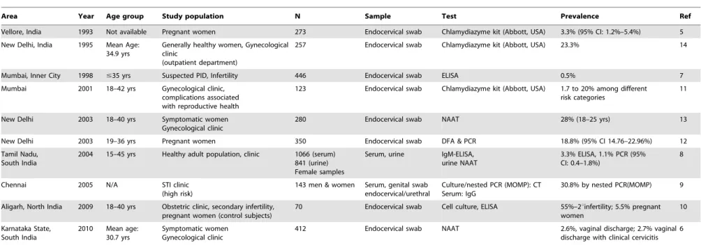
limited regions where most infants are born. Scarce information is available on laboratory-confirmed incidence and prevalence of chlamydia infection in otherwise healthy males and females in India. Furthermore, the available Indian data show a wide variation in CT prevalence and methods of laboratory confirma-tion [5–14]. Some studies have found infecconfirma-tion rates of Indian women ranging from 3.3% to 33% depending on the population sampled [5–14] (Table 1). Most of these studies focus on high risk groups (female sex workers, STI/infertility patients, and HIV-positive women) and were limited by small sample sizes [6,7,9– 11,13]. In Tamil Nadu, Joyeeet al.found the prevalence of active genital CT infection in a healthy adult female population by NAAT of the urine was 1.1% (95% CI: 0.4%–1.8%) [8]. However, among symptomatic men and women attending a STI clinic, the prevalence of confirmed CT infection by culture and/or nested polymerase chain reaction (PCR) detecting major outer mem-brane protein (MOMP) was 30.8% (95% CI: 23.4–38.6%) [9].
The prevalence of CT in pregnant women in India has been shown to vary by geographic region. In a study conducted in Vellore, TN India in 1993, a prevalence of CT in pregnant women was found to be 3.3% (95% CI: 1.2%–5.4%) using an
enzyme immunoassay (EIA) which detects the presence of chlamydia antigen (ChlamydiazymeH kit, AbbottH,USA) [5]. The authors also reported a higher prevalence of CT in rural women (5.9%) compared to urban women (1.8%) [5]. A 1999 study from New Delhi found the prevalence of CT infection in mid-pregnancy and at labor using the ChlamydiazymeHtest to be 17% and 18.6%, respectively [15]. In addition, the study also found neonates born to infected mothers experienced purulent conjunctivitis more frequently than those born to non-infected mothers, 12.5% versus 2.8% (p = 0.04) respectively [15]. Rastogiet al. also found a CT prevalence of 18.8% (95% CI: 14.76– 22.96%) among 350 pregnant women in New Delhi diagnosed using PCR and DFA [12]. One concern about comparing these studies is that the EIA test is estimated to be only 65–70% sensitive and 90–99% specific compared to nucleic acid amplification test (NAAT) methods. Thus the EIA estimates would be expected to underestimate the true prevalence of infection.
Current obstetric practice in southern India does not include universal prenatal screening for CT. This practice is based on evidence from older local data [5]. In contrast, the CDC recommends that pregnant women in the US be screened for CT during their first prenatal visit; and women who are at increased risk for infection (new or multiple sex partners and those under 25 years of age) should have a repeat screen during the third trimester [16–17]. This is based on an estimated CT infection rate of 5% among US women of reproductive age [18]. However, there are no data on the current CT prevalence among healthy pregnant women in Vellore, using more sensitive methods such as NAAT testing.
Furthermore, although there have been only a few studies documenting prevalence ofChlamydia trachomatisin southern India, there are no studies documenting the frequency of maternal to child transmission of CT in the Vellore region. The Alexanderet al
study [5] is more than a decade old and only documents the prevalence of maternal CT infection. In addition, since 1993, newer techniques for more accurate detection of CT such as NAAT have evolved and the prevalence of CT may have changed. The Clearview Chlamydia Test KitH (Inverness, Houston, TX 77038) is one of the few FDA approved rapid diagnostic tests (RDT) for CT but a wide range of sensitivity (53–73%) and specificity (68–99%) on endocervical specimens have been reported when compared to culture or DNA probe assay/NAAT [19–23]. Newer NAAT technology using the Roche AmplicorH CT/NG test (Roche Molecular Systems, Inc., Branchburg, NJ 08876) shows higher sensitivity of 93.3% and specificity of 99.7% on endocervical specimens compared to cell culture and RDT [24].
In order to provide data for development of evidence-based guidelines for resource-limited regions, the objectives of our study were to determine the prevalence of CT genital infection in delivering women using highly sensitive NAAT testing and to study the maternal to child transmission of CT in Vellore, India.
Specific Aims of Study
We aimed 1) to estimate the prevalence of genital CT infection among women presenting for delivery at Christian Medical College Hospital (CMCH) Vellore, India using a NAAT technique 2) to identify risk factors associated with chlamydia infections in pregnant and delivering women in order to guide development of screening policies if needed; and 3) to estimate the prevalence of CT infection at birth and to identify factors associated with maternal-infant transmission.
Methods
Ethics Statement
The institutional review boards at both Cincinnati Children’s Hospital Medical Center (CCHMC) and the Christian Medical College Hospital approved the study protocol. Written informed consent was obtained during enrollment for the subject and her neonate. All clinical investigation was conducted according to the principles expressed in the Declaration of Helsinki.
Study Design
This was a prospective hospital-based observational study.
Surveillance Area
CMCH, located in Vellore, India served as the surveillance site for this study. Vellore is a city in the Indian state of Tamil Nadu located in tropical southern India midway between the cities of Chennai (formerly Madras) and Bengaluru (formerly Bangalore). Vellore town has a population of 177,230 (2001 Indian census) of which 50.4% are female. CMCH is a private, non-profit, 2000+ bed tertiary referral hospital with primary care clinics (including pediatrics) and serves a diverse population of lower to middle class patients. The CMCH obstetrics department reports over 8,000 deliveries per year.
Study Population
From April 2009 to January 2010, delivering women were approached and recruited daily by a research staff member during an antenatal outpatient visit, a scheduled labor/induction, or in the postpartum ward. During the antenatal outpatient visit, a physician would send his/her patient, based on the study’s inclusion criteria, to the designated research office for the research assistant to discuss the study with the delivering woman. On the inpatient setting, the research assistant approached subjects daily on the obstetric wards that met inclusion criteria. After the research assistant obtained the subject’s consent to the study, a questionnaire on socio-demographic factors, prenatal history, and sexual history was administered by the research assistant on initial encounter during a private interview using a pen and paper format.
Enrollment Criteria
The inclusion criteria required that study participants be 18 to 45 years of age; greater or equal to 28 weeks gestation; and planning to deliver at CMCH and use the CMCH Vaccine or Child Health clinic for infant care. Pregnant mothers who had a history of chronic disease including diabetes, congestive heart failure, and renal failure were excluded. Stillborn infants and infants with congenital disorders or malformations were excluded.
Specimen Collection and Laboratory Testing
Endocervical swabs were collected by physicians or a research assistant during a pelvic exam performed either during an antenatal outpatient visit; prior to a scheduled labor/induction; or in the postpartum ward. The first swab was used to perform the rapid diagnostic test (RDT) and the second for NAAT testing. A third swab was stored frozen at270uC for future testing. Swabs were collected before use of lubricant or insertion of a prostaglandin tablet for induction to prevent the possibility of inhibition of the test.
were collected after 24 hours in order to decrease the chance that a positive test would represent contamination by maternal secretions. After collection, the swabs were stored in a freezer at 270uC for NAAT testing.
Lab Methods
Rapid Test. The RDT used in the study was the Clearview Chlamydia Test KitH(Inverness, Houston, TX 77038). This test is a one step immunochromatographic assay for direct antigen detection with an internal positive control indicator. The test was performed in the onsite research lab by study staff according to the package insert instructions.
NAAT. Endocervical specimens were stored in a freezer at CMCH at 270uC. The NAAT samples were tested at Y.R. Gaitonade Centre for AIDS Research and Education Laboratory in Chennai, India using the Roche AmplicorH CT/NG test for
Chlamydia trachomatis(Roche Molecular Systems, Inc., Branchburg, NJ 08876). The test was performed according to the package insert instructions. Internal positive controls all tested positive. In addition, blinded commercial positive controls (n = 5) (AccurunH 341, SeraCare Life Sciences, Inc., Milford, MA 01757) were included among the participant samples sent to the lab. All of these blinded positive controls were reported as positive results.
In addition, 102 samples from vials with remaining endocervical swab specimen had DNA extracted using the QIAamp DNA MinikitH (Qiagen, Hilden, Germany). These extracted samples were shipped, on dry ice, and retested independently in an established US laboratory using the Roche Amplicor PCRH (Roche Diagnostic Systems, Inc., Branchburg, N.J).
Treatment
Subjects’ positive rapid test results were reported to the attending physicians in both the obstetric and neonatology departments following birth. Treatment decisions were made by the primary physicians.
Statistical Methods
Sample size calculation. For sample size estimations, we used estimated prevalence of CT from previous studies (Table 1). It was assumed, from these studies, that the prevalence in our study population is about 3.3%. Using standard sample size calculations, a sample of 799 is sufficient to detect prevalence up to 9% with precision of 2%. For the neonate samples, a sample of 195 provides 80% power to detect a prevalence of 3% with precision of 2.5%.
Statistical analysis. Data were analyzed using Statistical Analysis Systems software (SAS Institute, Cary, NC, Version 9.2). Means and standard deviations were used to characterize normally distributed data. T-tests were used to compare means of normally distributed data. Chi-square and Fisher’s exact tests were used to assess differences in proportions. All reported P values were 2-sided and p values,0.05 were considered statistically significant. Concordance between the rapid test and NAAT was determined by the Kappa statistic and Kendall’s Tau B.
Results
Prevalence in Pregnant Women
From April 2009 to January 2010, 7955 women delivered during the recruitment period. Of these 7955 women, a convenience sample of 1367 (17%) pregnant and delivering women was approached for recruitment into the study. Of those, 1198 (88%) women were enrolled; 45% during an antenatal outpatient visit, 55% before a scheduled labor/induction, and
,1% in the postpartum ward. Figure 1 shows the study flow diagram.
From the initial 1367 participants approached, 169 delivering women were not enrolled. Of these, 131 women were not enrolled in the study after refusing consent or not planning on follow up care at CMC. Another 38 women did not participate in the study for other reasons. Inquiries were made to determine the reasons for declining; 85% indicated they declined because their family did not want them to participate. Overall, women who declined were similar to the women who did participate in the study in regards to urban/rural residence (p = 0.77), religion (p = 0.47), and gravida (p = 0.1).
Table 2 outlines the demographic characteristics of the enrolled study participants. The mean age of mothers was 25.6 years and 22% (95% CI: 19.7–24.4%) were primigravida. Mean income was 5064 Rupees/month and 50% of mothers had at least a secondary education or higher. The predominant religion of enrolled women was Hinduism which is a reflection of the population composition in Vellore, India. Urban and village residents were of equal distribution. All women enrolled were married; one reported .one sexual partner; and six reported prior STI.
We collected 799 endocervical samples from the 1198 enrolled subjects and data on 784 participants with both RDT and NAAT results are reported. 399 enrolled participants (33%) did not have endocervical samples collected after enrollment; see figure 1 for reasons. Tested mothers were significantly older, multiparous, and in the higher socio-economic group compared to untested mothers (p = 0.03, p =,0.0001, and p = 0.03; respectively). Please see table 2. All other demographic were similar. Fifteen (2%) endocervical samples were lost in processing after collection.
NAAT Versus Rapid Test
The prevalence detected using the NAAT (considered the gold standard for this study) was 0.1% (95% CI: 0–0.38%). The mother with the positive NAAT specimen had no significant character-istics or risk factors. All 71 RDT samples that were positive were considered to be false positives. Thus, compared to NAAT this RDT had a sensitivity of 0%, a specificity of 90%. As anticipated, the statistical analysis by Kappa and Kendall Tau B revealed no agreement between the RDT and NAAT (Table 3). The samples retested independently in an established laboratory confirmed the data found in India, including confirming the single positive. Both sets of laboratories confirmed that there is a low incidence of CT in this population of India.
Neonatal Data
There were 811 neonates enrolled which included 12 twin infants. There were 768 newborn specimens (NP and conjunctival) obtained from the neonates of the 784 enrolled mothers who had both NAAT and RDT results reported. During neonatal swab collection, there were 10 neonates who were lost to follow-up due to early discharge, 1 mother refused swab collection from her infant after delivery, and 5 neonates had incomplete swab collections. The neonatal characteristics included 52% born by normal vaginal delivery; mean gestation was 39 weeks, and mean birth weight was 2.99 kilograms (not including twins). Please see Table 4. Only 11% of neonates had newborn problems which included neonatal intensive care admission for risk of sepsis; prematurity, respiratory distress syndrome, gestational diabetes. We expected to have minimal transmission to the neonate since only one mother tested positive by NAAT. Because there was such a low prevalence of mothers with NAAT positive results; we decided to test only 25% of all neonatal samples collected which accounted for 195 samples. All neonatal NAAT specimens, PrevalenceChlamydia trachomatisin Women in India
Table 1.CT Prevalence Studies in India.
Area Year Age group Study population N Sample Test Prevalence Ref
Vellore, India 1993 Not available Pregnant women 273 Endocervical swab Chlamydiazyme kit (Abbott, USA) 3.3% (95% CI: 1.2%–5.4%) 5
New Delhi, India 1995 Mean Age: 34.9 yrs
Generally healthy women, Gynecological clinic
(outpatient department)
257 Endocervical swab Chlamydiazyme kit (Abbott, USA) 23.3% 14
Mumbai, Inner City 1998 #35 yrs Suspected PID, Infertility 446 Endocervical swab ELISA 0.5% 7
Mumbai 2001 18–42 yrs Gynecological clinic, complications associated with reproductive health
123 Endocervical swab Chlamydiazyme kit (Abbott, USA) 1.7 to 20% among different risk categories
11
New Delhi 2003 18–40 yrs Symptomatic women Gynecological clinic
280 Endocervical swab NAAT 28% (18–25 yrs) 13
New Delhi 2003 19–36 yrs Pregnant women 350 Endocervical swab DFA & PCR 18.8% (95% CI 14.76–22.96%) 12
Tamil Nadu, South India
2004 15–45 yrs Healthy adult population, clinic 1066 (serum) 841 (urine) Female samples
Serum, urine IgM-ELISA, urine NAAT
3.3% ELISA, 1.1% PCR (95% CI: 0.4–1.8%)
8
Chennai 2005 N/A STI clinic (high risk)
143 men & women Serum, genital swab endocervical/urethral
Culture/nested PCR (MOMP): CT Serum: IgG
30.8% by nested PCR(MOMP) 9
Aligarh, North India 2009 18–40 yrs Obstetric clinic, secondary infertility, pregnant women (control subjects)
70 Endocervical swab Cell culture, ELISA 55%–2uinfertility; 5.5% pregnant women
10
Karnataka State, South India
2010 Mean age: 30.7 yrs
Symptomatic women Gynecological clinic
412 Endocervical swab NAAT 2.6%, vaginal discharge; 2.7% vaginal discharge with clinical cervicitis
6
This table shows a review on Indian data which show a wide variation in CT prevalence and methods of laboratory confirmation. doi:10.1371/journal.pone.0034794.t001
Prevalen
ce
Chlamydia
trachomatis
in
Women
in
India
ONE
|
www.plos
one.org
4
May
2012
|
Volume
7
|
Issue
5
|
including NP and conjunctival swabs, were negative; including the infant of the mother with the positive endocervical NAAT result. The neonate born to the NAAT positive mother did not have any neonatal problems.
Discussion
This is the largest report from India on Chlamydia trachomatis
infection in healthy pregnant women in a non-urban setting using Figure 1. Study Flow Chart.April 2009 to January 2010, 7955 women delivered during the recruitment period. 1198 (88%) women were enrolled; 799 endocervical samples from the 1198 enrolled subjects were collected and data on 784 participants with both RDT and NAAT results are reported. doi:10.1371/journal.pone.0034794.g001
PrevalenceChlamydia trachomatisin Women in India
NAAT technique. Previous studies have shown the reported chlamydial infection rates in India have ranged as high as 33% to as low as 3.3% [5–14] (Table 1). Most of these studies however have focused on high risk population groups in urban settings, potentially could have underestimated prevalence compared to more sensitive NAAT testing [5,8,10,11,14]. Previous studies in normal pregnant women showed a lower range of infection (3.3– 18.8%) using EIA. We used NAAT for confirmatory testing and showed the prevalence of genital chlamydia infections amongst pregnant women to be 0.1% (95% CI: 0–0.38%). Based on this low prevalence it appears that routine prenatal screening in southern India does not appear to be indicated.
In our study, we found that 10% of specimens were positive when evaluated by RDT. We chose the Clearview Chlamydia
Test KitHbecause it is one of the few FDA approved RDT for CT, and it had reasonable reported sensitivity (53–73%) and specificity (68–99%) on endocervical specimens when compared to culture or DNA probe assay/NAAT [19,20,22,23]. However, in our low prevalence setting the sensitivity and specificity were lower (0% sensitive and 90% specific) and there was a major discrepancy between the RDT and NAAT results. Other studies have also noted lower sensitivity and specificity of RDT and EIA when compared to PCR, especially in low prevalence settings [25,26].
If the true specificity of the RDT is between 68 and 99% [19– 23], we would expect a range of 7 to 356 false positives in a sample size of 784 subjects from a population with a prevalence of 3.3%. We observed 71 such results. Similarly, if the true sensitivity is between 58–78%, it is not surprising that the one true positive case Table 2.Baseline characteristics of Enrolled Mothers, Enrolled Tested mothers, and Enrolled and not Tested.
Enrolled Mothers Enrolled Tested Enrolled Not Tested
P-Value of Tested mothers vs. Untested mothers
N Percent N Percent N Percent
Age Group 1198 783 415 0.03
18–23 years 384 32.05 231 29.50 153 36.87
24–29 years 625 52.17 425 54.28 200 48.19
$30 years 189 15.78 127 16.22 62 14.94
Parity 1198 783 415 ,0.0001
Other 935 78.05 649 82.89 286 68.91
Primigravida 263 21.90 134 17.11 129 31.08
Mother’s Education 1195 780 415 0.08
Illiterate 31 2.59 20 2.56 11 2.65
Primary Education 443 37.07 277 35.51 166 40
Secondary/Tertiary 293 24.52 183 23.46 110 26.51
University Diploma 428 35.82 300 38.46 128 30.84
Income Group* 1121 731 390 0.03
(,5000 Rs/mo) 795 70.92 499 68.26 296 75.9
(5000–10,000 Rs/mo) 229 20.43 162 22.16 67 17.18
(.10,000 Rs/mo) 97 8.65 70 9.58 27 6.92
Residence 1196 781 415 0.86
Village 604 50.50 393 50.32 211 50.84
Urban 592 49.50 388 49.68 204 49.16
Religion 1193 779 414 0.07
Hindu 964 80.80 635 81.51 329 79.47
Muslim 165 13.83 96 12.32 69 16.67
Christian 62 5.20 47 6.03 15 3.62
Jain 2 0.17 1 0.13 1 .24
History of STI 1183 775 408 0.64
No 1147 96.96 751 96.90 396 97.06
Yes 6 0.51 3 0.39 3 .74
Don’t Know 30 2.54 21 2.71 9 2.21
Sexual Partners 1190 775 415 1
Only Husband 1189 99.92 774 99.87 415 100
Others 1 0.08 1 0.13
*(50Rs = 1 USD).
This table shows that tested mothers were significantly older, multiparous, and higher socio-economic group compared to untested mothers (p = 0.03, p =,0.0001, and p = 0.03; respectively).
would be missed. The package insert states the antibody used in the RDT detects all 15 Chlamydia serovars in addition to
Chlamydia psittaci[27]. However, cross-reactivity is unlikely as the primary explanation for these results.
In newborns, the CT prevalence was found to be zero. Therefore, the transmission rate from Chlamydia positive mothers to infants could not be characterized because we did not have sufficient power to address this question. Further studies need to be done on pregnant mothers with a higher prevalence of CT infection to assess maternal to neonate transmission in this setting. There are some limitations to our study. First, due to limited staff, we were only able enroll 17% of delivering women during our study period. However, 88% of women approached were enrolled. Second, the project sample population may not represent the local delivering female population. CMCH is a tertiary, private hospital and some patients may go to a nearby government hospital, or deliver at home. In addition, not all endocervical samples were obtained from recruited participants due to logistical reasons. Few endocervical specimens (n = 2) were also obtained postpartum which may have changed the RDT sensitivity and/or specificity. Tested mothers were significantly older, multiparous, and in the higher socio-economic group versus untested mothers (p = 0.03, p =,0.0001, p = 0.03; respectively). One reason for this finding may be because younger women in the lower socio-economic group delivered their neonate at home (especially if primigravida) or at a nearby government hospital.
In summary, we found that CT infection appears to be relatively rare in women delivering in this private tertiary care hospital in Vellore, India. We found a prevalence of 0.1% (95% CI: 0–0.38%) by NAAT which was much lower than that noted in other international studies including pregnant women [28–31]. However, Joyeeet al.[8] found a prevalence of 1.1% (95% CI: 0.4–1.8%) in the healthy female population of Tamil Nadu using urine NAAT. Their result of a low CT prevalence supports our findings of a very low rate of CT infection in our similar
population. Furthermore, NAAT proved to be superior in the diagnosis of CT than the RDT in a low prevalence setting.
The local practice is to not perform prenatal screening in healthy pregnant women in this population based on evidence from twenty years ago which used older diagnostic methods. We conclude that our current study justifies that routine prenatal screening in this population would not be recommended given the low prevalence. However, future studies are important to reassess prevalence as sexual practices may change within this culture. Outpatient pre-natal testing may provide better information in all SES strata.
Acknowledgments
We gratefully acknowledge the Fogarty International Center and Vanderbilt University for allowing us this opportunity through the Fogarty International Research Clinical Fellows program. We thank the laboratory support of YRG Care (Dr. P. Balakrishnan and staff). We also thank CMC biostatistics department, Mrs. Alice Augustine, and Mrs. Geeta Marimuthu for recruitment and data management. We thank Stephanie Donnelly, Tasha Hughes, OG3 (CMCH) office for administrative support. This study would not have been possible without the help of the physicians (Dr. AK Jana, Dr. Ruby Jose, Dr. Gigi Matthews), OG residents, nurses, laboratory, and administrative staff affiliated with the Christian Medical College Hospital. We also thank Dr. Chuck Schubert,Dr. Connelly, Dr. David Bernstein, Gretchen Langdon, Dr. Joel Mortensen, and Amy Cassedy for their guidance with this project. We thank Dr. Charlotte Gaydos at Johns Hopkins University for her laboratory assistance. We deeply express our gratitude to the families who participated in the project.
Author Contributions
Conceived and designed the experiments: NKV MS JSH MAS AR VV. Performed the experiments: NKV RN SA. Analyzed the data: NKV MS JSH MAS CD RN. Contributed reagents/materials/analysis tools: AR SA VV CD. Wrote the paper: NKV MS JSH MAS RN AR. Study supervision: MS AR VV.
References
1. World Health Organization (2010) Sexually Transmitted Diseases -Chlamydia trachomatis, 2010. Available: http://www.who.int/vaccine_research/diseases/ soa_std/en/index1.html. Accessed 2010 Aug 6.
2. Chen CJ, Wu KG, Tang RB, Yuan HC, Soong WJ, et al. (2007) Characteristics ofChlamydia trachomatisinfection in hospitalized infants with lower respiratory tract infection. J Microbiol Immunol Infect. 40(3): 255–9.
3. Hammerschlag MR (1989) Chlamydial infections. J Pediatr. 114(5): 727–734.
4. Rosenman MB, Mahon BE, Downs SM, Kleiman MB (2003) Oral erythromycin prophylaxis vs watchful waiting in caring for newborns exposed toChlamydia trachomatis. Arch Pediatr Adolesc Med. 157(6): 565–71. 5. Alexander R, Mathai E, Nayyar V, Mathew M, Jasper P (1993) Low prevalence
of chlamydial endocervical infection in antenatal south Indian women. Genitourin Med. 69(3): 240–1.
6. Becker M, Stephen J, Moses S, Washington R, Maclean I, et al. (2010) Etiology and determinants of sexually transmitted infections in Karnataka state, south India. Sex Transm Dis. 37(3): 159–64.
Table 3.Comparison of NAAT test with rapid diagnostic test.
RDT
NAAT Positive Negative Total
Positive 0 1 1
Negative 71 712 783
Total 71 713 784
Cohen’s Kappa =20.0025 Kendall’s Tau B =20.0113
Cohen’s Kappa, which tests the agreement between two tests is negative, showing no agreement. Kendall’s Tau B is also negative, showing very slight negative association (inversion) between the two tests. The association here is very limited.
The prevalence detected using the NAAT (considered the gold standard for this study) was 0.1% (95% CI: 0–0.38%).
doi:10.1371/journal.pone.0034794.t003
Table 4.Neonate Characteristics.
N Percent
Delivery Type, n = 768
Forceps 164 21.35
C Section 201 26.17
Normal Vaginal 403 52.47
Newborn Problems, n = 768
No 681 88.67
Yes (RDS, Sepsis, IUGR, NICU) 87 11.33
Neonate N Mean Std Dev
Gestational Age (weeks) 768 39.0 1.359
Birth Weight (kilograms) 762 2.99 0.455 There were 768 newborn specimens (NP and conjunctival) obtained from the neonates of the 784 enrolled mothers who had both NAAT and RDT results reported. This table describes the neonatal characteristics.
doi:10.1371/journal.pone.0034794.t004
PrevalenceChlamydia trachomatisin Women in India
7. Brabin L, Gogate A, Gogate S, Karande A, Khanna R, et al. (1998) Reproductive tract infections, gynaecological morbidity and HIV seroprevalence among women in Mumbai, India. Bull World Health Organ. 76(3): 277–87. 8. Joyee AG, Thyagarajan SP, Rajendran P, Hari R, Balakrishnan P, et al. (2004)
Chlamydia trachomatisgenital infection in apparently healthy adult population of Tamil Nadu, India: a population-based study. Int J STD AIDS. 15(1): 51–5. 9. Joyee AG, Thyagarajan SP, Reddy EV, Venkatesan C, Ganapathy M (2005)
Genital chlamydial infection in STD patients: its relation to HIV infection. Indian J Med Microbiol. 23(1): 37–40.
10. Malik A, Jain S, Rizvi M, Shukla I, Hakim S (2009) Chlamydia trachomatis infection in women with secondary infertility. Fertil Steril. 91(1): 91–5. 11. Mania-Pramanik J, Meherji PK, Gokral JS, Donde UM (2001) Chlamydia
trachomatisinfection in an urban setting. Sex Transm Infect. 77(2): 141. 12. Rastogi S, Das B, Salhan S, Mittal A (2003) Effect of treatment forChlamydia
trachomatisduring pregnancy. Int J Gynaecol Obstet. 80(2): 129–37. 13. Singh V, Salhan S, Das BC, Mittal A (2003) Predominance of Chlamydia
trachomatisserovars associated with urogenital infections in females in New Delhi, India. J Clin Microbiol. 41(6): 2700–2.
14. Singh V, Sehgal A, Satyanarayana L, Gupta MM, Parashari A, et al. (1995) Clinical presentation of gynecologic infections among Indian women. Obstet Gynecol. 85(2): 215–9.
15. Paul VK, Singh M, Gupta U, Buckshee K, Bhargava VL, et al. (1999)Chlamydia trachomatis infection among pregnant women: prevalence and prenatal importance. Natl Med J India. 12(1): 11–4.
16. Berg A (2001) Screening for chlamydial infection: recommendations and rationale. Am J Prev Med. Apr; 20(3 Suppl): 90–4. Available: http://dx.doi.org/ 10.1016/S0749-3797(01)00254-9.
17. U.S. Preventive Services Task Force (2007)Screening for Chlamydial Infection: U.S. Preventive Services Task Force Recommendation Statement. First published in Ann Intern Med 2007;147: 128–33. Available: http://www.uspreventiveservicestaskforce. org/uspstf07/chlamydia/chlamydiars.htm. Accessed 2010 Jun 20.
18. Centers for Disease Control and Prevention (2008) Sexually Transmitted Disease Surveillance, Atlanta, GA: U.S. Department of Health and Human Services; November 2009.
19. Blanding J, Hirsch L, Stranton N, Wright T, Aarnaes S, et al. (1993) Comparison of the Clearview Chlamydia, the PACE 2 assay, and culture for detection ofChlamydia trachomatisfrom cervical specimens in a low-prevalence population. J Clin Microbiol. 31(6): 1622–5.
20. Hislop J, Quayyum Z, Flett G, Boachie C, Fraser C, et al. (2010) Systematic review of the clinical effectiveness and cost-effectiveness of rapid point-of-care
tests for the detection of genital chlamydia infection in women and men. Health Technol Assess. 14(29): 1–97, iii– iv.
21. Kluytmans JA, Goessens WH, Mouton JW, van Rijsoort-Vos JH, Niesters HG, et al. (1993) Evaluation of Clearview and Magic Lite tests, polymerase chain reaction, and cell culture for detection ofChlamydia trachomatisin urogenital specimens. J Clin Microbiol. 31(12): 3204–10.
22. Saison F, Mahilum-Tapay L, Michel CE, Buttress ND, Nadala EC Jr., et al. (2007) Prevalence ofChlamydia trachomatisinfection among low- and high-risk Filipino women and performance of Chlamydia rapid tests in resource-limited settings. J Clin Microbiol. 45(12): 4011–7.
23. Yin YP, Peeling RW, Chen XS, Gong KL, Zhou H, et al. (2006) Clinic-based evaluation of Clearview Chlamydia MF for detection ofChlamydia trachomatisin vaginal and cervical specimens from women at high risk in China. Sex Transm Infect. 82 Suppl 5: v33–7.
24. Livengood CH, Wrenn JW (2001) Evaluation of COBAS AMPLICOR (Roche): accuracy in detection of Chlamydia trachomatis and Neisseria gonorrhoeae by coamplification of endocervical specimens. J Clin Microbiol. 39(8): 2928–32. 25. Horner P, Skidmore S, Herring A, Sell J, Paul I, et al. (2005) Enhanced enzyme
immunoassay with negative-gray-zone testing compared to a single Nucleic Acid Amplification Technique for community-based chlamydial screening of men. J Clin Microbiol. 43(5): 2065–9.
26. Lauderdale TL, Landers L, Thorneycroft I, Chapin K (1999) Comparison of the PACE 2 assay, two amplification assays, and Clearview EIA for detection of Chlamydia trachomatis in female endocervical and urine specimens. J Clin Microbiol. 37(7): 2223–9.
27. The Inverness Medical Group of Companies (2006) ClearviewHChlamydia MF Available: http://www.clearview.com/pdf/CV_Ch211BBB_EN.pdf. Accessed 2010 Aug 30.
28. El-Qouqa IA, Shubair ME, Al Jarousha AM, Sharif FA (2009) Prevalence of Chlamydia trachomatisamong women attending gynecology and infertility clinics in Gaza, Palestine. Int J Infect Dis. 13(3): 334–41.
29. Sturm AW, Wilkinson D, Ndovela N, Bowen S, Connolly C (1998) Pregnant women as a reservoir of undetected sexually transmitted diseases in rural South Africa: implications for disease control. Am J Public Health. 88(8): 1243–5. 30. Wessel HF, Herrmann B, Dupret A, Moniz F, Brito C, et al. (1998) Genital
infections among antenatal care attendees in Cape Verde. Afr J Reprod Health. 2(1): 32–40.