An ANNEXIN-Like Protein from the Cereal
Cyst Nematode
Heterodera avenae
Suppresses Plant Defense
Changlong Chen1, Shusen Liu1,2, Qian Liu1, Junhai Niu3, Pei Liu1, Jianlong Zhao1, Heng Jian1*
1Department of Plant Pathology, China Agricultural University, Beijing, China,2Institute of Plant Protection and Agro-products Safety, Anhui Academy of Agricultural Sciences, Hefei, Anhui, China,3Tropical Crops Genetic Resources Institute, Chinese Academy of Tropical Agricultural Sciences, Danzhou, Hainan, China
Abstract
Parasitism genes encoding secreted effector proteins of plant-parasitic nematodes play im-portant roles in facilitating parasitism. An annexin-like gene was isolated from the cereal cyst nematodeHeterodera avenae(termedHa-annexin) and had high similarity toannexin 2, which encodes a secreted protein ofGlobodera pallida.Ha-annexinencodes a predicted 326 amino acid protein containing four conserved annexin domains. Southern blotting re-vealed that there are at least two homologies in theH.avenaegenome.Ha-annexin tran-scripts were expressed within the subventral gland cells of the pre-parasitic second-stage juveniles byin situhybridization. Additionally, expression of these transcripts were relatively higher in the parasitic second-stage juveniles by quantitative real-time RT-PCR analysis, coinciding with the time when feeding cell formation is initiated. Knockdown ofHa-annexin
by method of barley stripe mosaic virus-based host-induced gene silencing (BSMV-HIGS) caused impaired nematode infections at 7 dpi and reduced females at 40 dpi, indicating im-portant roles of the gene in parasitism at least in early stagein vivo. Transiently expression of Ha-ANNEXIN in onion epidermal cells andNicotiana benthamianaleaf cells showed the whole cell-localization. Using transient expression assays inN.benthamiana, we found that Ha-ANNEXIN could suppress programmed cell death triggered by the pro-apoptotic mouse protein BAX and the induction of marker genes of PAMP-triggered immunity (PTI) inN.
benthamiana. In addition, Ha-ANNEXIN targeted a point in the mitogen-activated protein ki-nase (MAPK) signaling pathway downstream of two kiki-nases MKK1 and NPK1 inN.
benthamiana.
Introduction
Heterodera avenaeis one of the most important cereal cyst nematodes (CCNs) in the world. This pathogen is distributed worldwide on cereal crops and occurs in approximately 80% of the total cereal growing areas in China [1]. Additionally,H.avenaecauses substantial economic
OPEN ACCESS
Citation:Chen C, Liu S, Liu Q, Niu J, Liu P, Zhao J, et al. (2015) An ANNEXIN-Like Protein from the Cereal Cyst NematodeHeterodera avenae
Suppresses Plant Defense. PLoS ONE 10(4): e0122256. doi:10.1371/journal.pone.0122256
Academic Editor:John Jones, James Hutton Institute, UNITED KINGDOM
Received:October 30, 2014
Accepted:February 10, 2015
Published:April 7, 2015
Copyright:© 2015 Chen et al. This is an open access article distributed under the terms of the Creative Commons Attribution License, which permits unrestricted use, distribution, and reproduction in any medium, provided the original author and source are credited.
Data Availability Statement:All relevant data are within the paper and its Supporting Information files.
Funding:This research was supported by funding from the National Key Basic Research Program of China (No. 2013CB127501) and the Special Fund for Agro-scientific Research in the Public Interest in China (Nos. 201503114 and 200903040). The funders had no role in study design, data collection and analysis, decision to publish, or preparation of the manuscript.
yield losses; in some wheat fields, the losses caused by this nematode can range from 30 to 100% [2,3].
H.avenaeis an obligate sedentary plant parasitic nematode that invades the roots of wheat and related cereals in the subfamily Pooideae. The second-stage juveniles (J2s) penetrate the root tip and migrate intracellularly through the cortex to the vascular cylinder, where the nem-atode inserts its stylet into a selected parenchyma cell and induces its transformation into a feeding site. The stylet is used to deliver secretions referred to as“effectors”into root tissue, which facilitates plant parasitism.
The identification of genes encoding candidate effector proteins has gained increasing atten-tion in molecular plant nematology research in the last two decades. However, only a few effec-tors have been reported inH.avenae. Threeβ-1,4-endoglucanase genes, whose transcripts accumulate specifically in the two subventral gland cells ofH.avenae, have been identified and suggested to play a crucial role in plant cell wall-degradation during the penetration and migra-tion of nematodes in the host roots [4,5]. A new expansin gene (Ha-expb1) expressed in the subventral glands ofH.avenaewas cloned and has been suggested to play a role in the early parasitic-stage process, most likely aiding migration within the plant [6]. Cathepsin S-like cys-teine proteinase ofH.avenaewas isolated, and its plausible mode of interaction was illustrated by docking analysis. Additionally, qRT-PCR analysis has suggested that this proteinase has an important role in both pre-parasitic and parasitic stages of the nematode life cycle [7]. Un-doubtedly, these findings are far from unraveling the parasitism mechanism ofH.avenae. Transcriptome data onH.avenaereported this year provide an opportunity to identify new ef-fectors that are specifically involved inH.avenae-host interactions [8].
It has been reported that annexins play important roles in animals and plants, including exocytosis, actin binding, peroxidase activity, callose synthase regulation, ion transport and the ability to link Ca2+, redox and lipid signaling to coordinate development with responses to the biotic and abiotic environment [9,10]. The diverse family of proteins in plant parasitic nema-todes has also been examined, and there have been some important findings. In 2001, an annexin named gp-nex was identified fromGlobodera pallidaand was found to be immunolo-calized in the amphids, genital primordium and constraining muscles above and below the metacorpus pump chamber [11]. In 2003,Hg4F01, a gene similar to annexins in the nematode Caenorhabditis elegans, was isolated from the parasitome ofHeterodera glycines, which is ex-pressed exclusively in the dorsal esophageal gland cell of the parasitic stages [12]. InHeterodera schachtii, the molecular function of annexin involved in the host-nematode interaction was de-scribed. The expression of annexin in wild-typeArabidopsispromoted hyper-susceptibility to H.schachtiiinfection, and in anAnnAt1(annexin-1ofArabidopsis) mutant reverted mutant sensitivity to 75 mM NaCl (high salt condition), suggesting a similar function toAnnAt1in the stress response by plant cells. Additionally, yeast two-hybrid assays showed that annexin inter-acted with an oxidoreductase member of the 20 G Fe (II) oxygenase family that is linked to host defense and stress response. All of this evidence suggests that annexin fromH.schachtii may mimic plant annexin function to modulate host defense responses to promote parasitism [13].
associated with the defense-related HR. As a result, the ability to suppress BAX-triggered PCD (BT-PCD) has been proven a valuable initial screening tool for pathogen effectors capable of suppressing defense-associated PCD [19–22]. With regard to PTI, the increased expression of defense-related genes is one of its phenotypes. Plant mitogen-activated protein kinase (MAPK) cascades also play a pivotal role in the PTI signaling pathway by transducing signals from pat-tern recognition receptors (PRRs) to downstream components [23–27]. MAPK cascades con-sist of at least three protein kinases: a MAPK kinase kinase phosphorylates and activates a MAPK kinase, which in turn activates a MAPK by phosphorylation [22]. MKK1 encodes a MAPK kinase, and NPK1 encodes a MAPK kinase kinase that functions to transduce PAMP-triggered signals [22,28–31]. Genes encoding full-length MKK1 and the N terminus of NPK1 (residues 1 to 373; NPK1Nt) could trigger PCD when introduced by agroinfiltration into Nicoti-ana benthamiNicoti-ana, and Wang found that Avh238P7076, an effector ofPhytophthora sojae, could suppress the PCD triggered by both MAPKs, suggesting that this effector acted at a point in the signaling pathway downstream of the two kinases [22].
In this study, we report the identification and certain functional characteristics of an annexin-like gene from the cereal cyst nematodeH.avenaethat most likely suppresses plant immunity to facilitate parasitism.
Materials and Methods
Nematodes
Heterodera avenaewas propagated on wheat (Triticum aestivumcv. Aikang 58) in an artificial environment. Embryo eggs were pipetted from crushed newly formed cysts. Infective second-stage juveniles were collected by hatching cysts at 15°C after at least 4 weeks incubation at 4°C. To obtain parasitic life stages, infected wheat roots were obtained at different days after inocu-lation, washed with tap water, cut into sections and digested at 28°C with 160 rpm in a 6% cel-lulose (Sinopharm Chemical Reagent Beijing Co., Ltd., China) water solution for 12 h. After digestion, the roots were placed on a sieve set and crushed by rapid-flow water to release nema-todes onto the sieve. Adult females were directly washed from the root surface.
Cloning of the
Ha-annexin
cDNA and gDNA sequence
and assembled with the sequence of the initial cloned fragment to obtain the full-length cDNA sequence ofHa-annexin.
For cloning the gDNA sequence ofHa-annexin, the primers annexinQCF4/annexinQCR4 (S1 Table) were used, and the product was also sequenced.
Bioinformation analysis
The predicted protein sequence of Ha-ANNEXIN was blasted for closed protein using BLASTP searching of the protein databases of the National Center for Biotechnology Information (NCBI) (http://blast.ncbi.nlm.nih.gov/Blast.cgi). Prediction of a signal peptide for secretion was performed using Signal P 4.0 (http://www.cbs.dtu.dk/services/SignalP/). Multiple amino acid sequence alignment of annexins fromH.avenae,G.pallida,Bursaphelenchus xylophilus, H.schachtiiandH.glycineswas conducted using DNAMAN V6, and conserved domains were searched using the Conserved Domain Search Service (CD Search) of the NCBI (http://www. ncbi.nlm.nih.gov/Structure/cdd/wrpsb.cgi).
Southern blot hybridization
H.avenaegenomic DNA was extracted using a QIAamp DNA Micro Kit (Qiagen, Germany). Five micrograms of genomic DNA was digested overnight at 37°C withBamHI orEcoRI (New England Biolabs, USA), separated by 0.8% agarose gel electrophoresis and transferred onto a Hybond-N membrane (GE Healthcare, USA) using standard protocols [32]. The membrane was hybridized with theHa-annexin-CDS DNA probe, which was random primed labeled with Digoxigenin-11-dUTP using DIG-High Prime (Roche). Hybridization (at 42°C) and de-tection were performed following the instruction manual of the DIG High Prime DNA Label-ing and Detection Starter Kit I (Roche, USA).
mRNA
in situ
hybridization
In situhybridization was performed as previously described [33] but with the hybridization temperature adjusted to 50°C. The specific primers anne-qRT-S and anne-qRT-A (S1 Table) were used to synthesize digoxigenin (DIG)-labeled sense (control) and antisense cDNA probes (Roche, USA) by asymmetric PCR [34].
Developmental expression analysis
Total RNA of different life stages ofH.avenaewas extracted using the RNeasy Plus Micro Kit (Qiagen, Germany), which includes gDNA eliminator columns to remove DNA. cDNA was prepared according to the instructions for the QuantiTect Whole Transcriptome Kit (Qiagen, Germany), which can provide sufficient cDNA for gene expression analysis by quantitative real-time PCR (qPCR). A SYBR Green assay was used to quantify the expression of Ha-annexinthroughout the nematode life cycle. qPCR was performed using a SYBR Premix Ex Taq (TaKaRa, Japan) in an ABI Prism 7000 instrument (Applied Biosystems, USA), with prim-ers anne-qRT-S/anne-qRT-A and GAPDH-qS1/ GAPDH-qAS1 designed (S1 Table), respec-tively, from theHa-annexincoding sequence and an endogenous control geneGAPDH-1 (sequence from internal data). Triplicate PCR reactions for each cDNA sample were complet-ed, and the assay consisted of three technical replicates. Data were analyzed using the 2-ΔΔCt
Subcellular localization
in planta
Subcellular localization in onion. Ha-annexinORF sequences (without a stop codon) were amplified using primers ann-XbaI-S and ann-XhoI-AS (S1 Table) for cloning into the pUC35SGFP vector to generate the 35S:ANNEXIN:GFP construct by the method of digestion and connection. Each construct plasmid (4–5μg/shot) was delivered into onion epidermal cells
through biolistic bombardment by vacuumizing to 27–28 in. Hg using a PDS1000/He system (Biolistic Particle Delivery System, Bio-Rad, USA). After bombardment, epidermal peels were incubated at 25°C for 24 h in the dark. The subcellular localization of the fused proteins was vi-sualized using laser confocal fluorescence microscopy (Nikon Eclipse TE300, Nikon, Japan) at an excitation wavelength of 488 nm.
Subcellular localization inN.benthamiana. Ha-annexinwas constructed into
pCamv35SGFP vector with GFP fused at the C-terminal, according to the user manual of the In-Fusion HD Cloning Kit (Clontech, USA), with the primers ann-if-pCam35SGFP-S1/AS1 (S1 Table). The construct was confirmed by sequencing and transformed intoAgrobacterium tumefaciensstrain EHA105.N.benthamianaplants were grown in a growth room for 4 weeks at approximately 25°C with a 14 h light/10 h dark cycle. For infiltration into leaves, recombi-nant strains ofA.tumefaciensof pCamv35SGFP-annexin and pCamv35SGFP-p19 (retained by the lab; p19, a RNA silencing suppressor) were cultured, collected, washed three times and re-suspended with infiltration buffer [10mM MgCl2in 10 mM 2-(N-morpholino) ethanesulfonic
acid (MES), pH 5.2, and 0.1 mM acetosyringone] to ~0.5 OD600, respectively. Then, 3:1
amounts of pCamv35SGFP-annexin and pCamv35SGFP-p19 were mixed, collected and resus-pended to 1/2 of the total volume, before infiltrated toN.benthamianaleaves. After infiltration, N.benthamianaplants were incubated at 22°C for 48 h with the same light/dark cycle.
pCamv35SGFP (retained by the lab) was used as a control. The subcellular localization was vi-sualized as described in“Subcellular localization in onion”. For verification of intact of annexin-GFP fusion, western blotting was performed. A primary mouse anti-GFP monoclonal antibody in 1: 5000 dilution (Medical & Biological Laboratories, Japan), a goat anti-IgG, (mouse) pAb-HRP secondary antibody in 1: 5000 dilution (Medical & Biological Laboratories, Japan) and a DAB Kit (Beijing ComWin Biotech Co., Ltd., China) for color visualization were used for detecting the expression of annexin-GFP fusion and GFP control.
Knockdown of
Ha-annexin
by BSMV-HIGS and infection assay
Cell-death suppression assay in
N
.
benthamiana
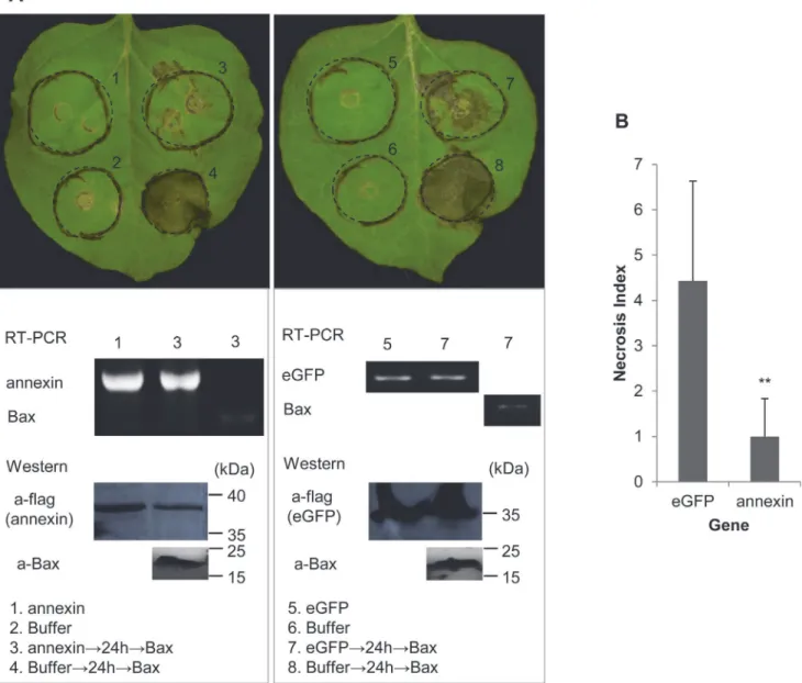
Ha-annexinwas constructed into PVX vector pGR107 with a flag-tag fused at the N-terminal, according to the user manual of the In-Fusion HD Cloning Kit (Clontech, USA), with the primers ann-if107f-S1 and ann-if107f-AS1 (S1 Table).eGFPwas constructed into the vector with a flag-tag fused at the N-terminal by the method of digestion and connection using the primersSmaI-GFP-ORF and GFP-ORF-SalI (S1 Table). The full-lengthMKK1and the N ter-minus ofNPK1(residues 1 to 373; NPK1Nt) were constructed into pGR107 with an HA-tag, ac-cording to the user manual of the In-Fusion HD Cloning Kit (Clontech, USA), with the primer pairs MKK1-if-p107HA-S1/AS1 and NPK1(Nt)-if-p107HA-S1/AS1 (S1 Table), respectively. The constructs were confirmed by sequencing and transformed intoA.tumefaciensstrain GV3101.N.benthamianaplants were grown in a growth room for 4 to 6 weeks at approxi-mately 25°C with a 14 h light/10 h dark cycle. Assays of the suppression of BT-PCD and M/ NT-PCD (MKK1/NPK1Nt-triggered PCD) were performed as previously described [22], ex-cept thatA.tumefacienscells carrying theBaxandMKK1/NPK1Ntgene were infiltrated only at 24 h and 16 h after the initial inoculation, respectively. The assays were repeated twice, with 3–7 plant replicates inoculated on three leaves each.
For verification of gene expression, reverse transcription-PCR (RT-PCR) or western blot-ting were performed. Total RNA and proteins were isolated from infiltrated parts of leaves of N.benthamianausing TRIzol Reagent (Invitrogen, USA). For RT-PCR, cDNA was synthesized as described in Section of“Cloning of theHa-annexincDNA and gDNA sequence”and PCR was performed using the gene-specific primers ann-if107f-S1/ ann-if107f-AS1,ClaI-Bax-ORF/ Bax-ORF-SalI, and eGFP-F1/ eGFP-F2 (S1 Table) forHa-annexin,BaxandeGFP, respectively. For western blotting, a primary rabbit anti-FLAG polyclonal antibody in 1: 6000 dilution (Cell Signaling Technology, USA), a goat anti-rabbit IgG, HRP-conjugated secondary antibody in 1: 3000 dilution (Beijing ComWin Biotech Co., Ltd., China) and a DAB Kit (Beijing ComWin Biotech Co., Ltd., China) for color visualization were used for detecting the expression of ANNEXIN and eGFP control, both fused with FLAG. A primary Bax monoclonal anti-body (Abcam, UK) in 1 mg/mL, a goat anti-IgG, (mouse) pAb-HRP secondary antianti-body in 1: 5000 dilution (Medical & Biological Laboratories, Japan) and a DAB Kit (Beijing ComWin Bio-tech Co., Ltd., China) for color visualization were used for detecting the expression of Bax.
Photos of phenotypes of infiltrated leaves ofN.benthamianawere taken approximately 7 days after the last infiltration. The degree of PCD of Ha-ANNEXIN and control eGFP followed by BAX, MKK1 or NPK1Nt, referred to as the“Necrosis Index”, was scored on a ten-point scale according to the size of the necrotic area (grade 1 for 10% necrosis of the whole circle area, grade 2 for 20%, and so on). Necrosis Indices of ANNEXIN and eGFP were compared.
PTI marker gene expression in
N
.
benthamiana
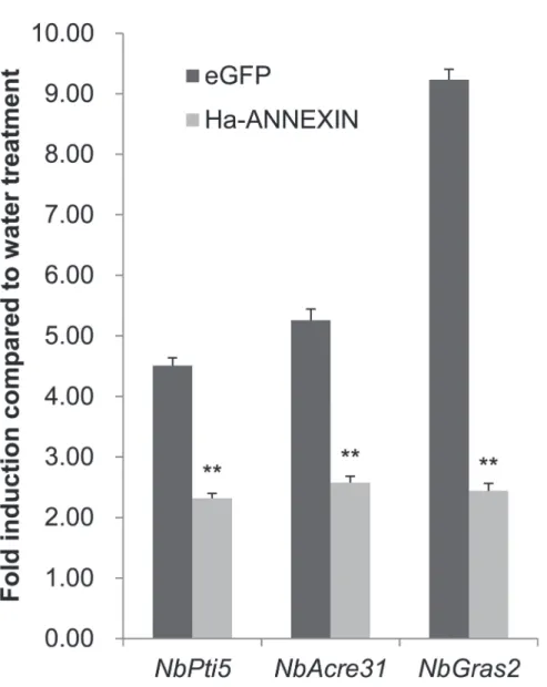
The experiment was conducted as previously reported [17]. The expression of three marker genes—NbPti5,NbAcre31andNbGras2—of PTI [38], induced by flg22 (SciLight Biotechnolo-gy, China) or distilled water inN.benthamianatissue infiltrated byA.tumefacienscells carry-ingannexinoreGFPcontrol, were evaluated by qPCR. Isolation of total RNA and cDNA synthesis was conducted in the same way as in the cell-death suppression assay. Gene expres-sion was determined by qPCR as described in Section of“Developmental expression analysis”, and the reference geneNbEF1α[39] was used. The qPCR primers forNbPti5,NbAcre31,
NbGras2andNbEF1αwere NbPti5-F/R, NbAcre31-F/R, NbGras2-F/R and NbEF1α-F/R (S1
Statistical analysis
Data difference of treatments was analyzed by a one-way ANOVA (Duncan test) or an inde-pendent-samples t-test performed in SPSS 13.0.
Results
Isolation and sequence analysis of the annexin-like gene from
H
.
avenae
The full-length cDNA ofHa-annexin(GenBank Accession KJ562871) obtained by the 5’and 3’ end amplification of cDNA is 1201 bp long, composed of a 981 bp open reading frame (ORF), a 63 bp 5’-untranslated region (5’-UTR) before the ATG initiation codon and a 157 bp 3’-UTR containing a polyA tail. The gDNA sequence ofHa-annexin(GenBank Accession KJ562872) lacks an intron.
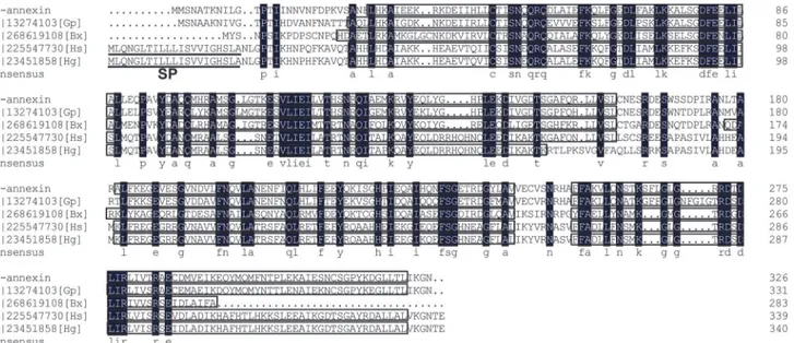
The protein encoded byHa-annexinwas predicted to have 326 amino acids and showed 76% identity to annexin 2, a secreted protein ofG.pallidaby using BLASTP. Similar to annex-ins fromG.pallidaandB.xylophilus, Ha-ANNEXIN had no secretion signal peptide at the N-terminal predicted by Signal P 4.0, but different from those of the plant-parasitic nematodes (PPNs)H.schachtiiandH.glycines. These annexins from different PPNs all have four con-served annexin domains (Fig 1), which is typical of the annexin family of calcium and phos-pholipid binding proteins.
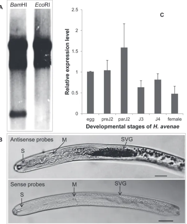
Southern blotting showed that DIG-labeledHa-annexinprobes hybridized to at least two DNA fragments on DNA gel blots (Fig 2A), which suggested that two or more members of an annexin gene family may exist inH.avenae.
Ha-annexin
expression is gland specific and relatively higher in parJ2
In situhybridization was performed in pre-parasitic second-stage juveniles (preJ2s) ofH. ave-naeto localize the expression ofHa-annexintranscripts. The hybridization signal of
DIG-Fig 1. Multiple alignment of annexins fromHeterodera avenaeand some other plant-parasitic nematodes.In the consensus, residues in the black background are totally identical; boxed areas are four conserved annexin domains predicted by the NCBI Conserved Domain Search Service; signal peptides ofH.schachtiiandH.glycinesanalyzed by Signal P 4.0 are underlined. SP, signal peptide; Gp,Globodera pallida; Bx,Bursaphelenchus xylophilus; Hs,H.schachtii; Hg,H.glycines.
Fig 2. Southern blot,in situhybridization and developmental expression pattern analysis ofHa-annexin.(A) Southern blot analysis ofHa-annexin. Genomic DNA fromH.avenaewas digested withBamHI andEcoRI, respectively, and probed with the Dig-labeled CDS ofHa-annexin. They had 3 and 2 signal strips, respectively. (B)In situhybridization of theHa-annexintranscripts in pre-parasitic second-stage juveniles. Signal of antisenseHa-annexin
DIG-Labeled cDNA probes localized within the subventral glands (SVGs), with sense probes as a negative control. The SVG, metacorpus (M), and stylet (S) are indicated with arrows. Scale bar = 20μm. (C) Developmental expression pattern ofHa-annexin. The relative expression level ofHa-annexinwas quantified using qPCR for six differentH.avenaestages. The fold change values were calculated using the 2-ΔΔCtmethod and presented as the change in
mRNA level in various nematode developmental stages relative to that of egg. Each column represents the mean of 3 independent assays with standard deviation. preJ2: pre-parasitic second-stage juvenile; parJ2, J3 and J4: parasitic second-, third- and fourth-stage juvenile, respectively.
labeled antisense probes was observed in the subventral gland cells ofH.avenae, and no signal was detected when using sense probes (Fig 2B).
The developmental expression pattern ofHa-annexinwas determined by qPCR analysis for six developmental stages (egg; preJ2; parasitic second-, third- and fourth- stage juvenile (parJ2, J3, and J4, respectively); and adult female). The mean values of the three independent replicates were presented inFig 2C, that is, the expression level ofHa-annexintranscripts in parJ2 was relatively higher than in other developmental stages.
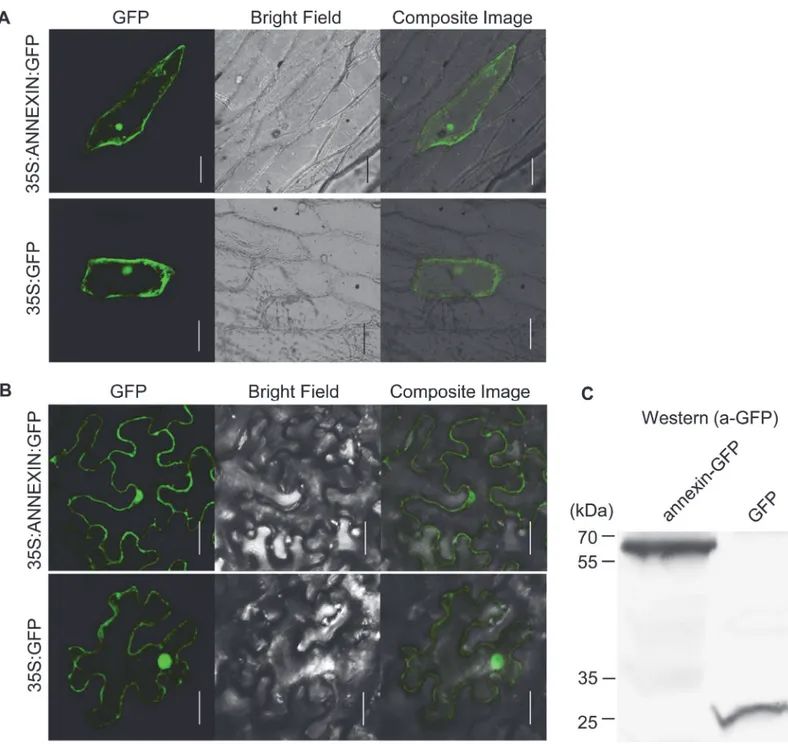
Ha-ANNEXIN is localized in the whole plant cell
Ha-ANNEXIN is expected to be delivered to the host cell through nematode stylets. To test where Ha-ANNEXIN localizes in plant cells, a protein transient expression assay was per-formed in both onion andN.benthamiana. The GFP signal was observed in the whole cell as the vector control (Fig 3), which indicated the whole cell-localization of Ha-ANNEXIN. West-ern blotting ofN.benthamianaleaves infiltrated with pCamv35SGFP-annexin showed ex-pected size of annexin-GFP fusion, which is larger than GFP (Fig 3B). This indicates that annexin-GFP fusion is intact fused.
BSMV-HIGS of
Ha-annexin
causes impaired nematode parasitism
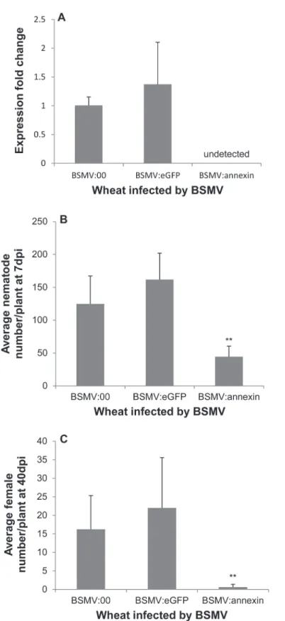
In recent years, BSMV has become a popular vector for virus-induced gene silencing (VIGS) in wheat, and a BSMV-HIGS system has emerged [40]. We used this system to conduct a loss-of-function study ofHa-annexinin wheat, and two repetitive experiments yielded consistent re-sults. This consistency demonstrated that the system worked well in silencing the expression of Ha-annexin(Fig 4A). The expression ofHa-annexinin nematodes recovered from wheat inoc-ulated by BSMV:annexinwas not detected, showing a significant reduction compared to that from BSMV:00 and BSMV:eGFPcontrols by qPCR (P<0.01) (Fig 4A). Additionally, due to the reduction inHa-annexinexpression, nematode infections of wheat inoculated by BSMV: annexincompared to the blank negative control BSMV:00 and the negative control BSMV: eGFPshowed a highly significant reduction in the number of juveniles/plant at 7 dpi by 73% (Fig 4B) and females/plant at 40 dpi by 97% (Fig 4C) (P<0.01). Off-target silencing effects on other genes fromH.avenae, BSMV and wheat should not happen because of the specificity of the selected fragment. Furthermore the BSMV infection symptom and root phenotype weren’t affected by the annexin silencing when compared to the controls. These results indicate that Ha-annexinplays important roles during the parasitism process at least in early stage ofH. avenae.
Ha-ANNEXIN suppresses plant defense
Two repetitive results of the BAX cell-death suppression assay inN.benthamianawere consis-tent; the infiltration spot of Ha-ANNEXIN followed by BAX was not as necrotic as that of eGFP followed by BAX (shown in representative photos,Fig 5A). RT-PCR and western blot-ting were conducted to verify the expression ofHa-annexin,eGFPandBaxfrom the transcrip-tional and translatranscrip-tional levels, respectively. For a quantitative comparison, the degree of PCD (referred to as the“Necrosis Index”) of Ha-ANNEXIN and the eGFP control followed by BAX was scored and compared. As shown inFig 5B, the Necrosis Index of Ha-ANNEXIN followed by BAX was low (0.9) compared with that of the negative eGFP control (4.1) (P<0.01). This finding suggested that Ha-ANNEXIN could suppress BT-PCD to some extent. Accordingly, ANNEXIN ofH.avenaemay play roles against plant defense.
controleGFPafter flg22 treatment were compared. Two repetitive assays were conducted with consistent results and the results of one were shown inFig 6. Three marker genes—NbPti5, NbAcre31andNbGras2—were induced by 1.3-, 1.6-, and 1.4-fold upregulation, respectively, in leaf tissues infiltrated byA.tumefacienscells carryingannexin, in contrast to 3.5-, 4.3-, and 8.2-fold upregulation ineGFPcontrol, respectively. An independent-samples t-test analysis Fig 3. Subcellular localization of Ha-ANNEXIN in the plant cell.(A) pUC35S:ANNEXIN:GFP fusion construct and pUC35S:GFP control construct were transformed into onion epidermal cells by bombardment. Scale bar = 100μm. (B)Agrobacterium tumefacienscells carrying pCamv35S:ANNEXIN:GFP fusion and pCamv35S:GFP were transiently expressed inNicotiana benthamianacells. Scale bar = 20μm. Western blotting ofN.benthamianaleaves infiltrated with pCamv35SGFP-annexin showed expected size of annexin-GFP fusion, which is larger than GFP control. In both (A) and (B), GFP signals were observed in the whole transformed cells for annexin-GFP fusion, which is the same as GFP control.
Fig 4. Effect of BSMV-HIGS ofHa-annexinon infection of wheat roots byH.avenae.(A) At 7 dpi, the expression ofHa-annexinin nematodes recovered from wheat inoculated by BSMV:annexinwas not detected by qPCR with BSMV:00 and BSMV:eGFPas positive controls. Nematode infection of wheat inoculated by BSMV:annexincompared to the blank negative control BSMV:00 and the negative control BSMV:eGFPshowed a highly significant reduction in the number of juveniles/plant at 7 dpi (B) and females/ plant at 40 dpi (C) by Duncan test (P<0.01). Each column represents the mean with standard deviation.
showed that all three marker genes were significantly less upregulated in leaf tissues infiltrated byA.tumefacienscells carryingannexinthan in those with theeGFPcontrol (P<0.01) (Fig 6). It means that Ha-ANNEXIN suppressed flg22-triggered PTI inN.benthamiana,which is one aspect of plant defense.
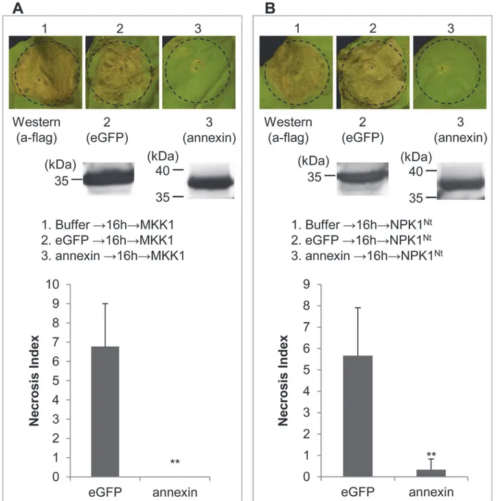
Target point of Ha-ANNEXIN in PTI suppression
Plant MAPK cascades play pivotal roles in the PTI signaling pathway [23–27]. MAPK cascades consist of at least three protein kinases: a MAPK kinase kinase phosphorylates and activates a Fig 5. Suppression of BT-PCD by Ha-ANNEXIN.(A) Assay for the suppression of BAX-triggered cell death (BT-PCD) inNicotiana benthamianaby Ha-ANNEXIN.N.benthamianaleaves were infiltrated with buffer orAgrobacterium tumefacienscells containing a vector carrying theHa-annexingene or the negative controleGFPgene, either alone or followed 24 h later withA.tumefacienscells carrying a mouseBaxgene. Photos of phenotypes of infiltrated leaves ofN.benthamianawere taken approximately 7 days after the last infiltration. Results of the verification of gene expression ofHa-annexin,eGFPand
Baxby RT-PCR or western blotting are shown below. (B) Necrosis Index of Ha-ANNEXIN and control eGFP followed by BAX. Each column represents the mean with standard deviation. The column with asterisks indicate a highly statistically significant reduction of the Necrosis Index of Ha-ANNEXIN compared with that of eGFP by t-test (P<0.01).
MAPK kinase, which subsequently activates a MAPK by phosphorylation [22]. To further study the mechanism of Ha-ANNEXIN in PTI suppression, we tested the suppression of PCD triggered by genes encoding MKK1 (a MAPK kinase) or the N terminus of NPK1 (residues 1 to 373; NPK1Nt) (a MAPK kinase kinase) when introduced by agroinfiltration intoN. benthami-ana. Two repetitive results of the cell-death suppression assay inN.benthamianawere consis-tent for both MKK1 and NPK1Nt. Western blotting was conducted to verify the protein expression ofHa-annexin and eGFP(Fig 7). As representative photos shown inFig 7A, the in-filtration spot of Ha-ANNEXIN followed by MKK1 was not as necrotic as that of eGFP fol-lowed by MKK1, which was the same as the assay for NPK1Ntshown in representative photos inFig 7B. Quantitatively, the Necrosis Index of Ha-ANNEXIN followed by MKK1 was low (0) compared with that of the negative eGFP control (6.8) (P<0.01) (Fig 7A), and the Necrosis Fig 6. Ha-ANNEXIN suppresses flg22-triggered upregulation of PTI marker genes inNicotiana benthamiana.The upregulation of three PTI marker genes—NbPti5,NbAcre31andNbGras2—after flg22
treatment inN.benthamianaleaf tissues expressing Ha-ANNEXIN or the negative control eGFP was compared using qPCR, respectively. Each column represents the mean with standard deviation. The column with asterisks indicate a highly statistically significant difference compared with the eGFP negative control by t-test (P<0.01).
Index of Ha-ANNEXIN followed by NPK1Ntwas also low (0.3) compared with that of the neg-ative eGFP control (5.7) (P<0.01) (Fig 7B). The results revealed that Ha-ANNEXIN could sup-press cell death triggered by the conditional exsup-pression of the MKK1 or NPK1NtinN.
benthamiana, which means it is targeted at a point in the signaling pathway downstream of the two kinases MKK1 and NPK1.
Fig 7. Ha-ANNEXIN can suppress MAPK-triggered cell death.Ha-ANNEXIN can suppress MKK1 (A)—or NPK1Nt(B)-triggered cell death. Results of the
verification of expression ofannexin and eGFPby western blotting are shown below. The Necrosis Index of Ha-ANNEXIN and control eGFP followed by MKK1 (A) or NPK1Nt(B) was scored. Each column represents the mean with standard deviation. The column with asterisks indicate a highly statistically
significant reduction of the Necrosis Index of Ha-ANNEXIN compared with that of eGFP by t-test (P<0.01).
Discussion
In this study, we cloned an annexin-like gene fromH.avenae. This gene encodes a protein with a significant similarity (up to 76%) to annexin 2, a secreted protein ofG.pallidaalso without signal peptide [11]. There is a group of secretory proteins without signal peptides, which are ex-ported by a non-classical secretion system independent of the classical ER-Golgi secretion pathway. There are increasing evidences about annexins without signal peptides that can be se-creted. For example, human prostate gland selectively secretes high concentrations of annexin 1 through a highly selective mechanism that does not involve targeting to the endoplasmic re-ticulum by a hydrophobic signal sequence [41]. AtANN1 and AtANN2 fromArabidopsis thali-anahave been predicted by SECRETOMEP software (http://www.cbs.dtu.dk/services/
SecretomeP/) to be non-classical secreted proteins that can potentially become extracellular [42]. And they have been identified in the cell wall [43,44] while15N metabolically labelled AtANN1 has been found in washing fluid from leaves, suggesting an apoplastic location [45]. One annexin-like protein from plant pathogenP.ramorum[46] was identified as a cell-wall as-sociated protein based on mass spectrometric sequence analysis of tryptic peptides obtained by proteolytic digestion of sodium dodecyl sulfate-treated mycelial cell walls. It’s an authentic se-cretory protein. Annexins inC.elegansandG.pallidaare no exceptionally secreted, either. In C.elegans, annexins are expressed in a variety of tissues, including the gland cells of the termi-nal bulb of the esophagus, where they may have a role in exocytosis [47,48]. gp-nex (annexin 2 fromG.pallida) was isolated by screening a mixed stageG.pallidaexpression library using the monospecific polyclonal antibody IACR-PC320 which was raised usingG.rostochiensis puri-fied protein. The antibody is able to bind to excretory/secretory products fromG.pallida sec-ond stage juveniles treated with the neurotransmitter 5 methoxy-N, N dimethyl tryptamine. It indicated that gp-nex is secreted out as one of the excretory/secretory products ofG.pallida. Besides, it could be immunolocalised in the amphid, a nematode secretory organ [11]. Ha-annexinin our study is most related to gp-nex (annexin 2 fromG.pallida) with 76% identities, which are higher than alignment with annexins ofC.elegans(50% identities),H.schachtii (50% identities), andH.glycines(47% identities). So it’s reasonable for us to hypothesize that Ha-annexinhas the secretory ability even without the classical secretion signal. Andin situ hy-bridization of the gene showing the subventral glands localization (Fig 2B) further supported our hypothesis. Most of the effectors involved in parasitism are produced in the pharyngeal gland cells and are secreted into the host through the stylet [49]. qPCR analysis ofHa-annexin for the six developmental stages ofH.avenaerevealed that its expression was relatively higher in parJ2 (Fig 2C). So it’s most likely Ha-ANNEXIN mainly functions in the early parasitic stage. In addition, silencing of the genein vivousing the BSMV-HIGS system caused signifi-cantly (P<0.01) impaired nematode infections at early parasitic stages (7 dpi) of wheat (Fig 4), which further confirmed that Ha-ANNEXIN played important roles during early parasitism.
H.schachtii) linked to plant defense [13]. In addition, plant MAPK cascades play pivotal roles in the PTI signaling pathway by transducing signals from PRRs to downstream components [23–
27]. We investigated if Ha-ANNEXIN involved in the MAPK signaling pathway. The results showed that Ha-ANNEXIN could suppress cell death triggered by the conditional expression of two kinases genes—MKK1andNPK1—inN.benthamiana, which encode a MAPK kinase and a MAPK kinase kinase that functions to transduce PAMP-triggered signals [22,28–31], respective-ly. This finding suggested thatHa-annexinis targeted at a point downstream of the two kinases MKK1 and NPK1 of the MAPK pathway to suppress PTI. It is reported that plants often rely on elaborate signaling networks regulated by phytohormones to defend themselves from pathogen attack. And pathogens including plant-parasitic nematodes have adopted strategies to manipu-late phytohormone-regumanipu-lated plant defenses [50,51]. While MAPK cascades play important roles in regulating plant defense hormone biosynthesis and signaling [23]. Previous report showed that Hs4F01 interacts with an oxidoreductase member of the 2OG-Fe (II) oxygenase family, which is linked to host defense and stress response and is involved in the biosynthesis and metabolism pathways of phytohormones [13,52]. Therefore, further research should be con-ducted to investigate whether and howHa-annexinregulates plant defense hormone biosynthesis and signaling downstream of MKK1 and NPK1 to modulate MAPK.
As direct genetics by the generation of mutant nematode lines for the knock-out of specific nematode genes is not feasible for plant parasitic nematodes at present, other strategies such as overexpressing effector proteins in the host plant and host-derived RNAi of specific effectors have been employed. However, genetic transformation of wheat is low efficacy and time con-suming andH.avenaehas a narrow host range, which limits the functional study of candidate effectors of the nematode. While VIGS, an alternate powerful tool to study the loss-of-function of target genes, lights the hope for us [40,53,54]. It is based on viruses activating the posttran-scriptional gene-silencing defense response during the infection of plants [55]. In VIGS, a short sequence fragment of the gene of interest is inserted into a cloned virus plasmid, and the re-combinant virus is then inoculated onto host plants. During multiplication and spread of the introduced virus, posttranscriptional gene silencing is triggered. The corresponding mRNAs of the targeted gene are selectively degraded to result in silencing of the gene [36,40]. In recent years, BSMV, a Hordeivirus type member, has become a popular VIGS vector for the study of gene function in many monocots, including wheat [40]. Additionally, the BSMV-HIGS system has emerged for gene silencing in plant-associated organisms by the recombinant virus deliver-ing double-stranded RNAs (dsRNAs) of targeted genes from host plants to pathogens. To our knowledge, only some researches on gene silencing of fungi are reported at present [40]. In one study [56], three predicted pathogenicity genes—a MAPK, a cyclophilin, and a calcineurin reg-ulatory subunit of the wheat leaf rust fungusPuccinia triticina—were silenced by BSMV-HIGS, resulting in a disease-suppressed phenotype ofP.triticina. The disease suppression indicated the likely involvement of these fungal genes in pathogenicity and demonstrated that
BSMV-HIGS in plant-generated RNAi is an effective strategy for functional genomics in rust fungi. Our research here first introduces the system for function analysis of the nematode effec-tor ofH.avenae. The results of two repetitive experiments were successful and consistent. We demonstrated that the system worked well with a highly significant reduction in the expression level ofHa-annexinin nematodes compared to the controls (P<0.01) (Fig 4A). Consequently, nematode infections of wheat showed a highly significant reduction in the number of juve-niles/plant at 7 dpi and females/plant at 40 dpi compared with the controls (P<0.01) (Fig 4B
Furthermore, overexpressing effector proteins in the host plant is another important gain-of-function study strategy for nematode effectors. For example,H.glycines HgSYV46gene en-codes a potentially secreted protein containing a CLE domain, and its overexpression in wild-type plants mimics expression of AtCVL3 (one CLE involved in maintaining the cell division/ cell differentiation balance at the Arabidopsis apical meristem), arresting meristem develop-ment. This finding indicates that Hg-CLE might impact the cell division/differentiation of the syncytium initials, influencing feeding site development [57]. The BSMV-mediated overex-pression of small heterologous proteins (BSMV-VOX), including pathogen effector proteins from plant-associated organisms in plants, has emerged [40]. For example, ToxA effector pro-tein fromPyrenophora tritici-repentiswas expressed using BSMV-VOX and induced cell death in wheat cultivars that are either sensitive or insensitive to the external application of ToxA. This finding confirms that the internal expression of ToxA in wheat induces cell death, regard-less of a cultivar’s sensitivity to externally delivered ToxA [58]. Though with significant size constraint of BSMV-VOX currently, next-generation derivatives of BSMV vectors will allow stable expression of larger proteins in plants for functional analyses in the near future [40], which will be helpful for the further functional study of effectors ofH.avenae, including Ha-annexin.
Supporting Information
S1 Table. List of primers used in this study. (DOCX)
Acknowledgments
The authors would like to thank Profs Wang Yuanchao, Nanjing Agricultural University, China, and Xie Bingyan, Chinese Academy of Agricultural Sciences, China, for providing the PVX vector pGR107 with flag-tag and the recombinant construct of pGR107 carrying theBax gene. Additionally, the authors are grateful to Prof Li Dawei, China Agricultural University, China, for providing the BSMV-HIGS system, and to Wang Gaofeng, Huangzhong Agricultur-al University, China, for experimentAgricultur-al technique helps.
Author Contributions
Conceived and designed the experiments: HJ CLC QL JHN. Performed the experiments: CLC SSL PL JLZ. Analyzed the data: CLC HJ SSL QL JHN. Contributed reagents/materials/analysis tools: CLC QL PL JHN. Wrote the paper: CLC HJ.
References
1. Peng D, Nicol JM, Li H, Hou S, Li H, Chen S, et al. Current knowledge of cereal cyst nematode ( Hetero-dera avenae) on wheat in China. In: Riley IT, Nicol JM, Dababat AA, editors. Cereal cyst nematodes: status, research and outlook. Proceedings of the First Workshop of the International Cereal Cyst Nema-tode Initiative; 2009 Oct 21–23; Antalya, Turkey. International Maize and Wheat Improvement Centre
(CIMMYT); 2009. pp. 29–34.
2. Bonfil DJ, Dolgin B, Mufradi I, Asido S. Bioassay to forecast cereal cyst nematode damage to wheat in fields. Precision Agriculture. 2004; 5(4): 329–344.
3. Nicol JM, Elekçioğlu IH, Bolat N, Rivoal R. The global importance of the cereal cyst nematode ( Hetero-deraspp.) on wheat and international approaches to its control. Communications in Agricultural and Ap-plied Biological Sciences. 2004; 72(3): 677–686. PMID:18399504
5. Long H, Peng H, Huang W, Wang G, Gao B, Moens M, et al. Identification and molecular characteriza-tion of a newβ-1,4-endoglucanase gene (Ha-eng-1a) in the cereal cyst nematodeHeterodera avenae. European Journal of Plant Pathology. 2012; 134(2): 391–400.
6. Long H, Peng D, Huang W, Liu Y, Peng H. Identification of a putative expansin gene expressed in the subventral glands of the cereal cyst nematodeHeterodera avenae. Nematology. 2012; 14(5): 571–577. 7. Thakur PK, Kumar M, Kumar J, Gantasala NP, Rao U. Structural and functional analysis of cathepsin S ofHeteroderaspp: a promising candidate for its control. Indian Journal of Experimental Biology. 2014; 52: 223–231. PMID:24669665
8. Kumar M, Gantasala NP, Roychowdhury T, Thakur PK, Banakar P, Shukla RN, et al. De novo transcrip-tome sequencing and analysis of the cereal cyst nematode,Heterodera avenae. PLoS ONE. 2014; 9 (5): e96311. doi:10.1371/journal.pone.0096311PMID:24802510
9. Gerke V, Moss SE. Annexins: from structure to function. Physiological Reviews. 2002; 82(2): 331–371.
PMID:11917092
10. Laohavisit A, Davies JM. Annexins. New Phytologist. 2011; 189(1): 40–53. doi:10.1111/j.1469-8137.
2010.03533.xPMID:21083562
11. Fioretti L, Warry A, Porter A, Haydock P, Curtis R. Isolation and localisation of an annexin gene ( gp-nex) from the potato cyst nematode,Globodera pallida. Nematology. 2001; 3(1): 45–54.
12. Gao B, Allen R, Maier T, Davis EL, Baum TJ, Hussey RS. The parasitome of the phytonematode Het-erodera glycines. Molecular Plant-Microbe Interactions. 2003; 16(8): 720–726. PMID:12906116 13. Patel N, Hamamouch N, Li C, Hewezi T, Hussey RS, Baum TJ, et al. A nematode effector protein
simi-lar to annexins in host plants. Journal of Experimental Botany. 2010; 61(1): 235–248. doi:10.1093/jxb/
erp293PMID:19887499
14. Chronis D, Chen S, Lu S, Hewezi T, Carpenter SC, Loria R, et al. A ubiquitin carboxyl extension protein secreted from a plant-parasitic nematodeGlobodera rostochiensisis cleaved in planta to promote plant parasitism. The Plant Journal. 2013; 74(2): 185–196. doi:10.1111/tpj.12125PMID:23346875 15. Hewezi T, Baum TJ. Manipulation of plant cells by cyst and root-knot nematode effectors. Molecular
Plant-Microbe Interactions. 2013; 26(1): 9–16. doi:10.1094/MPMI-05-12-0106-FIPMID:22809272 16. Postma WJ, Slootweg EJ, Rehman S, Finkers-Tomczak A, Tytgat TO, van Gelderen K, et al. The
effec-tor SPRYSEC-19 ofGlobodera rostochiensissuppresses CC-NB-LRR-mediated disease resistance in plants. Plant Physiology. 2012; 160(2): 944–954. doi:10.1104/pp.112.200188PMID:22904163 17. Chen S, Chronis D, Wang X. The novel GrCEP12 peptide from the plant-parasitic nematodeGlobodera
rostochiensissuppresses flg22-mediated PTI. Plant Signaling & Behavior. 2013; 8(9): e25359.
18. Jaouannet M, Magliano M, Arguel MJ, Gourgues M, Evangelisti E, Abad P, et al. The root-knot nema-tode calreticulin Mi-CRT is a key effector in plant defense suppression. Molecular Plant-Microbe Inter-actions. 2013; 26(1): 97–105. doi:10.1094/MPMI-05-12-0130-RPMID:22857385
19. Abramovitch RB, Kim YJ, Chen S, Dickman MB, Martin GB.Pseudomonastype III effector AvrPtoB in-duces plant disease susceptibility by inhibition of host programmed cell death. The EMBO Journal. 2003; 22(1): 60–69. PMID:12505984
20. Dou D, Kale SD, Wang X, Chen Y, Wang Q, Wang X, et al. Conserved C-terminal motifs required for avirulence and suppression of cell death byPhytophthora sojaeeffector Avr1b. The Plant Cell. 2008; 20(4): 1118–1133. doi:10.1105/tpc.107.057067PMID:18390593
21. Jamir Y, Guo M, Oh HS, Petnicki-Ocwieja T, Chen S, Tang X, et al. Identification ofPseudomonas syr-ingaetype III effectors that can suppress programmed cell death in plants and yeast. The Plant Journal. 2004; 37(4): 554–565. PMID:14756767
22. Wang Q, Han C, Ferreira A, Yu X, Ye W, Tripathy S, et al. Transcriptional programming and functional interactions within thePhytophthora sojaeRXLR effector repertoire. The Plant Cell. 2011; 23(6): 2064–
2086. doi:10.1105/tpc.111.086082PMID:21653195
23. Meng X, Zhang S. MAPK cascades in plant disease resistance signaling. Annual Review of Phytopa-thology. 2013; 51: 245–266. doi:10.1146/annurev-phyto-082712-102314PMID:23663002 24. Tena G, Boudsocq M, Sheen J. Protein kinase signaling networks in plant innate immunity. Current
Opinion in Plant Biology. 2011; 14(5): 519–529. doi:10.1016/j.pbi.2011.05.006PMID:21704551 25. Chisholm ST, Coaker G, Day B, Staskawicz BJ. Host-microbe interactions: shaping the evolution of the
plant immune response. Cell. 2006; 124(4): 803–814. PMID:16497589
26. Dodds PN, Rathjen JP. Plant immunity: towards an integrated view of plant-pathogen interactions. Na-ture Reviews Genetics. 2010; 11(8): 539–548. doi:10.1038/nrg2812PMID:20585331
28. Gao M, Liu J, Bi D, Zhang Z, Cheng F, Chen S, et al. MEKK1, MKK1/MKK2 and MPK4 function together in a mitogen-activated protein kinase cascade to regulate innate immunity in plants. Cell Research. 2008; 18(12): 1190–1198. doi:10.1038/cr.2008.300PMID:18982020
29. Jin H, Axtell MJ, Dahlbeck D, Ekwenna O, Zhang S, Staskawicz B, et al. NPK1, an MEKK1-like mito-gen-activated protein kinase kinase kinase, regulates innate immunity and development in plants. De-velopmental Cell. 2002; 3(2): 291–297. PMID:12194859
30. Kovtun Y, Chiu WL, Zeng W, Sheen J. Suppression of auxin signal transduction by a MAPK cascade in higher plants. Nature. 1998; 395(6703): 716–720. PMID:9790195
31. Mészáros T, Helfer A, Hatzimasoura E, Magyar Z, Serazetdinova L, Rios G, et al. The Arabidopsis MAP kinase kinase MKK1 participates in defence responses to the bacterial elicitor flagellin. The Plant Journal. 2006; 48(4): 485–498. PMID:17059410
32. Sambrook J, Fritsch EF, Maniatis T. Molecular cloning: A laboratory manual. New York: Cold Spring Harbor Laboratory Press; 1989.
33. De Boer JM, Yan Y, Smant G, Davis EL, Baum TJ. In-situ hybridization to messenger RNA in Hetero-dera glycines. Journal of Nematology. 1998; 30(3): 309–312. PMID:19274224
34. Huang G, Gao B, Maier T, Allen R, Davis EL, Baum TJ, et al. A profile of putative parasitism genes ex-pressed in the esophageal gland cells of the root-knot nematodeMeloidogyne incognita. Molecular Plant-Microbe Interactions. 2003; 16(5): 376–381. PMID:12744507
35. Livak KJ, Schmittgen TD. Analysis of relative gene expression data using real-time quantitative PCR and the 2-ΔΔCTmethod. Methods. 2001; 25(4): 402
–408. PMID:11846609
36. Yuan C, Li C, Yan L, Jackson AO, Liu Z, Han C, et al. A high throughput Barley stripe mosaic virus vec-tor for virus induced gene silencing in monocots and dicots. PloS ONE. 2011; 6(10): e26468. doi:10. 1371/journal.pone.0026468PMID:22031834
37. Bybd DW, Kirkpatrick T, Barker KR. An improved technique for clearing and staining plant tissues for detection of nematodes. Journal of Nematology. 1983; 15(1): 142–143. PMID:19295781
38. Nguyen HP, Chakravarthy S, Velásquez AC, McLane HL, Zeng L, Nakayashiki H, et al. Methods to study PAMP-triggered immunity using tomato andNicotiana benthamiana. Molecular Plant-Microbe In-teractions. 2010; 23(8): 991–999. doi:10.1094/MPMI-23-8-0991PMID:20615110
39. Heese A, Hann DR, Gimenez-Ibanez S, Jones AM, He K, Li J, et al. The receptor-like kinase SERK3/ BAK1 is a central regulator of innate immunity in plants. Proceedings of the National Academy of Sci-ences of the United States of America. 2007; 104(29): 12217–12222. PMID:17626179
40. Lee WS, Hammond-Kosack KE, Kanyuka K. Barley stripe mosaic virus-mediated tools for investigating gene function in cereal plants and their pathogens: virus-induced gene silencing, host-mediated gene silencing, and virus-mediated overexpression of heterologous protein. Plant Physiology. 2012; 160(2): 582–590. doi:10.1104/pp.112.203489PMID:22885938
41. Christmas P, Callaway J, Fallon J, Jones J, Haigler HT. Selective secretion of annexin 1, a protein with-out a signal sequence, by the human prostate gland. Journal of Biological Chemistry. 1991; 266(4): 2499–2507. PMID:1824943
42. Anuphon L, Davies JM. Multifunctional annexins. Plant Science. 2009; 177(6): 532–539.
43. Kwon HK, Yokoyama R, Nishitani K. A proteomic approach to apoplastic proteins involved in cell wall regeneration in protoplasts ofArabidopsissuspension-cultured cells. Plant and Cell Physiology. 2005; 46(6): 843–857. PMID:15769804
44. Bayer EM, Bottrill AR, Walshaw J, Vigouroux M, Naldrett MJ, Thomas CL, et al.Arabidopsiscell wall proteome defined using multidimensional protein identification technology. Proteomics. 2006; 6(1): 301–311. PMID:16287169
45. Bindschedler LV, Palmblad M, Cramer R. Hydroponic isotope labelling of entire plants (HILEP) for quantitative plant proteomics; an oxidative stress case study. Phytochemistry. 2008; 69(10): 1962–
1972. doi:10.1016/j.phytochem.2008.04.007PMID:18538804
46. Meijer HJ, van de Vondervoort PJ, Yin QY, de Koster CG, Klis FM, Govers F, et al. Identification of cell wall-associated proteins fromPhytophthora ramorum. Molecular Plant-Microbe Interactions. 2006; 19 (12): 1348–1358. PMID:17153919
47. Daigle SN, Creutz CE. Transcription, biochemistry and localization of nematode annexins. Journal of Cell Science. 1999; 112(12): 1901–1913.
48. Creutz CE. The annexins and exocytosis. Science. 1992; 258(5084): 924–931. PMID:1439804 49. Davis EL, Hussey RS, Baum TJ, Bakker J, Schots A, Rosso MN, et al. Nematode parasitism genes.
50. Li R, Rashotte AM, Singh NK, Weaver DB, Lawrence KS, Locy RD. Integrated signaling networks in plant responses to sedentary endoparasitic nematodes: a perspective. Plant Cell Reports. 2015; 34(1): 5–22. doi:10.1007/s00299-014-1676-6PMID:25208657
51. Kazan K, Lyons R. Intervention of phytohormone pathways by pathogen effectors. The Plant Cell. 2014; 26(6): 2285–2309. PMID:24920334
52. Farrow SC, Facchini PJ. Functional diversity of 2-oxoglutarate/Fe (II)-dependent dioxygenases in plant metabolism. Plant Metabolism and Chemodiversity. 2014; 5: 524.
53. Becker A, Lange M. VIGS-genomics goes functional. Trends in Plant Science. 2010; 15(1): 1–4. doi:
10.1016/j.tplants.2009.09.002PMID:19819180
54. Senthil-Kumar M, Mysore KS. New dimensions for VIGS in plant functional genomics. Trends in Plant Science. 2011; 16(12): 656–665. doi:10.1016/j.tplants.2011.08.006PMID:21937256
55. Waterhouse PM, Wang MB, Lough T. Gene silencing as an adaptive defence against viruses. Nature. 2001; 411(6839): 834–842. PMID:11459066
56. Panwar V, McCallum B, Bakkeren G. Host-induced gene silencing of wheat leaf rust fungusPuccinia triticinapathogenicity genes mediated by the Barley stripe mosaic virus. Plant Molecular Biology. 2013; 81(6): 595–608. doi:10.1007/s11103-013-0022-7PMID:23417582
57. Wang X, Mitchum MG, Gao B, Li C, Diab H, Baum TJ, et al. A parasitism gene from a plant-parasitic nematode with function similar toCLAVATA3/ESR (CLE)ofArabidopsis thaliana. Molecular Plant Pa-thology. 2005; 6(2): 187–191. doi:10.1111/j.1364-3703.2005.00270.xPMID:20565649