1 – MSc. Head Physician of the Spine group, Department of Orthopedics and Traumatology, Santa Casa da Misericórdia de Santos, Santos, SP, Brazil. 2 – Specialist Physician in the Spine group, Department of Orthopedics and Traumatology, Santa Casa da Misericórdia de Santos, Santos, SP, Brazil. 3 – PhD. Professor in the Physiotherapy Course, Federal university of São Paulo, São Paulo, SP, Brazil.
4 – Third-year Resident Physician in the Department of Orthopedics and Traumatology, Santa Casa da Misericórdia de Santos, Santos, SP, Brazil.
Work performed in the Department of Orthopedics and Traumatology, Santa Casa da Misericórdia de Santos.
Correspondence: Av. Ana Costa 259/51, Encruzilhada, 11060-001 Santos, SP. E-mail: albertocoluna@yahoo.com.br
Work received for publication: December 21, 2010; accepted for publication: March 25, 2011.
bending RadiogRaphs as a pRedictiVe factoR in
suRgical coRRection of adolescent
idiopathic scoliosis
Alberto Ofenhejm Gotfryd1, Fernando José Franzin2, Patrícia Rios Poletto3, Alexandre Spertini de Laura4, Luis Carlos Ferreira da Silva4
AbsTRACT
Objective: To evaluate the use of x-rays in dorsal decubitus, as a predictive factor for surgical correction of the main thoracic curve using pedicle screws, on patients with idiopathic adolescent scoliosis. Method: Twenty patients with idiopathic adolescent scoliosis of Lenke types 1A and 1B who were operated using a technique only involving pedicle screws by means of the posterior route were evaluated clinically and radiographically. The curve flexibility was calculated by means of active supine lateral oblique radiographs. The postoperative values for the main thoracic curve were included in a mathematical equation proposed
by Cheung et al., with the aim of predicting the
The authors declare that there was no conflict of interest in conducting this work
This article is available online in Portuguese and English at the websites:www.rbo.org.br and www.scielo.br/rbort
expected angular result from the surgical correction. The difference between the expected and actual postoperative results was then investigated regarding its statistical significance. Results: There was statistical significance for all the cases studied, between the values predicted before the operation and the radiographic findings immediately after the operation (p < 0.005). Conclusions: It is possible to predict the percentage surgical correction of the main thoracic curve that will be achieved using pedicle screws in patients with idiopathic adolescent scoliosis of Lenke types 1A and 1B, by means of preoperative supine oblique radiographs.
Keywords – Scoliosis; Arthodesis; Adolescent; Radiography, Bone Screw
INTRODUCTION
Adolescent idiopathic scoliosis (AIS) is a three-dimensional deformity of the spine. Curvature with angular values greater than 40 degrees, as measured
using Cobb’s method(1), is generally an indication
for surgical treatment in skeletally immature individuals(2,3).
Preoperative determination of curve flexibility is a fundamental stage in the surgical planning, since it
guides the selection of levels that are to be fused(2).
Furthermore, radiographic evaluation may suggest
a need to carry out additional procedures, such as osteotomy, anterior release or use of a greater
number of implants(4-6).
Several radiographic methods for determining the flexibility of scoliotic curves have been described, such as use of active lateral oblique views(7-10), views under traction in dorsal decubitus(6,11) and views with a fulcrum
at the apex of the deformity in lateral decubitus(4,12),
573
Although some authors(4,12) have previously
described use of radiographic views with a fulcrum in operative planning for patients with AIS, no data are available on the effectiveness of radiographs with lateral oblique views, among patients operated exclusively using pedicle screws.
ObJECTIVE
To evaluate the use of radiographs with the lateral oblique view in dorsal decubitus as a predictive factor for surgical correction of the main thoracic curvature in patients with AIS, solely using pedicle screws.
METHODs
The medical files of 20 patients with AIS who were treated surgically between July 2007 and July 2010 were retrospectively reviewed. All the operations were performed by the same team (senior surgeon and auxiliary), using the same material and technique. All the patients came from the spine outpatient clinic of the Department of Orthopedics and Traumatology, Santa Casa da Misericórdia de Santos.
The following were considered to be inclusion cri-teria: individuals of both sexes; with AIS; diagnosed and operated between the ages of 11 and 17 years; presentation of scoliotic deformity of more than 40
degrees, as measured using Cobb’s method(1);
classi-fied by Lenke et al(7) as type 1A or 1B; treated with
selective arthrodesis of the thoracic curve by means of a posterior access, with an upper anatomical limit for fusion at T4 and a lower limit at L1; and use of pedicle screws alone in the assemblies. Among the 20 patients included in this study, 17 (85%) were female and three (15%) were male; 45% presented Risser’s sign, which was classified as type 4 at the time of the surgery. The patients’ mean age was 14.1 years, and 70% of the patients presented a curve classified as Lenke 1A and 30%, as Lenke 1B.
Patients who presented scoliosis secondary to any other cause, patients whose operations involved im-plants other than solely pedicle screws and cases in which the arthrodesis involved vertebrae above T4 or below L1 were excluded. The operative technique for insertion of the pedicle screws was “freehand”, based on anatomical markers. Only monoaxial pedicle
screws (with a fixed head) were used, and the curves were reduced by means of the maneuver of derotation of the nail at the concavity. A second nail (positioned at the convexity of the curve) was fixed in situ. All the implants used were of the same brand and model (universal Spinal System 1, Synthes), and were made of titanium. The patients underwent operations con-sisting of an adaptation of the technique described by
Cotrel and Dubousset(13). In this, 10 pedicle screws
were positioned in vertebrae that were considered to be strategic: four at the base, three in the central verte-brae (or “heart” of the curvature) and three at the top. The patients were documented by means of panoramic radiographs while they were standing upright, in the anteroposterior (AP) and lateral (L) views, before the operation and during the immediate postoperative period (Figure 1).
Radiographs in lateral oblique view were pro-duced in accordance with the description by Moe and
Byrd(2), with the patient positioned in dorsal
decubi-tus and promoting the greatest possible active lateral flexion of the spine (Figure 2). This examination was monitored by a physician and was documented in two films: one to measure the main thoracic curve and the other to measure the proximal and lumbar thoracic curve (Figure 3).
The next step was to determine the pre and postoperative Cobb angles of the main thoracic curve and its lateral inclinations. The main thoracic curve values before and after the operation were used to obtain the flexibility rate of the curves (which was indicative of the possibility of curve correction based on
Figure 1 – Representative radiographic images of the study sample. A) Preoperative AP radiograph. B) Preoperative lateral radiograph. C) Immediate postoperative AP radiograph. D) Immediate postoperative lateral radiograph.
radiographs in lateral oblique view) and the surgical correction rate of the curves (which was indicative of the real correction of the curve that was obtained through the surgical treatment), in accordance with
the equations proposed by Cheung et al(4) (Figure 4).
The equation for predicting the postoperative Cobb
angle that was proposed by Cheung et al(4) could only
have been used if a correlation had been found in our study between the same variable used by these authors. Thus, the correlation analysis was done by means of Pearson’s correlation coefficient between
Figure 2 – Photographic image showing how lateral oblique radiographs were produced.
Figure 3 – Representative radiographic images of the study sample. A) Lateral oblique radiograph of main thoracic curve. B) Lateral oblique radiograph of lumbar curve.
Figure 4 – Equations for obtaining the flexibility rate (FR) and surgical correction rate (SCR).
the postoperative Cobb angle and: the patient’s age, the preoperative Cobb angle, the Cobb angles for the lateral inclinations and the flexibility rate.
The significance level was taken to be 5% for all the statistical analyses.
REsULTs
The radiographic description of the mean values for the pre and postoperative Cobb angles of the main thoracic curve and the preoperative lateral inclinations are presented in Table 1.
In this, it can be seen that there was a mean reduc-tion from the preoperative Cobb to the postoperative Cobb of 46º, with 6º of standard deviation.
The mean values for the flexibility rate and correc-tion rate are presented in Table 2.
It was found that all the correlations for the main thoracic curve (in relation to the age, preoperative Cobb angle, Cobb angle of lateral inclination and flexibility rate) were statistically significant (p < 0.05), as shown in Table 3.
The fact that all these correlations for the main tho-racic curve were statistically significant allowed us to
use the equation proposed by Cheung et al(4) to predict
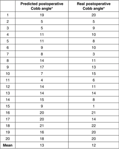
postoperative Cobb angles in our sample (Figure 5). After obtaining the results from applying the equa-tion, an analysis was done on the difference between the predicted result and the real result found for the postoperative Cobb angles (by means of the paired t test, with a significance level of 5%), in order to obtain confirmation of the prediction power of this equation (Box 1).
As can be seen in Box 1, there was no statistically significant difference between the predicted and real values for the postoperative Cobb angle of the main thoracic curve (p = 0.389), through applying the equa-tion to our sample.
FR = Preoperative Cobb angle – Cobb angle of lateral inclination x 100% Preoperative Cobb angle
575
DIsCUssION
Evaluation of the flexibility of all scoliotic curves (proximal thoracic, main thoracic and lumbar) forms part of the basic protocol for treating vertebral
deformities(7). Different radiographic methods have
been described for this purpose, such as use of active
lateral oblique views(7-10), views under traction in
dorsal decubitus(6,11) and views with a fulcrum at the
apex of the deformity, in lateral decubitus(4,12), among
Table 1 – Characterization of the curves according to Cobb angles of the main thoracic curve.
Mean DP 95% confidence interval
Preoperative Cobb
angle (º) 55 6 52-58
Postoperative Cobb
angle (º) 12 6 9-15
Lateral inclination (º) 26 12 20-30
Table 2 – Mean values, standard deviation and confidence interval of the flexibility rate and surgical correction rate.
Mean DP 95% confidence in-terval
Flexibility rate (%) 54* 18 48-63
Correction rate (%) 79 10 74-83
Table 3 – Results from Pearson’s correlation coefficient.
R p
Postoperative main thoracic Cobb angle
Age 0.52 0.018*
Preoperative
Cobb angle 0.51 0.021*
Cobb angle of lateral
inclination 0.71 0.000*
IF –0.65 0.002*
*Statistically signifi-cant correlations at p
< 0.05.
Figure 5 – Equation for predicting the postoperative Cobb angle, pro-posed by Cheung et al (4)
Box 1 – Comparison between predicted and real postoperative Cobb angles of the main thoracic curve.
Predicted postoperative Cobb angle*
Real postoperative Cobb angle*
1 19 20
2 5 5
3 5 9
4 11 10
5 11 8
6 9 10
7 8 3
8 14 11
9 17 13
10 7 15
11 4 6
12 14 11
13 14 14
14 15 8
15 9 1
16 20 21
17 20 14
18 21 22
19 16 20
20 18 20
Mean 13 12
others. The basic concept is that the compensatory curves tend to become corrected spontaneously and thus should not be subjected routinely to arthrodesis(2). In our service, we use radiographs with an active lateral oblique view in dorsal decubitus, because these are easy to perform and inexpensive.
Cheung et al(4) were able to produce an equation
for predicting the postoperative value of the main tho-racic scoliotic curve, and this may assist in choosing the best operative technique, according to the stiffness of the curve. This may be very valuable, especially in cases in which selective thoracic arthrodesis is indi-cated, since there is a risk of iatrogenic decompensa-tion of the lumbar curve. The results from the present study showed that there was no statistically significant difference between the value predicted by the equa-tion and the real postoperative value, thus confirming the validity of using the predictive equation proposed
by Cheung et al(4) among Brazilian adolescents.
Exclusive use of pedicle screws in the thoracic spine for surgical treatment of AIS gained popularity
following publication of the paper by Suk et al(3)
CORRECTION OF ADOLESCENT IDIOPATHIC SCOLIOSIS
and today it is considered to be the gold standard
for surgical correction of AIS(14). These implants
provide support in the three parts of the spine(15) and
have biomechanical characteristics that are superior to those of other types of materials such as hooks
and sublaminar wires(16-18). The main issue relating to
pedicle screws is perhaps whether the greater angular correction of the deformities would result in better clinical and functional results. Some authors have demonstrated a positive correlation is this regard, measured through questionnaires standardized by the
Scoliosis Research Society(19,20). Because we believe
that superior results are achieved through using assemblies consisting only of pedicle screws, as well
as the safety of this method at thoracic levels(3,4,6),
we use this type of instrumentation preferentially in our service.
Furthermore, in relation to the operative correction rate, we found values that were very close to those
described in other studies, such as Cheung et al(4). It
is noteworthy that these authors used assemblies with pedicle screws in all the vertebrae of the curve that underwent arthrodesis and, consequently, a greater number of anchorage points. In our sample, we used the philosophy of the method popularized by Cotrel
and Dubousset(13), which consists of instrumentation
of some vertebrae that are considered to be strategic, while leaving others free from any type of implant. Despite not being an objective of the present study, this allows us to suggest the hypothesis that, for this type of population, greater density of screws seems not to increase the corrective power. This matter seems to us to be fundamental and deserves to be evaluated in a more specific manner, through ran-domized prospective studies in order to confirm this hypothesis.
Assessing whether the correction obtained through the operation was maintained over the years, or whether it was lost, was not an objective of our study. Thus, we only used the angular values from the
im-mediate postoperative period. Other authors(4,18) have
reported that the mean loss of correction was minimal with exclusive use of pedicle screws, after two years of follow-up. Future analyses with this aim might answer this question in relation to our sample.
CONCLUsION
Through radiographs using a lateral oblique view obtained in the supine position, it is possible to predict the percentage operative correction achievable for the main thoracic curve, through using pedicle screws, in patients with AIS of Lenke types 1A and 1B.
REFERENCEs
1. Cobb JR. Outline for the study of scoliosis. Instr Course Lect. 1948; 5:261-75.
2. Moe JH, Byrd JA. Idiopathic scoliosis. In: Lonsteins JE, Winter RB, Bradford DS RB, Olgivie JW, editors. Moe’s textbook of scoliosis and other spinal deformities. 2nd ed. Philadelphia: Saunders; 1987. p.191-232.
3. Suk SI, Lee CK, Kim WJ, Chung YJ, Park YB. Segmental pedicle screw fixation in the treatment of thoracic idiopathic scoliosis. Spine (Phila Pa 1976). 1995;20(12):1399-405.
4. Cheung WY, Lenke LG, Luk KD. Prediction of scoliosis correction with thoracic segmental pedicle screw constructs using fulcrum bending radiographs. Spine (Phila Pa 1976). 2010;35(5):557-61.
5. Hamzaoglu A, Talu U, Tezer M, Mirzanli C, Domanic U, Goksan SB. Assessment of curve flexibility in adolescent idiopathic scoliosis. Spine (Phila Pa 1976). 2005;30(14):1637-42.
6. Watanabe K, Kawakami N, Nishiwaki Y, Goto M, Tsuji T, Obara T, et al. Traction versus supine side-bending radiographs in determining flexibility: what factors influence these techniques? Spine (Phila Pa 1976). 2007;32(23):2604-9.
7. Lenke LG, Betz RR, Harms J, Bridwell KH, Clements DH, Lowe TG, et al. Adolescent idiopathic scoliosis: a new classification to determine extent of spinal arthrodesis. J Bone Joint Surg Am. 2001;83-A(8):1169-81.
8. Large DF, Doig WG, Dickens DR, Torode IP, Cole WG. Surgical treatment of double major scoliosis. Improvement of the lumbar curve after fusion of the thoracic curve. J Bone Joint Surg Br. 1991;73(1):121-4.
9. McCall RE, Bronson W. Criteria for selective fusion in idiopathic scoliosis using Cotrel-Dubousset instrumentation. J Pediatr Orthop. 1992;12(4):475-9.
10. Vaughan JJ, Winter RB, Lonstein JE. Comparison of the use of supine bending and traction radiographs in the selection of the fusion area in adolescent idiopathic scoliosis. Spine (Phila Pa 1976). 1996;21(21):2469-73.
11. Polly DW Jr, Sturm PF. Traction versus supine side bending. Which technique best determines curve flexibility? Spine (Phila Pa 1976). 1998;23(7):804-8.
12. Cheung KM, Luk KD. Prediction of correction of scoliosis with use of the fulcrum bending radiograph. J Bone Joint Surg Am. 1997;79(8):1144-50.
13. Cotrel Y, Dubousset J. [A new technic for segmental spinal osteosynthesis using the posterior approach]. Rev Chir Orthop Reparatrice Appar Mot. 1984;70(6):489-94.
14. Lehman RA Jr, Lenke LG, Keeler KA, Kim YJ, Buchowski JM, Cheh G, et al. Operative treatment of adolescent idiopathic scoliosis with posterior pedicle screw-only constructs: minimum three-year follow-up of one hundred fourteen cases. Spine (Phila Pa 1976). 2008;33(14):1598-604.
15. Denis F. Spinal instability as defined by the three-column spine concept in acute spinal trauma. Clin Orthop Relat Res. 1984;(189):65-76.
16. Cheng I, Hay D, Iezza A, Lindsey D, Lenke LG. Biomechanical analysis of derotation of the thoracic spine using pedicle screws. Spine (Phila Pa 1976). 2010;35(10):1039-43.
17. Suk SI, Lee CK, Min HJ, Cho KH, Oh JH. Comparison of Cotrel-Dubousset pedicle screws and hooks in the treatment of idiopathic scoliosis. Int Orthop. 1994;18(6):341-6.
18. Dobbs MB, Lenke LG, Kim YJ, Kamath G, Peelle MW, Bridwell KH. Selective posterior thoracic fusions for adolescent idiopathic scoliosis: comparison of hooks versus pedicle screws. Spine (Phila Pa 1976). 2006;31(20):2400-4. 19. Haher TR, Merola A, Zipnick RI, Gorup J, Mannor D, Orchowski J. Meta-analysis
of surgical outcome in adolescent idiopathic scoliosis. A 35-year English literature review of 11,000 patients. Spine (Phila Pa 1976). 1995;20(14):1575-84. 20. Avanzi O, Landim E, Meves R, Caffaro MFS, Umeta RSG. Escoliose idiopática