Development of a Multivalent Subunit
Vaccine against Tularemia Using Tobacco
Mosaic Virus (TMV) Based Delivery System
Sukalyani Banik1☯, Ahd Ahmed Mansour1☯, Ragavan Varadharajan Suresh1, Sherri Wykoff-Clary2, Meenakshi Malik3, Alison A. McCormick2, Chandra Shekhar Bakshi1*
1Department of Microbiology and Immunology, New York Medical College, Valhalla, New York, United States of America,2College of Pharmacy, Touro University California, Mare Island, Vallejo, California, United States of America,3Albany College of Pharmacy and Health Sciences, Albany, New York, United States of America
☯These authors contributed equally to this work. *[email protected]
Abstract
Francisella tularensisis a facultative intracellular pathogen, and is the causative agent of a
fatal human disease known as tularemia.F.tularensisis classified as a Category A
Bio-threat agent by the CDC based on its use in bioweapon programs by several countries in the past and its potential to be used as an agent of bioterrorism. No licensed vaccine is currently available for prevention of tularemia. In this study, we used a novel approach for development of a multivalent subunit vaccine against tularemia by using an efficient tobacco mosaic virus (TMV) based delivery platform. The multivalent subunit vaccine was formulated to contain a combination ofF.tularensisprotective antigens: OmpA-like protein
(OmpA), chaperone protein DnaK and lipoprotein Tul4 from the highly virulentF. tularen-sisSchuS4 strain. Two different vaccine formulations and immunization schedules were
used. The immunized mice were challenged with lethal (10xLD100) doses ofF.
tularen-sisLVS on day 28 of the primary immunization and observed daily for morbidity and
mortal-ity. Results from this study demonstrate that TMV can be used as a carrier for effective delivery of multipleF.tularensisantigens. TMV-conjugate vaccine formulations are safe and
multiple doses can be administered without causing any adverse reactions in immunized mice. Immunization with TMV-conjugatedF.tularensisproteins induced a strong humoral
immune response and protected mice against respiratory challenges with very high doses ofF.tularensisLVS. This study provides a proof-of-concept that TMV can serve as a
suit-able platform for simultaneous delivery of multiple protective antigens ofF.tularensis.
Refinement of vaccine formulations coupled with TMV-targeting strategies developed in this study will provide a platform for development of an effective tularemia subunit vaccine as well as a vaccination approach that may broadly be applicable to many other bacterial pathogens.
OPEN ACCESS
Citation:Banik S, Mansour AA, Suresh RV, Wykoff-Clary S, Malik M, McCormick AA, et al. (2015) Development of a Multivalent Subunit Vaccine against Tularemia Using Tobacco Mosaic Virus (TMV) Based Delivery System. PLoS ONE 10(6): e0130858. doi:10.1371/journal.pone.0130858
Editor:Ashlesh K Murthy, Midwestern University, UNITED STATES
Received:April 2, 2015
Accepted:May 26, 2015
Published:June 22, 2015
Copyright:© 2015 Banik et al. This is an open access article distributed under the terms of the Creative Commons Attribution License, which permits unrestricted use, distribution, and reproduction in any medium, provided the original author and source are credited.
Data Availability Statement:All relevant data are within the paper.
Funding:This work was supported by Touro University Seed Grant to AAM and CSB. The funders had no role in study design, data collection and analysis, decision to publish, or preparation of the manuscript.
rently available in the USA for prevention of tularemia [8,9]. Considering the bioweapon potential ofF.tularensisand repercussions of 2001 anthrax attack in the USA, there has been an increased interest in development of vaccine and effective countermeasures against bioter-ror agents. An ideal solution for prevention of tularemia occurring naturally or consequent to the use ofFrancisellaas a bioweapon or an act of bioterrorism is to develop a safe and effective vaccine capable of inducing long lasting protection in a relatively short period of time [10].
In the last 100 years since the discovery ofF.tularensis, three broad approaches comprising of killed whole cell, live attenuated and subunit vaccines have been employed for vaccine devel-opment, but none of these have been successful [11]. Although, a Live Vaccine Strain (LVS) developed from the Russian strainF.holarcticaS15 is protective, it retains residual virulence in humans when immunized via aerosol or intranasal (i.n.) routes. Due to adverse reactions and residual virulence, LVS is not approved by the FDA for mass immunizations in the USA. Attenuated mutants ofF.tularensisSchuS4 or the LVS containing single gene deletions have shown better protective efficacy in mouse models of tularemia [12,13,14,15,16,17]. However, these mutants pose a potential possibility of reversion to fully virulent forms. Inactivated LVS or SchuS4 tularemia vaccines have demonstrated poor protective efficacies against challenges with virulentF.tularensis[11,18,19,20]. Several efforts to develop subunit tularemia vaccine have met with limited success. The primary shortcomings have been the constituents of sub-unit vaccines which contained either a single surface associated antigenic component ofF. tularensissuch as LPS or specific immunoreactive proteins such as GroEL, DnaK, FopA, KatG or aF.tularensisspecific lipoprotein Tul4 [21,22,23,24,25,26,27,28]. Despite being immuno-genic, these single subunit vaccines failed to provide protection against virulentF.tularensis strains. The possible explanations for their failure could be that single proteins are not suffi-cient or that the vaccine formulations lacked right combination of antigens required for induc-tion of a protective immune response. The challenges thus far in development of multivalent subunit vaccines have been the availability of suitable approaches for consistent preparation and efficient delivery of multiple antigens through mucosal routes.
non-functional) which is important for inducing cell mediated immunity (or both). Conjugat-ing an immunogenic subunit vaccine protein to the surface of TMV promotes antigen uptake and improves an antiviral response against the subunit protein. A recent study demonstrated single dose potency of a TMV-hemagglutinin (TMV-HA) vaccine in an influenza challenge model without the need for an adjuvant [32]. Because TMV is not a human pathogen [33], TMV is inherently safe. In addition, TMV does not show evidence of neutralizing antibodies in individuals, so it can be used repeatedly for boosting [31,32]. These characteristics of TMV are extremely important in producing a safe, effective vaccine that can stimulate protection against F.tularensischallenge.
We investigated the vaccine potential of a multivalent tularemia vaccine by chemically con-jugating TMV to multiple protective antigens ofF.tularensis. We used purified recombinant proteins DnaK (FTT1269c), OmpA (FTT0831c) and Tul4 (FTT0901) ofF.tularensisSchuS4 and determined the vaccine potential of TMV-F.tularensisprotein conjugate vaccine. When used in vaccine formulations, both DnaK and Tul4 have been shown to render some degree of protection againstF.tularensisLVS in vaccinated mice [34,35]. This was the rationale for inclu-sion of these proteins in our studies to investigate the efficacy of TMV-conjugate vaccine. In addition to DnaK and Tul4, we also included OmpA-like protein in the conjugate cocktail based on its surface exposed structures, role in innate immune subversion bothin vitroandin vivo[13,36,37] and exclusive reactivity of this protein with the serum from successfully vacci-nated individuals as well as mice [38,39,40,41]. This study demonstrates that TMV effectively delivers multipleF.tularensisantigens to induce protective immune responses in mouse model of respiratory tularemia and provide a proof-of-concept for the feasibility of TMV as a carrier for bacterial antigenic proteins.
Materials and Methods
Ethics Statement
This study was carried out in strict accordance with the recommendations and guidelines of National Council for Research (NCR) for care and use of animals. All the animal experiments were conducted in the centralized Animal Resources Facilities of Albany Medical College and New York Medical College licensed by the USDA and the NYS Department of Health, Division of Laboratories and Research and accredited by the American Association for the Accreditation of Laboratory Care. The use of animals and protocols were approved by the Institutional Ani-mal Care and Use Committee (IACUC) of New York Medical College (Protocol Number 30-2-0414H). Mice were administered an anesthetic cocktail consisting of ketamine (5 mg/kg) and xylazine (4 mg/kg) and underwent experimental manipulation only after they failed to exhibit a toe pinch reflex. Mice exhibiting more than 20% weight loss, anorexia, dehydration and impairment of mobility were removed from the study and euthanized by approved means. Humane endpoints were also necessary for mice which survived at the conclusion of the exper-iment. Mice were administered an anesthetic cocktail of ketamine and xylazine intraperitone-ally and then euthanized via cervical dislocation followed by cardiac puncture, a method that is consistent with recommendations of the Panel on Euthanasia of the American Veterinary Medical Association. In all experimental procedures, efforts were made to minimize pain and suffering.
Bacterial Strains
induced for expression by IPTG, and purified by metal affinity chromatography. The purity of the proteins was confirmed by SDS-PAGE and western blot analysis using anti-6His
antibodies.
Purification of TMV-Lysine Virus Particles
TMV was genetically engineered to express coat protein containing a surface exposed lysine [42]. Infectious TMV RNA was inoculated onto 30 dayNicotiana benthamianaplants, and harvested for virus 10 days later according to previously described protocols [42,43]. Briefly, plant tissue was homogenized in 0.86M NaCl, 0.04% w/v sodium metabisulfite (0.5 g of tissue/ ml of buffer), adjusted to pH 5.0, heated to 47°C for 5 min, and then chilled to 4°C. Homoge-nate was centrifuged at 6000 × g for 20 min, and then the clarified supernatant was precipitated with 5% Poly Ethylene Glycol (PEG) 8000 at 4°C, and spun at 12,000 × g for 10 min at 4°C to recover the virus. PEG pellets were resuspended in PBS, and re-precipitated with PEG a second time. Final PEG pellets were resuspended in PBS at 1:10th homogenization volume, and final protein concentration was measured by Bicinchoninic Acid (BCA). Purity was determined (typically>98%) by SDS-PAGE.
Conjugation of
F
.
tularensis
Proteins to TMV
In order to use TMV as a platform, recombinantF.tularensisproteins were chemically conju-gated to decorate the surface of TMV. For conjugation reaction, purified–TMV-Lysine and individual purified recombinant proteins OmpA, DnaK, and Tul4 were mixed at 1:1 molar ratios. The conjugation reaction was carried out by adding 5mM of 1- Ethyl-3-(3-dimethylami-nopropyl) carbodiimide (EDC) and 1mM of N-hydroxysuccinimide (NHS). The conjugation mixture was then incubated for various time intervals to achieve maximum conjugation effi-ciency. The reaction time that generated the least amount of free protein (2 hours) was used in scale up conjugation reactions (5mg antigen with 5mg TMV) for vaccine potency testing. For the TMV monoconjugate vaccine, where all three proteins were reacted together onto the same virus, each protein was mixed with TMV at a 30% molar ratio (1x TMV, 0.3x each protein) and reacted for 2 hours to ensure complete conjugation. The efficiency and successful conjuga-tion of recombinant proteins ofF.tularensisSchuS4 to the TMV virion was determined by 8–16% Tris-Glycine SDS-PAGE.
Safety, Immunogenicity and Protective Efficacy of TMV-Conjugate
Vaccines
environmentally controlled and pathogen-free animal facility of New York Medical College. All mice that were to be immunized or challenged were anesthetized by i. p. injection of a cock-tail of Ketamine and xylazine to facilitate delivery of the inoculum to the respiratory compart-ment. All mice experiments were performed according to the guidelines and protocols approved by the IACUC at New York Medical College.
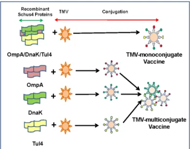
Vaccine formulations. Two different vaccine formulations were used. In the first formula-tion, all three recombinant proteins OmpA, DnaK and Tul4 were conjugated to a single TMV virion. This vaccine formulation was designated as TMV-monoconjugate vaccine. Mice were immunized with 60μg of TMV monoconjugate vaccine (~30μg TMV and 30μg of recombinant
proteins). In the second vaccine formulation, each individual protein was conjugated to the TMV individually (10μg TMV + 10μg recombinant protein) and then each of the three
TMV-protein conjugates were mixed in equal concentrations [20μg x 3 = 60μg (30μg TMV + 30μg
recombinant proteins)]. This formulation was designated as TMV-multiconjugate vaccine (Fig 1). Based on the amount of TMV that each of the vaccinated mouse received (30μg), mice
inoculated with an 30μg of TMV served as controls.
Immunization schedules. Two different immunization schedules were used. In the first immunization schedule (Schedule I) C57BL/6 mice were immunized intranasally (i.n.) with 60μg of TMV monoconjugate or TMV-multiconjugate vaccine. Mice were immunized i.n. with
30μl (15μl/ nostril) volume of each of the vaccine formulation or the TMV controls. Booster
vaccinations using dosages similar to the primary immunization were administered on days 7 and 14 after the primary immunization. Mice receiving 30μg of TMV alone and administered
in a fashion similar to the vaccine groups were kept as controls (Fig 2A). Mice were monitored for any adverse reaction following each vaccine administration.
Mice immunized employing schedule II were vaccinated only with TMV-multiconjugate vaccine. C57BL/6 mice were immunized i.n. on day 0, 5 and 14 with a total of 60μg of
TMV-multiconjugate vaccine. These mice also received similar vaccination dose of TMV-multiconju-gate vaccine subcutaneously (s.c.) on days 3, 7 and 14. Control mice received 30μg of TMV
Fig 1. Vaccine Formulations.Two different vaccine formulations were used. In the first vaccine formulation all three recombinant proteins OmpA, DnaK and Tul4 were conjugated to a single TMV virion (TMV-monoconjugate vaccine). The second vaccine formulation contained each recombinant protein ofF.tularensisconjugated individually to TMV and then mixed in equal concentrations to generate a TMV-multiconjugate vaccine.
following the immunization schedule identical to those for the TMV-multiconjugate vaccine group (Fig 2B). All vaccinated and control mice were monitored for any adverse reaction fol-lowing each primary and booster immunizations. All the vaccinated mice were bled periodi-cally to collect serum to determine antibody responses. The Schedule I immunizations were based on a number of vaccination studies using inactivated or subunit vaccines ofF.tularensis [18,34,44]. The aggressive immunization strategy (Schedule II) was based on our recent report indicating that alternate i.n. and s.c. booster immunizations enhanced protection in immu-nized mice against an i.n. challenge withF.tularensisSchuS4 strain [45] and the vaccination schedule recommended for commercially available oral typhoid vaccine, Vivotif.
Determination of antibody responses in immunized mice. For determination of anti— F.tularensisantibody levels in vaccinated mice following the immunization schedules described above, ELISA was performed using lysates made fromF.tularensisSchuS4 or LVS strains. The formalin fixed SchuS4 was obtained from BEI Resources, Manassas, VA. For ELISA 96-well microtiter plates were coated with 1×10⁷CFU/ml of eitherF.tularensisSchuS4 or LVS in bicarbonate buffer.F.tularensis-specific antibody levels for total IgG, IgG1, IgG2a and IgG2b in serum samples collected from immunized mice on 28 post-immunization were determined by ELISA. Serum collected from naïve mice or mice that received TMV were used as controls. To determine the level of antibodies induced against each individual protein of the TMV-monoconjugate or the TMV-multiconjugate vaccine each individual ELISA was per-formed by coating plates with 1μg of each individual purified recombinant OmpA, DnaK and
Tul4 proteins. The protein specific total IgG levels were determined in serum from vaccinated mice collected on day 28 post-immunization. Antibody titers were calculated from linear Fig 2. Immunization Schedules I and II. (A)C57BL/6 mice were immunized intranasally (i.n.) either with TMV-monoconjugate (60μg/mouse) or TMV-multiconjugate vaccine formulations (20μg each of
OmpA-TMV; DnaK-TMV and Tul4-TMV conjugates. Total 60μg/mouse) and booster vaccinations were
administered i.n. using dosages similar to those for primary immunization on days 7 and 14 of the post-primary immunization (Schedule I).(B)Alternatively, mice were administered TMV-multiconjugate vaccines with booster immunizations i.n. on day 5 and 14 and subcutaneously (s.c.) on days 7 and 14 post-primary immunization (Schedule II). The dosages used were similar to those described for TMV-multiconjugate vaccine inA. Mice inoculated with TMV (30μg/mouse) in a manner similar to the vaccinated groups were
kept as controls.
regression curves as the inverse of the serum dilution that showed an OD450value 2.5 times
above the controls, and expressed as Log10values.
To determine if antibodies generated in vaccinated mice are capable of identifying native and recombinantFrancisellaOmpA, DnaK and Tul4 proteins, western blot analysis was per-formed. Serum collected on day 28 from mice immunized with TMV-multiconjugate vaccine utilizing Schedule II was used for western blot analysis. Eight micrograms each ofF.tularensis LVS and SchuS4 lysates were resolved on SDS-PAGE, transferred to nitrocellulose membrane and blotted against pooled serum from immunized mice. Serum collected from mice immu-nized with TMV was used as a control. To determine if antibodies from immuimmu-nized mice reacted with recombinant proteins as well, 1μg of purified recombinant proteins were used in
western blot analysis.
Challenge studies. To determine the protective efficacy of TMV-vaccine against a high challenge dose of LVS, all immunized mice were challenged i.n. with 10×LD100(1×10⁵CFU)
dose ofF.tularensisLVS on day 28 of the primary immunization. The actual numbers ofF. tularensisinoculated into mice at that time of challenge were confirmed by plating serial dilu-tions on MH-chocolate agar plates and counting the colonies 48 hours later.
Post-challenge studies. All the challenged mice were observed daily for signs of morbidity and/or mortality for a period of 21 days. To monitor the progression of infection all challenged mice were weighed every day until they regained their original body weight.
Statistical Analysis
All data for antibody levels of immunized mice were statistically analyzed using InStat program (Graph-Pad Software). The results were expressed as Means ± S.D. The survivals data were expressed as Kaplan-Meier survival curves and statistical significance for survival results were evaluated by analyzing the mean time to death by the Log-Rank test.
Results
Purification, and Conjugation of DnaK, OmpA and Tul4 Proteins of
F
.
tularensis
SchuS4 to TMV
Immunization of Mice with TMV-Multiconjugate Vaccine Induces
Antibody Responses Capable of Recognizing both Native and
Recombinant DnaK, OmpA and Tul4 Proteins
Since purification of recombinant proteins may alter their confirmation or may result in dena-turation of immunogenic epitopes, we next investigated if vaccination of mice with TMV-mul-ticonjugate vaccine generates an antibody response capable of recognizing nativeFrancisella DnaK, OmpA and Tul4 proteins. Mice immunized with the TMV-multiconjugate vaccine fol-lowing immunization Schedule II in which mice were boosted by both the i.n. and s.c. routes were bled on day 28 post-immunization. The pooled sera from TMV-multiconjugate vaccine immunized mice specifically recognized DnaK, OmpA and Tul4 proteins inF.tularensisLVS and SchuS4 lysates indicating that all the antigenic epitopes in immunizing proteins are intact and are capable of recognizing native bacterial proteins (Fig 5A). Conversely, we also investi-gated if vaccination of mice with liveF.tularensisLVS induces antibody responses against native DnaK, OmpA and Tul4 proteins that can react with the purified recombinant forms of these three proteins. Our results show that sera from mice immunized with liveF.tularensis LVS recognized all three recombinant proteins similar to those observed for sera from mice immunized with TMV-multiconjugate vaccine (Fig 5B).
Immunization with TMV-Monoconjugate Vaccine Generates Antibody
Response Predominated by IgG1 Antibodies
We first investigated the antibody response in mice that received TMV-monoconjugate vaccine formulation in which all the three recombinant proteins were conjugated to a single TMV virion, and received boosters only on day 7 and 14 (Schedule I). Mice were bled on day 28 post-immunization and antibody responses were determined. Higher levels ofFrancisella specific total IgG levels were detected in TMV-monoconjugate vaccinated mice (Fig 6). Deter-mination of IgG isotypes on day 28 post-immunization revealed that mice vaccinated with TMV-monoconjugate vaccine induced higher IgG1 levels. However, very low to undetectable levels of IgG2a and IgG2b antibodies were observed in this group of vaccinated mice (Fig 6). Antigen specific ELISA indicated that antibodies were generated against OmpA, DnaK and Tul4 proteins (Fig 7). Collectively, these results indicated that a weak antibody response pre-dominated by a Th2 biased immune response is generated in mice immunized using Schedule I vaccination regimen with TMV-monoconjugate vaccine formulation.
Fig 3. Expression and Purification of Recombinant DnaK, OmpA and Tul4 Proteins ofF.tularensisSchuS4.Purification of recombinant OmpA, DnaK and Tul4 proteins ofF.tularensisSchuS4 proteins was confirmed by SDS-PAGE and western blot analysis using anti-His antibodies.
Immunization Schedule I with TMV-Multiconjugate Vaccine Generates
Stronger Antibody Responses than those Observed with
TMV-Monoconjugate Vaccine
We next examined antibody response generated following vaccination with TMV-multi conjugate vaccine in which individual TMV-protein conjugates were blended in a multivalent formulation. The vaccination schedule included booster immunizations on days 7 and 14 post-immunization (Schedule I). The total IgG responses observed on days 14 (not shown) and 28 Fig 4. Conjugation of DnaK, OmpA and Tul4 Proteins ofF.tularensisSchuS4 to TMV.Purified OmpA, DnaK and Tul4 proteins were combined with purified TMV and incubated with EDC and NHS for 0, 30 min, 1, or 2 hours as described in Methods section. Twoμg of TMV or recombinant proteins DnaK, OmpA, Tul4 or
4μg of the TMV-protein mixtures were resolved on an 8–16% SDS-PAGE gel to observe conjugation products indicated by changes in the molecular masses of the starting materials.(A)Conjugation of DnaK, OmpA and Tul4 to a single TMV virion to generate TMV-monoconjugate vaccine. The progress of conjugation process was observed over a period of time: Lane M = Precision Plus Dual Color standard (BioRad) Marker; Lane 1 = TMV-protein mix, 0 min; Lane 2 = TMV-protein mix, 30 min; Lane 3 = TMV-protein mix,1 hour; Lane 4 = TMV-protein mix, 2 hours.(B, C, D)Kinetics of DnaK, OmpA and Tul4 TMV-protein conjugations over a two hour incubation period to generate protein conjugates. The individual TMV-protein conjugates were then admixed to generate TMV-multiconjugate vaccine. Lane M = Precision Plus Dual Color standard (BioRad) Marker; Lane 1 = TMV; Lane 2 = Recombinant protein; Lane 3 = TMV-protein mix, 0 hour; Lane 4 = TMV-protein mix, 1 hour; Lane 5 = TMV-protein mix, 2 hours. In all cases, 2 hour time points were used for scale-up and vaccine preparation. Solid arrows indicate TMV-protein conjugate(s), dashed arrows indicate free TMV or free proteins.
were much higher than those observed with the TMV-monoconjugate vaccine (Fig 6). IgG isotype profiles were also different in TMV-multiconjugate vaccine than those observed for TMV-monoconjugate vaccine immunized mice. Higher levels ofFrancisellaspecific IgG2a and IgG2b antibodies were observed in immunized mice. However, as observed for TMV-monoconjugate vaccinated mice, higher levels of IgG1 antibodies were also observed in Fig 5. Immunization of Mice with TMV-Multiconjugate Vaccine Induces Antibody Responses Capable of Recognizing both Native and Recombinant OmpA, DnaK and Tul4 Proteins. (A)Serum collected on day 28 post-immunization from C57BL/6 mice immunized with TMV-multiconjugate vaccine (Schedule II) was pooled (n = 4) and blotted againstF.tularensisLVS and SchuS4 lysates.(B)Pooled serum from C57BL/6 mice (n = 4) immunized either with TMV-multiconjugate vaccine, or 100 CFU ofF.tularensisLVS were collected on day 28 post immunization and blotted against purified recombinant OmpA, DnaK and Tul4 proteins. Sera from mice inoculated with TMV alone were used as controls.
vaccinogen than the TMV-monoconjugate vaccine however, similar to the latter vaccine for-mulation, generates an immune response predominated by IgG1 antibodies.
An Aggressive Immunization with TMV-Multiconjugate Vaccine does not
Further Enhance Antibody Response
We further investigated if an aggressive immunization schedule consisting of TMV-multi con-jugate formulation administered by both the i.n. and s.c. routes with multiple booster vaccina-tions (Schedule II) improves the antibody response compared to the other two vaccination strategies. It was observed that the total IgG and IgG1 antibody responses did not differ from mice vaccinated with TMV-multiconjugate vaccine using Schedule I (Fig 8). However, the lev-els of antibody isotypes IgG2a and IgG2b were in fact significantly higher in mice vaccinated with the TMV-multiconjugate vaccine receiving Schedule I than those receiving Schedule II vaccinations (Fig 8). We further investigated if there are any differences in levels of antibodies generated against native proteins ofF.tularensisSchuS4 andF.tularensisLVS. No differences in IgG, IgG1, IgG2a and IgG2b antibody levels were observed when ELISAs were performed usingF.tularensisSchuS4 and LVS lysates (Fig 8). These results indicate that similar to the results obtained with western blot analysis, antibodies from TMV-multiconjugate vaccine are equally capable of recognizing native proteins of bothF.tularensisSchuS4 andF.tularensis LVS. The group of mice receiving Schedule II of TMV-multiconjugate vaccine showed signifi-cantly higher titers of OmpA antibodies, while titers of DnaK and Tul4 antibodies were similar to those receiving Schedule I vaccination with TMV-multiconjugate vaccine (Fig 9).
Protective Efficacy of TMV-Conjugate Vaccine
We investigated the protective efficacy of the TMV-monoconjugate and TMV-multiconjugate vaccine by vaccinating mice using immunization schedules I and II as described inFig 2. Mice were immunized with TMV-monoconjugate vaccine using only immunization schedule I; while both schedule I and II were used for TMV-multiconjugate vaccine. Mice were chal-lenged i.n. with 10×LD100dose ofF.tularensisLVS on day 28 post-primary immunization and
Fig 7. OmpA, DnaK and Tul4 Specific Antibody Responses in Mice Immunized with TMV-Monoconjugate and TMV-Multiconjugate Vaccines using Schedule I of Immunization.F.tularensisSchuS4 recombinant proteins OmpA, DnaK and Tul4 specific IgG antibody levels on day 28 in serum samples of
survival and body weight loss of mice receiving TMV-multiconjugate vaccine with Schedule II was similar to that observed for mice vaccinated with Schedule I regimen, however 50% of the vaccinated mice survived the challenge (Fig 10E and 10F). Additional booster vaccinations by i. n. and s.c. routes in Schedule II only slightly improved the level of protection in this group of vaccinated mice. Collectively, these results demonstrated thatFrancisellaproteins conjugated to TMV when used as vaccine induce protective immune response in mice. These results also indicated that TMV-monoconjugate vaccine in which all the three recombinant proteins OmpA, DnaK and Tul4 ofF.tularensisconjugated to a single TMV virion serves as a poor vac-cinogen. On the other hand, the vaccine formulation that contains a multivalent blend of all the three proteins conjugated individually to TMV induces a superior protective immune response that can marginally be improved further by increasing number of booster vaccina-tions by s.c. route.
Discussion
The possibility of usingF.tularensisas a bioterror agent has renewed attention towardsF. tularensisresearch and to develop a licensable vaccine for effective prevention of tularemia. The tularemia vaccine development research has largely been focused on development of live attenuated or inactivated tularemia vaccines. However, concerns about their efficacy and safety have halted the progress. A recent study confirms this notion and reported a variant ofF. tular-ensisLVS which is 100 times lethal than the standard ATCC strain indicating that, as feared,F. tularensisLVS may revert back to its virulent form [46]. Recombinant subunit vaccines obvi-ously have potential safety advantages over inactivated or live attenuated vaccines.
Ongoing studies for subunit vaccine development have identified a number ofF.tularensis antigens that are capable of inducing a partial protective immune response [47,48,49,50,51]. The possible explanation for limited protective efficacy of these subunit vaccines could be that a single protein or combinations of proteins used in vaccine formulations were not sufficient to induce an efficient protective immune response. Another shortcoming appeared to be the availability of a suitable platform for simultaneous delivery of antigens in a consistent fashion. It has been shown that the protective efficacy improves when multiple antigens are used in vac-cine formulations [52,53,54]. However, the biggest challenge thus far in the development of multivalent subunit vaccines has been the availability of suitable approaches for consistent preparation and efficient delivery of multiple antigens through mucosal route.
The goal of this study was to explore vaccine potential of a multivalent subunit vaccine against tularemia using an efficient TMV based delivery platform. The premise behind utilizing a novel TMV-conjugated vaccination strategy is based on the proven efficacy of TMV vaccines in stimulating robust immune response without the requirement of an additional adjuvant [29]. In order to provide a proof-of-concept and feasibility of TMV as a carrier forF.tularensis proteins, we used purified recombinant proteins OmpA, DnaK and Tul4 fromF.tularensis SchuS4 for conjugation studies and determined the vaccine potential of TMV-F.tularensis pro-tein conjugates. Two different vaccine formulations consisting either of all threeF.tularensis proteins conjugated to a single TMV virion (TMV-monoconjugate vaccine); or a mixture con-sisting of each protein individually conjugated to TMV (TMV-multiconjugate vaccine) were Fig 8. Antibody Responses in Mice Immunized with TMV-Multiconjugate Vaccines using Schedule I and II of Immunization.Francisellaspecific total IgG, IgG1, IgG2a and IgG2b levels on day 28 in serum samples of C57BL/6 mice immunized with TMV-multiconjugate vaccine using Schedule II were determined by ELISA. The plates were coated withF.tularensisSchuS4 or LVS lysates. Serum samples obtained from naïve mice or those inoculated with TMV alone were used as controls. The data are represented as Mean±S.D. of absorbance values measured at 450nm. The comparisons are shown with the
data obtained from mice immunized with TMV-multiconjugate vaccine using schedule I (shown inFig 6). Table shows comparison of antibody titers between groups of mice vaccinated with Schedule I and II vaccination regimens.
Fig 9. OmpA, DnaK and Tul4 Specific Antibody Responses in Mice Immunized with TMV-Multiconjugate Vaccines using Schedule I and II of Immunization.F.tularensisSchuS4 recombinant proteins OmpA, DnaK and Tul4 specific IgG, antibody levels on day 28 in serum samples of C57BL/6 mice
immunized with TMV-multiconjugate vaccine using Schedule II were determined by ELISA. The plates were coated with recombinantF.tularensisSchuS4 proteins. Serum samples obtained from naïve mice or those inoculated with TMV alone were used as controls. The data are represented as Mean±S.D. of absorbance values measured at 450nm. The comparisons are shown with the data obtained from mice immunized with TMV-multiconjugate vaccine using schedule I (shown inFig 7). Table shows comparison of antibody titers between groups of mice vaccinated with Schedule I and II vaccination regimens.
When immune responses were compared between mice receiving TMV-monoconjugate and TMV-multiconjugate vaccine using a similar vaccination regimen (Schedule I), a weaker antibody response was observed in mice vaccinated with monoconjugate formulation. These results indicate that conjugating all proteins to a single TMV virion is not an ideal approach for development of a TMV-based tularemia vaccine. The poor antibody response could be due to preferential conjugation of one of the three proteins in the conjugation mix or due to antigenic competition. The antibody responses observed against OmpA and DnaK proteins does support this notion. It was observed that TMV-monoconjugate vaccinated mice induced antibody response against Tul4 protein were similar to those observed for TMV-multiconjugate vaccine formulations however, the response against OmpA and DnaK were significantly lower than that observed for the latter vaccine formulation (Fig 7). The binding capacity of each of these proteins to TMV was not determined in the present study. However, these results do point to the fact that Tul4 due to its smaller size may have a preferential binding to the surface of TMV than OmpA or DnaK proteins. Moreover, the immune response was predominated by IgG1 antibodies and no IgG2a or IgG2b responses were observed in mice vaccinated with TMV-monoconjugate vaccine. These results indicate generation of a predominantly Th2 biased immune response in this group of vaccinated mice. Contrary to what was observed for TMV-monoconjugate vaccinated mice, the TMV-multiconjugate vaccinated mice mounted a very strongFrancisellaspecific total IgG response and the titers went up from days 14 to 28 post-immunization (not shown). Although, IgG1 was the most predominant antibody isotype, higher levels of IgG2a and IgG2b antibodies were also detected in this group of immunized mice. It is noteworthy that both the IgG2a and IgG2b antibodies have been shown to be protec-tive againstF.tularensisinfection [55,56]. Collectively, these results indicate that conjugating each protein individually to TMV and then blending them in equimolar concentration to gen-erate a multiconjugate composition is an ideal approach for the development of a TMV-based tularemia vaccine.
We further investigated if an enhanced immune response can be generated following an aggressive vaccination regimen with the TMV-multiconjugate vaccine. We administered booster vaccinations by alternating i.n. and s.c. routes. The intent was to induce potent sys-temic as well as local mucosal immune response by administering vaccine by both i.n. and s.c. routes (Schedule II). However, the immune responses did not differ from those observed in mice receiving only two booster vaccines (Schedule I). We speculate that the failure to observe further amplification following an aggressive vaccination could be due to an excessive antigenic overload. When vaccinated mice were challenged intranasally with 10LD100dose (1x10⁵CFU)
effective delivery of multipleF.tularensisantigens. The TMV-conjugate vaccine is safe and multiple doses can be administered in mice without any adverse reactions and immunization withF.tularensisantigens conjugated individually and blended in a multivalent composition induce a more potent immune response than the formulation in which all of the three proteins are conjugated to a single TMV virion. Most importantly immunization with TMV-conjugated F.tularensisproteins can protect mice against lethal doses ofF.tularensisLVS.
In conclusion, this study provides a proof-of-concept that TMV can serve as a suitable plat-form for simultaneous delivery of multiple protective antigens ofF.tularensis. Future studies to improve the level of protection would require generation of TMV-multiconjugate vaccine by incorporating additional immunoprotective antigens ofF.tularensisand inclusion of suitable adjuvant(s) to generate potent humoral and cell-mediated immune responses and induce long-lasting immunity against tularemia caused byF.tularensisSchuS4 strain.
Author Contributions
Conceived and designed the experiments: MM A. McCormick CSB. Performed the experi-ments: SB A. Mansour RVS SWC MM A. McCormick CSB. Analyzed the data: SB A. Mansour MM CSB. Contributed reagents/materials/analysis tools: A. McCormick MM CSB. Wrote the paper: MM A. McCormick CSB.
References
1. Dennis DT, Inglesby TV, Henderson DA, Bartlett JG, Ascher MS, Eitzen E et al. Tularemia as a biologi-cal weapon: medibiologi-cal and public health management. JAMA. 2001; 285: 2763–2773. PMID:11386933 2. Altman GB. Tularemia. A pathogen in nature and a biological weapon. AAOHN J. 2002; 50: 373–377.
PMID:12227212
3. Forsman M, Sandstrom G, Sjostedt A. Analysis of 16S ribosomal DNA sequences of Francisella strains and utilization for determination of the phylogeny of the genus and for identification of strains by PCR. Int J Syst Bacteriol. 1994; 44: 38–46. PMID:8123561
4. Hollis DG, Weaver RE, Steigerwalt AG, Wenger JD, Moss CW, Brenner DJ. Francisella philomiragia comb. nov. (formerly Yersinia philomiragia) and Francisella tularensis biogroup novicida (formerly Fran-cisella novicida) associated with human disease. J Clin Microbiol. 1989; 27: 1601–1608. PMID: 2671019
5. Svensson K, Larsson P, Johansson D, Bystrom M, Forsman M, Johansson A. Evolution of subspecies of Francisella tularensis. J Bacteriol. 2005; 187: 3903–3908. PMID:15901721
6. Atlas RM. Bioterriorism: from threat to reality. Annu Rev Microbiol. 2002; 56: 167–185. PMID: 12142472
7. Bossi P, Bricaire F. [Tularemia, a potential bioterrorism weapon]. Presse Med. 2003; 32: 1126–1130. PMID:12947746
8. Steiner DJ, Furuya Y, Metzger DW. Host-pathogen interactions and immune evasion strategies in Fran-cisella tularensis pathogenicity. Infect Drug Resist. 2014; 7: 239–251. doi:10.2147/IDR.S53700PMID: 25258544
9. Oyston PC. Francisella tularensis: unravelling the secrets of an intracellular pathogen. J Med Microbiol. 2008; 57: 921–930. doi:10.1099/jmm.0.2008/000653-0PMID:18628490
10. Barry EM, Cole LE, Santiago AE. Vaccines against tularemia. Hum Vaccin. 2009; 5: 832–838. PMID: 19923904
11. Barry EM, Cole LE, Santiago AE. Vaccines against tularemia. Hum Vaccin. 2009; 5: 832–838. PMID: 19923904
12. Twine S, Bystrom M, Chen W, Forsman M, Golovliov I, Johansson A et al. A mutant of Francisella tular-ensis strain SCHU S4 lacking the ability to express a 58-kilodalton protein is attenuated for virulence and is an effective live vaccine. Infect Immun. 2005; 73: 8345–8352. PMID:16299332
18. Baron SD, Singh R, Metzger DW. Inactivated Francisella tularensis live vaccine strain protects against respiratory tularemia by intranasal vaccination in an immunoglobulin A-dependent fashion. Infect Immun. 2007; 75: 2152–2162. PMID:17296747
19. Eyles JE, Hartley MG, Laws TR, Oyston PC, Griffin KF, Titball RW. Protection afforded against aerosol challenge by systemic immunisation with inactivated Francisella tularensis live vaccine strain (LVS). Microb Pathog. 2008; 44: 164–168. PMID:17904793
20. Lavine CL, Clinton SR, ngelova-Fischer I, Marion TN, Bina XR, Bina JE et al. Immunization with heat-killed Francisella tularensis LVS elicits protective antibody-mediated immunity. Eur J Immunol. 2007; 37: 3007–3020. PMID:17960662
21. Ashtekar AR, Zhang P, Katz J, Deivanayagam CC, Rallabhandi P, Vogel SN et al. TLR4-mediated acti-vation of dendritic cells by the heat shock protein DnaK from Francisella tularensis. J Leukoc Biol. 2008; 84: 1434–1446. doi:10.1189/jlb.0308215PMID:18708593
22. Noah CE, Malik M, Bublitz DC, Camenares D, Sellati TJ, Benach JL et al. GroEL and lipopolysaccha-ride from Francisella tularensis live vaccine strain synergistically activate human macrophages. Infect Immun. 2010; 78: 1797–1806. doi:10.1128/IAI.01135-09PMID:20123721
23. Cole LE, Yang Y, Elkins KL, Fernandez ET, Qureshi N, Shlomchik MJ et al. Antigen-specific B-1a anti-bodies induced by Francisella tularensis LPS provide long-term protection against F. tularensis LVS challenge. Proc Natl Acad Sci U S A. 2009; 106: 4343–4348. doi:10.1073/pnas.0813411106PMID: 19251656
24. Gregory SH, Chen WH, Mott S, Palardy JE, Parejo NA, Heninger S et al. Detoxified endotoxin vaccine (J5dLPS/OMP) protects mice against lethal respiratory challenge with Francisella tularensis SchuS4. Vaccine. 2010; 28: 2908–2915. doi:10.1016/j.vaccine.2010.01.067PMID:20170768
25. Khlebnikov VS, Golovliov IR, Kulevatsky DP, Tokhtamysheva NV, Averin SF, Zhemchugov VE et al. Outer membranes of a lipopolysaccharide-protein complex (LPS-17 kDa protein) as chemical tularemia vaccines. FEMS Immunol Med Microbiol. 1996; 13: 227–233. PMID:8861034
26. Mohapatra SK, Cole LE, Evans C, Sobral BW, Bassaganya-Riera J, Hontecillas R et al. Modulation of hepatic PPAR expression during Ft LVS LPS-induced protection from Francisella tularensis LVS infec-tion. BMC Infect Dis. 2010; 10: 10- doi:10.1186/1471-2334-10-10PMID:20082697
27. Hickey AJ, Hazlett KR, Kirimanjeswara GS, Metzger DW. Identification of Francisella tularensis outer membrane protein A (FopA) as a protective antigen for tularemia. Vaccine. 2011; 29: 6941–6947. doi: 10.1016/j.vaccine.2011.07.075PMID:21803089
28. Kaur R, Chen S, Arevalo MT, Xu Q, Chen Y, Zeng M. Protective immunity against tularemia provided by an adenovirus-vectored vaccine expressing Tul4 of Francisella tularensis. Clin Vaccine Immunol. 2012; 19: 359–364. doi:10.1128/CVI.05384-11PMID:22278325
29. McCormick AA, Corbo TA, Wykoff-Clary S, Palmer KE, Pogue GP. Chemical conjugate TMV-peptide bivalent fusion vaccines improve cellular immunity and tumor protection. Bioconjug Chem. 2006; 17: 1330–1338. PMID:16984144
30. McCormick AA, Corbo TA, Wykoff-Clary S, Nguyen LV, Smith ML, Palmer KE et al. TMV-peptide fusion vaccines induce cell-mediated immune responses and tumor protection in two murine models. Vaccine. 2006; 24: 6414–6423. PMID:16860441
31. Smith ML, Corbo T, Bernales J, Lindbo JA, Pogue GP, Palmer KE et al. Assembly of trans-encapsi-dated recombinant viral vectors engineered from Tobacco mosaic virus and Semliki Forest virus and their evaluation as immunogens. Virology. 2007; 358: 321–333. PMID:17014881
33. Liu R, Vaishnav RA, Roberts AM, Friedland RP. Humans have antibodies against a plant virus: evi-dence from tobacco mosaic virus. PLoS ONE. 2013; 8: e60621- doi:10.1371/journal.pone.0060621 PMID:23573274
34. Ashtekar AR, Katz J, Xu Q, Michalek SM. A mucosal subunit vaccine protects against lethal respiratory infection with Francisella tularensis LVS. PLoS ONE. 2012; 7: e50460- doi:10.1371/journal.pone. 0050460PMID:23209745
35. Kaur R, Chen S, Arevalo MT, Xu Q, Chen Y, Zeng M. Protective immunity against tularemia provided by an adenovirus-vectored vaccine expressing Tul4 of Francisella tularensis. Clin Vaccine Immunol. 2012; 19: 359–364. doi:10.1128/CVI.05384-11PMID:22278325
36. Mahawar M, Atianand MK, Dotson RJ, Mora V, Rabadi SM, Metzger DW et al. Identification of a novel Francisella tularensis factor required for intramacrophage survival and subversion of innate immune response. J Biol Chem. 2012; 287: 25216–25229. doi:10.1074/jbc.M112.367672PMID:22654100 37. Dotson RJ, Rabadi SM, Westcott EL, Bradley S, Catlett SV, Banik S et al. Repression of Inflammasome
by Francisella tularensis during Early Stages of Infection. J Biol Chem. 2013; 288: 23844–23857. doi: 10.1074/jbc.M113.490086PMID:23821549
38. Eyles JE, Unal B, Hartley MG, Newstead SL, Flick-Smith H, Prior JL et al. Immunodominant Francisella tularensis antigens identified using proteome microarray. Crown Copyright 2007 Dstl. Proteomics. 2007; 7: 2172–2183. PMID:17533643
39. Havlasova J, Hernychova L, Brychta M, Hubalek M, Lenco J, Larsson P et al. Proteomic analysis of anti-Francisella tularensis LVS antibody response in murine model of tularemia. Proteomics. 2005; 5: 2090–2103. PMID:15892173
40. Havlasova J, Hernychova L, Halada P, Pellantova V, Krejsek J, Stulik J et al. Mapping of immunoreac-tive antigens of Francisella tularensis live vaccine strain. Proteomics. 2002; 2: 857–867. PMID: 12124931
41. Janovska S, Pavkova I, Hubalek M, Lenco J, Macela A, Stulik J. Identification of immunoreactive anti-gens in membrane proteins enriched fraction from Francisella tularensis LVS. Immunol Lett. 2007; 108: 151–159. PMID:17241671
42. Smith ML, Lindbo JA, llard-Telm S, Brosio PM, Lasnik AB, McCormick AA et al. Modified tobacco mosaic virus particles as scaffolds for display of protein antigens for vaccine applications. Virology. 2006; 348: 475–488. PMID:16466765
43. Smith ML, Corbo T, Bernales J, Lindbo JA, Pogue GP, Palmer KE et al. Assembly of trans-encapsi-dated recombinant viral vectors engineered from Tobacco mosaic virus and Semliki Forest virus and their evaluation as immunogens. Virology. 2007; 358: 321–333. PMID:17014881
44. Rawool DB, Bitsaktsis C, Li Y, Gosselin DR, Lin Y, Kurkure NV et al. Utilization of Fc receptors as a mucosal vaccine strategy against an intracellular bacterium, Francisella tularensis. J Immunol. 2008; 180: 5548–5557. PMID:18390739
45. Suresh RV, Ma Z, Sunagar R, Bhatty V, Banik S, Catlett SV et al. Preclinical Testing of a Vaccine Can-didate against Tularemia. PLoS ONE. 2015; 10: e0124326- doi:10.1371/journal.pone.0124326PMID: 25897786
46. Griffin AJ, Crane DD, Wehrly TD, Bosio CM. Successful Protection against Tularemia in C57BL/6 Mice Is Correlated with Expansion of Francisella tularensis-Specific Effector T Cells. Clin Vaccine Immunol. 2015; 22: 119–128. doi:10.1128/CVI.00648-14PMID:25410207
47. Holm SE, Tarnvik A, Sandstrom G. Antigenic composition of a vaccine strain of Francisella tularensis. Int Arch Allergy Appl Immunol. 1980; 61: 136–144. PMID:6153169
48. Barker JH, Weiss J, Apicella MA, Nauseef WM. Basis for the failure of Francisella tularensis lipopoly-saccharide to prime human polymorphonuclear leukocytes. Infect Immun. 2006; 74: 3277–3284. PMID:16714555
49. Huntley JF, Conley PG, Rasko DA, Hagman KE, Apicella MA, Norgard MV. Native outer membrane proteins protect mice against pulmonary challenge with virulent type A Francisella tularensis. Infect Immun. 2008; 76: 3664–3671. doi:10.1128/IAI.00374-08PMID:18505805
50. Apicella MA, Post DM, Fowler AC, Jones BD, Rasmussen JA, Hunt JR et al. Identification, characteri-zation and immunogenicity of an O-antigen capsular polysaccharide of Francisella tularensis. PLoS One. 2010; 5: e11060- doi:10.1371/journal.pone.0011060PMID:20625403
51. Fulop M, Manchee R, Titball R. Role of lipopolysaccharide and a major outer membrane protein from Francisella tularensis in the induction of immunity against tularemia. Vaccine. 1995; 13: 1220–1225. PMID:8578807