ABSTRACT: The transmission of tobamovirus by tomato and pepper seeds is an important mean of virus introduction in crops. Therefore, detecting its presence in the seed becomes essential for the preventive control of virus diseases. In this study, a method was proposed for the detection of Tobacco mosaic virus (TMV) and
Tomato mosaic virus (ToMV) in tomatoes (Solanum lycopersicum) and Pepper mild mottle virus (PMMoV) in pepper (Capsicum annum) seeds. Seed lots with different levels of incidence were analyzed by biological, serological, and molecular methods. Using DAS-ELISA technique, it was possible to detect TMV up to the limit rate of 1:170 (1 contaminated seed: total seeds) and the ToMV up to 1:200 in tomato seeds; PMMoV in pepper seeds was detected up to 1:140. The IC-RT-PCR detected the TMV and ToMV up to the
PLANT PROTECTION - Article
Procedure for detecting tobamovirus in tomato
and pepper seeds decreases the cost analysis
João Eduardo Melo Almeida, Antonia dos Reis Figueira*, Priscilla de Sousa Geraldino Duarte, Mauricio Antônio Lucas, Nara Edreira AlencarUniversidade Federal de Lavras - Departamento de Fitopatologia - Lavras (MG), Brazil.
*Corresponding author: [email protected]
Received: Sep. 19, 2017 – Accepted: Mar. 26, 2018
limit of 1:400 and PMMoV up to the limit of 1:300. The assembled lots containing only 1 contaminated seed in 1000 (1:1000) were combined into 30 sub lots for DAS-ELISA analysis and 10 sub lots for IC-RT-PCR analysis. Both techniques, DAS-ELISA and IC-RT-PCR, were efficient to detect the three viruses in all analyzed samples, but the detection of tobamoviruses with RT-PCR and biological tests was not reliable. Based on the results of this study, in which a combination of seeds in sub lots was made to reduce the number of tests performed, it is possible to make significant savings in the cost of the diagnostic methods routinely conducted in official laboratories, with high efficiency and reliability.
Key words: Tobacco mosaic virus(TMV), Tomato mosaic virus(ToMV),
INTRODUCTION
The virus species of Tobamovirus genus have worldwide distribution and can occur in different environments, in tropical as well as in temperate climates. In Brazil the most frequently found of tobamoviruses species are Tobacco mosaic virus (TMV), type species of the genus; Tomato mosaic virus
(ToMV), which is present in solanaceous crops, including vegetable and ornamentals plants; and Pepper mild mottle virus (PMMoV CP) in pepper (Moreira et al. 2003; Eiras et al. 2004; Duarte et al. 2007; Cezar et al. 2009; Silva et al. 2008; 2011).
The tobamoviruses have no vectors in nature; however, because of their high stability, they are easily transmitted mechanically during the cultivation process, and can remain viable in soil for long periods in crop residues (Gülser et al. 2008; Candemir et al. 2012). Thus, they are capable of infecting newly established plants through possible roots injuries, which are common during root growth and expansion in the soil. They can also be transmitted by seeds. Although the majority of tobamoviruses do not infect the embryo or endosperm, viral particles remain externally attached to the integument and can infect plants during the germination process (Chitra et al. 1999; Genda et al. 2011; Sevik and Kose-Tohumcu 2011; Liu et al. 2014).
Considering the lack of vectors in nature, the most important measures for the control of tobamoviruses should include the elimination of a source of inoculum for culture, such as contaminated crop residues, and the use of virus-free seed. This requires that the seeds for planting be analyzed by means of efficient and reliable techniques, requiring accurate and easily applied methods. The use of resistant or tolerant varieties would be the most desirable method, but these are not always commercially available. Several methods for the diagnosis of tobamovirus in seeds have been described. The simplest, called the biological method, consists of planting the seeds and observing the symptoms in infected plants, which also can be analyzed by other diagnostic methods. Serological methods such as the DAS and the PTAS ELISA have been extensively used for the detection of several species of virus with a satisfactory result (Chitra et al. 1999; Eiras et al. 2004; Sevik and Kose-Tohumcu 2011). Among the molecular methods, the most widely used is the RT-PCR (Eiras et al. 2004; Lee et al. 2004; Kumar et al. 2011; Liu et al. 2014). A combination of serologic and molecular
methods, called IC-RT-PCR, has also been widely used (Berniak et al. 2009; Mulholland 2009; Chikh-Ali and Karasev 2015).
Although it is known that these techniques are efficient for the detection of tobamovirus in seeds, there are quite few studies involving the procedure for processing the seed lots that are submitted to phytosanitary tests in official laboratories. Therefore, the objective of this study was contribute to the operationalization of these tests in laboratory routines, investigating the efficiency, sensitivity, and repeatability of DAS-ELISA, RT-PCR, and IC-RT-PCR techniques for the detection of tobamoviruses in the seed lots of tomatoes and peppers with a known virus incidence. Based on the results obtained, some procedures and techniques were suggested to reduce the amount of reagents, materials and hand labor employed, consequently reducing the cost of routine analyzes.
MATERIAL AND METHODS
Viral Isolates’ Origin and Maintenance
The TMV and ToMV isolates were retrieved from the DFP/UFLA collection and multiplied in tobacco plants (Nicotiana tabacum), and in tomato plants (Solanum lycopersicum), respectively. The PMMoV isolate was kindly provided by Sakata Seed Sudamerica Ltda., and multiplied in pepper plants (Capsicum. bacatum).
Seeds with Different Rates of Infection
described above. The immersion times were 0, 1, 30, 60, 90, 120, 150, 180, 210, 240 and 270 seconds, with 2 replicates. The contamination was checked by mechanical inoculation in tobacco plants cv. Turkish, which reacts with hypersensitivity to tobamoviruses, and by serologic testing DAS-ELISA. The resulting absorbances obtained in this test were statistically analyzed by the Sisvar® program
(Ferreira 2011), and the means were compared by the Scott-Knott test (p ≤ 5%). After obtaining the contaminated seeds, several lots were assembled with different levels of infection for testing.
The investigation of the sensitivity of IC-RT-PCR and DAS-ELISA to detect tobamovirus was investigated in tomato seeds artificially contaminated with ToMV and/or TMV, and pepper seeds artificially contaminated with PMMoV. Initially ten levels of infection were used to test the sensitivity of the DAS-ELISA – 1:10 (1 contaminated seed in a total of 10 seeds); 1:100; 1:150; 1:160; 1:170; 1:180; 1:190; 1:200; 1:210; and 1:220 – and ten levels to test the sensitivity of the IC-RT-PCR – 1:100; 1:150; 1:200; 1:250; 1:300; 1:350; 1:400; 1:500; 1:600; and 1:700. Seed lots were also mounted containing one contaminated seed in 1000 seeds, using seeds naturally and artificially contaminated, to undergo tests reproducing the methodology used in routine diagnostic laboratories. These batches were analyzed with three replicates per lot on three lots with the same level of infection.
Obtaining the Antigen for Serologic Testing
Since the viral particles of the studied tobamoviruses were adhered externally to the seed, we tested two methods for obtaining the antigen. In the first method, seeds were macerated in an extraction buffer at a ratio of 1 seed/15 μL of extraction buffer, and the resulting extracts were used as an antigen in serological tests. In the second, the seeds were just dipped into the extraction buffer, in the same proportion as above, to remove viral particles adhering to the seed coat. The assay tubes were then shaken for 30 min, the seeds were removed and the buffers with the particles, presumably in suspension, were used as an antigen.
In the tests investigating the sensitivity of the DAS-ELISA technique, each lot containing all the seeds, with its specific level of contaminated seed incidence, was placed into a test tube containing 15 μL of extraction buffer/seed, and agitated for 30 min. Then, 100 μL of
this extract were transferred into a microwell of ELISA plate, covered before with the capture antibody. For example, in seeds with a 1:100 incidence the 100 seeds were placed in 1.5 mL of extraction buffer, and after 30 min of stirring, 100 μL of the resulting suspension were placed in a plate microwell. The same methodology was employed in the IC-RT-PCR, transferring 400 µL of the suspension to each Eppendorf tube previously coated with the antibody specific for each virus. In the tests with 1000 seeds containing only one contaminated seed, they were subdivided, considering the sensitivity limits investigated in the previous experiment. For the DAS-ELISA, 1000 seeds were divided into 30 sub lots, 20 containing 33 and 10 containing 34 seeds. This allowed the analysis of three lots of 1000 seeds each in only one ELISA microplate, using the same methodology for obtaining the antigen described before. The IC-RT-PCR batches were divided into 10 sub lots, each containing 100 seeds.
Diagnosis of Tobamovirus in Seeds by the Biological Method
In the biological method, lots of 1000 seeds of tomato with 0.1% incidence of TMV and/or ToMV and of 100 seeds of pepper with 0.1% of PMMoV were used, with three replicates for each virus. They were planted in clean trays, measuring 50 × 30 cm, containing sterile substrate with the necessary nutrients. In all cases, each sample was distributed over 10 trays, each containing 100 seeds. These trays were kept in the greenhouse until a period of approximately 30 days after germination, with daily visual assessment of seedlings to identify the symptoms. The presence or absence of viruses was confirmed by DAS-ELISA.
DAS-ELISA
The DAS-ELISA was performed with polyclonal antisera of Bioreba, specific for each virus, following the manufacturer’s recommended protocol. The buffers used were prepared in the laboratory: coating buffer (carbonate-bicarbonate 0.025 M, pH 9.6, containing 0.02 g∙L–1 NaN
3); extraction buffer (20 mM Tris-HCl pH 7.4,
Tween 20 and 0.02 g∙L–1 NaN
3); conjugate buffer [phosphate
buffered saline (PBS) containing 2% bovine serum albumin (BSA), 2 % of PVP and 0.02 g∙L–1 NaN
3]; substrate buffer
(1 M diethanolamine, pH 9.8, containing 0.02 g∙L–1 NaN 3),
and wash buffer (PBS containing 0.05% Tween 20).
RT-PCR
RNA Extraction
Total RNA was extracted from the seeds using the Trizol method (AFGC Protocols 2002) and analyzed on agarose gel at 0.7% gel stained with GelRed™ from Biotium, Inc.
cDNA Synthesis and Polymerase Chain Reaction (PCR)
The pairs of pr imers for detec ting t he t hree tobamoviruses were designed to amplify the genomic fragments, located in the capsid gene by Geraldino Duarte et al. (2016), as detailed in Table 1. The cDNA were synthesized from the total RNAs extracted as described before, by reverse transcription using the M-MLV RT reverse transcriptase from Promega®, following the
manufacturer’s instructions and the reverse primers for each virus.
The cDNA amplification was done with a 25 μL PCR reaction, containing: 1,5 μL of cDNA, 5 μL of 5X PCR buffer, 1.5 μL of MgCl2 (25 mM), 1.0 μL of dNTPs (10 mM)
0.75 μL of each primer (sense and antisense 10 pM), 0.125 μL of GoTaq® Flexi DNA Polymerase (Promega®) and
14.375 μL of DEPC-treated ultrapure water. For TMV and ToMV amplification, the tubes were incubated at 95 °C for 2 min followed by 35 amplification cycles: 95 °C for 45 s; 49 °C for 1 min and 72 °C, for 1 min, with a final extension for 5 min at 72 °C. The same conditions
were used to amplify PMMoV, changing the melting temperature to 57 °C. The amplified products were analyzed by electrophoresis in 0.7% agarose gel, contrasted with GelRed® (Biotium, Inc.).
IC-RT-PCR
In IC-RT-PCR polypropylene Eppendorf microtubes manufactured by Axygen Scientific, with a volume of 500 µL were first coated with 400 µL of polyclonal antibody from Bioreba, for each virus, diluting 1000 times the concentration standard used in the DAS-ELISA test. The tubes were incubated in an oven at 37 °C for 2 h and 30 min. After this step, the microtubes were washed three times with a wash buffer and 400 µL of the antigen obtained in the same manner as described for the DAS-ELISA were added, followed by incubation at 4 °C overnight. The tubes were then again washed with a wash buffer for three times, and the viral cDNA synthesis was performed using this tube and the same methodology described for RT-PCR.
RESULTS AND DISCUSSION
Production of Naturally Contaminated Seeds
The tomato plants cv. Santa Clara inoculated with ToMV, and pepper cultivar Ikeda inoculated with PMMoV showed typical mosaic symptoms on leaves and fruits and produced 100% of contaminated seeds, as confirmed by DAS-ELISA. The tomato plants inoculated with TMV, even showing typical symptoms of virus, produced 98% of contaminated seeds.
Similar results for tomatoes has been found by other authors, such as Van Winckel and Gcypens (1965), who found the presence of ToMV in 94% of the seeds collected from fruit
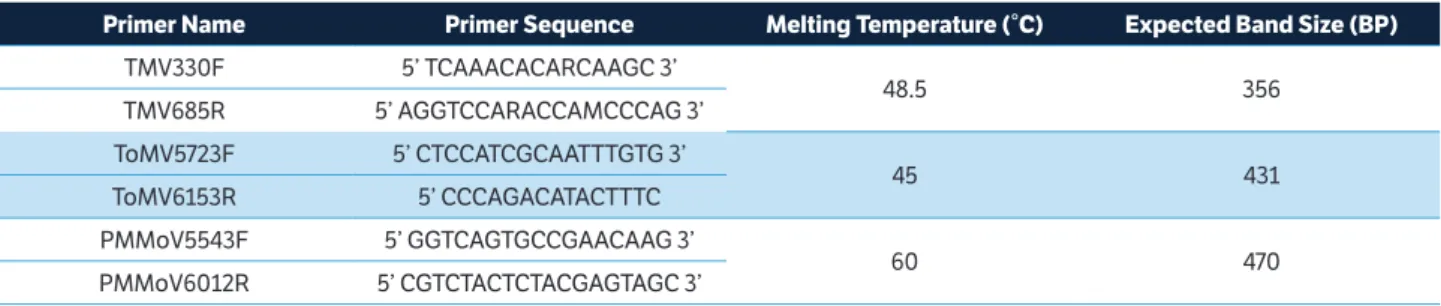
Table 1. Primers used for tobamoviruses detection.
Primer Name Primer Sequence Melting Temperature (°C) Expected Band Size (BP)
TMV330F 5’ TCAAACACARCAAGC 3’
48.5 356
TMV685R 5’ AGGTCCARACCAMCCCAG 3’
ToMV5723F 5’ CTCCATCGCAATTTGTG 3’
45 431
ToMV6153R 5’ CCCAGACATACTTTC
PMMoV5543F 5’ GGTCAGTGCCGAACAAG 3’
60 470
produced by infected plants. However, the same did not happen with the occurrence of infected seeds produced by pepper plants infected with PMMoV, which in this study was much higher than those found by other authors. Tošić et al. (1980) detected the presence of PMMoV CP in 29% of seeds produced by infected plants and McKinney (1952) detected the virus in only 22% of the seeds harvested from Capsicum frutescens infected plants. Probably, as observed in other viruses, such transmission depends on the viral isolate and susceptibility of the cultivar of sweet pepper in addition to environmental conditions, which can play a significant role in viral multiplication and consequent systemic invasion of the plant.
Seed Production Artificially Contaminated with Tobamovirus
The presence of viral particles in the artificially inoculated seeds of pepper and tomato plants was checked by DAS-ELISA, and the average absorbance obtained in this experiment is discribed in Table 2. There was no significant statistical difference in absorbance of all treatments, indicating that for obtaining pepper seeds contaminated with PMMoV and tomato seeds contaminated with TMV and ToMV, immersion in the extract containing the virus for only 1 second was enough for the adherence of viral particles to the surface of seeds.
Extracts of tomato seeds infected with TMV showed a higher absorbance in DAS-ELISA when compared to
pepper seeds. This should be related to the presence of trichomes, which occurs only in tomato seeds. Other authors have found that these trichomes provide greater adhesion of pathogens, especially bacteria, on the seed surface (Cevallos-Cevallos et al. 2012). The same must have happened with the viral particles.
The methodology for inoculation and production of the artificially contaminated seeds used in this study was shown to be an excellent tool to assist future research aiming at the detection of tobamovirus in tomato and pepper seeds.
Sensitivity of Biological, Serological and Molecular Techniques in the Detection of TMV and ToMV in Tomato Seeds and Pmmov CP in Pepper
When the seeds with 0.1% of virus contamination were planted in the greenhouse, it was not possible to observe the presence of common symptoms of viral diseases in tomato and pepper seedlings. This may have occurred because the contamination of TMV, ToMV, and PMMoV is external to the seeds, and not always enough to infect the plant during the process of germination and seedling emergence, through an injury due to a wound in the roots (Güsler et al. 2008) or mechanical damage to the seedlings (Broadbent 1976). Damage to seedlings most commonly occurs during the handling in seedling production nursery because after germination the integument remains adhered to the first pair of leaves, from where the virus can be transmitted to the seedling. Therefore,
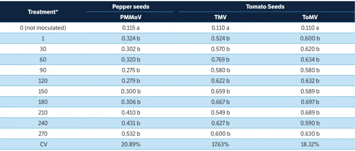
Table 2. Average absorbance obtained in the DAS-ELISA test using pepper seeds, artificially inoculated with Pepper mild motle virus (PMMoV), and tomato seeds artificially inoculated with Tobacco mosaic virus (TMV) and Tomato mosaic virus (ToMV).
Treatment* Pepper seeds Tomato Seeds
PMMoV TMV ToMV
0 (not inoculated) 0.115 a 0.110 a 0.110 a
1 0.324 b 0.524 b 0.600 b
30 0.302 b 0.570 b 0.620 b
60 0.320 b 0.769 b 0.634 b
90 0.275 b 0.580 b 0.580 b
120 0.279 b 0.622 b 0.632 b
150 0.300 b 0.659 b 0.589 b
180 0.306 b 0.667 b 0.697 b
210 0.410 b 0.549 b 0.689 b
240 0.431 b 0.627 b 0.590 b
270 0.532 b 0.600 b 0.630 b
CV 20.89% 17.63% 18.32%
although the symptoms may not be noticeable in seedlings, this does not mean that the virus cannot be spread during transplanting to the field.
In DAS-ELISA it was possible in all analyzed lots to detect the TMV incidence up to 1:170 and ToMV up to 1:200. Probably this small difference was due more to a variation in the antiserum sensitivity than the concentration of the virus itself, since these results were repeated. In pepper seeds, PMMoV was detected until the incidence of 1:140.
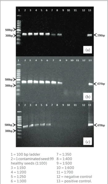
The IC-RT-PCR, as expected, showed greater sensitivity in detecting these tobamoviruses. Both TMV and ToMV viruses could be detected in seeds with the incidence rate of 1:400 and PMMoV was detected in pepper seeds up to a rate of 1:300. The Fig. 1 shows the bands that were amplified
in the PCR reaction: 356 bp to TMV, 431 bp for ToMV, and 471 bp for PMMoV. The lower sensitivity for detection of tobamovirus observed in pepper seeds can be explained by the higher adhesion of viral particles on the surface of tomato seeds due to existing trichomes on their surface, as observed when evaluating the absorbance obtained from virus-contaminated seeds of tomatoes and peppers in the earlier tests. The sensitivity of this technique can be further increased when combined with real time PCR. Yang et al. (2012) demonstrated that the IC-RT-qPCR technique was capable of detecting TMV-infected soil at a dilution of 1.106, while the DAS-ELISA only detected the virus up to
the dilution of 1.102.
The usefulness of knowing the sensitivity of these techniques is to provide subsidies that can decrease the cost of routine analysis. In countries like Brazil, where virtually all the materials and reagents used are imported, there is a great demand for cheaper and affordable tests to producers and importers of seeds for planting. The combination of seeds for analysis does not allow the calculation of incidence of infected seeds, but allows verifying the presence or absence of viruses. In our study, tests conducted with RT-PCR using the Trizol protocol for obtaining the total RNA from seeds were ineffective for detecting all tobamoviruses, even when 100% of infected seeds were employed. This should be related to the possible existence of compounds that could act as inhibitors of the enzymes responsible for the synthesis of viral cDNA or amplification of this cDNA in the PCR reaction (Schrader et al. 2012). The seeds are mostly rich in phenolic compounds, mainly phenolic acids, tannins, and coumarins (Veronezi and Jorge 2012). The IC-RT-PCR technique has as an advantage the fact that crushing of the seeds for the viral RNA extraction is unnecessary, since only the viral particles that lie on the outside are captured by the specific antiserum (Mulholland 2009). Another advantage of IC-RT-PCR, compared to RT-PCR, would be the speed of testing. The RNA obtained by IC-RT-PCR is performed in fewer steps when compared with RT-PCR (Kamenova and Adkins 2004; Chikh-Ali and Karasev 2015).
Diagnosis of Tobamoviruses in Lots of Tomato and Pepper Seeds
All lots of 1000 seeds containing one seed, infected naturally and/or artificially, which were divided into 30 sub lots, when analyzed by DAS-ELISA, were positive.
Figure 1. IC-RT-PCR detection of: (a) Tobacco mosaic virus (TMV); (b) Tomato mosaic virus (ToMV) in lots of tomato seeds and (c) Pepper mild mottle virus (PMMoV) in lots of pepper seeds.
1 = 100 bp ladder 2 = 1 contaminated seed:99 healthy seeds (1:100) 3 = 1:150
4 = 1:200 5 = 1:250 6 = 1:300
7 = 1:350 8 = 1:400 9 = 1:500 10 = 1:600 11 = 1:700
12 = negative control 13 = positive control.
(a)
(b)
Therefore, one out of the 30 sub lots placed in 30 wells was positive, indicating the presence of the contaminated seed. As noted above, the sensitivity of this technique allows contaminated seeds in the proportion of 1:33 or 1:34 to be efficiently detected, since the sensitivity of the technique was 1:170 for TMV, 1:200 for ToMV and 1:140 for PMMoV. In this experiment we chose to use the fractionation in 30 batches, which allows for analysis of up to three lots of 1000 seeds in each ELISA microplate, obtaining an average absorbance greater than 0.5 for positive samples. Considering that naturally infected seeds could present a lower concentration of particles depending on the condition of the infected plant such as time of infection, resistance, etc., it was considered that the division into 30 batches would be able to provide reliable and reproducible results. It is known, however, that if necessary, the division into a larger number of batches can still provide a good idea about the quality of seeds.
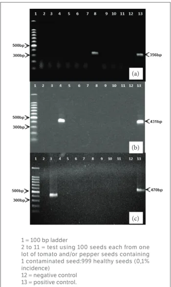
In IC-RT-PCR, using lots of 100 seeds was possible to detect one contaminated seed (naturally and/or artificially contaminated) of tomato with TMV and ToMV and of pepper with PMMoV. One of the 10 tests employing 100 tomato or pepper seeds was positive in all analyzed lots by any of the tobamoviruses tested. Electrophoretic analysis in 0.7% agarose gel showed the amplification of a band with sizes specific to each virus, as previously mentioned (Fig. 2).
When comparing the diagnostic methods tested between each other, it was observed that the RT-PCR and the biological method were the most unsuitable for the detection of seeds infected with tobamovirus. Although both DAS-ELISA and IC-RT-PCR have been effective, DAS-ELISA was less sensitive. However, using a smaller number of seeds per batch to be analyzed, its performance is quite satisfactory. The chosen test can then be limited to hand labor and infrastructure available resources, since both require the use of specific antibodies for each virus. Due to the fact that the virus does not possess a curative control point to the need to take all the possible measures for preventive control, in order to prevent or delay as much as possible the introduction of the virus in the field, the use of virus-free seeds becomes a major control measure to prevent an epidemic (Silva et al. 2011). This study proposes an efficient methodology for tobamovirus detection in tomato and pepper seeds, which allows considerable savings in the use of labor and inputs, reducing the cost of analysis without losing the quality of the desired results.
Figure 2. IC-RT-PCR detection of (a) Tobacco mosaic virus (TMV); (b) Tomato mosaic virus (ToMV) in lots of tomato seeds and (c) Pepper mild mottle virus (PMMoV) in lots of pepper seeds.
CONCLUSION
The RT-PCR technique and biological tests were not reliable for the tobamoviruses detection in tomato and pepper seeds.
The studies performed allowed us to determine the sensitivity of the DAS-ELISA and IC-RT-PCR tests to detect TMV and ToMV in tomato and pepper seeds, and PMMoV in pepper seeds.
Based on the tests’ sensitivity, rather than employing a seed per test, performing 1000 tests, it was possible to suggest a combination of seeds from a lot with 1000 seeds, containing the minimum of one infected seed, as follows:
1 = 100 bp ladder
2 to 11 = test using 100 seeds each from one lot of tomato and/or pepper seeds containing 1 contaminated seed:999 healthy seeds (0,1% incidence)
12 = negative control 13 = positive control.
(a)
(b)
• in DAS-ELISA, 30 tests containing the extracts of 33 to 34 seeds per well can be done with high sensitivity, allowing reducing 33 times the number of DAS-ELISA tests performed;
• in IC-RT-PCR, the extracts of 100 seeds per test can be employed, performing only 10 tests without losing the efficiency, which allows reducing 100 times the number of tests performed.
Besides ensuring reliable results with excellent efficiency and repeatability, a significant saving in the cost of routine analysis can be achieved, by reducing the number of tests necessary to analyze one lot containing 1000 seeds.
ORCID IDS
J. E. M. Almeida
https://orcid.org/0000-0001-8118-5630
A. R. Figueira
https://orcid.org/0000-0002-6907-1160
P. S. G. Duarte
https://orcid.org/0000-0002-7873-785X
M. A. Lucas
https://orcid.org/0000-0001-9536-8470
N. E. Alencar
https://orcid.org/0000-0002-3121-6707
AFGC protocols (2002). Total RNA isolation;[accessed 2012 April
4]. http://www. arabidopsis.org/portals/masc/AFGC/RevisedAFGC/ site2RnaL.htm#isolation
Berniak, H., Malinowski, T. and Kamińska, M. (2009). Comparison of ELISA and RT-PCR assays for detection and identification of
Cucumber mosaic virus (CMV) isolates infecting horticultural crops in Poland. Journal of Fruit and Ornamental Plant Research, 17, 5-20.
Broadbent, L. (1976). Epidemiology and control of Tomato mosaic
virus. Annual Review of Phytopathology, 14, 75-96. https://doi. org/10.1146/annurev.py.14.090176.000451
Candemir, F., Kutluk-Yilmaz, N. D. and Gülser, C. (2012). The effect of tobacco waste application on Tobacco mosaic virus (TMV) concentration in the soil. Žemdirbystė=Agriculture, 99, 99-104.
Cevallos-Cevallos, J. M., Gu, G., Danyluk, M. D. and van Bruggen, A. H. C. (2012). Adhesion and splash dispersal of Salmonella enterica Typhimurium on tomato leaflets: effects of rdar morphotype and trichome density. International Journal of Food Microbiology, 160,
58-64. https://doi.org/10.1016/j.ijfoodmicro.2012.09.021
Cezar, M. A., Krause-Sakate, R., Pavan, M. A. and Costa, C. P.
(2009). Evaluation of resistance of Capsicum spp. genotypes to
tobamovirus.Summa Phytopathologica, 35, 39-43. http://dx.doi.
org/10.1590/S0100-54052009000100006
Chikh-Ali, M. and Karasev, A. V. (2015). Immunocapture-multiplex RT-PCR for the simultaneous detection and identification of plant
viruses and their strains: study case, Potato Virus Y (PVY). In C.
Lacomme (Ed.), Plant Pathology. Methods in Molecular Biology,
REFERENCES
vol. 1302 (p. 177-186). New York: Humana Press. https://doi.
org/10.1007/978-1-4939-2620-6_14
Chitra, T. R., Prakash, H. S., Albrechtsen, S. E., Shetty, H. S. and Mathur, S. B. (1999). Infection of tomato and bell pepper by ToMV and TMV at different growth stages and establishment of virus in seeds. J. Plant Pathology, 81, 123-126.
Duarte, L. M. L., Alexandre, M. A. V., Rivas, E. B., Cattai, M. B., Soares, R. M., Harakava, R. and Fernandes, F. M. C. (2007). Phylogenetic
analysis of Tomato mosaic virus from Hemerocallissp. and Impatiens
hawker. Summa Phytopathologica, 33, 409-413. http://dx.doi. org/10.1590/S0100-54052007000400016
Eiras, M., Chaves, A. L. R., Moreira, S. R., Araujo, J. and Colariccio, A. (2004). Characterization of a non-L3 gene-resistance breaking
Pepper mild mottle virus isolate in Capsicum. Fitopatologia Brasileira,
29, 670-675. http://dx.doi.org/10.1590/S0100-41582004000600014
Ferreira, D. F. (2011). SISVAR: a computer statistical analysis system.
Ciência e Agrotecnologia, 35, 1039-1042. http://dx.doi.org/10.1590/
S1413-70542011000600001
Genda, Y., Sato, K., Nunomura, O., Hirabayashi, T. and Tsuda, S.
(2011). Immunolocalization of Pepper mild mottle virus in developing
seeds and seedlings of Capsicum annuum. Journal of General Plant
Pathology, 77, 201-208. https://doi.org/10.1007/s10327-011-0307-0
and RT-PCR. Crop Protection, 86, 31- 41. https://doi.org/10.1016/j. cropro.2016.03.007
Gülser, C., Yılmaz, K. N. and Candemir, F. (2008). Accumulation of tobacco mosaic virus (TMV) at different depths clay and loamy sand textural soils due to tobacco waste application. Environ Monit
Assess, 146, 235-242. https://doi.org/10.1007/s10661-007-0075-7
Kamenova, I. and Adkins, S. (2004). Comparison of detection methods for a novel tobamovirus isolated from Florida Hibiscus.
Plant Disease, 88, 34-40. https://doi.org/10.1094/PDIS.2004.88.1.34
Kumar, S., Udaya Shankar, A. C., Nayaka, S. C., Lund, H. S. and Prakash, O. S. (2011). Detection of Tobacco mosaic virus and Tomato mosaic virus in pepper and tomato by multiplex RT–
PCR. Letters in Applied Microbiology, 53, 359-363. https://doi.
org/10.1111/j.1472-765X.2011.03117.x
Lee, B. Y., Lim, H. R., Choi, J. Y. and Ryu, K. H. (2004). Development of molecular detection of three species of seed-transmissible virus
useful for plant quarantine. The Plant Pathology Journal, 20,
302-307. http://doi.org/10.5423/PPJ.2004.20.4.302
Liu, H. W., Luo, L. X., Li, J. Q., Liu, P. F., Chen, X. Y. and Hao, J. J. (2014). Pollen and seed transmission of Cucumber green mottle
mosaic virus in cucumber. Plant Pathology, 63, 72-77. https://doi.
org/10.1111/ppa.12065
Mckinney H. H. (1952). Two strains of Tobacco-mosaic virus, one of which is seed borne in an etch-immune pungent Pepper. Plant Disease Reporter, 36, 184-187.
Moreira, S.R., Eiras, M., Chaves, A.L.R., Galleti, S.R. and Colariccio, A.
(2003). Characterization of a new Tomato mosaic virus strain isolated
from tomato in the State of São Paulo, Brazil. Fitopatologia Brasileira,
28, 602-607. http://dx.doi.org/10.1590/S0100-41582003000600004
Mulholland, V. (2009). Immunocapture-PCR for plant virus detection. In R. Burns (Ed.), Plant Pathology. Methods in Molecular
Biology, vol. 508 (p. 183-192). Totowa: Humana Press. https://doi.
org/10.1007/978-1-59745-062-1_15
Schrader, C., Schielke, A., Ellerbroek, L. and Johne, R. (2012). PCR inhibitors – occurrence, properties and removal.
Journal of Applied Microbiology, 113, 1014-1026. https://doi.
org/10.1111/j.1365-2672.2012.05384.x
Sevik, M. A. and Kose-Tohumcu, E. (2011). The ELISA analysis
results in tomato (Lycopersicon esculentum Mill.) seed health testing
for Tobacco mosaic virus. Žemdirbystė=Agriculture, 98, 301-306.
Silva, P. P., Freitas, R. A. and Nascimento, W. M. (2011). Detection of Tomato mosaic virusin Tomato Seed and Treatment by
Thermotherapy. Acta Horticulturae, 917, 303-307. http://doi.
org/10.17660/ActaHortic.2011.917.43
Silva, R. M., Souto, E. R., Pedroso, J. C., Arakava, R., Almeida, A. M. R., Barboza, A. A. L. and Vida, J. B. (2008). Detection and identification of TMV infecting tomato under protected cultivation in Paraná State. Brazilian Archives of Biology Technology, 51,
903-909. http://dx.doi.org/10.1590/S1516-89132008000500005
Tošić, M., Šutić, D. and Pešić, Z. (1980). Transmission of Tobbacco
mosaic virus through pepper (Capsicum annum L.) seed. Journal of
Phytopathology, 97, 10-13. https://doi.org/10.1111/j.1439-0434.1980.
tb04595.x
Van Winckel, A. and Gcypens, M. (1965). Inactivering van Tabakmosaiekvirus in de grond. Parasitica, 11, 124-137.
Veronezi, C. M. and Jorge, N. (2012). Bioa ctive compounds
in lipid fractions of pumpkin (Cucurbitasp) seeds for use
in food. Journal of Food Science, 77, C653-C657. https://doi.
org/10.1111/j.1750-3841.2012.02736.x
Yang, J. G., Wang, F. L., Chen, D. X., Shen, L. L., Qian ,Y. M., Liang, Z. Y, Zhou, W. C. and Yan, T. H. (2012). Development of a one-step immunocapture Real-Time RT-PCR assay for detection of
Tobacco Mosaic Virusin soil. Sensors, 12, 16685-16694. http://doi.