The Effect of Post-Exercise Cryotherapy on
Recovery Characteristics: A Systematic
Review and Meta-Analysis
Erich Hohenauer1,4*, Jan Taeymans2,3,5☯, Jean-Pierre Baeyens3,4,6☯, Peter Clarys3☯, Ron Clijsen1,3,4☯
1Department of Business Economics, Health and Social Care, University of Applied Sciences and Arts of Southern Switzerland, Landquart / Manno, Switzerland,2Health Department, Bern University of Applied Sciences, Berne, Switzerland,3Faculty of Physical Education and Physiotherapy, Vrije Universiteit Brussel, Brussels, Belgium,4University College Physiotherapy Thim van der Laan, Landquart (GR), Switzerland, 5Faculty of Medicine and Health Sciences, University of Antwerp, Antwerp, Belgium,6Faculty of Applied Engineering, University of Antwerp, Antwerp, Belgium
☯These authors contributed equally to this work. *[email protected](EH)
Abstract
The aim of this review and meta-analysis was to critically determine the possible effects of different cooling applications, compared to non-cooling, passive post-exercise strategies, on recovery characteristics after various, exhaustive exercise protocols up to 96 hours (hrs). A total of n = 36 articles were processed in this study. To establish the research ques-tion, the PICO-model, according to the PRISMA guidelines was used. The Cochrane’s risk of bias tool, which was used for the quality assessment, demonstrated a high risk of perfor-mance bias and detection bias. Meta-analyses of subjective characteristics, such as delayed-onset muscle soreness (DOMS) and ratings of perceived exertion (RPE) and objective characteristics like blood plasma markers and blood plasma cytokines, were per-formed. Pooled data from 27 articles revealed, that cooling and especially cold water immer-sions affected the symptoms of DOMS significantly, compared to the control conditions after 24 hrs recovery, with a standardized mean difference (Hedges’g) of -0.75 with a 95% confidence interval (CI) of -1.20 to -0.30. This effect remained significant after 48 hrs (Hedges’g: -0.73, 95% CI: -1.20 to -0.26) and 96 hrs (Hedges’g: -0.71, 95% CI: -1.10 to -0.33). A significant difference in lowering the symptoms of RPE could only be observed after 24 hrs of recovery, favouring cooling compared to the control conditions (Hedges’g: -0.95, 95% CI: -1.89 to -0.00). There was no evidence, that cooling affects any objective recovery variable in a significant way during a 96 hrs recovery period.
Introduction
For decades, different cryotherapies–such as cold-water immersions (CWI) and ice packs–have
been used for post-exercise recovery in a variety of sports to cope with fatigue and/or
delayed-onset muscle soreness (DOMS) [1]. Exercising at different intensity levels induces various
OPEN ACCESS
Citation:Hohenauer E, Taeymans J, Baeyens J-P, Clarys P, Clijsen R (2015) The Effect of Post-Exercise Cryotherapy on Recovery Characteristics: A Systematic Review and Meta-Analysis. PLoS ONE 10(9): e0139028. doi:10.1371/journal.pone.0139028
Editor:Stephen E Alway, West Virginia University School of Medicine, UNITED STATES
Received:January 14, 2015
Accepted:September 7, 2015
Published:September 28, 2015
Copyright:© 2015 Hohenauer et al. This is an open access article distributed under the terms of the Creative Commons Attribution License, which permits unrestricted use, distribution, and reproduction in any medium, provided the original author and source are credited.
Data Availability Statement:All relevant data are within the paper and its Supporting Information files.
Funding:The authors received no specific funding for this work.
degrees of fatigue to the musculoskeletal, nervous and metabolic systems [2]. Exercise is also associated with microscopic tears in the muscle tissue, commonly described as exercise induced
muscle damage, leading to DOMS [3].
In sports medicine, cryotherapy, as a post-exercise intervention, has been investigated by means of subjective ratings of DOMS and general ratings of perceived exertion (RPE), objective attenuations of blood plasma markers such as creatine-kinase (CK) and lactate-levels, or blood
plasma cytokines encompassing Interleukines (IL) and C-reactive protein (CRP) [1,2,4–37].
Cryotherapy is commonly described as a procedure to relieve pain and to decrease
inflamma-tion in musculoskeletal problems [38]. The mechanism of cold therapy for recovery after
exer-cise is predominantly attributed to its vasoconstrictive effect, which reduces the inflammation
reactions through a decrease of the cell metabolism [39]. In sports, cooling represents a
consid-erable therapy for athletes. A recently published meta-analysis from Leeder et al. (2012) showed that CWI is an effective strategy to reduce the symptoms of DOMS following a range
of strenuous and exhaustive exercise types [40]. These results are in line with those of Bleakley
et al. (2012). These authors recently demonstrated significant differences in their meta-analysis,
favouring cooling compared to passive interventions up to 96 hrs [3]. In their review article,
Banfi et al. (2010) recently indicated, that whole-body cooling (WBC) is a possible application to treat muscle injuries, muscle syndromes of overuse and to reduce the recovery time between training sessions. The WBC treatment did not induce modifications of biochemical and hae-matological parameters. The authors concluded that WBC is not harmful and does not induce
general or specific negative effects in athletes [38]. However, the negative effects of cooling and
the area of application must also be critically examined. Takagi et al. (2011) recently showed, that applying ice bags for 20 minutes (min) after a crush injury to the extensor digitorum longus muscle in rats, led to a delay in muscle regeneration, impairment of muscle regeneration and redundant collagen synthesis due to reduced calpain activity. This enzyme is necessary for the primary reaction of muscle-degeneration. A decreased number of macrophages and sup-pressed inflammation after local cooling applications might be closely related to the retarded
healing processes [41–43]. The effects of post-exercise cryotherapy on recovery dependent
var-iables, such as DOMS, blood plasma markers, blood plasma cytokines, different performance parameters and RPE in non-injured participants are still unclear. A lack of appropriate recov-ery may as a result prevent the athlete of training at a required intensity or, moreover, of achieving the required load during the consecutive training session(s). Furthermore, full
recov-ery is necessary for optimal competition performance [44]. The aim of this study was to
criti-cally determine the effects of different cooling applications compared to non-cooling, passive post-exercise strategies by means of recovery characteristics after various exhaustive exercise protocols up to 96 hours.
Research Question and Search Algorithm
The research question was defined by the PICO-model in accordance with the Preferred
Reporting Items for Systematic Reviews and Meta-Analyses (PRISMA) statement [45]:
analogue scales (VAS), BORG scales or Likert-scales. RPE was rated by various scaling systems, such as BORG scales or VAS. CK-levels, lactate-levels, CRP and IL-6 were defined as objective recovery characteristics in this study. The rationale for inclusion of these outcome variables was comparability. Numerous researchers who tried to identify a subjective and objective recovery profile after exhaustive exercises previously used these variables in their studies.
Methods
Literature Search Strategies and Data Sources
A systematic search was accomplished electronically between October 2013 and August 2014 in the MEDLINE (PubMed), SportDiscus and PEDro databases according to the PRISMA
statement [45]. The following key words and their combinations were used without using any
automatic filters:“Exercise AND Cold OR Cooling OR Cryotherapy AND Recovery OR
Recov-ery Strategy OR RecovRecov-ery Modality”. Seven additional articles were included after having read
the reference list of the eligible studies (n = 49).
Study Selection Criteria
Inclusion criteria for the articles were: (1) the study design was randomized or quasi random-ized into an intervention group and a non-cooling, passive control group; (2) the cold therapy had to be an external application form without combinations, focused on the mentioned out-come variables after the exercise bouts; (3) the cold interventions had to be compared to at
least one control condition; (4) the outcome variables had to be measured immediately (0–24
hrs), and / or 24 hrs, and / or 48 hrs, and / or 72 hrs and/or 96 hrs post-exercise; (5) the data of the outcome variables had to be processed in the articles; (6) the human volunteers had to be healthy without any physical infirmity; (7) the human volunteers could be of any athletic train-ing status; (8) there were no sex-defined inclusion or exclusion criteria; (9) at least one of the subjective or objective recovery characteristics had to be evaluated in the articles; (10) the cold therapy had to be applied within one hour after the end of the exercise protocol; (11) all exhaustive exercise types or muscle damaging protocols were accepted; (12) English and Ger-man language restrictions. Articles, which did not meet these criteria were excluded from this
study.Fig 1shows the systematic search strategy and selection process.
Data Analysis
General characteristics of the studies and results of the individual studies were extracted inde-pendently by two researchers (EH, RC). In case of a lack of information, the statistics were extrapolated from the given figures and graphs. In case of disagreement, consensus was sought by involving a third researcher (JPB). Meta-analyses with a random-effects model (specified a
priori), allowing for possible between–studies heterogeneity, were used to examine the overall
effect size of cooling applications on the subjective and objective recovery characteristics. The inversed variance method was used to calculate the weighting factors for the included,
individ-ual study results. The different individindivid-ual study effect sizes were standardized using Cohen’s d,
by dividing the effect sizes by the pooled standard deviations, as proposed by Borenstein et al.
(2009) [46]. The effect sizes were expressed as Hedges’g, to account for possible overestimation
of the true population effect size in small studies. The magnitude of“g”was interpreted
accord-ing to the followaccord-ing scale [47]:<.20 = negligible effect, .20 - .49 = small effect, .50 - .79 =
moder-ate effect,.80 = large effect. The Cochran’s Q statistics and its corresponding p-Value, as well
as the I2,were calculated to assess across studies heterogeneity and its degree respectively [48].
Fig 1. Flow-chart describing the systematic review procedure.
analyses were performed to assess the potential influence of different co-variates, such as cool-ing modality (CWI, WBC, cold air, cold pack), sex (discrete data) and coolcool-ing temperature (continuous data) on the pooled effect size. Two of the 36 studies used extreme low cooling
temperatures (up to -110°C) [1,8]. To assess the robustness of the overall weighted estimate
against these extreme cooling temperatures, a sensitivity analysis was conducted. Publication bias was assessed using visual analysis of the funnel plot and formal testing for funnel plot
asymmetries using the“trim and fill”and“fail and safe”algorithms [46]. The Comprehensive
Meta-Analysis 2 software (CMA- Version 2 Professional, Biostat Inc., Englewood, USA) was used for the calculations of the weighted overall estimates, the corresponding 95% confidence intervals (95% CI) and to establish the forest plots.
Data Synthesis
In this article the subjective variables under investigation were DOMS and RPE. The objective outcomes of interest for the meta-analyses were blood plasma markers (CK, blood/muscle lac-tate, LDH) and blood plasma cytokines (IL-6, CRP). Meta-analyses were conducted using means ± standard deviations (SD) or standard errors (SE).
Risk of Bias
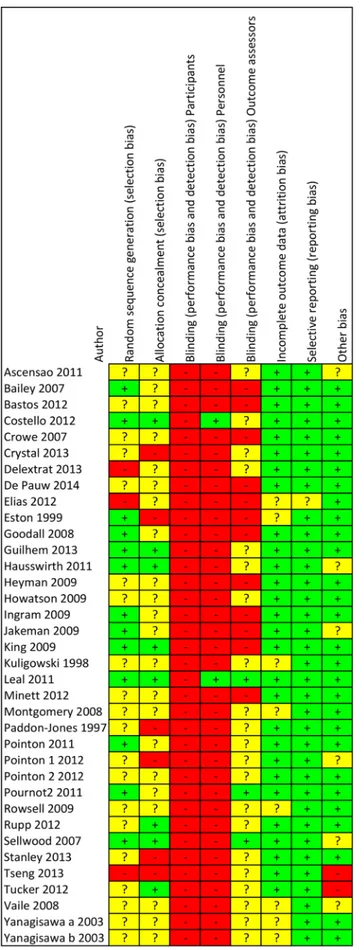
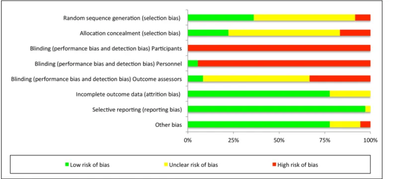
The systematic error of the 36 articles was assessed using the Cochrane’s risk-of-bias tool. [49].
Two researchers (EH, RC) independently scored each trial for the risk of bias. In case of dis-agreement, a third researcher (PC) rated the questionable item and agreement was sought by consensus. Each study was graded for the following domains: random sequence generation, allocation concealment, blinding participants, blinding personnel, blinding outcome assessors,
incomplete outcome data, selective reporting and other bias. They were rated“low”(+) if the
risk of bias for this item was low, or“high”(-) if the risk of bias for this item was high. In case
of insufficient reported information, or information which made an interpretation
question-able and thus unclear, the risk of bias item was rated as“unclear”(?).
Results
Risk of Bias Analysis
The risk of bias analysis demonstrated a high risk of bias for the blinding procedures. Blinding
of the participants was only described in two studies [1,9]. Personnel blinding was also
described in two studies [9,30] only. Blinding of outcome assessors was only found in two
studies [29,31]. The selection bias ratings stayed unclear because of insufficient or unclear
information. A low risk of reporting bias and other bias could be observed throughout the stud-ies. The full details of the risk of bias analysis for all and individual single studies can be
observed in Figs2and3.
Study and Population Characteristics
A total of n = 36 articles met the inclusion criteria. These trials were processed in the present article as sources of primary research data, while the systematic review articles and meta-analy-ses were used in the introduction and the discussion part. Characteristics of the included
stud-ies are summarized inTable 1. The total study population of all selected articles comprised 574
healthy volunteers with an unequal distribution of sex. The selected articles comprised 412 male and 72 female volunteers. In three articles, with a total population of 90 male and female
volunteers, it was not possible to identify the sex distribution [23,30,31]. Four studies included
Fig 2. Risk of bias graph for each included study.
In one study, female and male were equally distributed [16]. Throughout all studies, the mean
sample size was 16 volunteers (range = 6 to 40 volunteers) [9,31]. The mean age of the total
study population was 22 years (yrs). In one article, the age of the study population was not
mentioned [33]. There were 18 randomized controlled trials [1,4,6,7,10,12,14,15,18,19,23,
25,26,29–31,34,35] and 18 randomized crossover trials [2,5,8,9,11,16,17,20–22,24,27,
28,32,33,36,37,50]. The experimental trials were conducted in Belgium (n = 2), France
(n = 3), Ireland (n = 1), Portugal (n = 1), United Kingdom (n = 6), Australia (n = 14), Brazil (n = 2), United States (n = 4), Taiwan (n = 1) and Japan (n = 2). Eight studies involved
well-trained/elite volunteers [4,7–9,17,29,30,50]. Six of these studies included soccer / football
players [4,7,9,17,29,30]. One study worked with well-trained male runners [8] and another
study chose well-trained female climbers [50]. The remaining 28 studies used healthy / trained
volunteers [1,2,5,6,10–12,14–16,18–28,31–37]. Detailed information about the training
sta-tus can be observed inTable 1.
Characteristics of the Exercise Protocols
The exercise protocols encompassed soccer-games or tournaments [4,7]. Six authors used
cycle ergometer tests [5,9,12,32,36,37]. Two of these cycle tests were sprint tests, while the
remaining four were endurance tests [5,9]. A total of n = 14 studies used functional exercises
to create the pre-cooling load. This involved ten sets of ten counter movement jumps within
ten min [6], 100 eccentric contractions of the knee extensors in a 20 set design [1], six sets of 25
maximal concentric and eccentric contractions for the knee extensors [2], five sets of ten
eccen-tric contractions of the knee extensors [31], seven sets of ten eccentric knee extensions on a leg
press [33], five sets of twelve respectively 20 eccentric ankle plantar flexion [34,35] five sets of
20 drop jumps [19,20], eight sets of five maximal eccentric contractions of the upper arm
flex-ors, respectively extensors [18], 50 eccentric contractions of the elbow flexors [23], eight sets of
eight eccentric contractions of the elbow flexors [26], three sets of maximum (max) 20
Fig 3. Risk of bias summary for all included studies.
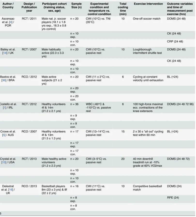
Table 1. Summary of the used studies for the meta-analysis. A Author / Country Design / Publication year Participant cohort (training status, sex, age) Sample Size (n) Experimental condition and temperature vs. control condition Total cooling time (min)
Exercise Intervention Outcome variables and time of measurement post exercise (hrs) Ascensao
et al. [4] / POR
RCT / 2011 Male nat. jr. soccer players (18.1±1.8
yrs exp., 18.3±0.8
yrs control)
n = 20 CWI (10°C) vs. TNI (35°C)
10 One-off soccer match DOMS (24 48)
n = 10 exp.
CK (24 48)
n = 10 con.
CRP (24 48)
Bailey et al. [14] / UK
RCT / 2007 Male habitually active (22.3±3.3 yrs)
n = 20 CWI (10°C) vs. passive rest
10 Loughborough intermittent shuttle test
DOMS (24 48)
n = 10 exp.
CK (24 48)
n = 10 con. Bastos et al.
[36] / BRA
RCO / 2012 Male active subjects (21±2 yrs)
n = 20 CWI (11±2°C) vs. passive rest
6 Cycling at constant velocity until exhaustion
BL (<24)
n = 20 exp. n = 20 con. Costello et al.
[1] / IRL
RCT / 2012 Healthy volunteers 4f & 14m (21.2±2.1 yrs)
n = 36 WBC (-60°C & -110°C) vs. passive rest
6 100 high-force maximal ecc. contractions of the knee extensors
DOMS (24 48 72 96)
n = 9 exp. n = 9 con. Crowe et al.
[5] / AUS
RCO / 2007 Healthy volunteers 4f & 13m (21.5±1.3 yrs)
n = 17 CWI (13–14°C) vs. passive rest
15 2 x 30 s "all out" cycling test within 60 min
BL (<24)
n = 17 exp. n = 17 con. Crystal et al.
[15] / USA
RCT / 2013 Male healthy active volunteers (21.2±2.3 yrs)
n = 20 CWI (3–5°C) vs. passive rest
20 40 min downhill treadmill run at -10% grade at 60% VO2max
DOMS (24 48 72)
n = 10 exp. n = 10 con. Delextrat
et al. [16] / UK
RCO / 2013 Basketball players 8m (23±3 yrs) & 8f (22±2 yrs)
n = 16 CWI (11°C) vs. passive rest
10 Competitive basketball match
DOMS (24)
n = 8 exp.
RPE (24)
n = 8 con. B
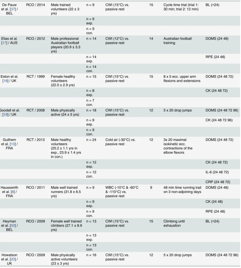
Table 1. (Continued)
De Pauw et al. [37] /
BEL
RCO / 2014 Male trained volunteers (22±3
yrs)
n = 9 CWI (15°C) vs. passive rest
15 Cycle time trial (trial 1: 30 min; trial 2: 12 min)
BL (<24)
n = 9 exp. n = 9 con. Elias et al.
[17] / AUS
RCO / 2012 Male professional Australian football players (20.9±3.3 yrs)
n = 14 CWI (12°C) vs. passive rest
14 Australian football training
DOMS (24 48)
n = 14 exp.
RPE (24 48)
n = 14 con. Eston et al.
[18] / UK
RCT / 1999 Female healthy volunteers (22.0±2.9 yrs)
n = 15 CWI (15°C) vs. passive rest
15 8 x 5 ecc. upper arm
flexions and extensions
DOMS (24 48 72)
n = 8 exp.
CK (24 48 72)
n = 7 con. Goodall et al.
[19] / UK
RCT / 2008 Male physically active (24±5 yrs)
n = 18 CWI (15°C) vs. passive rest
12 5 x 20 drop jumps DOMS (24 48 72 96)
n = 9 exp.
CK (24 48 72 96)
n = 9 con. Guilhem
et al. [10] / FRA
RCT / 2013 Male healthy volunteers (25.2±1.1 yrs in exp., 23.9±1.4 yrs in con.)
n = 24 Cold air (-30°C) vs. passive rest
12 3x 20 maximal isokinetic ecc. contractions of the elbowflexors
DOMS (24 48 72)
n = 12 exp.
CK (24 48 72)
n = 12 con.
IL-6 (24 48 72)
CRP (24 48 72) Hausswirth
et al. [8] / FRA
RCO / 2011 Male well trained runners (31.8±6.5 yrs)
n = 9 WBC (-10°C & -60°C & -110°C) vs. passive rest
9 48 min time running trail on 3 non-adjoining days
DOMS (24 48)
n = 9 exp.
CK (24 48)
n = 9 con.
RPE (24 48)
Heyman et al. [50] /
BEL
RCO / 2009 Female well trained climbers (27.1±8.9 yrs)
n = 13 CWI (15°C) vs. passive rest
15 Climbing until exhaustion
BL (<24)
n = 13 exp. n = 13 con. Howatson
et al. [20] / UK
RCO / 2009 Male physically active volunteers (23±3 yrs)
n = 16 CWI (15°C) vs. passive rest
12 5 x 20 drop jumps DOMS (24 48 72 96)
eccentric contractions of the elbow flexors with a three min passive recovery period between
the sets and six sets of five eccentric elbow flexor contractions [10,11]. Furthermore, the
exer-cise protocols encompassed various endurance running protocols [8,14,15] and sprint
run-ning protocols [27,28]. Other authors used sport specific training or match exercises. These
involved basketball [16,25], Australian football [17], climbing [50], netball [22], bowling [24],
simulated team sport exercises [21] and exhaustive intermittent exercise tasks [29].Table 1
gives a detailed overview of the conducted exercise protocols.
Characteristics of the Cooling Applications
In the present article, the most common cold therapy application (28 studies) was CWI of the
legs [4–7,9,14–23,25,27–37,50]. Water temperature in the leg immersing studies was 5°C to
10°C in 15 articles [4,6,7,9,14,15,21,22,27–29,31,32,34,35] and 11°C to 15°C in 13 articles
[5,16–20,23,25,30,33,36,37,50]. One author used an arm immersing protocol with a water
temperature of 5°C [26] instead of a leg immersing protocol.
Table 1. (Continued)
n = 16 exp.
CK (24 48 72 96)
n = 16 con. C
Ingram et al. [21] / AUS
RCO / 2009 Male athletes (27.5±6.0 yrs)
n = 11 CWI (10°C) vs. passive rest
10 80 min of simulated team sport exercises
DOMS (24 48)
n = 11 exp.
CK (24 48)
n = 11 con.
CRP (24 48)
Jakeman et al. [6] / UK
RCT / 2009 Female physically active (19.9±0.97 yrs)
n = 18 CWI (10°C) vs. passive rest
10 10 sets of 10 countermovement jumps within 10 min
DOMS (24 48 72 96)
n = 9 exp.
CK (24 48 72 96)
n = 9 con. King et al.
[22] / AUS
RCO / 2009 Female trained netball players (19.5±1.5 yrs)
n = 10 CWI (9.3°C) vs. passive rest (15 min)
10 Simulated netball match DOMS (24)
n = 10 exp.
BL (24)
n = 10 con. Kuligowski
et al. [23] / USA
RCT / 1998 Healthy volunteers, 28m (21.1±3.1 yrs) 28f (20.1±2.1 yrs)
n = 56 CWI (12.8C) vs. passive rest
24 50 ecc. contractions of the elbowflexors
DOMS (24 48 72 96)
n = 14 exp. n = 14 con.
Values are means±SD; CWI = cold water immersion, TNI = thermoneutral immersion, exp. = experimental, con. = control, nat. jr. = national junior, f = female, m = male, ecc. = eccentric, conc. = concentric, DOMS = delayed-onset muscle soreness, RPE = ratings of perceived exhaustion, CK = creatine-kinase, BL = blood lactate, ML = muscle lactate, LDH = lactate dehydrogenase, IL-6 = interleukine-6, CRP = C-reactive protein.
WBC with sequential entering -110°C, -60°C and -10°C cold chambers was used in two
studies [1,8]. One author used a cold air application of -30°C [10]. Local ice cuff/ cold pack or
ice bag applications were used in three studies [2,11,12]. Pointon et al. (2011) used ice cuffs
with 0.5°C, which covered the entire surface of the exercised leg [2]. Tseng et al. (2013) and
Tucker et al. (2012) did not mention the temperature of their cold packs / ice bags, which
cov-ered the exercised legs [11,12]. One author used a mixed method WBC application, comprised
of a towel soaked in cold water (5°C) and worn over the head, neck and shoulders. Addition-ally, the participants had to wear an ice-vest and received ice packs, which were placed on the quadriceps and the hamstrings muscles. The ice-vest and ice packs were kept frozen at -20°C
before application [24].
Characteristics of the Passive Control Interventions
The predominant passive control condition, used in 30 studies, was sitting, standing or resting
supine in a room with comfortable temperatures, ranging from 15°C to 24°C [1,2,5,6,8,10,
12,14–24,26–30,32–37,50]. Another passive control condition, used in three studies, was the use of a thermoneutral immersion (TNI). The volunteers who received this control condition were immersed until the same levels, as described in the experimental group. Ascensao et al. (2011) submerged the volunteers to the iliac crest, Rowsell et al. (2009) to the mesosternal level
and Sellwood et al. (2007) to the anterior iliac crest [4,7,31]. Leal et al. (2011) and Tseng et al.
(2013) used a passive sham and placebo application as their control condition [9,11]. Leal et al.
(2011) used a placebo light emitting diode therapy (PLEDT) as their control condition [9].
Tseng et al. (2013) used a sham application for their control group [11]. Montgomery et al.
(2008) used carbohydrate intake and a stretching protocol for the control group [25]. The
vol-unteers were directed through a standardized programme of ten stretches completed twice for
15 s to the legs and the lower back bilaterally, and consumed a carbohydrate (1 gκγbody mass
-1) snack, a carbohydrate bar (PowerbarTM) and 600 ml of fluid in form of a sports drink
(GatoradeTM). We decided not to exclude this study because of the stretching protocol. In all
studies the control and experimental groups received interventions of the same average duration.
Cooling Effects on Subjective Recovery Characteristics
Cooling Effects on DOMS. We obtained the data from 27 articles which revealed the
effectiveness of cooling on DOMS [1,2,4,6,8,10,11,14–27,29–32,34,35].
Only two of these studies used a pain pressure algometer to measure DOMS [18,26]. Six
articles used a scale rating procedure, ranging from 0–10 [2,4,6,10,24,29], one study a 0–50
scale [1], one study a 0–7 scale [11], one study a 1–12 scale [23], three studies a 1–10 scale [14,
25,32] and two studies a 1–5 scale [34,35]. Three studies used a 10-point Likert scale for their
assessment [21,22,27]. Six studies used a 100 mm scale [8,15–17,30,31] and two studies used
a 200 mm scale [19,20]. All of the above mentioned 27 articles measured the effect of cooling–
compared to the control condition for DOMS–at least 24 hrs after the cooling application. The
pooled results are presented in four subcategories, based on the follow-up time. A
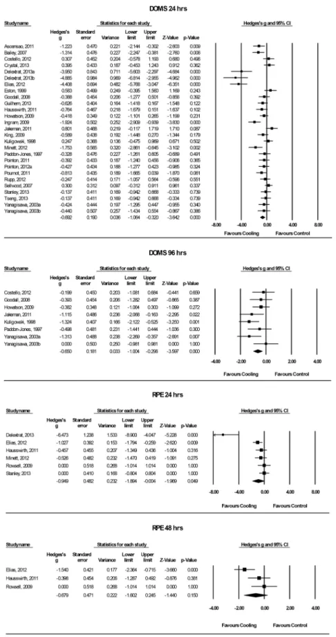
meta-analy-sis from 24 hrs up to 96 hrs could be performed for DOMS. Looking at the results inFig 4, one
can observe that cooling lowered the symptoms of DOMS 24 hrs after the application
signifi-cantly compared to the control conditions (Hedges’g: -0.69, 95% CI: -1.06 to -0.32; Cochran’s
Q: 120.3, df (Q): 26, p<0.001; I2: 78.4%). The classic fail and safe model analysis showed, that
317 negative studies would be needed before alpha increases above the 5% level for DOMS, measured 24 hrs after the cold application, indicating a low risk of publication bias. Cold
Fig 4. Forest plot of the meta-analysis illustrating the comparison of cooling versus control for measurement of DOMS AND RPE.DOMS = delayed-onset muscle soreness, RPE = ratings of perceived exertion.
g: -0.62, 95% CI: -1.00 to -0.25; Cochran’s Q: 71.2, df (Q): 19, p<0.001; I2: 73.3%); (72 hrs:
Hedges’g: -0.18, 95% CI: -0.53 to 0.17; Cochran’s Q: 24.0, df (Q): 11, p = 0.013; I2: 54.3%); (96
hrs: Hedges’g: -0.65, 95% CI: -1.00 to -0.30; Cochran’s Q: 9.2, df (Q): 7, p = 0.238; I2: 24.0%).
Up to 72 hrs, a moderate to very high heterogeneity was observed [48]. The subgroup analysis
according to different cooling modalities, sex or cooling temperature decreased the heterogene-ity somewhat, but could not significantly explain it. The subgroup analysis for different cooling modalities showed that CWI significantly revealed reduced DOMS compared to cold air, cold
pack and WBC up to 96 hrs (24 hrs: Hedges’g: -0.75, 95% CI: -1.20 to -0.30; Cochran’s Q:
110.9, df (Q): 20, p<0.001; I2: 82.0%); (48 hrs: Hedges’g: -0.73, 95% CI: -1.20 to -0.26;
Cochran’s Q: 63.3, df (Q): 14, p<0.001; I2: 77.9%); (72 hrs: Hedges’g: -0.32, 95% CI: -0.74 to
0.10; Cochran’s Q: 18.4 df (Q): 8, p = 0.018; I2: 56.7%); (96 hrs: Hedges’g: -0.71, 95% CI: -1.10
to -0.33; Cochran’s Q: 8.1, df (Q): 6, p = 0.233; I2: 25.7%). These results indicate, that the
find-ings in favour of cooling are dependent on the cooling modality. Subgroup analysis according to sex showed significant different effects, favouring cooling for male participants compared to
female participants, up to 48 hrs (24 hrs: Hedges’g: -0.92, 95% CI: -1.37 to -0.48; Cochran’s Q:
72.4, df (Q): 18, p<0.001; I2: 75.2%); (48 hrs: Hedges’g: -0.76, 95% CI: -1.21 to -0.32;
Cochran’s Q: 47.6, df (Q): 16, p<0.001; I2: 66.9%). To analyse the effect of the different
cool-ing temperatures on the attenuation of the symptoms of DOMS, a meta-regression analysis between these parameters was performed. The meta-regression for the cooling temperature across the recovery time showed no significant correlation between cooling temperature and relieving symptoms of DOMS (48 hrs: Slope = -0.001, 95% CI: -0.007 to 0.004, p = 0.532; 72 hrs: Slope = -0.005, 95% CI: -0.01 to 0.00, p = 0.148). Visual analysis of the regression showed two extreme low temperatures used in the study from Hausswirth et al. (2011) and Costello
et al. (2012) [1,8]. These two authors used cold chambers with the coldest chamber set at
-110°C. After excluding these two authors for the sensitivity analysis, the results still did not correlate significantly after performing the meta-regression (48 hrs: Slope = 0.002, 95% CI: -0.02 to 0.02, p = 0.78; 72 hrs: Slope = -0.005, 95% CI: -0.03 to 0.01, p = 0.552; 96 hrs: Slope = 0.007, 95% CI: -0.07 to 0.08, p = 0.854).
Cooling Effects on RPE. In this composed article, data of n = 6 studies were pooled up to
48 hrs for the analysis of the subjective RPE [7,8,16,17,24,32]. The authors used various
rat-ing systems, such as 0–10 scales [24], 0–100 mm scales [8,16], 1–10 scales [17,32] and 6–20
scales [7]. The subjective ratings of perceived exertion demonstrated significantly different
results favouring cooling compared to the control condition after 24 hrs of recovery (Hedges’
g: -0.95, 95% CI: -1.89 to -0.00; Cochran’s Q: 27.3, df (Q): 5, p<0.001; I2: 81.7%). The
meta-regression according to cooling temperature could not explain this high heterogeneity, nor
show a significant correlation between cooling temperature and Hedges’g (Slope = -0.001, 95%
CI: 0.009 to 0.006, p = 0.707). Two cooling modalities (CWI: n = 4, WBC: n = 2) were used to assess the effect of cooling on RPE. The subgroup analysis according to cooling modality showed no significant differences between the cooling modalities, measured after 24 hrs of
recovery (Cochran’s Q: 0.51, df (Q): 1, p = 0.477). No significant differences between cooling
and the control condition could be observed after 48 hrs of recovery. (Hedges’g: -0.68, 95% CI:
-1.60 to 0.25; Cochran’s Q: 6.2, df (Q): 2, p = 0.044; I2: 67.9%).
Cooling Effects on Objective Recovery Characteristics
Cooling Effects on Blood Plasma Markers. The effect of post-exercise cooling on various
blood plasma markers was analysed in 26 studies [2,4–12,14,18–22,24,27–29,31,33,35–37,
50]. From those, 13 studies measured lactate-levels [5,7,9,12,22,28,29,33,35–37,50] and 19
both lactate- and CK-levels [7,9,27–29,33,35]. Detailed information about the results of the
meta-analysis of the objective recovery variables can be observed inFig 5.
Cooling Effects on Lactate-Levels. Eight studies analysed blood lactate-levels [5,9,22,27,
28,36,37,50], four studies LDH-levels [7,29,33,35] and one study muscle lactate-levels [12].
Data, measured at follow-up times up to 24 hrs and exactly 24 hrs after the exhaustive exercise, was pooled. Within the first 24 hrs of recovery, the volunteers in the cryotherapy group did not
show significant differences compared to the control group. (Hedges’g: -0.28, 95% CI: -0.64 to
0.08; Cochran’s Q: 33.6, df (Q): 14, p = 0.002; I2: 58.3%). The classic fail and safe model analysis
suggested, that 6 studies would be needed to bring alpha above the 5% level, indicating a
poten-tial risk of publication bias. Subgroup analysis according to time (<24 hrs vs. 24 hrs) and sex
could not explain the heterogeneity. However, the cooling temperature and also the diagnostic procedure to obtain blood lactate or muscle lactate results could significantly explain parts of
the heterogeneity (p<0.001, p = 0.003). Tucker et al. (2012) were the only authors who
ana-lysed the lactate-levels by measuring local muscle lactate [12]. After performing the subgroup
analyses, one could observe, that only this study showed significant effects, favouring cooling
compared to blood lactate and LDH measurements (Hedges’g: -2.20, 95% CI: -3.24 to -1.17;
Cochran’s Q: 0, df (Q): 0.0, p = 1.0; I2: 0%). A meta-regression according to cooling
tempera-ture showed no significant differences between cooling and alleviated lactate-levels during the 24 hrs recovery time (Slope = 0.04, 95% CI: -0.00 to 0.08, p = 0.079).
Cooling Effects on CK-Levels. The possible effects of post-exercise cooling on the
CK-lev-els was investigated in 19 studies up to 72 hrs [2,4,6–8,10,11,14,18–21,24,27–29,31,33,
35]. The pooled data showed no significant results, favouring cooling compared to the control
condition during the 72 hrs recovery time (24 hrs: Hedges’g: -0.002, 95% CI: -0.33 to 0.32;
Cochran’s Q: 49.7, df (Q): 18, p<0.001; I2: 63.8%; 48 hrs: Hedges’g: -0.19, 95% CI: -0.46 to
0.08; Cochran’s Q: 18.4, df (Q): 13, p = 0.143; I2: 29.4%; 72 hrs: Hedges’g: -0.10, 95% CI: -0.46
to 0.27; Cochran’s Q: 10.3, df (Q): 7, p = 0.175; I2: 31.7%). The subgroup analyses could not
sig-nificantly explain the high heterogeneity, which was only found after 24 hrs of recovery. A meta-regression according to cooling temperature was performed at 24 hrs. The results showed
no significant slope between cooling temperature and Hedges’g (Slope = -0.003, 95% CI: -0.01
to 0.00, p = 0.388). After excluding the extreme low temperatures, presented in the study from Hausswirth et al. (2011), the results of the meta-regression still did not change significantly
(Slope<-0.000, 95% CI: -0.01 to 0.01, p = 0.992) [8]. No publication bias could be observed
during the visual analysis of the funnel plot.
Cooling Effects on Blood Plasma Cytokines. Ten studies used plasma cytokines (IL-6
and CRP)–amongst other–as a recovery variable [2,4,7,10,11,21,24,27,28,33]. Four studies
measured the IL-6 levels up to 72 hrs [7,10,11,33] while seven studies measured the CRP
lev-els up to 48 hrs [2,4,10,21,24,27,28].
There were no significant main effects, favouring cooling compared to the control
condi-tions during the 72 hrs recovery period, according to the IL-6 levels (24 hrs: Hedges’g: -0.33,
95% CI: -0.76 to 0.09; Cochran’s Q: 2.5, df (Q): 3, p = 0.473; I2: 0%; 48 hrs: Hedges’g: -0.35,
95% CI: -0.85 to 0.15; Cochran’s Q: 0.9, df (Q): 2, p = 0.646; I2: 0%; 72 hrs: Hedges’g: -0.43,
95% CI: -0.93 to 0.07; Cochran’s Q: 0.54, df (Q): 2, p = 0.764; I2: 0%). Similar results could be
observed for the CRP values. No significant results favouring cooling compared to the control
conditions could be observed during the 24 hrs recovery period (24 hrs: Hedges’g: -0.55, 95%
CI: -1.24 to 0.15; Cochran’s Q: 25.3, df (Q): 6, p<0.001; I2: 76.3%). The meta-regression for
cooling temperature showed no significant slope after 24 hrs of recovery (Slope<0.000, 95%
CI: -0.70 to -0.03, p = 0.032). However, there were significant differences after 48 hrs of
recov-ery, favouring cooling compared to the control condition (48 hrs: Hedges’g: -0.73, 95% CI:
to cooling modality or cooling temperature was not possible because a meaningful grouping was not applicable.
Discussion
Quality of Evidence and Limitations
The majority of the studies had a high or unclear risk of bias, which made the interpretation of the results uncertain. The main bias was produced by means of the blinding procedure. The possibilities for blinding cooling applications stay limited. Another problem was the descrip-tion of the random sequence generadescrip-tion and the procedure of allocadescrip-tion concealment. The power of the selected articles was mainly small according to the individual study population. Bleakly et al. (2012) examined the effects of CWI for preventing and treating muscle soreness
after exercise and reported similar limitations according to the individual study population [3].
Another limitation of this composed article is variation in exercise protocols: exercise induced muscle damage protocols varied over sprint- and endurance protocols to various sports proto-cols. A subgroup analysis according to the various exercise protocols was considered, but not possible because of the wide range of performed exercise protocols. The processed studies used only female, only male or both (female and male) participants. The sex specific factor might have influenced the results in the selected studies. In this context it can be advised to take the amount of adipose tissue over the affected cooling area into account, because it can
signifi-cantly affect the rate of intramuscular cooling [51]. These findings are important for further
studies because the effect of cooling could be highly influenced by the amount of adipose tissue. One needs to evaluate if there is a possibility to create an algorithm for calculating the ideal cooling temperature, respectively cooling duration for specific amounts of adipose tissue. Fur-ther studies, which evaluate the effect of cooling, should at least consider the body composi-tions of the selected study participants. However, when performing the sex specific subgroup analyses across this study, significant differences could only be observed for DOMS up to 48 hrs, favouring men compared to women for cooling applications. The authors of this study are aware of the observed heterogeneity between the studies. The heterogeneity during the meta-analyses was analysed by performing subgroup meta-analyses, sensitivity meta-analyses and meta-regres-sion analyses. However, these analyses according to cooling modality, sex and cooling tempera-ture could not, or only partly explain the high heterogeneity. The various exercise protocols and also the different passive non-cooling recovery strategies, used in the included studies, may be additional factors that lead to the heterogeneity. Subgroup analyses were considered, but a meaningfully grouping according to these factors was not possible. The authors of this com-posed article are aware that publication bias might occur, due to the fact, that no gray literature was analysed.
Subjective Recovery Characteristics
One main finding of this study was, that cold therapy (CWI) significantly alleviated the symp-toms of DOMS 24 hrs, 48 hrs and 96 hrs after the cooling application. This significant effect was not present at 72 hrs after the cooling application. By performing subgroup analysis, sensi-tivity analyses and meta-regressions, we could not detect the reason for this non-significant result. However, Tseng et al. (2013) showed a trend, favouring the sham application compared Fig 5. Forest plot of the meta-analysis illustrating the comparison of cooling versus control for measurement of lactate, CK, IL-6 and CRP.CK = creatine-kinase, IL-6 = interleukine-6, CRP = C-reactive protein.
to cooling application [11]. These authors did not mention the cooling temperature of the applied cold pack or the size of the affected skin area. CWI was the most used cooling applica-tion across the studies and showed the best effect between cold air, cold pack and WBC. It should be considered, that the treated surface of the body might influence the cooling results.
Grahn et al. (2009) showed that the effect of treating multiple body surfaces was additive [52].
Montgomery et al. (2008) included stretching protocols in their control conditions. This active
therapy form could have influenced the results [25]. The results of the studies from Tseng et al.
(2013) and Montgomery et al. (2008) might be the reasons for the non-significant effects of cooling compared to the control condition in this meta-analysis for DOMS 72 hrs. Leeder et al.
(2012) and Bleakley et al. (2012) conducted a meta-analysis for CWI on DOMS. [3,40]. The
results of these authors are in line with the findings of this composed article and confirm the conclusion from Grahn et al. (2009). Furthermore, in this composed article, a step forward was made according to the question from earlier researchers. These researchers concluded, that fur-ther studies are needed to support the hypothesis that cryofur-therapy for well-trained and high-trained individuals is effective to alleviate the symptoms of DOMS during the first 48 hrs of
recovery [44]. Another finding was, that cooling significantly reduced the RPE after 24 hrs of
recovery. It seems that cooling positively affects the local and overall subjective ratings. In the results of the study and population characteristics could be observed, that n = 8 articles were included with well-trained/elite athletes. The results of these athletes were analysed together with the results of the healthy / trained volunteers. All well-trained volunteers showed results that favoured cooling compared to the control conditions. This was true for DOMS 24 hrs and 48 hrs and RPE 24 hrs and 48 hrs, where these volunteers were present. However, it has to be mentioned, that the results favouring cooling compared to the control condition for RPE 48
hrs were only significant when using the fixed model analysis (Hedges’g: -0.75, 95% CI: -1.27
to -0.23; Cochran’s Q: 6.23, df (Q): 2, p = 0.044; I2: 67.88%). A possible explanation could be
that the well-trained volunteers can provide more power and more DOMS and RPE than the normal trained volunteers. These findings can be useful for physiotherapists, coaches and phy-sicians who work with athletes. Especially athletes, who perform exhaustive exercises or sports and possess only short recovery periods between training sessions or competitions, could bene-fit from these findings.
Objective Recovery Characteristics
Cooling did not affect objective recovery variables such as lactate-levels, CK-levels or IL6-le-vels. However, significant differences could be observed favouring cooling at CRP 48 hrs. Because of the high risk of bias according to the blinding procedures and the unclear selection bias, it is not certain that these results represent the real effects of cooling at this point. Further-more, only four studies with a total of n = 65 volunteers represent this significant outcome. No inflammatory item was significantly lower at any time in this meta-analysis except the CRP value measured at 48 hrs, which made this result questionable. Although the effect size was
moderate (Hedges’g: -0.72), a high heterogeneity could be observed (I2: 56.52%) according to
Cohen et al. (1992) and Higgins et al. (2002), which could not be explained by meta-regressions
[47,48]. An interesting result could be observed in the subgroup analysis according to the
literature as an indicator for muscle damage, together with the determination of CK-levels [53,
54]. The“American Association for Clinical Chemistry”shows, that measuring LDH-level is a
non-specific test that may be used in the evaluation of different diseases, and that temporary
elevations after strenuous exercises are possible [55]. The clinical relevance of lactate-levels as
indicators of physical recovery is questionable. However, the overall effect of cooling compared to the control condition for reducing lactate-levels was only significantly different at 24 hrs
post-exercise, when using the fixed model analysis (Hedges’g: -0.244, 95% CI: -0.472 to -0.016;
Cochran’s Q: 33.57, df (Q): 14, p = 0.002; I2: 58.30%). All meta-regressions according to cooling
temperature indicated, that the cooling temperature did not correlate to Hedges’g. Especially
very low temperatures (-110°) which were used in cold chambers, seem to negatively influence the effect of cooling. One needs to consider that very low cooling temperatures were only used for a short period or without direct skin contact. Gregson et al. (2011) recently showed that CWI induced a reduction of femoral artery blood flow and muscle temperature after a 10 min
(8°C to 22°C) immersion [56]. From these results one could conclude, that the blood plasma
markers and the blood plasma cytokines would change due to blood flow and temperature dif-ferences. In this composed articles, neither the blood plasma markers nor the blood plasma cytokines significantly changed between cooling and the control conditions. More studies with a minimum risk of bias are needed to be able to explain the real physiological mechanisms of cold therapy. Moreover, further studies should clarify the real physiological effect, or if the effect of cooling simply reflects the placebo effect. The phenomenon of the placebo effect can easily occur due to the fact that cooling applications are used from many famous athletes dur-ing public events. Broatch et al. (2014) recently published results which indicate that an admin-istered placebo immersion is superior in the recovery of muscle strength over 48 hrs compared to TNI and is as effective as CWI. The authors concluded that this result can be attributed to improved subjective ratings, suggesting that the commonly hypothesized physiological benefits
surrounding CWI are at least partly placebo related [57]. WBC and cold air applications
proba-bly do not cool as deep as cold applications with direct contact to the skin, but only affect skin temperature. Bleakley et al. (2012) recently indicated the possibility, that cryotherapy tech-niques used in humans do not sufficiently cool muscle tissue to produce any physiological
effect [58]. However, the effect of cooling has not been clearly established despite the large
vol-ume of research in this area. Another proposed mechanism of cold therapy is a cold-induced nerve-conduction adaption. Herrera et al. (2010) recently showed, that CWI changed the sen-sory conduction at a physiological level that is sufficient to induce a hypoalgesic effect. How-ever, negative effects of cooling, like inhibition of healing due to inhibition of inflammatory
responses and healing related signalling are reported. [41,43,59]. Tiidus et al. (2015) recently
concluded that cryotherapy or icing, as currently practiced, will not likely be successful in cool-ing human muscle sufficiently to have any significant influence on muscle repair regardless of the degree of injury. Based on studies in animal models, it may be that if sufficient muscle cool-ing could be achieved in humans, it could actually delay repair and increase muscle scarrcool-ing
following recovery from significant muscle damage [60].
Future Study
with respect to the sex. The selected articles mainly used male participants. More studies with female participants are needed to evaluate, if there is a sex specific difference for cooling effects and how the amount of adipose tissue influence the effects of cooling. We had to spend much time for extracting data out of graphs or to contact researchers to obtain their data. Future studies should present their data for example as mean ± SD and not only present graphs with-out detailed description. Furthermore, the scaling system for rating DOMS and RPE showed a wide range. Future studies should consider their rating scales and use sensitive and common scaling systems. The extraction of data from graphs implies a potential risk of under- or overes-timation of the individual treatment effect. Further studies should consider the use of cheap and mobile cooling equipment, because it might be clinically more relevant for an advisor to use such devices compared to fixed and/or expensive cooling devices.
Conclusion
From the current results, one can conclude, that cooling is superior compared to passive recov-ery strategies after various exhaustive or muscle damaging exercise protocols. These results relate to the subjective effects of different cooling applications. Cooling showed significant effects in reducing the symptoms of DOMS (up to 96 hrs) and RPE (up to 24 hrs) compared to passive control interventions. CWI achieved the best effect with respect to the other cooling applications. To sum up the results of the individual studies: the mean temperature of the stud-ies, showing a significant result favouring cooling compared to the passive recovery interven-tion, was 10°C (range: 5°C to 13°C). The reported and suggested cooling time for alleviating the subjective symptoms is 13 min (range: 10 min to 24 min). Cooling did not significantly affect the objective recovery outcomes compared to passive control interventions. Despite the detailed analysis of the individual study results, it must be viewed with caution, because the risk of bias was high according to the blinding procedures and remained unclear for the selec-tion bias. The between-studies heterogeneity could be partly explained. Other factors (various pre-cooling exercises and various passive non-cooling recovery strategies) might influence the different recovery characteristics.
Supporting Information
S1 PRISMA Checklist.(PDF)
Acknowledgments
The authors express their gratitude to Pat Viroux for his insightful advice.
Author Contributions
Conceived and designed the experiments: EH JT JPB PC RC. Performed the experiments: EH RC JPB. Analyzed the data: EH JT RC. Contributed reagents/materials/analysis tools: EH JT RC. Wrote the paper: EH JT JPB PC RC.
References
1. Costello JT, Algar LA, Donnelly AE. Effects of whole-body cryotherapy (-110 degrees C) on propriocep-tion and indices of muscle damage. Scandinavian journal of medicine & science in sports. 2012; 22 (2):190–8. doi:10.1111/j.1600-0838.2011.01292.xPMID:21477164.
3. Bleakley C, McDonough S, Gardner E, Baxter GD, Hopkins JT, Davison GW. Cold-water immersion (cryotherapy) for preventing and treating muscle soreness after exercise. The Cochrane database of systematic reviews. 2012; 2:CD008262. doi:10.1002/14651858.CD008262.pub2PMID:22336838. 4. Ascensao A, Leite M, Rebelo AN, Magalhaes S, Magalhaes J. Effects of cold water immersion on the
recovery of physical performance and muscle damage following a one-off soccer match. Journal of sports sciences. 2011; 29(3):217–25. doi:10.1080/02640414.2010.526132PMID:21170794. 5. Crowe MJ, O'Connor D, Rudd D. Cold water recovery reduces anaerobic performance. International
journal of sports medicine. 2007; 28(12):994–8. doi:10.1055/s-2007-965118PMID:17534786. 6. Jakeman JR, Macrae R, Eston R. A single 10-min bout of cold-water immersion therapy after strenuous
plyometric exercise has no beneficial effect on recovery from the symptoms of exercise-induced muscle damage. Ergonomics. 2009; 52(4):456–60. doi:10.1080/00140130802707733PMID:19401897. 7. Rowsell GJ, Coutts AJ, Reaburn P, Hill-Haas S. Effects of cold-water immersion on physical
perfor-mance between successive matches in high-perforperfor-mance junior male soccer players. Journal of sports sciences. 2009; 27(6):565–73. doi:10.1080/02640410802603855PMID:19308790.
8. Hausswirth C, Louis J, Bieuzen F, Pournot H, Fournier J, Filliard JR, et al. Effects of whole-body cryo-therapy vs. far-infrared vs. passive modalities on recovery from exercise-induced muscle damage in highly-trained runners. PloS one. 2011; 6(12):e27749. doi:10.1371/journal.pone.0027749PMID: 22163272; PubMed Central PMCID: PMC3233540.
9. Leal Junior EC, de Godoi V, Mancalossi JL, Rossi RP, De Marchi T, Parente M, et al. Comparison between cold water immersion therapy (CWIT) and light emitting diode therapy (LEDT) in short-term skeletal muscle recovery after high-intensity exercise in athletes—preliminary results. Lasers in medi-cal science. 2011; 26(4):493–501. doi:10.1007/s10103-010-0866-xPMID:21088862; PubMed Central PMCID: PMC3119799.
10. Guilhem G, Hug F, Couturier A, Regnault S, Bournat L, Filliard JR, et al. Effects of air-pulsed cryother-apy on neuromuscular recovery subsequent to exercise-induced muscle damage. The American jour-nal of sports medicine. 2013; 41(8):1942–51. doi:10.1177/0363546513490648PMID:23739686. 11. Tseng CY, Lee JP, Tsai YS, Lee SD, Kao CL, Liu TC, et al. Topical cooling (icing) delays recovery from
eccentric exercise-induced muscle damage. Journal of strength and conditioning research / National Strength & Conditioning Association. 2013; 27(5):1354–61. doi:10.1519/JSC.0b013e318267a22c PMID:22820210.
12. Tucker TJ, Slivka DR, Cuddy JS, Hailes WS, Ruby BC. Effect of local cold application on glycogen recovery. The Journal of sports medicine and physical fitness. 2012; 52(2):158–64. PMID:22525651. 13. Bacon NT, Wingo JE, Richardson MT, Ryan GA, Pangallo TC, Bishop PA. Effect of two recovery
meth-ods on repeated closed-handed and open-handed weight-assisted pull-ups. Journal of strength and conditioning research / National Strength & Conditioning Association. 2012; 26(5):1348–52. doi:10. 1519/JSC.0b013e318231a610PMID:22516908.
14. Bailey DM, Erith SJ, Griffin PJ, Dowson A, Brewer DS, Gant N, et al. Influence of cold-water immersion on indices of muscle damage following prolonged intermittent shuttle running. Journal of sports sci-ences. 2007; 25(11):1163–70. doi:10.1080/02640410600982659PMID:17654228.
15. Crystal NJ, Townson DH, Cook SB, LaRoche DP. Effect of cryotherapy on muscle recovery and inflam-mation following a bout of damaging exercise. European journal of applied physiology. 2013; 113 (10):2577–86. doi:10.1007/s00421-013-2693-9PMID:23873339.
16. Delextrat A, Calleja-Gonzalez J, Hippocrate A, Clarke ND. Effects of sports massage and intermittent cold-water immersion on recovery from matches by basketball players. Journal of sports sciences. 2013; 31(1):11–9. doi:10.1080/02640414.2012.719241PMID:22935028.
17. Elias GP, Varley MC, Wyckelsma VL, McKenna MJ, Minahan CL, Aughey RJ. Effects of water immer-sion on posttraining recovery in Australian footballers. International journal of sports physiology and performance. 2012; 7(4):357–66. PMID:22645174.
18. Eston R, Peters D. Effects of cold water immersion on the symptoms of exercise-induced muscle dam-age. Journal of sports sciences. 1999; 17(3):231–8. doi:10.1080/026404199366136PMID:10362390. 19. Goodall S, Howatson G. The effects of multiple cold water immersions on indices of muscle damage.
Journal of sports science & medicine. 2008; 7(2):235–41. PMID:24149455; PubMed Central PMCID: PMC3761456.
20. Howatson G, Goodall S, van Someren KA. The influence of cold water immersions on adaptation fol-lowing a single bout of damaging exercise. European journal of applied physiology. 2009; 105(4):615– 21. doi:10.1007/s00421-008-0941-1PMID:19034491.
22. King M, Duffield R. The effects of recovery interventions on consecutive days of intermittent sprint exer-cise. Journal of strength and conditioning research / National Strength & Conditioning Association. 2009; 23(6):1795–802. doi:10.1519/JSC.0b013e3181b3f81fPMID:19675481.
23. Kuligowski LA, Lephart SM, Giannantonio FP, Blanc RO. Effect of whirlpool therapy on the signs and symptoms of delayed-onset muscle soreness. Journal of athletic training. 1998; 33(3):222–8. PMID: 16558514; PubMed Central PMCID: PMC1320427.
24. Minett GM, Duffield R, Kellett A, Portus M. Effects of mixed-method cooling on recovery of medium-fast bowling performance in hot conditions on consecutive days. Journal of sports sciences. 2012; 30 (13):1387–96. doi:10.1080/02640414.2012.709267PMID:22867101.
25. Montgomery PG, Pyne DB, Hopkins WG, Dorman JC, Cook K, Minahan CL. The effect of recovery strategies on physical performance and cumulative fatigue in competitive basketball. Journal of sports sciences. 2008; 26(11):1135–45. doi:10.1080/02640410802104912PMID:18608847.
26. Paddon-Jones DJ, Quigley BM. Effect of cryotherapy on muscle soreness and strength following eccentric exercise. International journal of sports medicine. 1997; 18(8):588–93. doi: 10.1055/s-2007-972686PMID:9443590.
27. Pointon M, Duffield R. Cold water immersion recovery after simulated collision sport exercise. Medicine and science in sports and exercise. 2012; 44(2):206–16. doi:10.1249/MSS.0b013e31822b0977PMID: 21716151.
28. Pointon M, Duffield R, Cannon J, Marino FE. Cold water immersion recovery following intermittent-sprint exercise in the heat. European journal of applied physiology. 2012; 112(7):2483–94. doi:10. 1007/s00421-011-2218-3PMID:22057508.
29. Pournot H, Bieuzen F, Duffield R, Lepretre PM, Cozzolino C, Hausswirth C. Short term effects of vari-ous water immersions on recovery from exhaustive intermittent exercise. European journal of applied physiology. 2011; 111(7):1287–95. doi:10.1007/s00421-010-1754-6PMID:21132438.
30. Rupp KA, Selkow NM, Parente WR, Ingersoll CD, Weltman AL, Saliba SA. The effect of cold water immersion on 48-hour performance testing in collegiate soccer players. Journal of strength and condi-tioning research / National Strength & Condicondi-tioning Association. 2012; 26(8):2043–50. doi:10.1519/ JSC.0b013e318239c3a1PMID:21986695.
31. Sellwood KL, Brukner P, Williams D, Nicol A, Hinman R. Ice-water immersion and delayed-onset mus-cle soreness: a randomised controlled trial. British journal of sports medicine. 2007; 41(6):392–7. doi: 10.1136/bjsm.2006.033985PMID:17261562; PubMed Central PMCID: PMC2465319.
32. Stanley J, Peake JM, Buchheit M. Consecutive days of cold water immersion: effects on cycling perfor-mance and heart rate variability. European journal of applied physiology. 2013; 113(2):371–84. doi:10. 1007/s00421-012-2445-2PMID:22752345.
33. Vaile J, Halson S, Gill N, Dawson B. Effect of hydrotherapy on the signs and symptoms of delayed onset muscle soreness. European journal of applied physiology. 2008; 102(4):447–55. doi:10.1007/ s00421-007-0605-6PMID:17978833.
34. Yanagisawa O, Niitsu M, Takahashi H, Goto K, Itai Y. Evaluations of cooling exercised muscle with MR imaging and 31P MR spectroscopy. Medicine and science in sports and exercise. 2003; 35(9):1517– 23. doi:10.1249/01.MSS.0000084418.96898.2EPMID:12972871.
35. Yanagisawa O, Niitsu M, Yoshioka H, Goto K, Kudo H, Itai Y. The use of magnetic resonance imaging to evaluate the effects of cooling on skeletal muscle after strenuous exercise. European journal of applied physiology. 2003; 89(1):53–62. doi:10.1007/s00421-002-0749-3PMID:12627305. 36. Bastos FN, Vanderlei LC, Nakamura FY, Bertollo M, Godoy MF, Hoshi RA, et al. Effects of cold water
immersion and active recovery on post-exercise heart rate variability. International journal of sports medicine. 2012; 33(11):873–9. doi:10.1055/s-0032-1301905PMID:22722961.
37. De Pauw K, Roelands B, Vanparijs J, Meeusen R. Effect of recovery interventions on cycling perfor-mance and pacing strategy in the heat. International journal of sports physiology and perforperfor-mance. 2014; 9(2):240–8. doi:10.1123/ijspp.2012-0366PMID:24571917.
38. Banfi G, Lombardi G, Colombini A, Melegati G. Whole-body cryotherapy in athletes. Sports medicine. 2010; 40(6):509–17. doi:10.2165/11531940-000000000-00000PMID:20524715.
39. White GE, Wells GD. Cold-water immersion and other forms of cryotherapy: physiological changes potentially affecting recovery from high-intensity exercise. Extreme physiology & medicine. 2013; 2 (1):26. doi:10.1186/2046-7648-2-26PMID:24004719; PubMed Central PMCID: PMC3766664. 40. Leeder J, Gissane C, van Someren K, Gregson W, Howatson G. Cold water immersion and recovery
41. Takagi R, Fujita N, Arakawa T, Kawada S, Ishii N, Miki A. Influence of icing on muscle regeneration after crush injury to skeletal muscles in rats. Journal of applied physiology. 2011; 110(2):382–8. doi:10. 1152/japplphysiol.01187.2010PMID:21164157.
42. Schaser KD, Disch AC, Stover JF, Lauffer A, Bail HJ, Mittlmeier T. Prolonged superficial local cryother-apy attenuates microcirculatory impairment, regional inflammation, and muscle necrosis after closed soft tissue injury in rats. The American journal of sports medicine. 2007; 35(1):93–102. doi:10.1177/ 0363546506294569PMID:17197574.
43. Meeusen R, Lievens P. The use of cryotherapy in sports injuries. Sports medicine. 1986; 3(6):398–414. PMID:3538270.
44. Barnett A. Using recovery modalities between training sessions in elite athletes: does it help? Sports medicine. 2006; 36(9):781–96. PMID:16937953.
45. Moher D, Liberati A, Tetzlaff J, Altman DG, Group P. Preferred reporting items for systematic reviews and meta-analyses: the PRISMA statement. Journal of clinical epidemiology. 2009; 62(10):1006–12. doi:10.1016/j.jclinepi.2009.06.005PMID:19631508.
46. Borenstein MH, L. Higgins JP, Rothstein H. Introduction to Meta-analysis (Statistics and Practice). John Wiley & Sons. 2009.
47. Cohen J. A power primer. Psychological bulletin. 1992; 112(1):155–9. PMID:19565683.
48. Higgins JP, Thompson SG. Quantifying heterogeneity in a meta-analysis. Statistics in medicine. 2002; 21(11):1539–58. doi:10.1002/sim.1186PMID:12111919.
49. Higgins JPT, Green S. Cochrane Handbook for Systematic Reviews of Interventions Version 5.1.0. [updated March 2011]. The Cochrane Collaboration 2011. Available:http://www.cochrane-handbook. org.
50. Heyman E, B DEG, Mertens I, Meeusen R. Effects of four recovery methods on repeated maximal rock climbing performance. Medicine and science in sports and exercise. 2009; 41(6):1303–10. doi:10. 1249/MSS.0b013e318195107dPMID:19461534.
51. Myrer WJ, Myrer KA, Measom GJ, Fellingham GW, Evers SL. Muscle Temperature Is Affected by Over-lying Adipose When Cryotherapy Is Administered. Journal of athletic training. 2001; 36(1):32–6. PMID: 12937512; PubMed Central PMCID: PMC155399.
52. Grahn DA, Dillon JL, Heller HC. Heat loss through the glabrous skin surfaces of heavily insulated, heat-stressed individuals. Journal of biomechanical engineering. 2009; 131(7):071005. doi:10.1115/1. 3156812PMID:19640130.
53. Koch AJ, Pereira R, Machado M. The creatine kinase response to resistance exercise. Journal of mus-culoskeletal & neuronal interactions. 2014; 14(1):68–77. PMID:24583542.
54. Marin DP, dos Santos Rde C, Bolin AP, Guerra BA, Hatanaka E, Otton R. Cytokines and oxidative stress status following a handball game in elite male players. Oxidative medicine and cellular longevity. 2011; 2011:804873. doi:10.1155/2011/804873PMID:21922038; PubMed Central PMCID:
PMC3172986.
55. AACC. 2015; American Association for Clinical Chemistry. Available:https://www.aacc.org/orhttp:// labtestsonline.org/understanding/analytes/ldh/tab/test/.
56. Gregson W, Black MA, Jones H, Milson J, Morton J, Dawson B, et al. Influence of cold water immersion on limb and cutaneous blood flow at rest. The American journal of sports medicine. 2011; 39(6):1316– 23. doi:10.1177/0363546510395497PMID:21335348.
57. Broatch JR, Petersen A, Bishop DJ. Postexercise cold water immersion benefits are not greater than the placebo effect. Medicine and science in sports and exercise. 2014; 46(11):2139–47. doi:10.1249/ MSS.0000000000000348PMID:24674975.
58. Bleakley CM, Glasgow P, Webb MJ. Cooling an acute muscle injury: can basic scientific theory trans-late into the clinical setting? British journal of sports medicine. 2012; 46(4):296–8. doi:10.1136/bjsm. 2011.086116PMID:21677317.
59. Herrera E, Sandoval MC, Camargo DM, Salvini TF. Motor and sensory nerve conduction are affected differently by ice pack, ice massage, and cold water immersion. Physical therapy. 2010; 90(4):581–91. doi:10.2522/ptj.20090131PMID:20185615.
![Table 1. (Continued) n = 16 exp. CK (24 48 72 96) n = 16 con. C Ingram et al. [21] / AUS](https://thumb-eu.123doks.com/thumbv2/123dok_br/16365437.190589/10.918.56.866.132.685/table-continued-exp-ck-c-ingram-et-aus.webp)