Bovine Viral Diarrhea Virus Type 2 Impairs
Macrophage Responsiveness to Toll-Like
Receptor Ligation with the Exception of
Toll-Like Receptor 7
Robert G. Schaut1,2, Julia F. Ridpath2, Randy E. Sacco1,2*
1Immunobiology Graduate Program, Iowa State University, Ames, Iowa, United States of America,
2Ruminant Diseases and Immunology Research Unit, National Animal Disease Center, ARS, USDA, Ames, Iowa, United States of America
*randy.sacco@ars.usda.gov
Abstract
Bovine viral diarrhea virus(BVDV) is a member of theFlaviviridaefamily. BVDV isolates are classified into two biotypes based on the development of cytopathic (cp) or non-cytopathic (ncp) effects in epithelial cell culture. BVDV isolates are further separated into species, BVDV1 and 2, based on genetic differences. Symptoms of BVDV infection range from sub-clinical to severe, depending on strain virulence, and may involve multiple organ systems and induction of a generalized immunosuppression. During BVDV-induced immune sup-pression, macrophages, critical to innate immunity, may have altered pathogen recognition receptor (PRR) signaling, including signaling through toll-like receptors (TLRs). Comparison of BVDV 2 strains with different biotypes and virulence levels is valuable to determining if there are differences in host macrophage cellular responses between viral phenotypes. The current study demonstrates that cytopathic (cp), noncytopathic (ncp), high (hv) or low viru-lence (lv) BVDV2 infection of bovine monocyte-derived macrophages (MDMΦ) result in dif-ferential expression of pro-inflammatory cytokines compared to uninfected MDMΦ. A hallmark of cp BVDV2 infection is IL-6 production. In response to TLR2 or 4 ligation, as might be observed during secondary bacterial infection, cytokine secretion was markedly decreased in BVDV2-infected MDMΦ, compared to non-infected MDMΦ. Macrophages were hyporesponsive to viral TLR3 or TLR8 ligation. However, TLR7 stimulation of BVDV2-infected MDMΦinduced cytokine secretion, unlike results observed for other TLRs. Together, these data suggest that BVDV2 infection modulated mRNA responses and induced a suppression of proinflammatory cytokine protein responses to TLR ligation in MDMΦwith the exception of TLR7 ligation. It is likely that there are distinct differences in TLR pathways modulated following BVDV2 infection, which have implications for macro-phage responses to secondary infections.
a11111
OPEN ACCESS
Citation:Schaut RG, Ridpath JF, Sacco RE (2016) Bovine Viral Diarrhea Virus Type 2 Impairs Macrophage Responsiveness to Toll-Like Receptor Ligation with the Exception of Toll-Like Receptor 7. PLoS ONE 11(7): e0159491. doi:10.1371/journal. pone.0159491
Editor:Pierre Roques, CEA, FRANCE
Received:November 9, 2015
Accepted:July 5, 2016
Published:July 15, 2016
Copyright:This is an open access article, free of all copyright, and may be freely reproduced, distributed, transmitted, modified, built upon, or otherwise used by anyone for any lawful purpose. The work is made available under theCreative Commons CC0public domain dedication.
Data Availability Statement:Data are all contained within the paper and/or Supporting Information files.
Funding:The authors received no specific funding for this work.
Introduction
Bovine viral diarrhea virus (BVDV) is a single stranded, positive sense RNA virus of the Pesti-virusgenus, in theFlaviviridaefamily. BVDV shares many germline similarities to related Fla-viviridaeviruses such ashepatitis C virus(HCV) [1]. BVDV can be categorized into two species BVDV1 and BVDV2 as determined by genetic analysis of the 5’UTR [2]. Symptoms associated with BVDV infection of both species can range from mild to severe fatal acute dis-ease with high mortality rates. Strains of low virulence causing subclinical infection may circu-late undetected in a herd for an extended period. Recently, an increased number of severe, acute BVDV field cases have been reported, which in North America, are predominately BVDV2. Infection with viruses isolated from these outbreaks reproducibly resulted in severe disease marked by marked drops in circulating white blood cells, platelets and lymphoid tissues not seen with previously isolated field strains [3]. Therefore, BVDV isolates may be classified as atypical/high virulence (HV) or typical/low virulence (LV), with disease severity correlating to virulence [3]. Cytopathogenicity is anin vitrotrait of BVDV in which strains belonging to cp biotype induce a cytopathic effect on epithelial cell culture [4]. Cytopathogenicity does not always correlate with disease severity and the majority of BVDV infections involve ncp strains [5]. Isogenic strains of cytopathic and non-cytopathic BVDV isolated from cases of mucosal disease, differ in the cleavage of the viral NS2/3 protein [6]. Evidence suggests that highly viru-lent ncp strains of BVDV2 induce a cytopathic effect on lymphoid cells that does not involve NS2/3 protein production; therefore via a differing mechanism than cp strains [7]. The two biotypes of BVDV differ in their detection and elimination by the immune system [8]. Few studies have explored immunological differences between BVDV2 HV and LV strains and potential differences in immune modulation are unknown. Specifically, immune responses induced by these various strains of BVDV2 are yet to be fully explored.
BVDV infection may predispose the host to a prolonged susceptibility to secondary bacte-rial or viral infection, even following recovery. The mechanisms of this immune modulation are unknown to date [9]. In cattle, BVDV is a contributing factor to bovine respiratory disease
complex (BRDC), and may predispose to secondary infection byMannheimia haemolytica,
Pasteurella multocidaor involve co-infection with viral agents such asbovine respiratory syncy-tial virus(bRSV),bovine parainfluenza virustype 3 (BPIV3) andbovine herpes virus 1
(BHV-1) [10–13]. Thus, there is a need to understand the underlying mechanisms of disease and
immune modulation to better develop anti-viral treatment modalities against BVDV and relatedFlaviviridaemembers.
Monocytes and tissue resident macrophages (MF) are essential cells of the innate immune
response to pathogens. MFcan recognize conserved molecules or components of
microorgan-isms termed pathogen associated molecular patterns (PAMPs) [14]. PAMPs are recognized by pattern recognition receptors (PRRs), which will initiate proinflammatory and anti-viral immune responses. More specifically, Toll-like receptors (TLRs) are a class of PRRs, which are critical to viral and bacterial sensing. As TLRs are conserved over mammalian species, patho-gens have developed similar ways to thwart immune recognition by these receptors, [15–17] which in turn lead to a more successful infection with respect to the pathogen.
To our knowledge, there have been no published comparative studies examining the effect of BVDV2 strains of varying cytopathogenicity and virulence on MFcytokine responses or the effect of these strains on subsequent TLR responsiveness. It was hypothesized that BVDV2 will
supress MFresponses, and secondly BVDV2 strains of varying pathogenicity may induce
dif-ferential responses in inoculated MFcells. BVDV2 infection may impact the ability of MF TLRs to appropriately respond to secondary infections. Modulation of responsiveness to viral or bacterial PAMPS could result in enhanced susceptibility to secondary bacterial or viral infec-tion. In this study, we investigated the effects of varying strains of BVDV2 on MDMFcytokine expression prior to and following TLR ligation.
Materials and Methods
Animals
12 clinically healthy Holsteins of approximately 1–2 years of age were used for blood donors in order to have cells from 9 donor animals per experiment for the four experiments. Animals were negative for BVDV as measured by an immunohistochemistry (IHC) ear notch and RT-PCR of peripheral blood mononuclear cells [22]. Animal procedures employed in these studies were approved by the National Animal Disease Center Institutional Animal Care and Use Committee.
Monocyte derived macrophage culture
Peripheral blood mononuclear cells were isolated and red blood cells (RBC) lysed with buffered ammonium chloride salt solution [21]. After RBC lysis, cells were washed with sterile phos-phate-buffered saline (PBS). Cells were resuspended in complete RPMI 1640 (cRPMI) contain-ing 10% fetal bovine serum (tested commercially and in house to be free of BVDV and
antibodies against BVDV), 2 mM L-glutamine, 1% antibiotic–antimycotic solution, and genta-micin sulfate (Life Technologies—Gibco, Carlsbad, CA). Monocytes were isolated by plastic adherence for 2 h as previously described [21]. Ice-cold PBS was added to the plate and adher-ent cells were removed with a cell scraper. Cells were cadher-entrifuged at 1180 x g (Sorvall RC3C Plus, Thermo Scientific, Waltham, MA), and resuspended in cRPMI 1640 at 5x10⁶cells per mL. Monocytes were plated in 96-well round bottom tissue culture plates in 100μL medium. Monocytes were cultured for 7 days with media change every 2–3 days to derive MFas previ-ously described [21,23]. Monocytes and subsequently derived macrophages were uniformly positive for CD14 and MHC class II surface expression as determined by flow cytometry (data not shown).
Viral inoculum and TLR agonists
BVDV strains were cultured as previously described [3,7,24]. Briefly, Madin-Darby bovine kidney (MDBK) monolayers were inoculated with the respective viruses when the cells were approximately 70% confluent. After inoculation, cultures were incubated at 37°C for 72–96 hr. Cultures were harvested by freezing at–20°C. After a freeze–thaw cycle, followed by centrifuga-tion for 10 min at 1,000 ×g, supernatants were collected, aliquoted, and stored at–80°C until use. BVDV2 strains had the following viral titers as determined by histological staining:
BVDV2-296c [TCID506.8x106], BVDC2-296nc [TCID503.8x108], BVDV2-1373 [TCID50
6.22x10⁶], BVDV2-28508-5 [TCID502.37x106]. Cell counts were determined using a Coulter Z2 Particle Count and Size Analyzer (BD Biosciences) and inoculated at an MOI of 1. Cells were incubated for 90 minutes with viral inoculum before they were washed twice with 37°C–
for 48 h prior to addition of TLR treatments. TLR agonists were used at the following
concen-trations: Pam3Cys [5μg/mL] (Invivogen, San Diego, CA),Mannheimia haemolyticaLPS
[10μg/mL] (produced in house),Escherichia coliLPS [1μg/mL] (055:B5 Sigma, St. Louis,
MO), Poly I:C [50μg/mL, Sigma], Imiquimod [10μg/mL] (Invivogen), ssRNA40 LyoVec
[10μg/mL] (Invivogen). Cell viability was measured by acridine orange staining prior to and after virus and TLR treatment and was observed to be>90%. All non-LPS containing reagents were certified free of endotoxin by the manufacturer.
RNA extraction, cDNA synthesis and qPCR
At 2, 6, 18, and 24 h post BVDV inoculation, cells were lysed with Buffer RLT containing 2-mercaptoethanol (Qiagen, Valencia, CA) and stored at−80°C. Total RNA was isolated using
the RNeasy Mini RNA Isolation Kit (Qiagen) and genomic DNA was removed during RNA isolation using an on-column RNase-Free DNase I digestion kit (Qiagen) per manufacturer’s instructions. 500 ng of total RNA from each sample was reverse transcribed using random primers/hexamers and Superscript III (Life Technologies) to generate first strand cDNA. Prim-ers were designed specifically for SYBR Green chemistry with the use of Primer Express v 3.0 (Applied Biosystems, Foster City, CA) or NCBI Primer Blast. Primer annealing temperature was set at 60°C with product size of 100–200 base pairs. Bovine ribosomal protein S9 (RPS9) was used as the endogenous control [25]. Primer set sequences designed for this study are indi-cated inTable 1or utilized from previous publications [26,27]. An Applied Biosystems 7300 Real Time PCR Systems thermocycler was used. Amplification conditions for all genes were the same: 2 min at 50°C, 10 min at 95°C, 40 cycles of 15 s 95°C and 1 min 60°C (measure flores-cence step) and a dissociation step of 15 s 95°C, 1 min 60°C, 15 s 95°C, 15 s 60°C. Ct values were calculated and normalized to the endogenous control and expressed relative to medium only treatment using the 2-ΔΔCTmethod [28].
Searchlight cytokine multiplex assay
50μL of macrophage (MF) supernatants or cytokine standards were incubated for 3 h in dupli-cate in 96 well, immobilized antibody 4-plex array plates utilizing reagents supplied in the kits
Table 1. Primer sequence for bovine targets and control genes.
Target Sequence (5'-3') PCR Product Length Accession no.
IL-1βF TTC TGT GTG ACG CAC CCG TGC 88 NM_174093.1
IL-1βR AGC ACA CAT GGG CTA GCC AGC
TNFαF CGG GGT AAT CGG CCC CCA GA 281 NM_173966.3
TNFαR GGC AGC CTT GGC CCC TGA AG
IL-6 F TGA GTC TGA AAG CAG CAA GGA 138 NM_173923.2
IL-6 R TAC TCC AGA AGA CCA GCA GTG G
IL-8 F CGC TGG ACA GCA GAG CTC ACA AG 105 NM_173925.2
IL-8 R GCC AAG AGA GCA ACA GCC AGC T
IL-12p40 F TCA AAC CAG ACC CAC CCA AG 201 NM_174356.1
IL-12p40 R TGT GGC ATG TGA CTT TGG CT
IL-10 F TTA CCT GGA GGA GGT GAT G 64 NM_174088.1
IL-10 R GTT CAC GTG CTC CTT GAT G
RPS9 F CGC CTC GAC CAA GAG CTG AAG 66 NM_001101152.2
RPS9 R CCT CCA GAC CTC ACG TTT GTT CC
F = forward; R = reverse.
(SearchLight, Aushon Biosystems, Billerica, MA). Wells of each plate were commercially coated with antibodies specific for bovine interleukin (IL)-1β, tumor necrosis factor (TNF)-α, and IL-6. The assay was conducted according to manufacturer’s instructions. After chemilumi-nescent substrate, plates were immediately read on a SearchLight Plate Reader (Aushon Biosys-tems). The concentrations of cytokines in each sample were determined from standard curves using SearchLight Array Analyst Software v 2.6.2.0 (Aushon Biosystems). The lower limits of detection for the cytokines were: IL-1β1.9, TNFα2.4, and IL-6 0.9 pg/mL, respectively.
Statistical analysis
qPCR cytokine data was analyzed with the outcome variable (2-ΔΔCT) log transformed.ΔΔCt values were analyzed using one-way ANOVA (Prism, GraphPad, LaJolla, CA). Bonferroni post test was used to compare replicate means by row to uninfected controls. Cytokine protein data was analyzed using one-way ANOVA (Prism, GraphPad). Results are expressed as means +/-standard errors of the means (SEM).
Results
The cytopathic strain BVDV2-296c induced higher levels of
proinflammatory cytokine mRNA in bovine MDM
Φ
than the
noncytopathic strain BVDV2-296nc
BVDV2-296c is a cp strain which is genetically identical to BVDV2-296nc except for an inserted sequence in the coding region of the NS2/3 gene of the BVDV2-296c strain [6,28–32].
The effects of infection with these BVDV strains on MDMFcytokine expression have not been
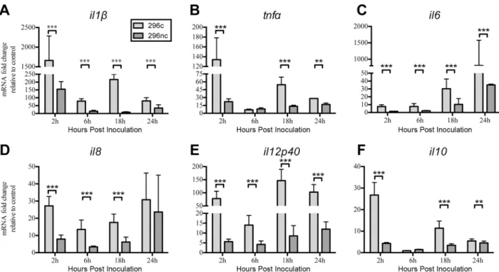
examined. Bovine MDMFs were differentiated for 7 days in 96 well plates and inoculated with an MOI of 1 for each strain and RNA isolated at 2 h, 6 h, 18 h, and 24 h post- inoculation. Cytokines measured displayed similar kinetics throughout the duration of experiments with an initial induction of mRNA followed by another induction of message at 18 h post BVDV2 inoc-ulation with either strain (Fig 1).il1β(Fig 1A),il6(Fig 1C) andil12p40(Fig 1E) were upregu-lated to a greater extent in MDMFs inocuupregu-lated with BVDV2-296c compared to BVDV2-296nc at each time point measured.tnfα(Fig 1B) andil10(Fig 1F) were induced to a greater extent in BVDV2-296c infected MDMFs compared to BVDV2-296nc inoculation except for 6 h post treatment. BVDV2-296c inoculated MDMFs exhibited greater levels ofil8(Fig 1D) than BVDV2-296nc inoculated MDMFs except for 24 h post treatment. MDMFs infected with BVDV2-296c exhibited greatly enhanced levels of proinflammatory cytokines. In contract, BVDV2-296nc inoculation of MDMFs only modestly induced proinflammatory cytokines and was, in general, significantly (P<0.0001) less than the cytokines induced by BVDV2-296c.
Infection of MDM
Φ
s with a non-cytopathogenic high virulence BVDV2
strain induced greater proinflammatory cytokine mRNA responses
compared to a non cytopathogenic typical/low virulence strain
in inoculated cells compared to LV BVDV2-28508-5 (Fig 2). However, HV BVDV2-1373 induced an initial mRNA expression of cytokines in MDMFs at 6h post inoculation as opposed to the other BVDV2 strains in which cytokine expression was beginning to be induced as early as 2 h post inoculation (Figs1and2).il1β(Fig 2A),tnfα(Fig 2B),il6(Fig 2C),il8(Fig 2D), and
il12p40(Fig 2E) were enhanced to a greater extent in MDMFs infected with HV BVDV2-1373 at 6, 18, 24 h post inoculation compared to LV BVDV2-28508 infected MDMFs. Additionally,
il10(Fig 2F) was increased in MDMFs inoculated with HV BVDV2-1373 at 18 and 24 h post
inoculation compared to LV BVDV2-28508 inoculated cells. Overall, HV BVDV2-1373 infected MDMFs demonstrated a greater level of proinflammatory cytokine expression start-ing at 6h post infection compared to LV BVDV2-28508 infected cells. It is of note that HV BVDV2-1373 did not induce a proinflamatory cytokine response at 2 h post inoculation com-pared to the other three strains of BVDV2 used in our experiments.
Cytopathic strain BVDV2-296c induced IL-6 secretion in infected
MDM
Φ
s, whereas all other strains did not induce secretion of
proinflammatory cytokines
Many studies of BVDV focus on cytokine mRNA expression in response to viral inoculationin
vitro. There have been no comparative studies of BVDV2 strainsin vitroin which MDMF cytokine secretion is explored. As we observed an induction in cytokine mRNA, we
investi-gated the protein secretion of MDMFs in response to 296c, 296nc,
BVDV2-1373 and BVDV2-28508. Interestingly, each of the viral strains did not induce cytokine Fig 1. Expression of proinflammatory cytokine gene transcription in MDMΦs inoculated with cytopathic or non-cytopathic BVDV2 strains.MDMΦs were differentiated in 96 well plates for 7 days and inoculated with BVDV2 strains in duplicate at an MOI of 1 with RNA harvested at 2, 6, 18, and 24 h after inoculation. Cytokine mRNA was analyzed by qPCR using ribosomal protein s9 (RPS9) as an endogenous control with fold change expressed relative to uninfected control MDMΦs harvested at the corresponding time points.il1β(A),
tnfα(B),il6(C),il8(D),il12p40(E),il10(F) were measured using primer sets specific for bovine genes using SYRB Green chemistry. Bars represent the mean value±SEM from four different experiments with 9 total donor cattle.***P<0.001;**P<0.005.
secretion, except for BVDV2-296c (Fig 3). LPS was used as a positive control in these experi-ments. IL-1β(Fig 3A), and TNFα(Fig 3C) secretion was not increased in the viral treated groups compared to control. Interestingly, IL-6 (Fig 3B) was increased only in the BVDV2-296c-treated MDMFs compared to both LPS stimulated cells or cells inoculated with other virus strains. These results suggest that there is a disconnect between mRNA expression and Fig 2. Expression of proinflammatory cytokine gene transcription in MDMΦs inoculated with high and low virulence BVDV2 strains.MDMΦs were differentiated in 96 well plates for 7 days and inoculated with BVDV2 strains in duplicate at an MOI of 1 with RNA harvested at 2, 6, 18, and 24 h after inoculation. Cytokine mRNA was analyzed by qPCR using ribosomal protein s9 (RPS9) as an endogenous control with fold change expressed relative to uninfected control MDMΦs harvested at the corresponding time points.il1β(A),
tnfα(B),il6(C),il8(D),il12p40(E),il10(F) were measured using primer sets specific for bovine genes using SYRB Green chemistry. Bars represent the mean value±SEM from four different experiments from 9 total donor cattle.***P<0.001.
doi:10.1371/journal.pone.0159491.g002
Fig 3. Proinflammatory cytokine secretion of BVDV2 inoculated or LPS stimulated MDMΦs 24 h after treatment.MDMΦs were differentiated in 96 well plates for 7 days and inoculated with BVDV2 strains in duplicate at an MOI of 1 or 2μg/mL LPS with cell
supernatants harvested at 24 h after treatment. Cytokine protein was analyzed by Searchlight Array using analytes specific for bovine IL-1β (A), IL-6 (B) and TNFα(C) with 50μL of cell supernatant analyzed in duplicate. Cytokines were quantified by generation of standard curves against recombinant bovine cytokines provided by the manufacturer of the Searchlight platform. Bars represent the mean value±SEM from
four different experiments from 9 total donor cattle.**P<0.001.
cytokine secretion in BVDV2 inoculated MDMFs. It is noteworthy that although BVDV2-296c did induce IL-6 secretion, which mirrors gene transcription, IL-1βand TNFαwere not induced. It is possible that the cytopathic nature of BVDV2-296c may be a factor in the induc-tion of IL-6. Addiinduc-tionally, examinainduc-tion of later time points of 48 h and 72 h post inoculainduc-tion did not yield any differences in expression of secreted cytokine protein (S1 Fig).
BVDV2 infected MDM
Φ
s stimulated with bacterial TLR agonists
exhibited reduced proinflammatory cytokine secretions
As the majority of BVDV2 strains alone did not induce proinflammatory cytokine secretion, this interesting observation lead us to explore whether BVDV2 would inhibit cytokine secre-tion in response to bacterial TLRs. Few studies have investigatedE.coliLPS responsiveness in
BVDV1 inoculated MFderivedin vitro[33–36] and there have been no studies comparing
varying strain of BVDV2 to multiple types of bacterial TLRs. TNFαprotein secretion was decreased in virus inoculated MDMFs in response to TLR stimulation compared to
TLR-stim-ulated uninfected controls (Fig 4). For MDMFs inoculated with 296c or
BVDV2-296nc, there were no dramatically different levels of suppression between the treatment groups
(Fig 4A). Noteworthy is that bothE.coliandM.haemolyticaLPS stimulation of MDMFs
Fig 4. TNFαprotein secretion from BVDV2 infected monocyte derived macrophages after stimulation with bacterial TLR agonists.
MDMΦs were differentiated in 96 well plates for 7 days and inoculated with BVDV2 strains with an MOI of 1 for 48 h prior to stimulation with Pam3Cys [5μg/mL],M.haemolyticaLPS [10μg/mL], orE.coli(055:B5) LPS [1μg/mL]. Cell supernatants were analyzed for TNFαprotein concentration 24 h after TLR stimulation and measured by Searchlight Array platform. Data from infection with cytopathic or noncytopathic strains are in the top panel (A) and from high or low virulence strains indicated in the lower panel (B). Bars represent the mean value±SEM
from four different experiments from 9 total donor cattle.**P<0.001 compared to uninfected, TLR stimulated cells.
inoculated with BVDV2-296c or BVDV2-296nc exhibited less TNFαprotein secretion than those stimulated with Pam3Cys. Similarly, MDMFs inoculated with 1373 or BVDV2-28508, there were no statistically different levels of suppression between the inoculated cells
(Fig 4B). Interestingly,M.haemolyticaLPS stimulation was not as suppressed in BVDV2
inoc-ulated BVDV2-1373 or BVDV2-28508 MDMFs as with the BVDV2-296c or BVDV2-296nc
inoculated cells. In general, virulence and cytopathogenicity do not necessarily impact the level of suppression in BVDV2 infected MDMFs stimulated with bacterial TLRs, as all groups were diminished in their cytokine response to bacterial TLR ligation. Furthermore, levels of secreted IL-1βand IL-6 were similarly reduced in BVDV2 inoculated and TLR-stimulated cells (S2and
S3Figs).
BVDV2 infected MDM
Φ
s respond to TLR7 ligation; however other viral
TLR agonists failed to induce similar cytokine responses
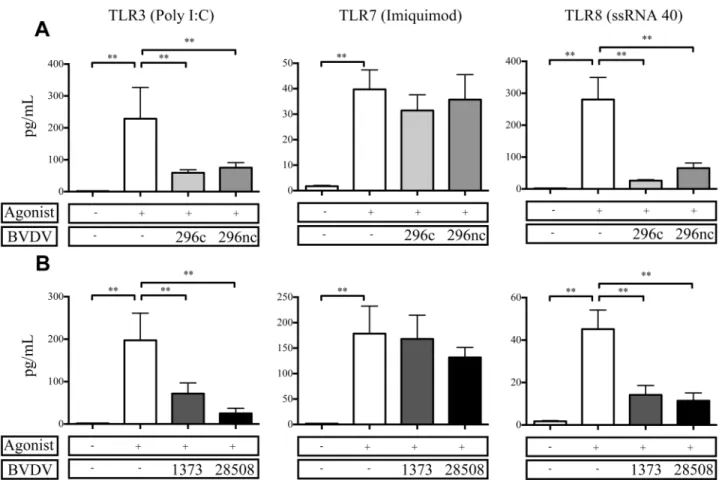
As we observed a decrease in cytokine protein expression from BVDV2 inoculated MDMFs in response to bacterial TLRs, we decided to explore TLRs specific to viral PAMPs. BVDV infec-tionin vivomay predispose an animal to secondary viral infection, and a deficit in viral TLR responsiveness could potentially contribute to this observation. Previous comparative studies of BVDV2 strains and the effects of various viral TLRsin vitrohave been limited. Interestingly,
TNFαprotein was not suppressed in BVDV2 inoculated MDMFs in response to TLR7;
how-ever responses to both TLR3 and TLR8 were suppressed (Fig 5). There were no statistically dif-ferent levels of suppression for TNFαexpression in either cytopathic BVDV2-296c or non-cytopathic BVDV2-296nc inoculated MDMFs (Fig 5A). Similarly, there were no differences in
the amount of suppression of TNFαprotein secretion between the MDMFs infected with
either HV BVDV2-1373 or LV BVDV2-28508 (Fig 5B) and stimulated with TLR agonists;
however TLR7 agonists were able to induce a response in each of the virus infected treatment groups, with each of them statistically similar to agonist stimulation alone. Similar to findings with bacterial TLR agonists, cytopathogenicity or virulence does not effect the level of suppres-sion for BVDV2 infected MDMFs stimulated with viral TLR agonists and in which TLR3 and TLR8 were both diminished in their ability to respond in virally inoculated cells. Likewise, IL-1βand IL-6 from virally inoculated cells stimulated with TLR3 and TL8 agonists demonstrated a reduction in cytokine secretion (S2andS3Figs).
Discussion
This study demonstrated that BVDV2 infection of MDMFinduces robust proinflammatory
cytokine mRNA responses, which differ dependent on the cytopathogenicity and virulence of the viral strains. Nevertheless, there was no enhancement of TNFαor IL-1βprotein secretion, whereas IL-6 was only detected in response to the cp strain utilized in this study. Future experi-ments should examine the mechanism whereby BVDV2 infection inhibits secretion of specific cytokines. It is noteworthy that the response to secondary bacterial or viral TLR stimuli after BVDV2 infection was generally reduced compared to uninfected cells. However, TLR7 signal-ing after BVDV infection was not abrogated.
46,47]. This observation suggests that monocytes or macrophages may play a critical role in recovery from BVDV infection, as they are not depleted throughout the course of infection.
This study compared differences between BVDV2 isogenic strains or strains of varying
viru-lence and the impact on infected MDMFcytokine mRNA responses. In the experimental
design of these experiments, MDMFwere inoculated with BVDV2 strains, followed 90 min
later by removal of the inoculum, washing and addition of fresh medium. Therefore; any non-specific activating factors would likley not illicit a measurable response in the inoculated MDMF, which have been shown return to a quiescent state rapidly after removal of these non-specific activating factors [48]. It is noteworthy that we saw a robust cytokine mRNA response, which was more pronounced in the cp and hv BVDV2 strains (Figs1and2). Of note, the work of others have demonstrated thattnfαgene transcription is enhanced in PBMCs infected with cp BVDV1 strains, however comparison to non-cytopathic strains was not examined [49]. Our findings suggest only an increase in expression of mRNA, not protein secretion, from infected MDMF. It is possible that BVDV2 can impact TNFαprotein expression by modification to the 3’UTR of thetnfαmRNA sequence eliciting it to degrade posttranscriptionally as observed in natural control of inflammatory processes by the cell [50]. The cytopathic BVDV2-296c strain Fig 5. TNFαprotein secretion from BVDV2 infected MDMΦafter stimulation with viral TLR agonists.MDMΦs were differentiated in 96 well plates for 7 days and inoculated with BVDV2 strains with an MOI of 1 for 48 h prior to stimulation with Poly I:C [50μg/mL], Imiquimod [10μg/mL], ssRNA40 LyoVec [10μg/mL]. Cell supernatants were analyzed for TNFαprotein concentration 24 h after TLR stimulation and measured by Searchlight Array platform. Incubation with cytopathic and noncytopathic strains are in the top panel (A) or high and low virulence strains indicated in the lower panel (B). Bars represent the mean value±SEM from four different experiments from 9 total donor
cattle.**P<0.001 compared to uninfected, TLR stimulated cells.
did induce IL-6 secretion in MDMF, which can help to induce apoptosis under certain condi-tions [51], therefore the induction of this cytokine may be criticial to cp responses of MDMF. We demonstrated a disconnect between mRNA expression and cytokine protein secretion in each of the BVDV2 inoculated MDMF. Previous studies have demonstrated a disassociation
between mRNA expression and protein production [52–54]. Differences between mRNA
expression and protein production may be related to cellular physiological functions, such as post-translational modifications that inhibit protein production [55,56], sequestering of pro-tein which blocks secretion [57] or production of inactive forms of cytokines which require sec-ondary mechanisms for activation and secretion [58]. Nonetheless, it should be noted that the dissociation between cytokine mRNA expression and protein secretion may be a trait of BVDV2 pathogenesis.
Similar to our finding, ncp BVDV1 strains have been shown to decrease intracellular pro-tein expression of IL-1βand TNFα[26]. It is of note, anotherFlaviviridaefamily member, den-gue virus, demonstrated reduced expression of proinflammatory cytokines IL-1β, IL-8 and TNFαin whole blood from infected children; however this decrease in expression correlated to heightened clinical disease severity [59]. Overall, our research and the findings of others sug-gest that BVDV reduces or inhibits cytokine protein secretion from infected cells.
Noteworthy is the observation that hv BVDV2 strain BVDV2-1373 induced proinflamma-tory cytokine gene transcription begining at 6 h post inoculation compared to the MDMF response to the other strains which occurred beginning at 2 h post inoculation. This observa-tion may indicate that this particular strain of BVDV2 is unique compared to the other strains we studied, as it is either not recognized as early by the MDMFto induce cytokine mRNA at 2 h or is recognized by differing innate mechanisms.
As far as we are aware, this is the only published study to date in which cytokine responses
of BVDV2-infected MDMFto multiple TLR agonists has been explored. However, a previous
study demonstrated that bone marrow derived macrophages (BMMF) infected with BVDV1
in vitrohave been shown to secrete less TNFαprotein in response to LPS or bacterial infection
compared to uninfected BMMFs [36]. Similarly, TLR4 hyporesponsiveness has been
demon-strated in MDMFfrom chronically infected HCV patients whereas this observation correlated to more severe disease and worse clinical outcome [60,61]. Likewise,Flaviviralinfections includingyellow fever virus(YFV) orSt.Louis encephalitis virus(SLEV), reduced IL-1βlevels in LPS-stimulated MDMFcompared to uninfected control cells [62]. Similar to these findings, we demonstrated that varying TLR4 agonists as well as TLR2/1 agonists are diminished in their
ability to promote pro-inflammatory cytokine secretion in MDMFafter BVDV2 infection.
Interestingly, each biotype suppressed MDMFcytokine secretion in response to bacterial TLR stimulation.
As with bacterial TLR ligation, there is evidence that BVDV infection can decrease respon-siveness to a double-stranded (ds)-RNA viral recognition receptor, TLR3 [26,63]. The damp-ened TLR3 response can be attributed to the BVDV ubiquination and degradation of
interferon regulatory factor (IRF)-3 directly downstream of TLR3 activation by means of inter-action with Npro protein of BVDV [64–66]. Thus, we demonstrated a reduction in viral TLR
responsiveness in MDMFevidenced by a reduction in cytokine secretion by both TLR3 and
TLR8 stimulation after BVDV2 infection (Fig 5). Intriguingly, TLR7 ligation was not damp-ened after BVDV2 infection and was the only TLR tested that was statistically similar or enhanced in cytokine secretion compared to uninfected control cells. These findings along with the data from this study of bacterial and viral TLR ligation (Figs4and5), demonstrate that there is a TLR hyporesponsivness induced byFlaviviridae, including BVDV infection.
not observe differences in TLR gene expression in MDMFinfected with several strains of BVDV2 (data not shown). In contrast, others have demonstrated differences in type-1 inter-feron responses in monocyte and monocyte derived dendritic cells infected with two ncp BVDV strains, with a suggested role for TLR7 [68]. Furthermore, microarray analysis which compared BVDV1-ncp to BVDV1-cp strains did find evidence of differential expression of TLR 2, TLR 3, TLR4, and TLR7 genes [69]. Interestingly the authors also reported that BCL2
was increased in the BVDV1-cp inoculated MDMF. BCL2 is a molecule important to
recep-tor-activated JNK signaling and cell survival [70]. In that regard, viral modulation of TLR receptor expression and associated signaling complexes would impact cytokine responses to subsequent ligation.
Taken together, the findings in this study demonstrate that the BVDV2 strains vary in their induction of cytokine mRNA responses from MDMF. Although a robust mRNA response was observed, protein secretion was not. In addition, subsequent TLR stimulation of
BVDV2-in-fected MDMFresulted in a hyporesponsive cytokine response compared to uninfected
MDMF. However, TLR7 ligation induced cytokine responses in infected MDMF.
Interest-ingly, MDMFinfected with isogenic strains of cp/ncp BVDV2 did not differ in suppression of TLR stimulated cytokine responses, indicating that NS2, NS3 or NS2/3 may not be an explana-tion of this observaexplana-tion; however IL-6 secreexplana-tion may be mediated by NS2 or NS3. These results demonstrate that BVDV2 infection generally blocks TLR responsiveness and provide informa-tion on how this virus may contribute to susceptibility to secondary infecinforma-tion.
Supporting Information
S1 Fig. IL-1βand IL-6 and protein expression of MDMFinfected with BVDV2 strains at 48 and 72 h post infection.MDMFs were differentiated in 96 well plates for 7 days and inocu-lated with BVDV2 strains with an MOI of 1. Cell supernatants were analyzed for protein con-centration 48 h (A) or 72 h (B) after infection and measured by Searchlight Array platform. Bars represent the mean value ± SEM from four different experiments from 9 total donors. ND = no cytokine detected.P<0.001,P<0.05 compared to uninfected, TLR stimulated cells.
(TIF)
S2 Fig. IL-1βand IL-6 protein expression in response to TLR stimulation in MDMF
infected with BVDV2 strains 296c or 296nc.MDMFs were differentiated in 96 well plates for 7 days and inoculated with BVDV2 strains with an MOI of 1 for 48 h prior to stimulation with Pam3Cys [5μg/mL],M.haemolyticaLPS [10μg/mL],E.coli(055:B5) LPS [1μg/mL], Poly I:C [50μg/mL], Imiquimod [10μg/mL], or ssRNA40 LyoVec [10μg/mL]. Cell supernatants were analyzed for protein concentration 24 h after TLR stimulation and measured by Searchlight Array platform. Addition of agonist or cytopathic or noncytopathic strains are indicated below each bar graph. Bars represent the mean value ± SEM from four different experiments from 9 total donors.P<0.001,P<0.05 compared to uninfected, TLR stimulated cells.
(TIF)
S3 Fig. IL-1βand IL-6 protein expression in response to TLR stimulation in MDMF
below each bar graph. Bars represent the mean value ± SEM from four different experiments from 9 total donors.P<0.001,P<0.05 compared to uninfected, TLR stimulated cells. (TIF)
Acknowledgments
The authors would like to thank Theresa Waters, Kathy McMullen, and Patricia Federico for their excellent technical assistance. The authors would also like to thank Todd Pille, Paul Amundson and Dr. Rebecca Cox for their excellent animal care and assistance. The authors would like to thank Dr. Christine Petersen for her critical reading and comments on the manuscript.
Author Contributions
Conceived and designed the experiments: RGS JFR RES. Performed the experiments: RGS JFR RES. Analyzed the data: RGS RES. Wrote the paper: RGS JFR RES.
References
1. Buckwold VE, Beer BE, Donis RO. Bovine viral diarrhea virus as a surrogate model of hepatitis C virus for the evaluation of antiviral agents. Antiviral Res. 2003; 60(1):1–15. PMID:14516916
2. Ridpath JF. Practical significance of heterogeneity among BVDV strains: impact of biotype and geno-type on U.S. control programs. Prev Vet Med. 2005; 72(1–2):17–30; discussion 215–9. PMID:
16183156
3. Liebler-Tenorio EM, Ridpath JF, Neill JD. Lesions and tissue distribution of viral antigen in severe acute versus subclinical acute infection with BVDV2. Biologicals. 2003; 31(2):119–22. PMID:12770542 4. Ammari M, McCarthy FM, Nanduri B, Pinchuk LM. Analysis of Bovine Viral Diarrhea Viruses-infected
monocytes: identification of cytopathic and non-cytopathic biotype differences. BMC Bioinformatics. 2010; 11 Suppl 6:S9. doi:10.1186/1471-2105-11-S6-S9PMID:20946620
5. Fulton RW, Ridpath JF, Ore S, Confer AW, Saliki JT, Burge LJ, et al. Bovine viral diarrhoea virus (BVDV) subgenotypes in diagnostic laboratory accessions: distribution of BVDV1a, 1b, and 2a subge-notypes. Vet Microbiol. 2005; 111(1–2):35–40. PMID:16263224
6. Donis RO, Dubovi EJ. Differences in virus-induced polypeptides in cells infected by cytopathic and non-cytopathic biotypes of bovine virus diarrhea-mucosal disease virus. Virol. 1987; 158(1):168–73. 7. Ridpath JF, Bendfeldt S, Neill JD, Liebler-Tenorio E. Lymphocytopathogenic activity in vitro correlates
with high virulence in vivo for BVDV type 2 strains: Criteria for a third biotype of BVDV. Vir Res. 2006; 118(1–2):62–9.
8. Peterhans E, Schweizer M. Pestiviruses: how to outmaneuver your hosts. Vet Microbiol. 2010; 142(1–
2):18–25. doi:10.1016/j.vetmic.2009.09.038PMID:19846261
9. Ridpath JF. The Contribution of Infections with Bovine Viral Diarrhea Viruses to Bovine Respiratory Dis-ease. Vet Clin North Am Food Anim Pract. 2010; 26:335–48. doi:10.1016/j.cvfa.2010.04.003PMID:
20619188
10. Houe H. Economic impact of BVDV infection in dairies. Biologicals. 2003; 31(2):137–43. PMID:
12770546
11. Liu L, Lehmkuhl HD, Kaeberle ML. Synergistic effects of bovine respiratory syncytial virus and non-cytopathic bovine viral diarrhea virus infection on selected bovine alveolar macrophage functions. Can J Vet Res. 1999; 63(1):41–8. PMID:9918333
12. Peterhans E, Jungi TW, Schweizer M. BVDV and innate immunity. Biologicals. 2003; 31(2):107–12.
PMID:12770540
13. Stott AW, Humphry RW, Gunn GJ. Modelling the effects of previous infection and re-infection on the costs of bovine viral diarrhoea outbreaks in beef herds. Vet J. 2010; 185(2):138–43. doi:10.1016/j.tvjl.
2009.05.020PMID:19709915
14. Medzhitov R. Approaching the asymptote: 20 years later. Immunity. 2009; 30(6):766–75. doi:10.1016/
j.immuni.2009.06.004PMID:19538928
15. Biasin M, Piacentini L, Lo Caputo S, Naddeo V, Pierotti P, Borelli M, et al. TLR activation pathways in HIV-1-exposed seronegative individuals. J Immunol. 2010; 184(5):2710–7. doi:10.4049/jimmunol.
16. Wortham BW, Eppert BL, Motz GT, Flury JL, Orozco-Levi M, Hoebe K, et al. NKG2D mediates NK cell hyperresponsiveness and influenza-induced pathologies in a mouse model of chronic obstructive pul-monary disease. J Immunol. 2012; 188(9):4468–75. doi:10.4049/jimmunol.1102643PMID:22467655 17. Yonkers NL, Rodriguez B, Milkovich KA, Asaad R, Lederman MM, Heeger PS, et al. TLR
ligand-depen-dent activation of naive CD4 T cells by plasmacytoid dendritic cells is impaired in hepatitis C virus infec-tion. J Immunol. 2007; 178(7):4436–44. PMID:17372001
18. Rathinam VAK, Fitzgerald KA. Inflammasomes and anti-viral immunity. J Clin Immunol. 2010; 30 (5):632–7. doi:10.1007/s10875-010-9431-4PMID:20665097
19. Rogez-Kreuz C, Manéglier B, Martin M, Dereuddre-Bosquet N, Martal J, Dormont D, et al. Involvement of IL-6 in the anti-human immunodeficiency virus activity of IFN-tau in human macrophages. Int Immu-nol. 2005; 17(8):1047–57. PMID:15976033
20. Wong KL, Chen W, Balakrishnan T, Toh YX, Fink K, Wong S-C. Susceptibility and response of human blood monocyte subsets to primary dengue virus infection. PLoS ONE. 2012; 7(5):e36435. doi:10. 1371/journal.pone.0036435PMID:22574162
21. Schaut RG, McGill JL, Neill JD, Ridpath JF, Sacco RE. Bovine viral diarrhea virus type 2 in vivo infec-tion modulates TLR4 responsiveness in differentiated myeloid cells which is associated with decreased MyD88 expression. Vir Res. 2015; 208:44–55.
22. Fulton RW, Hessman B, Johnson BJ, Ridpath JF, Saliki JT, Burge LJ, et al. Evaluation of diagnostic tests used for detection of bovine viral diarrhea virus and prevalence of subtypes 1a, 1b, and 2a in per-sistently infected cattle entering a feedlot. J Am Vet Med Assoc. 2006; 228(4):578–84. PMID:
16478438
23. Duvel A, Frank C, Schnapper A, Schuberth HJ, Sipka A. Classically or alternatively activated bovine monocyte-derived macrophages in vitro do not resemble CD163/Calprotectin biased macrophage pop-ulations in the teat. Innate Immun. 2012; 18(6):886–96. doi:10.1177/1753425912446954PMID:
22627785
24. Bauermann FV, Flores EF, Ridpath JF. Antigenic relationships between Bovine viral diarrhea virus 1 and 2 and HoBi virus: possible impacts on diagnosis and control. J Vet Diagn Invest. 2012 Mar; 24 (2):253–61. doi:10.1177/1040638711435144PMID:22379042
25. Nelson CD, Reinhardt TA, Thacker TC, Beitz DC, Lippolis JD. Modulation of the bovine innate immune response by production of 1alpha,25-dihydroxyvitamin D(3) in bovine monocytes. J Dairy Sci. 2010; 93 (3):1041–9. doi:10.3168/jds.2009-2663PMID:20172224
26. Lee SR, Pharr GT, Boyd BL, Pinchuk LM. Bovine viral diarrhea viruses modulate toll-like receptors, cytokines and co-stimulatory molecules genes expression in bovine peripheral blood monocytes. Comp Immunol Microbiol Infect Dis. 2008; 31(5):403–18. PMID:17706777
27. Taraktsoglou M, Szalabska U, Magee DA, Browne JA, Sweeney T, Gormley E, et al. Transcriptional profiling of immune genes in bovine monocyte-derived macrophages exposed to bacterial antigens. Vet Immunol Immunopathol. 2011; 140(1–2):130–9. doi:10.1016/j.vetimm.2010.12.002PMID:
21242003
28. Livak KJ, Schmittgen TD. Analysis of relative gene expression data using real-time quantitative PCR and the 2(-Delta Delta C(T)) Method. Methods. 2001; 25(4):402–8. PMID:11846609
29. Baroth M, Orlich M, Thiel HJ, Becher P. Insertion of cellular NEDD8 coding sequences in a pestivirus. Virology. 2000; 278(2):456–66. PMID:11118368
30. Becher P, Thiel HJ, Collins M, Brownlie J, Orlich M. Cellular sequences in pestivirus genomes encoding gamma-aminobutyric acid (A) receptor-associated protein and Golgi-associated ATPase enhancer of 16 kilodaltons. J Virol. 2002; 76(24):13069–76. PMID:12438634
31. Ridpath JF, Bolin SR. The genomic sequence of a virulent bovine viral diarrhea virus (BVDV) from the type 2 genotype: detection of a large genomic insertion in a noncytopathic BVDV. Virology. 1995; 212 (1):39–46. PMID:7676648
32. Kummerer BM, Tautz N, Becher P, Thiel H, Meyers G. The genetic basis for cytopathogenicity of pesti-viruses. Vet Microbiol. 2000; 77(1–2):117–28. PMID:11042405
33. Qi F, Ridpath JF, Lewis T, Bolin SR, Berry ES. Analysis of the bovine viral diarrhea virus genome for possible cellular insertions. Virology. 1992; 189(1):285–92. PMID:1318605
34. Jensen J, Schultz RD. Effect of infection by bovine viral diarrhea virus (BVDV) in vitro on interleukin-1 activity of bovine monocytes. Vet Immunol Immunopathol. 1991; 29(3–4):251–65. PMID:1949588 35. Franchini M, Schweizer M, Mätzener P, Magkouras I, Sauter K-S, Mirkovitch J, et al. Evidence for
36. Adler H, Jungi TW, Pfister H, Strasser M, Sileghem M, Peterhans E. Cytokine regulation by virus infec-tion: bovine viral diarrhea virus, a flavivirus, downregulates production of tumor necrosis factor alpha in macrophages in vitro. J Virol. 1996; 70(4):2650–3. PMID:8642701
37. Brown GB, Bolin SR, Frank DE, Roth JA. Defective function of leukocytes from cattle persistently infected with bovine viral diarrhea virus, and the influence of recombinant cytokines. Am J Vet Res. 1991; 52(3):381–7. PMID:1852143
38. Ketelsen AT, Johnson DW, Muscoplat CC. Depression of bovine monocyte chemotactic responses by bovine viral diarrhea virus. Infect Immun. 1979; 25(2):565–68. PMID:226479
39. Atluru D, Gudapaty S, Xue W, Gurria F, Chengappa MM, McVey DS, et al. In vitro inhibition of 5-lipoxy-genase metabolite, leukotriene B4, in bovine mononuclear cells by bovine viral diarrhea virus. Vet Immunol Immunopathol. 1992; 31(1–2):49–59. PMID:1315086
40. Roth JA, Kaeberle ML, Griffith RW. Effects of bovine viral diarrhea virus infection on bovine polymor-phonuclear leukocyte function. Am J Vet Res. 1981; 42(2):244–50. PMID:6266288
41. Roth JA, Bolin SR, Frank DE. Lymphocyte blastogenesis and neutrophil function in cattle persistently infected with bovine viral diarrhea virus. Am J Vet Res. 1986; 47(5):1139–41. PMID:3717739 42. Welsh M, Adair B. Effect of BVD virus infection on alveolar macrophage functions. Vet Immunol
Immu-nopathol. 1995.
43. Bielefeldt Ohmann H. In situ characterization of mononuclear leukocytes in skin and digestive tract of persistently bovine viral diarrhea virus-infected clinically healthy calves and calves with mucosal dis-ease. Vet Pathol. 1988; 25(4):304–9. PMID:3261471
44. Bielefeldt Ohmann H, Rønsholt L, Bloch B. Demonstration of bovine viral diarrhoea virus in peripheral
blood mononuclear cells of persistently infected, clinically normal cattle. J Gen Virol. 1987; 68 (Pt 7):1971–82. PMID:3037018
45. Lambot M, Hanon E, Lecomte C, Hamers C, Letesson JJ, Pastoret PP. Bovine viral diarrhoea virus induces apoptosis in blood mononuclear cells by a mechanism largely dependent on monocytes. J Gen Virol. 1998; 79 (Pt 7):1745–9. PMID:9680138
46. Pedrera M, Gómez-Villamandos JC, Romero-Trevejo JL, Risalde MA, Molina V, Sánchez-Cordón PJ. Apoptosis in lymphoid tissues of calves inoculated with non-cytopathic bovine viral diarrhea virus geno-type 1: activation of effector caspase-3 and role of macrophages. J Gen Virol. 2009; 90(Pt 11):2650–9.
doi:10.1099/vir.0.012021-0PMID:19641046
47. Liebler-Tenorio EM, Ridpath JF, Neill JD. Distribution of viral antigen and tissue lesions in persistent and acute infection with the homologous strain of noncytopathic bovine viral diarrhea virus. J Vet Diagn Invest. 2004; 16(5):388–96. PMID:15460320
48. Porcheray F, Viaud S, Rimaniol AC, Léone C, Samah B, Dereuddre-Bosquet N, Dormont D, Gras G. Macrophage activation switching: an asset for the resolution of inflammation. Clin Exp Immunol. 2005 Dec; 142(3):481–9. PMID:16297160
49. Liebler-Tenorio EM, Ridpath JF, Neill JD. Distribution of viral antigen and development of lesions after experimental infection of calves with a BVDV 2 strain of low virulence. J Vet Diagn Invest. 2003; 15 (3):221–32. PMID:12735344
50. Yamane D, Nagai M, Ogawa Y, Tohya Y, Akashi H. Enhancement of apoptosis via an extrinsic factor, TNF-alpha, in cells infected with cytopathic bovine viral diarrhea virus. Microbes Infect. 2005; 7 (15):1482–91. PMID:16055364
51. Anderson P. Post-transcriptional regulons coordinate the initiation and resolution of inflammation. Nat Rev Immunol. 2010; 10(1):24–35. doi:10.1038/nri2685PMID:20029446
52. Afford SC, Pongracz J, Stockley RA, Crocker J, Burnett D. The induction by human interleukin-6 of apo-ptosis in the promonocytic cell line U937 and human neutrophils. J Biol Chem. 1992; 267(30):21612–6.
PMID:1400472
53. Raqib R, Ljungdahl A, Lindberg AA, Wretlind B, Andersson U, Andersson J. Dissociation between cyto-kine mRNA expression and protein production in shigellosis. Eur J Immunol. 1996; 26(5):1130–8.
PMID:8647178
54. Schindler R, Clark BD, Dinarello CA. Dissociation between interleukin-1 beta mRNA and protein syn-thesis in human peripheral blood mononuclear cells. J Biol Chem. 1990; 265(18):10232–7. PMID:
2354999
55. Ruzek MC, Pearce BD, Miller AH, Biron CA. Endogenous glucocorticoids protect against cytokine-mediated lethality during viral infection. J Immunol. 1999; 162(6):3527–33. PMID:10092810
57. Tserel L, Kolde R, Rebane A, Kisand K, Org T, Peterson H, et al. Genome-wide promoter analysis of histone modifications in human monocyte-derived antigen presenting cells. BMC Genomics. 2010; 11:642. doi:10.1186/1471-2164-11-642PMID:21087476
58. Re F, Sironi M, Muzio M, Matteucci C, Introna M, Orlando S, et al. Inhibition of interleukin-1 responsive-ness by type II receptor gene transfer: a surface "receptor" with anti-interleukin-1 function. J Exp Med. 1996; 183(4):1841–50. PMID:8666940
59. Broz P, Monack DM. Molecular mechanisms of inflammasome activation during microbial infections. Immunol Rev. 2011; 243(1):174–90. doi:10.1111/j.1600-065X.2011.01041.xPMID:21884176 60. de Kruif MD, Setiati TE, Mairuhu ATA, Koraka P, Aberson HA, Spek CA, et al. Differential gene
expres-sion changes in children with severe dengue virus infections. PLoS Negl Trop Dis. 2008; 2(4):e215. doi:10.1371/journal.pntd.0000215PMID:18398488
61. Chung H, Watanabe T, Kudo M, Chiba T. Correlation between hyporesponsiveness to Toll-like receptor ligands and liver dysfunction in patients with chronic hepatitis C virus infection. J Viral Hepat. 2011; 18 (10):e561–e7. doi:10.1111/j.1365-2893.2011.01478.xPMID:21914077
62. Liu B-S, Groothuismink ZMA, Janssen HLA, Boonstra A. Role for IL-10 in inducing functional impairment of monocytes upon TLR4 ligation in patients with chronic HCV infections. J Leukoc Biol. 2011; 89(6):981–8. doi:10.1189/jlb.1210680PMID:21385948
63. Barros VED, Ferreira BR, Livonesi M, Figueiredo LTM. Cytokine and nitric oxide production by mouse macrophages infected with Brazilian flaviviruses. Rev Inst Med Trop Sao Paulo. 2009; 51(3):141–7.
PMID:19551288
64. Schweizer M, Peterhans E. Noncytopathic bovine viral diarrhea virus inhibits double-stranded RNA-induced apoptosis and interferon synthesis. J Virol. 2001; 75(10):4692–8. PMID:11312340
65. Baigent SJ, Zhang G, Fray MD, Flick-Smith H, Goodbourn S, McCauley JW. Inhibition of beta interferon transcription by noncytopathogenic bovine viral diarrhea virus is through an interferon regulatory factor 3-dependent mechanism. J Virol. 2002; 76(18):8979–88. PMID:12186882
66. Chen Z, Rijnbrand R, Jangra RK, Devaraj SG, Qu L, Ma Y, et al. Ubiquitination and proteasomal degra-dation of interferon regulatory factor-3 induced by Npro from a cytopathic bovine viral diarrhea virus. Virology. 2007; 366(2):277–92. PMID:17531282
67. Hilton L, Moganeradj K, Zhang G, Chen Y-H, Randall RE, McCauley JW, et al. The NPro product of bovine viral diarrhea virus inhibits DNA binding by interferon regulatory factor 3 and targets it for protea-somal degradation. J Virol. 2006; 80(23):11723–32. PMID:16971436
68. Franchini M, Schweizer M, Mätzener P, Magkouras I, Sauter KS, Mirkovitch J, Peterhans E, Jungi TW. Evidence for dissociation of TLR mRNA expression and TLR agonist-mediated functions in bovine macrophages. Vet Immunol Immunopathol. 2006; 110(1–2):37–49. PMID:16216336
69. Gibson A, Larsson J, Bateman M, Brownlie J, Werling D. Bovine viral diarrhea virus strain- and cell type-specific inhibition of type I interferon pathways. J Virol. 2011; 85(7):3695–7. doi:10.1128/JVI.
02626-10PMID:21270161