Associated Gene Clusters and Candidate Determinants of
Tissue-Specificity in
Xanthomonas
Hong Lu1., Prabhu Patil2., Marie-Anne Van Sluys3,9
, Frank F. White4, Robert P. Ryan5, J. Maxwell Dow5, Pablo Rabinowicz6,7, Steven L. Salzberg8, Jan E. Leach9, Ramesh Sonti2, Volker Brendel1,10, Adam J. Bogdanove11*
1Department of Genetics Development and Cell Biology, Iowa State University, Ames, Iowa, United States of America,2Centre for Cellular and Molecular Biology, Council of Scientific and Industrial Research, Hyderabad, India,3Departamento de Botaˆnica, IB-USP, Sao Paulo, Sao Paulo, Brazil,4Department of Plant Pathology, Kansas State University, Manhattan, Kansas, United States of America,5BIOMERIT Research Centre, BioSciences Institute, University College Cork, Cork, Ireland,6The Institute for Genomic Research, Rockville, Maryland, United States of America,7Institute for Genome Sciences, University of Maryland, Baltimore, Maryland, United States of America,
8Center for Bioinformatics and Computational Biology, University of Maryland, College Park, Maryland, United States of America,9Department of Bioagricultural Sciences and Pest Management, Colorado State University, Fort Collins, Colorado, United States of America,10Department of Statistics, Iowa State University, Ames, Iowa, United States of America,11Department of Plant Pathology, Iowa State University, Ames, Iowa, United States of America
Abstract
Background:Xanthomonasis a large genus of plant-associated and plant-pathogenic bacteria. Collectively, members cause diseases on over 392 plant species. Individually, they exhibit marked host- and tissue-specificity. The determinants of this specificity are unknown.
Methodology/Principal Findings:To assess potential contributions to host- and tissue-specificity, pathogenesis-associated gene clusters were compared across genomes of eight Xanthomonas strains representing vascular or non-vascular pathogens of rice, brassicas, pepper and tomato, and citrus. Thegumcluster for extracellular polysaccharide is conserved except forgumNand sequences downstream. Thexcsandxpsclusters for type II secretion are conserved, except in the rice pathogens, in whichxcsis missing. In the otherwise conservedhrpcluster, sequences flanking the core genes for type III secretion vary with respect to insertion sequence element and putative effector gene content. Variation at the rpf (regulation ofpathogenicityfactors) cluster is more pronounced, though genes with established functional relevance are conserved. A cluster for synthesis of lipopolysaccharide varies highly, suggesting multiple horizontal gene transfers and reassortments, but this variation does not correlate with host- or tissue-specificity. Phylogenetic trees based on amino acid alignments ofgum,xps,xcs,hrp, andrpfcluster products generally reflect strain phylogeny. However, amino acid residues at four positions correlate with tissue specificity, revealinghpaAandxpsDas candidate determinants. Examination of genome sequences of xanthomonads Xylella fastidiosa and Stenotrophomonas maltophilia revealed that the hrp, gum, and xcs clusters are recent acquisitions in theXanthomonaslineage.
Conclusions/Significance: Our results provide insight into the ancestral Xanthomonas genome and indicate that differentiation with respect to host- and tissue-specificity involved not major modifications or wholesale exchange of clusters, but subtle changes in a small number of genes or in non-coding sequences, and/or differences outside the clusters, potentially among regulatory targets or secretory substrates.
Citation:Lu H, Patil P, Van Sluys M-A, White FF, Ryan RP, et al. (2008) Acquisition and Evolution of Plant Pathogenesis–Associated Gene Clusters and Candidate Determinants of Tissue-Specificity inXanthomonas. PLoS ONE 3(11): e3828. doi:10.1371/journal.pone.0003828
Editor:Ping Wang, Research Institute for Children and the Louisiana State University Health Sciences Center, United States of America
ReceivedAugust 22, 2008;AcceptedNovember 3, 2008;PublishedNovember 27, 2008
Copyright:ß2008 Lu et al. This is an open-access article distributed under the terms of the Creative Commons Attribution License, which permits unrestricted use, distribution, and reproduction in any medium, provided the original author and source are credited.
Funding:Support for this study was provided by the U.S. Department of Agriculture-National Science Foundation Microbial Genome Sequencing Program (20043560015022 to AB, JL, SS, and FW) and through a Principal Investigator Award from the Science Foundation of Ireland to JD. The funders had no role in study design, data collection and analysis, decision to publish, or preparation of the manuscript.
Competing Interests:The authors have declared that no competing interests exist.
* E-mail: [email protected]
.These authors contributed equally to this work.
Introduction
Comparative genomics is a powerful approach to discovering genetic features of related bacteria that have been acquired, modified, or lost during adaptation to particular environmental niches. Identification of such features is a first step toward
and provide clues to the evolution of pathogenesis or specific pathogenesis-related functions [for a review, see 5]. Few comparative genomics studies have been carried out on plant pathogenic bacteria. In 2002, Van Sluys et al. [6] identified nineteen genes (encoding conserved hypothetical genes, iron transporters, and cell-wall modifying enzymes) common to all sequenced plant-associated bacterial genomes available at the time. More recently, a comparative analysis of sequenced Enterobacteriaceae identified genes specific to the plant pathogen
Erwinia carotovora [7]. Comparative genomics has also provided novel insight into the role of horizontal gene transfer in shaping genomes of plant pathogenic xanthomonads [8–10].
Xanthomonas is a large genus of Gram-negative, yellow-pigmented, plant-associated bacteria. Pathogenic species and pathovars (pathogenic varieties, pv.) within species show a high degree of host plant specificity and combined are known to cause diseases on nearly 400 plant hosts, including both eudicots and monocots [11]. Many exhibit tissue-specificity, invading either host xylem vessels or the interveinal mesophyll apoplast of their host. Thus, the genus is a compelling subject for comparative genomics, as such analyses should shed light on how this group of bacteria has adapted to exploit an extraordinary diversity of plant hosts and host tissues. Understanding pathogenic adaptations of
Xanthomonaswill foster the development of needed improvements in bacterial plant disease control and prevention.
The genus Xanthomonas resides at the base of the gamma subdivision of the proteobacteria. The current taxonomic status of the genus is based on analysis of 16S–23S rDNA intergenic spacer sequences [12] and a combination of molecular markers such as rep-PCR, AFLP and other fingerprints [13,14]. Twenty DNA homology groups (species) have been distinguished, comprising 80 pathovars [14,15]. A species can encompass pathovars that infect diverse plant hosts and/or exhibit different patterns of plant colonization. For instance, Xanthomonas campestris includes patho-vars that (collectively) infect different brassicaceous, solanaceous, and other plant species, andXanthomonas oryzae, a species specific to rice and some wild relatives, comprises pathovars that either invade through the vascular system (X. oryzaepv.oryzae) or colonize the intercellular spaces of the parenchyma tissue (X . oryzaepv.
oryzicola) [16]. LikeX. oryzae, theX. campestrisgroup also includes vascular and non-vascular colonizers [11,17].
Complete genome sequences of sixXanthomonasstrains had been published at the commencement of the present study. These are strains ATCC33913 and 8004 ofX. campestrispv. campestris (XccA and Xcc8, respectively), a vascular pathogen of cabbage and other brassicas, including the model plantArabidopsis thaliana; strain 306 ofX. axonopodispv. citri (Xac), the causal agent of citrus canker, a non-vascular disease; strain 85-10 of X. axonopodispv. vesicatoria (Xav; formerly X. campestris pv. vesicatoria), a non-vascular pathogen that causes leaf spot on pepper and tomato; and strains KACC10331 and MAFF311018 of X. oryzae pv. oryzae (XooK and XooM, respectively), the vascular pathogen of rice [18–22]. During the course of this study, we finished and deposited in a public database the genome sequences of strain 756C [23] ofX. campestris pv. armoraciae (Xca), a non-vascular pathogen with a host range similar to that of Xcc, and strain BLS256 [24] ofX. oryzae pv. oryzicola (Xoc), the non-vascular counterpart of Xoo (AB et al., unpublished). We used these eight genome sequences in the analyses presented here. Subsequently, we completed the genome sequence of a third Xoo strain, PXO99A, and Vorholter et al. have recently published the genome sequence of a third Xcc strain, B100 [25,26]. Complete or near complete genome data are also available for representatives of the closely related xanthomo-nads, Xylella fastidiosa(Xf) [27–30] and Stenotrophomonas maltophilia
(Sma) [31, The Joint Genome Institute, US Dept of Energy, Genbank accession AAVZ00000000]. Xf is a group of fastidious, xylem-limited and insect-vectored plant pathogens with genomes roughly half the size of a typicalXanthomonasgenome. Xf strains collectively cause disease on diverse hosts, with some specificity [32]. Sma is a non-plant pathogenic species that includes free-living as well as endophytic isolates and opportunistic human pathogens [33].
The Xanthomonas genome sequences we examined represent plant pathogens that are closely related but distinct in their host and tissue-specificity and that include paired vascular and non-vascular pathogens (Xcc and Xca, and Xoo and Xoc, respectively) of the leading models for plant biology, A. thaliana and rice. Furthermore, the sequencedXanthomonasstrains span three of the 20 homology groups (species) defined by Rademaker et al. [14], providing good representation of the genus as a whole. Our objective was to determine whether differentiation of species and pathovars with respect to host- and tissue-specificity is reflected across genomes in content and structure of several gene clusters that are known to be involved in pathogenesis inXanthomonas spp.
or are implicated in pathogenesis based on functions of homologous gene clusters in other pathogenic bacterial species.
Results and Discussion
Xanthomonasgenomes and gene clusters examined The Xanthomonas genome sequences examined are given in Table 1, grouped by strain host- and tissue-specificity. The general features of the genomes are similar (Table S1). Each includes a circular chromosome of approximately 5 Mb. The Xav genome includes four plasmids, and the Xac genome two. Average G+C content ranges from 63.6% (XooK and XooM) to 65.3% (Xca). The percent of genome that is predicted coding sequence ranges from 83.9% (XooM) to 90.3% (Xac). The number of predicted genes ranges from 4,598 (Xca) to 5,832 (XccA). Each genome harbors two ribosomal RNA operons.
We examined thegumgene cluster for extracellular polysaccha-ride synthesis [34,35], the xps and xcs gene clusters for type II secretion [19,36], thehrpgene cluster for type III secretion [37], therpfgene cluster for regulation of pathogenicity factors [38], and an unnamed gene cluster involved in synthesis of lipopolysaccha-ride, which we hereafter refer to as the LPS gene cluster [39,40] (the coordinates of the clusters in each genome are given inTable S2). For each cluster, we sought correlations of gene content and structure with host- and tissue-specificity, and we examined phylogenetic relationships by comparing concatenated sequences of the deduced gene products within the cluster across genomes.
Thegumgene cluster
The ability to produce capsular extracellular polysaccharide (EPS) is correlated with virulence in several plant pathogenic bacteria [41], and the importance of EPS to pathogenicity in
Table 1. Classification of the examinedXanthomonasstrains by host- and tissue-specificitya.
Vascular Non-Vascular
Monocot XooK, XooM Xoc
Dicot Xcc8, XccA Xca, Xac, Xav
aAbbreviations are as in the text.
Xanthomonas has been demonstrated with EPS-deficient mutant strains of Xoo, Xcc, and Xac [35,42,43]. EPS is important in biofilm formation and epiphytic fitness [43,44]. It is postulated to promote colonization of plant tissues and to provide protection from harsh environmental conditions, and it contributes to occlusion of vascular elements in wilts and blights [41,45]. Synthesis of the Xanthomonas capsular EPS, xanthan, is carried out primarily by the twelve products of the roughly 16 kb gumB-gumM operon [34,35]. Additional open reading frames (ORFs) designated asgumgenes,gumAandgumN,-O, and-P, reside up and downstream (respectively) ofgumB-gumM[34], but a role for these genes in xanthan biosynthesis has not been demonstrated. Recently, gumNand an intervening ORF were shown to be co-transcribed with gumB-gumM in Xoo, but gumA is clearly in a distinct operon [46].
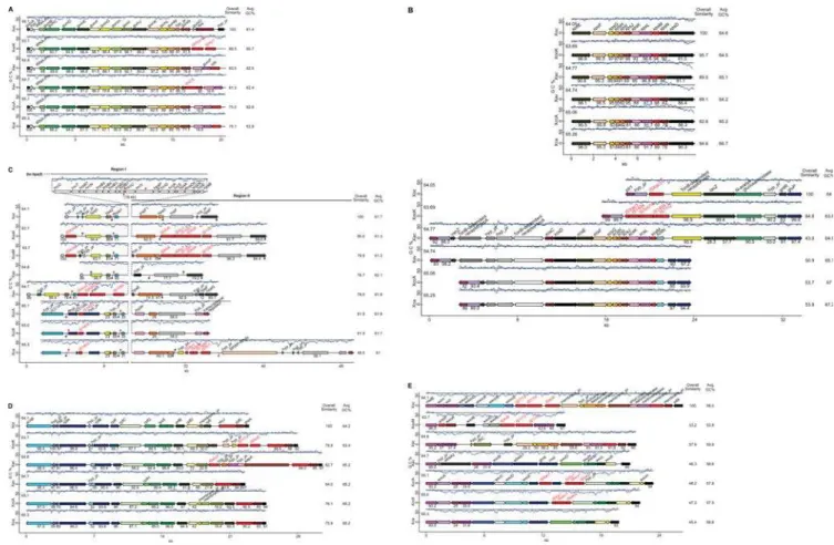
The nucleotide sequences of thegumcluster, delimited by and including gumA and gumN, and approximately 4 kb of sequence downstream of gumN, were retrieved from each genome and compared (Figure 1A). The cluster is highly conserved with
respect to overall gene content and order, including the ORF betweengumMandgumN. Differences among strains are limited to insertion sequence (IS) elements in or neargumNand differential content of genes outside the core cluster, includinggumO, gumP, andchd2. None of these genes, however, have been shown to play a role in xanthan biosynthesis. (For a complete discussion of differences observed at thegumgene cluster, seeText S1.)
As noted by Lima et al. [10], thegumgene cluster has features of a pathogenicity island, including a lower than average G+C content and a flanking tRNA gene (Figure 1A). Indeed, the cluster is absent from the Sma genome sequences, suggesting that acquisition ofgumgenes was an important adaptation toward plant pathogenicity. Consistent with the notion that gum genes were acquired subsequent to the divergence of the Xanthomonas and
Stenotrophomonaslineages, the regions flanking thegumlocus in the
Xanthomonas genomes are conserved and colinear in the Sma genomes, including the tRNA gene. A cluster containing gumB
though gumF and gumH is present in the Xf genomes, but the genomic context is distinct, suggesting independent acquisition of
Figure 1. Comparison of six clusters of genes involved or implicated in pathogenesis amongXanthomonasstrains representing three species and six pathovars.Sequences ofX. oryzaepv. oryzicola strain BLS256 (Xoc),Xanthomonas oryzaepv. oryzae KACC100331 (XooK) and MAFF311018 (XooM),X. axonopodis citristrain 306 (Xac),X. axonopodispv. vesicatoria strain 85-10 (Xav),X. campestrispv. campestris strains ATCC33913 (XccA) and 8004 (Xcc8), andX. campestrispv. armoraciae strain 756C (Xca) were used. Arrows represent individual genes. For each cluster across genomes, homologs are shown in like colors. Gene identities are given (non-redundantly) above each gene. The blue trace above each cluster represents GC content (window size: 160 bp, step: 40 bp). The black line above each cluster marks the average GC content of the genome, specified below and to the left of the line. Shown for each gene is the percent identity of the predicted product to that of the corresponding gene (if present) in the genome shown at the top. The overall similarity of each cluster to the cluster at the top is given at the near right. The average GC content of each cluster is given at the far right. Where clusters from different strains of the same pathovar are essentially identical, only one is represented. Insertion sequence elements are indicated by red rectangles, tRNA genes by blue triangles and plant-inducible promoter sequences (PIP boxes) by red or blue flags. A red flag represents a perfect PIP box and a blue flag represents an imperfect one. The orientation of the PIP box is represented by the orientation of the flag above or below the cluster. A, thegumgene cluster; B, thexpsandxcsgene clusters; C, thehrpgene cluster; D, therpfgene cluster; E, the lipopolysaccharide biosynthesis gene cluster bordered by theetfAandmetBgenes.
these genes in the Xylellalineage. Interestingly, in the sugarcane pathogenX. albilineans, PCR failed to detect any of ten gum genes assayed, using primer sequences conserved across Xac, Xcc, and Xoo [47]. This species produces an exopolysaccharide structurally related to but distinct from xanthan and compositionally more similar to the exopolysaccharide produced by Xf [48,49]. Production has only been observed in infected sugarcane stalks and appears to require plant components [50]. Direct comparison of theX. albilineansgenes for exopolysaccharide production with thegumgenes awaits completion of the firstX. albilineansgenome sequence, which is underway (P. Rott, personal communication). Nevertheless, the data available suggest that, as in Xf, these genes were acquired independently. The gum genes therefore, likely represent a relatively late adaptation in the lineage that gave rise to theX. axonopodis,X.campestris, andX. oryzaeclades.
Thexpsandxcsgene clusters
The type II secretion (T2S) system is the main terminal branch of the general secretory pathway in proteobacteria, mediating the transport of proteins into the extracellular space following their N-terminal signal peptide–dependent deposition into the periplasm. The T2S system was discovered inKlebsiella oxytoca, in which at least thirteen linked pul genes are required for secretion of the starch-hydrolyzing lipoprotein pullulanase [51,52]. It has since been found important to the virulence of many animal and plant pathogens, including Xcc [36,53] and Xoo [54,55], exporting proteins such as toxins, proteases, lipases, and phospholipases, as well plant cell wall–degrading enzymes such as cellulases, pectinases and xylanases [56]. Two T2S system gene clusters,
xpsandxcs[19], are represented among the sequencedXanthomonas
strains (Figure 1B). The xps cluster consists of 11 genes in two predicted transcriptional units, the first of which contains genes
xpsEandxpsFand the secondxpsGthrough xpsNandxpsD [57]. The xcs cluster consists of one predicted operon containing 12 genes, xcsC through xcsN [57]. Corresponding xps and xcs gene names indicate homology, with the exception ofxpsN, which is a homolog of xcsC [58]. No homolog ofxcsNis present in thexps
cluster. Thexpscluster should not be confused with loci involved in synthesis of xanthan precursors, designated as xpsI, xpsII,etc. [59]. Thexpscluster is present in all eightXanthomonasgenomes, as well as the Xf and Sma genomes. In contrast,xcsgenes are present only in the Xac, Xav, Xcc, and Xca strains, each of which infects eudicots (Figure 1B). As noted previously [60], thexcs cluster sequences are more similar to the T2S gene cluster inCaulobacter crescentus, a member of the alpha subdivision of the proteobacteria, than to thexpscluster. The average G+C content of the cluster is also slightly above average for each genome (Figure 1B). Further highlighting the distinction, XpsE and XcsE belong to distinct T2S:E subfamilies, which differ by an N-terminal domain, N0, that is present and essential in XpsE [61], but missing from XcsE. In strains with anxcscluster (Xac, Xav, Xca, and Xcc), a TonB-dependent receptor (TBDR) gene and two hypothetical protein genes are located upstream ofxcsC(leftward in the figure). In Xac and Xav only, beyond these genes is another TBDR gene. The region beyond that is again colinear across genomes, beginning with a homolog of the teicoplanin resistance genevanZ, followed (to the left) by a hypothetical protein gene and the pteridine reductase geneptr1. Downstream of xcsNin Xac are four genes (five ORFs, since the second gene is split by a frame shift), including another TBDR gene, that are absent from Xav and the
X. campestrisstrains. Following the four-gene insertion/deletion, the genomes resume colinearity, starting with thegntRgene and (to the right) the glucose/galactose transporter genegluP. Moreira et al. [60] reported that the three upstream and four downstream genes
flanking the xcs cluster in Xac are conserved flanking the T2S genes inC. crescentusand that genes up- and downstream of these in Xac are conserved and linked in Xf, suggesting that the region constitutes an island that was inserted inXanthomonasor deleted fromXylella. Specifically, we observed that the genes between the
vanZ homolog and gluP are missing in Xf, replaced by a glucokinase gene and a short non-coding region. This arrange-ment is conserved in the Sma genomes except for the replacearrange-ment of the non-coding region with an acetylhexosaminidase gene and a TBDR gene. This similarity suggests that theXylellalocus rather than the Xanthomonas locus more closely reflects the ancestral arrangement and that the xcs cluster is in fact an insertion in
Xanthomonas. Consistent with this conclusion, the absence of thexcs
cluster from the Xoc and Xoo genomes presents an arrangement distinct from that in Xylella. Colinearity of the Xoc and Xoo genomes with the region upstream ofxcsCin the otherXanthomonas
genomes exists, extending (from the left towardxcsC) up to but not including the vanZ homolog, which is missing. This region is followed by transposase genes and IS element sequences that are different between Xoc and Xoo. Thereafter, the Xoc and Xoo genomes are colinear with the Xac genome beginning with the TBDR gene immediately downstream of xcsN in Xac and extending through gntR (Figure 1B). The distinct endpoints of colinearity with the other genomes and differences in intervening gene content between the Xoc and Xoo vs. the Xf and Sma genomes (not shown) strongly suggest that the xcs cluster was present in but subsequently lost from theX. oryzaelineage.
Because severalxpsmutations that reduce virulence have been isolated in Xcc, the xcs genes, despite their similarity to xps
counterparts, clearly are not functionally redundant. And, no mutations that affect virulence have been reported in the xcs
cluster in any strain. Thexcscluster may play a role in processes not associated with pathogenesis, or in fact may be non-functional. Even presuming a role in pathogenesis, the fact that the Xf strains infect dicots, but lack thexcscluster, argues against a host-specific role for these genes.
Thehrpgene cluster
Thehrp(hypersensitivereaction andpathogenicity) gene cluster encodes components of the T3S system [62] and constitutes an important contributor to plant colonization by many plant pathogenic species. Individual genes have been classified and named as hrp, hrp-conserved (hrc), or hrp-associated (hpa). In the strains compared here, the cluster generally comprises 24 genes located in or adjacent to two designated subregions [63], the core
hrpcluster (Region I), extending fromhpa2tohpaB, and thehrpF
peninsula (Region II), a more variable subregion centered onhrpF
(Figure 1C). Originally, the designations ‘‘hrp’’ and ‘‘hrc’’ indicated loci that are required for induction of non-host hypersensitive reaction and for pathogenicity, and individual genes in the loci were given these designations. However, not all
hrpandhrcgenes have this phenotype. With some exceptions, the
hrp gene sequences are unique to Xanthomonas and some other genera with related hrp clusters, while hrc genes are clearly conserved among the xanthomonads and many other pathogens of animals and plants [64].hpagenes localize to the cluster and are important in pathogenicity to differing degrees, depending on the gene. Some have no known roles, some function in targeting type III secreted proteins to the secretion apparatus, and some are themselves secreted and in some cases translocated into host cells [63,65–67]. For some hrp-associated genes whose products are secreted, names that reflect this fact have been adopted (e.g.
The hrp cluster Region I and II sequences and structures support a model of monophyletic inheritance followed by fraying of the outer ends of the regions by insertions, deletions, and rearrangements (for details, see Text S1). The presence of remnants of several ORFs, includingxopF1andhpa5, in most or all of the genomes also supports this model. Xca and the Xcc strains share the same left and right border sequences of Regions I and II, again reflecting the close relationship between these two pathovars. The remaining genomes share a common left border for Region I. At the same time, rearrangements have obscured the ancestral right boundaries of thehrpcluster (Region II) in all but the X. campestris strains. Ten kilobases of sequence immediately adjacent to the end ofhrpFare unique to these strains. No evidence was found to indicate whether the left border sequences of theX. campestrisstrains or those in the Xav, Xoo, Xoc, and Xac genomes represent the ancestral border. All borders may have been the result of rearrangements after the divergence of the lines, and the ancestral borders may, in fact, have been deleted in all lines. Strain-specific genes in the left border of Region I or in Region II,
e.g. xopDin Xav, or the candidate SKWP family effector gene in Xca, may represent pathovar-specific adaptations, but no clear general correlations of gene content with host tissue- or class-(monocot vs. eudicot) specificity are apparent.
Thehrpgene cluster was previously identified as a pathogenicity island in Xanthomonas [68]. It is absent from the Xf and Sma genomes. Interestingly, it is missing also fromX. albilineansand two otherXanthomonas spp. [69]. These three species form a distinct phylogenetic clade based on 16s rDNA sequence alignment [70]. Thus it seems likely that acquisition of thehrpgene cluster, like the
gumcluster, was a relatively late adaptation in the lineage that led to theXanthomonasstrains in the present study.X. albilineanshas a genome roughly two-thirds the size of the sequencedXanthomonas
genomes and depends largely on a single toxin for pathogenesis [71,72]. The X. albilineans genome may represent a primitive genome that lacks many of the adaptations present in other
Xanthomonas strains, or, as postulated for Xylella [73], it may represent a reduced and highly adapted genome with a minimal complement of genes needed for survival within a plant. Another possibility is that X. albilineans is an evolutionary intermediate between Xf and otherXanthomonas spp. [47]. Once aX. albilineans
genome sequence is complete, comparisons with the genomes of other strains will shed light on this question as well as the question of whetherhrp(andgum) genes were present in and then lost from theX. albilineanslineage, or were never introduced into it. Therpfgene cluster
The rpf gene cluster positively regulates the synthesis of extracellular enzymes, extracellular polysaccharide, biofilm dis-persal and virulence in Xcc [38,74–77]. Several of the Rpf proteins are involved in an intercellular signal-response system that links perception of the diffusible signal factor (DSF)cis -11-methyl-2-dodecenoic acid [78] to the regulation of virulence factor synthesis and biofilm dispersal. RpfB and RpfF direct the synthesis of DSF, whereas the hybrid sensor kinase RpfC and the HD-GYP domain regulator RpfG are implicated in DSF signal perception and signal transduction [74,76,79]. In Xcc,rpfHis transcribed as part of the rpfGHC operon, though the function of RpfH is unknown. Otherrpfgenes (rpfADEI) are not implicated in the DSF regulatory system and have minor regulatory roles in Xcc [75]. The DSF regulatory system is also implicated in virulence in other xanthomonads. Mutation ofrpfCorrpfFinXoo and Xacleads to loss of virulence on rice and citrus, respectively [76,80–82], and disruption ofrpfGreduces virulence in Xoc [83]. DSF has not been isolated from any of these strains but it is likely to be highly similar
if not identical to DSF from Xcc. DSF from X. fastidiosa and
Burkholderia cenocepaciaare structurally only slightly different from Xcc DSF, and they are functionally conserved, inducing DSF-responsive reporter genes when added to cultures of DSF-deficient Xcc [84–86].
Significantly, allrpfgenes with an established role in the DSF regulatory system in Xcc (rpfBFCG) are intact in all theXanthomonas
genomes (Figure 1D). This is true also of the Xf and Sma genomes, indicating that therpfcluster is ancestral. In fact, therpfF
gene was recently shown to be important in Sma virulence and resistance to antibiotics [87]. RpfE is also conserved. As noted for thehrp cluster, minor, strain-specific differences in gene content exist in the rpf cluster, but correlations with host- or tissue-specificity are not readily apparent (for details, seeText S1).
The LPS gene cluster
LPS is a component of the bacterial cell surface that comprises three covalently linked structures: an outer membrane–bound moiety called lipid A, a core oligosaccharide, and an outermost polysaccharide known as the O-chain [88]. Structural variations in LPS, in particular the O-chain (also ‘‘O-antigen’’), often account for variations in serotype as well as the emergence of new virulent strains associated with epidemics of human and livestock disease [89,90]. LPS has been implicated previously in plant pathogenesis owing to the isolation of reduced virulence mutants that exhibited LPS deficiencies [for a review, see 91]. Plants recognize LPS or LPS components as pathogen-associated molecular patterns (PAMPs) [92], which trigger innate defense responses [91,93– 95]. In theRhizobium-legume symbiosis, structural changes in the O-chain take place during nodulation, suggesting an adaptive role [96]. A cluster of 15 genes in Xcc strain B100 governs the synthesis of the core and O-chain of LPS [40]. This locus has G+C content markedly lower than the average for the genome. In Xoo strain BXO1, the locus is substituted by a largely divergent and apparently non-homologous set of genes for LPS core and O-chain synthesis, also with atypical G+C content [39,97]. Two other Xoo strains (BXO8 and Nepal624) and a Xoc strain (BXOR1) contain yet other distinct clusters, based on PCR and Southern hybridization results [97]. All clusters are flanked by the highly conservedetfAandmetBgenes [97]. Mutations at this locus in BXO8 (PP and RS, unpublished), in Xcc8 [18], and in Xoc [83] are associated with reductions in virulence.
Comparison of gene content betweenetfAand metBacross the sequenced genomes revealed a remarkably high degree of variation both in number and in identity of genes (Figure 1E). The sizes of the clusters range from 14.4 kb (XooK and XooM) to 26.5 kb (Xoc). The number of genes varies from seven (the Xoo strains) to 15 (Xoc and the Xcc strains). The G+C content of each cluster is low compared to the average for each genome, ranging from 55% (in XooK) to 60.3% (in Xac). Five of the seven genes in the XooK and XooM cluster do not have orthologs in the Xoc cluster. The exceptions arewzmandwzt, which are predicted to encode components of an ABC transporter system for export of LPS. Interestingly, these genes exhibit only 48.2 and 41.7 % identity to their Xoc orthologs at the amino acid level. Similarly, though Xoc and the Xcc clusters all contain 15 ORFs, again, only
substitution at the end of the putative transcriptional unit proximal toetfAin Xca. This half of the Xca cluster is identical to that of Xav, except that Xav is missing one gene, wxocH. The metB
proximal part of the cluster is largely similar between Xav and the
X. campestrisstrains, except for a substitution in Xav that replaces three genes,wxcC,-D, and-E, with one,wbdA1. Between Xac and Xoc, theetfA proximal half of the cluster is essentially identical. Outside of the cluster, upstream of etfA, a distinct region that contributes to LPS biosynthesis spans approximately 11 kb [98,99]. This locus is highly conserved, differing only in Xac and Xav by the insertion of a UDP-glucose dehydrogenase– encoding gene roughly in the center (not shown).
Overall, there is no apparent correlation of the content of the LPS biosynthetic gene cluster betweenetfAandmetBwith host- or tissue-specificity. Indeed, though the cluster of the BXO1 strain of Xoo mentioned above is essentially identical to that of XooK and XooM, the cluster in Xoo strain BXO8 is similar to that of Xac [97,100], and the cluster in the B100 strain of Xcc shows near 100% identity to that of Xca [100]. Also, the Xav and Xac clusters are distinct. Thus, interspecies, interpathovar, and even interstrain variation is evident, suggesting that changes at this locus have not been strictly coincident with differentiation of species and pathovars. Rather, this locus seems to have been under intense diversifying selection and subject to frequent exchanges mediated by horizontal transfer and recombination. The atypical G+C content of the locus in each genome is consistent with this deduction. Examination of the two Sma genomes indicates that variability at this locus is not unique to theXanthomonasclade. The locus is 30 kb in strain K279a and only 15 kb in strain R551, with only four genes common to both. Forces driving variability as this locus are therefore not likely limited to interactions with plant hosts, but may include interactions with animal hosts or phage, or other environmental interactions.
Relationships of gene clusters across strains relative to ribosomal RNA sequences
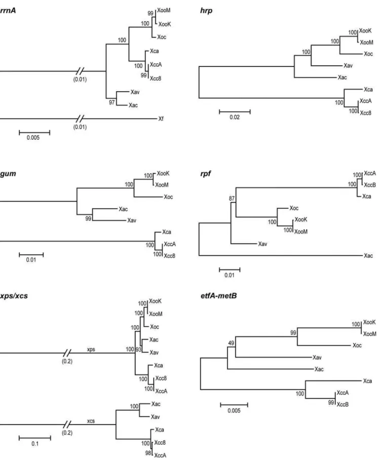
To assess the extent to which relatedness of cluster sequences across strains reflects shared host-or tissue-specificity, a phyloge-netic tree for each cluster was built based on alignments of concatenated sequences of the predicted gene products of that cluster (Figure 2). Thus, for thegumgene cluster, the amino acid sequences of GumB through GumJ in each strain were joined end to end, and the joined sequences from each strain aligned to one another. The xcs and xps gene clusters were analyzed together, using concatenated sequences of XcsC through XcsM, and of XpsN (the ortholog of XcsC) and XpsD with XpsE through XpsM. For the hrp gene cluster, HpaB through Hpa2 were concatenated and aligned. For therpfcluster, sequences of AcnB through RpfD were used. Differences in gene content precluded alignment of the LPS biosynthetic gene cluster sequences across all genomes. Instead, the predicted amino acid sequences of the bordering etfA and metBgenes were examined. For reference, a phylogenetic tree was generated from an alignment of the rrnA
operon of each strain, rooted by including the rrnA operon of
Xylella fastidiosastrain 9a–5c.
TherrnAtree groups theXanthomonasstrains into three distinct clades consisting of theX. campestrisstrains, the X. oryzae strains, and theX. axonopodisstrains, in agreement with Rademaker et al. [14]. TheX. axonopodisclade is basal, suggesting that these strains most closely resemble the common ancestor of the three clades. In the trees derived for each gene cluster, though the trees are not rooted and relative distances among sequences from different strains vary from those in the rrnA tree, the three clades are generally preserved. This shared overall topology indicates that the
most recent common ancestor of the strains examined contained each of the clusters and that the current sequences are the result of evolution over the course of direct transmission. Exceptions to the shared topology are the positions of Xav and Xac in thehrpandrpf
trees. In these trees, Xav lies between theX. oryzaeclade and Xac, and Xac occupies a distinct, more distant branch. The nucleotide sequence of the corehrpgenes (hrcCthroughhpaB) of Xav is more similar to that of Xoo (94% identity) than to that of Xac (92% identity). The Xac sequence is 99% identical to that of strain 8ra of
X. axonopodispv. glycines (GenBank accession AF499777). In the
rpftree, Xac is markedly distant from the other strains. Individual Rpf protein trees (not shown) indicate that this is due to highly distinct sequences for RpfF and RpfC in Xac. Also, the Xac cluster is missingrpfHand lacks any intergenic space between rpfCand
rpfG. Thus, lateral acquisition and substitution of or within thehrp
andrpfclusters may have taken place in the Xac or Xav lineages. It is also possible, though less likely based on the degree of divergence of the sequences, that theX. axonopodisclade, being the most basal in the phylogeny, acquired a greater degree of sequence diversity at these loci independent of lateral transfer.
Nevertheless, the exceptional sequence relationships forhrpand
rpf genes in Xac and Xav do not correlate with host-or tissue-specificity within the group of strains examined. Also, with regard to host specificity, except for theX. axonopodisstrains, which infect citrus and pepper, respectively, strains within clades infect the same or closely related hosts, so the topology where it is shared with that of therrnAtree is not informative, except that there is no robust clustering of pathogens of monocots versus pathogens of eudicots. That is, there is no correlation of gene cluster sequence with the general class of host colonized, arguing against a defining role of any particular cluster in determining host specificity. With regard to tissue specificity,X. campestrisandX. oryzaeeach contain vascular and non-vascular pathogens, yet across these species, none of the trees group pathogens that colonize the same tissues and therefore provide no evidence of a role in tissue specificity for any of the clusters. The similarity of theetfAandmetBtree to the others indicates that the recombination that gave rise to the observed diversity of gene content at the LPS biosynthesis locus took place within or between these genes.
Sequences predicted to be under selection
Figure 2. Relationships acrossXanthomonasstrains of ribosomal RNA sequences and sequences of pathogenesis-associated gene clusters.Phylogenetic trees generated as described in Materials and Methods are shown.rrnA, therrnAoperon (nucleotide alignment);gum, GumB through GumJ;xps/xcs, XpsD plus XpsE through XpsN, and XcsD through XpsM plus XpsC;hrp, HpaB through Hpa2;rpf, AcnB through RpfD; etfA-metB, EtfA and MetB, which flank the LPS biosynthetic locus (see Figure 1E). Strain abbreviations are as in the text. Sequence fromXylella fastidiosa 9a–5c was used to root therrnAtree. Numbers above and below branch points are bootstrap values (as percent) for neighbor-joining with 1000 replicates. Scale represents relative distance as a function of substitutions over time.
and translocators, as well as the T3S pilus component HrpE [67,102]. Binding of HpaA to HpaB is thought to block effector secretion and allow passage of non-effectors; secretion of HpaA is
postulated then to liberate HpaB and initiate effector secretion [67]. In light of its effector non-discriminatory role in secretion, divergence among HpaA sequences likely relates to its plant Figure 3. Sequences predicted to be under selection within the gene clusters examined.Based on the multiple alignments for each cluster, the Ka/Ks score for each codon was calculated with Selecton [101]. Shown is a plot of the Ka/Ks scores across each cluster using a window size of 50 with an offset of 20 residues, drawn using custom software. The vertical scales refer to the number of residues predicted to be under purifying (left) or positive (right) selection in each window. Evidence for selction (Ka/Ks ratios) is color coded, as shown at upper right, with yellow representing evidence of strong positive selection (high Ka/Ks ratio) and purple purifying selection (low Ka/Ks ratio). Raw selecton output for each alignment is available asData S1.
intracellular function. Positive selection inhrpE, which encodes the T3S pilin subunit, was identified and discussed previously [103].
To address whether the evidence of positive selection we detected might relate to host- or tissue-specificity, we aligned sequences, both nucleotide and predicted amino acid, and constructed trees for each of the xcs and hrp genes individually. Except for HrcS and HrcN, none of the trees show relationships different from strain phylogeny, including the trees forhpaP,hpaA, and hrpE. The HrcS tree groups Xca more closely with the X. axonopodis strains than with the Xcc strains, suggestive of a correlation to tissue-specificity for those strains, but Xoc in that tree groups with the Xoo strains, and HrcS, as a predicted inner membrane, core component of the T3S apparatus, would not be expected to play a direct role in host interactions. The HrcN tree places the X. oryzae strains between Xav and Xac, but this relationship does not reflect host- or tissue-specificity, and HrcN, a cytoplasmic ATPase that drives T3S, like HrcS would not be expected to play a direct role in host interaction.
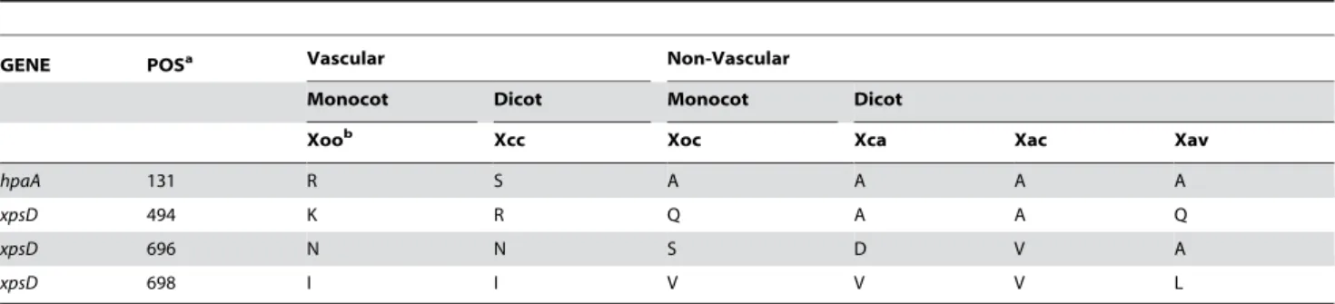
Gene product polymorphisms correlated to tissue-specificity
Irrespective of evidence for positive selection, in the multiple alignment for each cluster, individual residues at each position were examined for polymorphism that correlated to tissue-specificity. To maximize the likelihood of detecting correlations, residues were scored for similarity using several different amino acid substitution matrices (see Materials and Methods). Across all alignments, four positions correlated with tissue-specificity, based on any matrix. These correspond to residue 131 in HpaA and residues 494, 696, and 698 in XpsD (Table 2; positions given relative to the Xoc sequences). The same analysis but with Xoc and Xoo switched in the groupings served as a control to assess the significance of the observed numbers of residues potentially involved in tissue-specificity. Because the control also indicated two positions (one in hrcU and one in XpsD), we cannot exclude the possibility that the residue differences correlated to tissue-specificity listed above are chance events. However, the identity of the genes in which the correlated positions are located, and the concentration of possible tissue-specificity determinants in the C-terminal domain of XpsD are intriguing.
As discussed earlier, HpaA is a substrate of the T3S system that also plays a role in controlling secretion of type III effector and translocator proteins, via interaction with HpaB. Residue 131 in HpaA (a 275 amino acid protein) is between the N-terminal secretion and translocation domain and the C-terminal HpaB-binding domain [67]. An effector function for HpaA has not yet
been identified, but the abundance of positions showing evidence of positive selection and the correlation of residues at position 131 with tissue-specificity are consistent with an important, host-interactive role, and potential for residue 131 in particular, in determining the ability of the bacterium to colonize different host tissues.
XpsD is an outer membrane protein [104]. Members of the T2S:D protein family, to which XpsD belongs, are postulated to function as gatekeepers for type II secretion, demonstrating species-specific function for different type II secretion substrates [105,106]. XpsD in different strains could direct the secretion of different sets of proteins adapted for function in different tissues, or, as a bacterial outer membrane protein, XpsD could function as an elicitor of tissue-specific plant responses that confer interaction specificity. Consistent with the latter hypothesis, two of the three positions in XpsD that correlate with tissue-specificity reside in the hypervariable C-terminal S domain. As demonstrated with PulD ofKlebsiella, the S domain interacts with a specific lipoprotein (PulS in the case of PulD) that pilots it to the outer membrane and is thought to aide in homo-oligodimerization. In complex with this lipoprotein, the S domain is predicted to be largely exposed on the bacterial cell surface [107].
Conclusion
Several pathogenesis-associated gene clusters across eight
Xanthomonas strains were compared to assess potential contribu-tions of these clusters to host- and tissue-specificity. The strains fall into three clades, corresponding to species, each containing two pathovars. Included in these pathovar pairs are pathogens that infect the same host with different tissue-specificity, as well as pathogens that infect different hosts, with shared tissue-specificity. One of the clades is made up of monocot pathogens, and the other two are pathogens of eudicots. One of the eudicot pathogen clades is more closely related to the monocot pathogen clade than to the other eudicot pathogen clade (Figure 2). For this broadly representative group of plant pathogenic Xanthomonas strains, adaptation to different plant hosts and specific tissues within a host does not include major alteration or exchange of content within any of the gene clusters examined. Complex relationships within an LPS biosynthesis gene cluster indicate a history of horizontal transfer events and diversifying selection, suggesting an adaptive role, but these relationships do not correlate with host- or tissue-specificity.
Positive selection is evident at sites in several genes in thexcsand
hrpclusters. Nevertheless, none of thexcsorhrpgenes individually, when compared across strains, showed relationships that group
Table 2. Amino acid residues in alignments of pathogenesis-associated gene products that correlate with tissue-specificity across eightXanthomonasstrains.
GENE POSa Vascular Non-Vascular
Monocot Dicot Monocot Dicot
Xoob Xcc Xoc Xca Xac Xav
hpaA 131 R S A A A A
xpsD 494 K R Q A A Q
xpsD 696 N N S D V A
xpsD 698 I I V V V L
aPosition in the Xoc gene product.
bStrain abbreviations are as in the text. The Xoo and Xcc strains are vascular pathogens; Xoc, Xca, and theX. axonopodisstrains are non-vascular pathogens.
pathogens from different clades based on host specificity (i.e., eudicot vs. monocot pathogens) or tissue-specificity (i.e., vascular vs. non-vascular pathogens). Across all alignments, however, four positions showed correlation of amino acid residue identity with tissue specificity, revealing the T3S regulatory and putative effector gene hpaA and the type II secretory pathway genexpsD
as candidate tissue-specificity determinants.
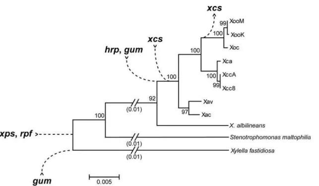
Comparison with other members of the Xanthomonadaceae revealed that therpfandxpsgene clusters were present early in the evolution of the group, that thehrp,gum, andxcsgene clusters were acquired later, and that thexcscluster was subsequently lost in the lineage that gave rise to the X. oryzae clade (Figure 4). This pattern of acquisition and loss, coupled with the demonstrated importance of thehrpandgumclusters to pathogenesis in several
Xanthomonasspp. and the lack of evidence for an important role of
xcsgenes in plant pathogen interactions, suggest that acquisition of thehrpandgumclusters were critical steps in the evolution of plant pathogenicity inXanthomonas.
The results of our study provide insight into the nature of the first
Xanthomonasgenome, and suggest that differentiation ofXanthomonas
species and pathovars with respect to host and tissue specificity resulted from subtle changes in a small number of individual genes in thegum, hrp, xps,xcs, orrpfclusters, modifications in non-coding, regulatory sequences in the clusters, and/or differences outside the clusters. Functional characterization of the differences discovered in
hpaAandxpsD, expression analysis of the genes in each cluster, and examination of differences outside the clusters that correlate to host and tissue-specificity, particularly among regulatory targets or secretory substrates, or genes for environmental sensing, are important next steps.
Methods
Genome sequences and annotation
The genome sequences and annotation used are presented in Table S1. For some sequences, annotation was confirmed and refined manually by performing BLASTP comparisons against the non-redundant protein database (National Center for Biotechnol-ogy Information) and against otherXanthomonasgenomes directly. Orthologs were defined as reciprocal best matches by BLASTP with an e-value minimum of e220
and 60% coverage [19].
Gene cluster comparisons
The genome clusters and corresponding genes were retrieved from genome sequences by referencing genome annotation. The coordinates for each cluster in each strain are presented inTable S2. Orthologous genes were grouped together, and in each group similarity to the gene in Xoc (if present) was calculated based on predicted amino acid sequence using the needle program of the EMBOSS package [108]. IS elements and tRNA genes were identified and mapped with BLASTN. Custom software was used to scan genome sequences to detect perfect and imperfect plant-inducible promoter sequences [PIP boxes, 109]. Overall sequence similarity of each cluster sequence to the Xoc sequence for that cluster was calculated using the stretcher program of the EMBOSS package [108]. GC content was plotted using a window size of 160 bp and a step size of 40 bp. In select comparisons, the genome ofXylella fastidiosastrain 9a–5c was included in the analysis but not shown. Schematic representations of the clusters were generated using custom software with the pre-calculated informa-tion above.
Figure 4. Inferred pattern of acquisition or loss of five pathogenesis-associated gene clusters inXanthomonas.Based on comparison of genome sequences and other data amongXanthomonasstrains and the close relativesXylella fastidiosaandStenotrophomonas maltophilia(see text), an inferred pattern of acquisition or loss of five pathogenesis associated gene clusters during the evolution of differentXanthomonaslineages is shown, superimposed on a phylogenetic tree drawn from an alignment of 16s rRNA gene sequences. Potential horizontal exchange ofhrpandrpf sequences affecting theX. axonopodisclade, discussed in the text, is not depicted. Strain abbreviations are as in the text. ForX. fastidiosa, the strain 9a–5c sequence was used. ForS. maltophilia, the strain K279a sequence was used.
Phylogenetic analyses
Concatenated protein sequences for each cluster and nucleotide sequences for the rrnA operon were aligned using ClustalW, Version 1.83, with default parameters [110]. Aligned sequences were inspected and manually adjusted when necessary. Regions with gaps between the strains were excluded to avoid problems reflecting start codon misassignment. Neighbor-joining trees were generated using PHYLIP [111] and displayed using Mega 3.1 [112]. Bootstrap values were derived from 1,000 replicates in each case to validate tree topology and are expressed as percent. Sequence from Xylella fastidiosa strain 9a–5c was used as an outgroup. For individual genes with codons showing evidence of positive selection, protein sequences were aligned using ClustalW, and trees were generated using PHYLIP. The PHYLIP programs PROTDIST and DNADIST, which use maximum likelihood estimates, were used to calculate distances, FITCH was used to estimate phylogenies from the distance matrices, and DRAW-TREE was used to draw unrooted trees.
Analysis of synonymous and non-synonymous substitutions
For analysis of synonymous and non-synonymous substitutions, nucleotide sequences of genes conserved across strains for each cluster were concatenated, using a 99 N spacer between individual gene sequences, and submitted to Selecton Version 2.4 (http:// selecton.tau.ac.il) [101] with the Xoc sequence as consensus. The Mechanistic Empirical Combination (MEC) model was used with the ‘‘high precision’’ option selected. With custom software, Ka/ Ks scores calculated by Selecton were plotted using a 50 amino acid window size and 20 amino acid offset. For each cluster that contained residues with evidence of positive selection (high Ka/Ks ratio), multiple alignments of each gene in the cluster across strains were generated and submitted to Selecton individually for confirmation, using the same parameters as above. For each of these multiple alignments, trees were also generated, as described above.
Identification of gene product polymorphisms correlated to tissue-specificity
The multiple alignment of each cluster was scanned for positions at which residues in the Xcc and Xoo sequences vs. residues in the Xoc, Xca, Xac, and Xav sequences were more similar within these groups than across them. For this analysis, gaps in the alignments were retained. To minimize artifacts of alignment, only positions with at least one completely conserved
neighbor were taken into consideration. The amino acid substitution matrices BLOSUM45, BLOSUM62, BLOSUM80, PAM30, PAM70 were used to assign substitution scores at each position in all pairwise comparisons, and then for each position the mean of the substitution scores within the groups of strains with like tissue-specificity, i.e., (Score(Xoo, Xcc)+Score(Xoc, Xca))/2, was compared with the mean of the scores for substitutions across groups of shared tissue specificity, i.e., (Score(Xoo, Xoc)+ Scor-e(Xoo, Xca)+Score(Xcc, Xoc)+Score(Xcc, Xca))/4. Any positions for which the mean within-group score was greater than the mean across-group score and at which there were no identical residues in any of the across-group comparisons were retained. For comparison to assess significance, these calculations were repeated using a tissue non-specific grouping of strains formed by switching Xoo with Xoc.
Supporting Information
Supporting information includes 1) Table S1, Xanthomonas
genome sequences examined in this study, 2) Table S2, Coordinates (bp) of the gene clusters examined in the eight
Xanthomonas genomes, 3) Text S1, Additional details of gene cluster comparisons, and 4)Data S1, Raw Selecton output used to generate Figure 3.
Text S1 Additional details of gene cluster comparisons Found at: doi:10.1371/journal.pone.0003828.s001 (0.12 MB PDF)
Table S1 Xanthomonasgenome sequences examined in this study. Found at: doi:10.1371/journal.pone.0003828.s002 (0.10 MB PDF)
Table S2 Coordinates (bp) of the gene clusters examined in the eightXanthomonasgenomes.
Found at: doi:10.1371/journal.pone.0003828.s003 (0.06 MB PDF)
Data S1 Selecton results.
Found at: doi:10.1371/journal.pone.0003828.s004 (0.10 MB ZIP)
Author Contributions
Conceived and designed the experiments: MAVS FW JL VB AJB. Performed the experiments: HL PBP MAVS. Analyzed the data: HL PBP MAVS FW RR JMD JL RVS VB AJB. Contributed reagents/materials/ analysis tools: HL PBP RVS VB. Wrote the paper: HL PBP MAVS FW RR JMD JL RVS VB AJB. Contributed data: PDR SLS.
References
1. Perna NT, Plunkett G III, Burland V, Mau B, Glasner JD, et al. (2001) Genome sequence of enterohaemorrhagicEscherichia coliO157:H7. Nature 409: 529–533.
2. Parkhill J, Wren BW, Mungall K, Ketley JM, Churcher C, et al. (2000) The genome sequence of the food-borne pathogen Campylobacter jejuni reveals hypervariable sequences. Nature 403: 665–668.
3. Deng W, Burland V, Plunkett G 3rd, Boutin A, Mayhew GF, et al. (2002) Genome sequence ofYersinia pestisKIM. J Bacteriol 184: 4601–4611. 4. Brosch R, Pym AS, Gordon SV, Cole ST (2001) The evolution of
mycobacterial pathogenicity: clues from comparative genomics. Trends Microbiol 9: 452–458.
5. Raskin DM, Seshadri R, Pukatzki SU, Mekalanos JJ (2006) Bacterial genomics and pathogen evolution. Cell 124: 703–714.
6. Van Sluys MA, Monteiro-Vitorello CB, Camargo LEA, Menck CFM, da Silva ACR, et al. (2002) Comparative genomic analysis of plant-associated bacteria. Annu Rev Phytopathol 40: 169–189.
7. Toth IK, Pritchard L, Birch PR (2006) Comparative genomics reveals what makes an enterobacterial plant pathogen. Annu Rev Phytopathol 44: 305–336. 8. Comas I, Moya A, Azad RK, Lawrence JG, Gonzalez-Candelas F (2006) The evolutionary origin of Xanthomonadales genomes and the nature of the horizontal gene transfer process. Mol Biol Evol 23: 2049–2057.
9. Lima WC, Sluys M-AV, Menck CFM (2005) Non-gamma-proteobacteria gene islands contribute to theXanthomonasgenome. OMICS: J Integrative Biol 9: 160–172.
10. Lima WC, Paquola AC, Varani AM, Van Sluys MA, Menck CF (2008) Laterally transferred genomic islands in Xanthomonadales related to pathogenicity and primary metabolism. FEMS Microbiol Lett 281: 87– 97.
11. Hayward A (1993) The hosts ofXanthomonas. In: Swings J, Civerolo EL, eds. Xanthomonas. London: Chapman and Hall. pp 1–18.
12. Goncalves ER, Rosato YB (2002) Phylogenetic analysis ofXanthomonasspecies based upon 16S-23S rDNA intergenic spacer sequences. Int J Syst Evol Microbiol 52: 355–361.
13. Rademaker JLW, Hoste B, Louws FJ, Kersters K, Swings J, et al. (2000) Comparison of AFLP and rep-PCR genomic fingerprinting with DNA-DNA homology studies:Xanthomonasas a model system. Int J Syst Evol Microbiol 50: 665–677.
14. Rademaker JLW, Louws FJ, Schultz MH, Rossbach U, Vauterin L, et al. (2005) A comprehensive species to strain taxonomic framework forXanthomonas. Phytopathology 95: 1098–1111.
16. Nino-Liu DO, Ronald PC, Bogdanove AJ (2006)Xanthomonas oryzaepathovars: model pathogens of a model crop. Mol Plant Pathol 7: 303–324.
17. Daniels MJ (1989) Pathogenicity ofXanthomonasand related bacteria towards plants. In: Hopwood DA, Chater KF, eds. Genetics of Bacterial Diversity. San Diego: Academic Press Ltd. pp 353–372.
18. Qian W, Jia Y, Ren SX, He YQ, Feng JX, et al. (2005) Comparative and functional genomic analyses of the pathogenicity of phytopathogenXanthomonas campestrispv. campestris. Genome Res 15: 757–767.
19. da Silva AC, Ferro JA, Reinach FC, Farah CS, Furlan LR, et al. (2002) Comparison of the genomes of twoXanthomonaspathogens with differing host specificities. Nature 417: 459–463.
20. Lee BM, Park YJ, Park DS, Kang HW, Kim JG, et al. (2005) The genome sequence ofXanthomonas oryzaepathovar oryzae KACC10331, the bacterial blight pathogen of rice. Nucleic Acids Res 33: 577–586.
21. Ochiai H, Inoue Y, Takeya M, Sasaki A, Kaku H (2005) Genome sequence of Xanthomonas oryzaepv. oryzae suggests contribution of large numbers of effector genes and insertion sequences to its race diversity. Jpn Agr Res Q 39: 275–287. 22. Thieme F, Koebnik R, Bekel T, Berger C, Boch J, et al. (2005) Insights into genome plasticity and pathogenicity of the plant pathogenic bacterium Xanthomonas campestris pv. vesicatoria revealed by the complete genome sequence. J Bacteriol 187: 7254–7266.
23. Kamoun S, Kado CI (1990) Phenotypic switching affecting chemotaxis, xanthan production, and virulence inXanthomonas campestris. Appl Environ Microbiol 56: 3855–3860.
24. Raymundo AK, Leach JE (1993) Amplification and sequence analysis of the upstream region of thehrpXogene homolog inXanthomonas oryzaepv. oryzicola. Philippine J Biotechnol 4: 29–38.
25. Salzberg SL, Sommer DD, Schatz MC, Phillippy AM, Rabinowicz PD, et al. (2008) Genome sequence and rapid evolution of the rice pathogenXanthomonas oryzaepv. oryzae PXO99A
. BMC Genomics 9: 204.
26. Vorholter FJ, Schneiker S, Goesmann A, Krause L, Bekel T, et al. (2008) The genome of Xanthomonas campestris pv. campestris B100 and its use for the reconstruction of metabolic pathways involved in xanthan biosynthesis. J Biotechnol 134: 33–45.
27. Gupta RS, Sneath PH (2007) Application of the character compatibility approach to generalized molecular sequence data: branching order of the proteobacterial subdivisions. J Mol Evol 64: 90–100.
28. Bhattacharyya A, Stilwagen S, Ivanova N, D’Souza M, Bernal A, et al. (2002) Whole-genome comparative analysis of three phytopathogenicXylella fastidiosa strains. Proc Natl Acad Sci USA 99: 12403–12408.
29. Simpson AJ, Reinach FC, Arruda P, Abreu FA, Acencio M, et al. (2000) The genome sequence of the plant pathogenXylella fastidiosa. Nature 406: 151–159. 30. Van Sluys MA, de Oliveira MC, Monteiro-Vitorello CB, Miyaki CY, Furlan LR, et al. (2003) Comparative analyses of the complete genome sequences of Pierce’s disease and citrus variegated chlorosis strains ofXylella fastidiosa. J Bacteriol 185: 1018–1026.
31. Crossman LC, Gould VC, Dow JM, Vernikos GS, Okazaki A, et al. (2008) The complete genome, comparative and functional analysis of Stenotrophomonas maltophiliareveals an organism heavily shielded by drug resistance determi-nants. Genome Biol 9: R74.
32. Purcell AH, Hopkins DL (1996) Fastidious xylem-limited bacterial plant pathogens. Annu Rev Phytopathol 34: 131–151.
33. Berg G, Roskot N, Smalla K (1999) Genotypic and phenotypic relationships between clinical and environmental isolates ofStenotrophomonas maltophilia. J Clin Microbiol 37: 3594–3600.
34. Vanderslice RW, Doherty DH, Capage MA, Betlach MR, Hassler RA, et al. (1989) Genetic engineering of polysaccharide structure inXanthomonas campestris. In: Crescenzi V, Dea ICM, Paoletti S, Stivala SS, Sutherland IW, eds. Biomedical and Biotechnological Advances in Industrial Polysaccharides. New York: Gordon and Breach Science Publishers. pp 145–156.
35. Katzen F, Ferreiro DU, Oddo CG, Ielmini MV, Becker A, et al. (1998) Xanthomonas campestris pv. campestris gum mutants: effects on xanthan biosynthesis and plant virulence. J Bacteriol 180: 1607–1617.
36. Dow JM, Daniels MJ, Dums F, Turner PC, Gough C (1989) Genetic and biochemical analysis of protein export fromXanthomonas campestris. J Cell Sci Suppl 11: 59–72.
37. Arlat M, Gough CL, Barber CE, Boucher C, Daniels MJ (1991)Xanthomonas campestriscontains a cluster ofhrpgenes related to the largerhrpcluster of Pseudomonas solanacearum. Mol Plant-Microbe Interact 4: 593–601.
38. Dow JM, Crossman L, Findlay K, He YQ, Feng JX, et al. (2003) Biofilm dispersal inXanthomonas campestrisis controlled by cell-cell signaling and is required for full virulence to plants. Proc Natl Acad Sci USA 100: 10995–11000.
39. Dharmapuri S, Yashitola J, Vishnupriya MR, Sonti RV (2001) Novel genomic locus with atypical G+C content that is required for extracellular polysaccha-ride production and virulence inXanthomonas oryzae pv. oryzae. Mol Plant-Microbe Interact 14: 1335–1339.
40. Vorholter FJ, Niehaus K, Puhler A (2001) Lipopolysaccharide biosynthesis in Xanthomonas campestrispv. campestris: a cluster of 15 genes is involved in the biosynthesis of the LPS O-antigen and the LPS core. Mol Genet Genomics 266: 79–95.
41. Denny TP (1995) Involvement of bacterial polysaccharides in plant pathogenesis. Annu Rev Phytopathol 33: 173–197.
42. Dharmapuri S, Sonti RV (1999) A transposon insertion in thegumGhomologue of Xanthomonas oryzaepv. oryzae causes loss of extracellular polysaccharide production and virulence. FEMS Microbiol Lett 179: 53–59.
43. Rigano LA, Siciliano F, Enrique R, Sendin L, Filippone P, et al. (2007) Biofilm formation, epiphytic fitness, and canker development inXanthomonas axonopodis pv. citri. Mol Plant-Microbe Interact 20: 1222–1230.
44. Dunger G, Relling VM, Tondo ML, Barreras M, Ielpi L, et al. (2007) Xanthan is not essential for pathogenicity in citrus canker but contributes toXanthomonas epiphytic survival. Arch Microbiol 188: 127–135.
45. Kiraly Z, El-Zahaby HM, Klement Z (1997) Role of extracellular polysaccharide (EPS) slime of plant pathogenic bacteria in protecting cells to reactive oxygen species. J Phytopathol 145: 59–68.
46. Yoon KH, Cho JY (2007) Transcriptional analysis of thegumgene cluster from Xanthomonas oryzaepathovar oryzae. Biotechnol Lett 29: 95–103.
47. Champoiseau P, Daugrois JH, Pieretti I, Cociancich S, Royer M, et al. (2006) High variation in pathogenicity of genetically closely related strains of Xanthomonas albilineans, the sugarcane leaf scald pathogen, in Guadeloupe. Phytopathology 96: 1081–1091.
48. Fontaniella B, Rodriguez CW, Pinon D, Vicente C, Legaz M-E (2002) Identification of xanthans isolated from sugarcane juices obtained from scalded plants infected byXanthomonas albilineans. J Chromatogr B 770: 275–281. 49. da Silva FR, Vettore AL, Kemper EL, Leite A, Arruda P (2001) Fastidian gum:
theXylella fastidiosaexopolysaccharide possibly involved in bacterial pathoge-nicity. FEMS Microbiol Lett 203: 165–171.
50. Blanch M, Legaz M-E, Vicente C (2008) Xanthan production byXanthomonas albilineansinfecting sugarcane stalks. J Plant Physiol 165: 366–374. 51. d’Enfert C, Ryter A, Pugsley AP (1987) Cloning and expression inEscherichia
coliof theKlebsiella pneumoniaegenes for production, surface localization and secretion of the lipoprotein pullulanase. EMBO J 6: 3531–3538.
52. Possot O, d’Enfert C, Reyss I, Pugsley AP (1992) Pullulanase secretion in Escherichia coliK-12 requires a cytoplasmic protein and a putative polytopic cytoplasmic membrane protein. Mol Microbiol 6: 95–105.
53. Dums F, Dow JM, Daniels MJ (1991) Structural characterization of protein secretion genes of the bacterial phytopathogenXanthomonas campestrispathovar campestris: relatedness to secretion systems of other gram-negative bacteria. Mol Gen Genet 229: 357–364.
54. Ray SK, Rajeshwari R, Sharma Y, Sonti RV (2002) A high-molecular-weight outer membrane protein ofXanthomonas oryzaepv. oryzae exhibits similarity to non-fimbrial adhesins of animal pathogenic bacteria and is required for optimum virulence. Mol Microbiol 46: 637–647.
55. Sun QH, Hu J, Huang GX, Ge C, Fang RX, et al. (2005) Type-II secretion pathway structural genexpsE, xylanase- and cellulase secretion and virulence in Xanthomonas oryzaepv. oryzae. Plant Pathol 54: 15–21.
56. Sandkvist M (2001) Type II secretion and pathogenesis. Infect Immun 69: 3523–3535.
57. Brunings AM, Gabriel DW (2003)Xanthomonas citri: breaking the surface. Mol Plant Pathol 4: 141–157.
58. Peabody CR, Chung YJ, Yen MR, Vidal-Ingigliardi D, Pugsley AP, et al. (2003) Type II protein secretion and its relationship to bacterial type IV pili and archaeal flagella. Microbiology 149: 3051–3072.
59. Harding NE, Cleary JM, Cabanas DK, Rosen IG, Kang KS (1987) Genetic and physical analyses of a cluster of genes essential for xanthan gum biosynthesis inXanthomonas campestris. J Bacteriol 169: 2854–2861.
60. Moreira LM, De Souza RF, Almeida Jr NF, Setubal JC, Oliveira JC, et al. (2004) Comparative genomics analyses of citrus-associated bacteria. Annu Rev Phytopathol 42: 163–184.
61. Chen Y, Shiue SJ, Huang CW, Chang JL, Chien YL, et al. (2005) Structure and function of the XpsE N-terminal domain, an essential component of the Xanthomonas campestristype II secretion system. J Biol Chem 280: 42356–42363. 62. Cornelis GR (2006) The type III secretion injectisome. Nat Rev Microbiol 4:
811–825.
63. Sugio A, Yang B, White FF (2005) Characterization of thehrpFpathogenicity peninsula of Xanthomonas oryzaepv. oryzae. Mol Plant-Microbe Interact 18: 546–554.
64. Bogdanove AJ, Beer SV, Bonas U, Boucher C, Collmer A, et al. (1996) Unified nomenclature for broadly conservedhrpgenes of phytopathogenic bacteria. Mol Microbiol 20: 681–683.
65. Kim JG, Park BK, Yoo CH, Jeon E, Oh J, et al. (2003) Characterization of the Xanthomonas axonopodispv. glycines Hrp pathogenicity island. J Bacteriol 185: 3155–3166.
66. Buttner D, Lorenz C, Weber E, Bonas U (2006) Targeting of two effector protein classes to the type III secretion system by a HpaC- and HpaB-dependent protein complex fromXanthomonas campestrispv. vesicatoria. Mol Microbiol 59: 513–527.
67. Lorenz C, Kirchner O, Egler M, Stuttmann J, Bonas U, et al. (2008) HpaA from Xanthomonasis a regulator of type III secretion. Mol Microbiol 69: 344–360.
68. Noel L, Thieme F, Nennstiel D, Bonas U (2002) Two novel type III-secreted proteins ofXanthomonas campestrispv. vesicatoria are encoded within thehrp pathogenicity island. J Bacteriol 184: 1340–1348.
70. Moore ER, Kruger AS, Hauben L, Seal SE, Daniels MJ, et al. (1997) 16S rRNA gene sequence analyses and inter- and intrageneric relationships of Xanthomonasspecies andStenotrophomonas maltophilia. FEMS Microbiol Lett 151: 145–153.
71. Birch RG, Patil SS (1987) Correlation between albicidin production and chlorosis induction byXanthomonas albilineans, the sugarcane leaf scald pathogen. Physiol Mol Plant Pathol 30: 199–206.
72. Birch RG, Patil SS (1987) Evidence that an albicidin-like phytotoxin induces chlorosis in sugarcane leaf scald disease by blocking plastid DNA replication. Physiol Mol Plant Pathol 30: 207–214.
73. Dow JM, Daniels MJ (2000)Xylellagenomics and bacterial pathogenicity to plants. Yeast 17: 263–271.
74. Barber CE, Tang JL, Feng JX, Pan MQ, Wilson TJ, et al. (1997) A novel regulatory system required for pathogenicity of Xanthomonas campestris is mediated by a small diffusible signal molecule. Mol Microbiol 24: 555–566. 75. Dow JM, Feng JX, Barber CE, Tang JL, Daniels MJ (2000) Novel genes
involved in the regulation of pathogenicity factor production within therpfgene cluster ofXanthomonas campestris. Microbiology 146: 885–891.
76. Slater H, Alvarez-Morales A, Barber CE, Daniels MJ, Dow JM (2000) A two-component system involving an HD-GYP domain protein links cell-cell signalling to pathogenicity gene expression in Xanthomonas campestris. Mol Microbiol 38: 986–1003.
77. Wilson TJG, Bertrand N, Tang JL, Feng JX, Pan MQ, et al. (1998) TherpfA gene ofXanthomonas campestrispathovar campestris, which is involved in the regulation of pathogenicity factor production, encodes an aconitase. Mol Microbiol 28: 961–970.
78. Wang LH, He YW, Gao YF, Wu JE, Dong YH, et al. (2004) A bacterial cell-cell communication signal with cross-kingdom structural analogues. Mol Microbiol 51: 903–912.
79. Ryan RP, Fouhy Y, Lucey JF, Crossman LC, Spiro S, et al. (2006) Cell-cell signaling inXanthomonas campestrisinvolves an HD-GYP domain protein that functions in cyclic di-GMP turnover. Proc Natl Acad Sci USA 103: 6712–6717. 80. Chatterjee S, Sonti RV (2002)rpfFmutants ofXanthomonas oryzaepv. oryzae are deficient for virulence and growth under low iron conditions. Mol Plant-Microbe Interact 15: 463–471.
81. Siciliano F, Torres P, Sendı´n L, Bermejo C, Filippone P, et al. (2006) Analysis of the molecular basis ofXanthomonas axonopodispv.citripathogenesis inCitrus limon. Electron J Biotechnol 9: 199–204.
82. Tang JL, Feng JX, Li QQ, Wen HX, Zhou DL, et al. (1996) Cloning and characterization of therpfCgene ofXanthomonas oryzaepv oryzae: involvement in exopolysaccharide production and virulence to rice. Mol Plant-Microbe Interact 9: 664–666.
83. Wang L, Makino S, Subedee A, Bogdanove AJ (2007) Novel candidate virulence factors in rice pathogenXanthomonas oryzaepv.oryzicolaas revealed by mutational analysis. Appl Environ Microbiol 73: 8023–8027.
84. Boon C, Deng Y, Wang LH, He Y, Xu JL, et al. (2008) A novel DSF-like signal from Burkholderia cenocepacia interferes with Candida albicans morphological transition. ISME J 2: 27–36.
85. Scarpari LM, Lambais MR, Silva DS, Carraro DM, Carrer H (2003) Expression of putative pathogenicity-related genes inXylella fastidiosagrown at low and high cell density conditionsin vitro. FEMS Microbiol Lett 222: 83–92. 86. Simionato AVC, da Silva DS, Lambais MR, Carrilho E (2007) Characteriza-tion of a putativeXylella fastidiosadiffusible signal factor by HRGC-EI-MS. J Mass Spectrom 42: 490–496.
87. Fouhy Y, Scanlon K, Schouest K, Spillane C, Crossman L, et al. (2007) Diffusible signal factor-dependent cell-cell signaling and virulence in the nosocomial pathogenStenotrophomonas maltophilia. J Bacteriol 189: 4964–4968. 88. Raetz CR, Whitfield C (2002) Lipopolysaccharide endotoxins. Annu Rev
Biochem 71: 635–700.
89. Mooi FR, Bik EM (1997) The evolution of epidemic Vibrio choleraestrains. Trends Microbiol 5: 161–165.
90. Reeves PP, Wang L (2002) Genomic organization of LPS-specific loci. Curr Top Microbiol Immunol 264: 109–135.
91. Newman MA, Dow JM, Daniels MJ (2001) Bacterial lipopolysaccharides and plant-pathogen interactions. Eur J Plant Pathol 107: 95–102.
92. Medzhitov R, Janeway CA (1997) Innate Immunity: The virtues of a nonclonal system of recognition. Cell 91: 295–298.
93. Mackey D, McFall AJ (2006) MAMPs and MIMPs: proposed classifications for inducers of innate immunity. Mol Microbiol 61: 1365–1371.
94. Silipo A, Molinaro A, Sturiale L, Dow JM, Erbs G, et al. (2005) The elicitation of plant innate immunity by lipooligosaccharide ofXanthomonas campestris. J Biol Chem 280: 33660–33668.
95. Bedini E, De Castro C, Erbs G, Mangoni L, Dow JM, et al. (2005) Structure-dependent modulation of a pathogen response in plants by synthetic O-antigen polysaccharides. J Am Chem Soc 127: 2414–2416.
96. Lerouge I, Vanderleyden J (2002) O-antigen structural variation: mechanisms and possible roles in animal/plant-microbe interactions. FEMS Microbiol Rev 26: 17–47.
97. Patil PB, Sonti RV (2004) Variation suggestive of horizontal gene transfer at a lipopolysaccharide (lps) biosynthetic locus inXanthomonas oryzaepv. oryzae, the bacterial leaf blight pathogen of rice. BMC Microbiol 4: 40.
98. Koplin R, Arnold W, Hotte B, Simon R, Wang G, et al. (1992) Genetics of xanthan production inXanthomonas campestris: thexanAandxanB genes are involved in UDP-glucose and GDP-mannose biosynthesis. J Bacteriol 174: 191–199.
99. Steinmann D, Koplin R, Puhler A, Niehaus K (1997)Xanthomonas campestrispv. campestris lpsI and lpsJ genes encoding putative proteins with sequence similarity to the alpha- and beta-subunits of 3-oxoacid CoA-transferases are involved in LPS biosynthesis. Arch Microbiol 168: 441–447.
100. Patil PB, Bogdanove AJ, Sonti RV (2007) The role of horizontal transfer in the evolution of a highly variable lipopolysaccharide biosynthesis locus in xanthomonads that infect rice, citrus and crucifers. BMC Evol Biol 7: 243. 101. Stern A, Doron-Faigenboim A, Erez E, Martz E, Bacharach E, et al. (2007)
Selecton 2007: advanced models for detecting positive and purifying selection using a Bayesian inference approach. Nucleic Acids Res 35: W506–511. 102. Huguet E, Hahn K, Wengelnik K, Bonas U (1998)hpaAmutants ofXanthomonas
campestrispv. vesicatoria are affected in pathogenicity but retain the ability to induce host-specific hypersensitive reaction. Mol Microbiol 29: 1379–1390. 103. Weber E, Koebnik R (2005) Domain structure of HrpE, the Hrp pilus subunit
ofXanthomonas campestrispv. vesicatoria. J Bacteriol 187: 6175–6186. 104. Chen LY, Chen DY, Miaw J, Hu NT (1996) XpsD, an outer membrane
protein required for protein secretion byXanthomonas campestrispv. campestris, forms a multimer. J Biol Chem 271: 2703–2708.
105. Lindeberg M, Salmond GP, Collmer A (1996) Complementation of deletion mutations in a cloned functional cluster ofErwinia chrysanthemi outgenes with Erwinia carotovora out homologues reveals OutC and OutD as candidate gatekeepers of species-specific secretion of proteins via the type II pathway. Mol Microbiol 20: 175–190.
106. Bouley J, Condemine G, Shevchik VE (2001) The PDZ domain of OutC and the N-terminal region of OutD determine the secretion specificity of the type II out pathway ofErwinia chrysanthemi. J Mol Biol 308: 205–219.
107. Nouwen N, Ranson N, Saibil H, Wolpensinger B, Engel A, et al. (1999) Secretin PulD: association with pilot PulS, structure, and ion-conducting channel formation. Proc Natl Acad Sci U S A 96: 8173–8177.
108. Rice P, Longden I, Bleasby A (2000) EMBOSS: the European Molecular Biology Open Software Suite. Trends Genet 16: 276–277.
109. Tsuge S, Terashima S, Furutani A, Ochiai H, Oku T, et al. (2005) Effects on promoter activity of base substitutions in the cis-acting regulatory element of HrpXoregulons inXanthomonas oryzaepv. oryzae. J Bacteriol 187: 2308–2314. 110. Thompson JD, Higgins DG, Gibson TJ (1994) CLUSTAL W: improving the
sensitivity of progressive multiple sequence alignment through sequence weighting, position-specific gap penalties and weight matrix choice. Nucleic Acids Res 22: 4673–4680.
111. Felsenstein J (2007) PHYLIP (Phylogeny Inference Package) version 3.67. Distributed by the author. Department of Genome Sciences, University of Washington, Seattle.