INTRODUCTION
Metallothioneins (MTs) are an extensive and diverse family of small cysteine-rich proteins found through-out the animal and plant kingdoms, as well as in some prokaryotes. MTs can sequester several metal ions, primarily Cu++ and Zn++ under normal
physi-ological conditions, and also more toxic metals (like Cd++ or Hg++) following exposure (K a g i and
Va l l e e , 1960). Their metal-binding ability suggests that MTs are involved in metal homeostasis and detoxification, but their definite physiological func-tion is still a matter of debate (F i s h e r et al., 1998; P a l m i t e r,1998).
The available scarce data on plant metallothio-neins were mainly obtained in gene expression studies. Plant MT genes have been found to be expressed in all plant organs - leaves, seeds, roots, fruits, and are also induced by various abiotic and biotic factors, such as metal ions and hormones (C o b b e t and G o l d s b r o u g h , 2002). Plant MTs have been suggested to play important roles in maintaining the homeostasis of essential transition metals, detoxification of toxic metals, and protec-tion against oxidative stress (P a l m i t e r , 1998, C o y l e et al., 2002; Wo n g et al., 2004, C o b b e t and G o l d s b r o u g h , 2002). Moreover, the diverse
expression patterns of MT genes suggest that MT-like protein variants differing in sequence may also have different functions in specific tissues (C o b b e t and G o l d s b r o u g h , 2002). A number of investi-gations demonstrated that these proteins could have distinct but overlapping functions in the multigene family in a single species (G u o et al., 2003).
The main approach in investigations of gene expression regulation in general is analysis of pro-moter activity at the transcriptional level. However, the increasing evidence that post-transcriptional mechanisms are critical in gene regulation leads to the conclusion that study of gene expression should include consideration of RNA-based gene regulation involving the 5’- and 3’- untranslated regions (UTRs) (H e s k e t h et al., 1996, 2002). Given that, there is considerable scope for regula-tory motifs within 3’-UTRs. In different animal and plant mRNAs, 3’-UTRs have been reported to be involved in regulation of mRNA stability, transla-tion, and localization (H e s k e t h and V i l l e t t e , 2002). There are some results on animal MTs which indicate that the association of MT mRNA with the cytoskeleton around the nucleus, depending upon its 3’-UTRs, is essential for efficient shuttling of the protein into the nucleus during the G1 to S phase transition (C h a b a n o n et al., 2004).
TWO METALLOTHIONEIN GENE FAMILY MEMBERS IN BUCKWHEAT:
EXPRESSION ANALYSIS IN FLOODING STRESS USING REAL TIME RT-PCR TECHNOLOGY
DRAGANA B. MAJIĆ, JELENA T. SAMARDŽIĆ, MIRA Đ. MILISAVLJEVIĆ, A. M. KRSTIĆ, and VESNA R. MAKSIMOVIĆ
Institute of Molecular Genetics and Genetic Engineering, 11010 Belgrade, Serbia.
Abstract — Metallothioneins (MTs) are an extensive and diverse family of small cysteine-rich proteins with metal-bind-ing ability that are involved in metal homeostasis and detoxification. Two cDNA clones of the MT3 type, differmetal-bind-ing in 3’ UTRs, were isolated from the developing buckwheat seed cDNA library. Following sequence analyses, expression profiles during flooding stress were monitored by Real Time RT PCR technology.
Key words: Buckwheat, flooding, metallothionein, Real Time RT-PCR
UDC 633.12:577.218:575
Buckwheat (Fagopyrum esculentum Moench) cDNA clone pBM 290 (GenBank accession num-ber AF056203) coding for a metallothionein-like protein was isolated from the developing buck-wheat seed cDNA library (B r k l j a č i ć et al., 1999). The deduced amino acid sequence showed the highest homology with the MT3-like protein from
A. thaliana (M u r p h y et al.., 1997). Expression analysis showed that buckwheat MT3 mRNAs are discernible in buckwheat root and leaf tissue and throughout seed development in normal conditions. Since metal ions and oxidative stress conditions influenced the MT3 expression levels in both leaf and seed (B r k l j a č i ć et al.., 2004), there was great interest in identifying new MT3 family members in order to elucidate their possible involvement in plant protection during particular environmental stress conditions.
A common disaster that greatly reduces the survival and yield of many commercially important plant species is flooding, with consequent hypoxic stress. Since a protective role of MT in hypoxia has been shown for mammalian MTs (L i et al., 2004), we examined whether expression of buckwheat MT3 transcripts changes under the influence of flooding stress.
MATERIAL AND METHODS
cDNA synthesis
cDNA synthesis using poly-T primer (5’-TTCT AGAATTCAGCGGCCGC(T)30-N-1N-3’, where N-1 is G, A or C, N is G, A, C or T-3’) and adaptor prim-er AP1 (5’-CCATCCTAATACGACTCACTATAGG GC-3’) was carried out according to C h e n c h i k et al. (1996) with some modifications as described by S a m a r d z i c et al. (2004).
Non-radioactive Southern blot analysis
Previously synthesized cDNA clones were digest-ed by EcoRI endonuclease, and the obtained inserts were purified on agarose gels using the Wizard DNA Gel Purification Kit (Promega) and run on new agarose gel for Southern blot. After electrophoresis, cDNA was depurinated in 3.25% HCl for 15 min and denaturated in 1.5 M NaCl and 0.5 M NaOH
for 30 min. Neutralization was performed twice in a neutralizing buffer (1.5 M NaCl; 0.5 M Tris-HCl, pH 7.2; 0.0001 M EDTA, pH 8.0) for 15 min. cDNA was then blotted on the Pall Biodyne® A Membrane (Pall Corporation) in 10xSSC buffer (1.5 M NaCl; 0.15 M Na-citrate) overnight. cDNA was fixed on the membrane by baking at 80ºC for 2 h.
Hybridization was performed overnight in a hybridization buffer (5xSSC buffer; 0.1% N-lau-rylsarcozine; 0.02% SDS; and 1% casein) at 65ºC, using as a probe pBM290 labeled by the BioPrime® DNA Labeling System (Invitrogen). After washing the membrane in SSC buffers of decreasing ionic strengths, followed by incubation in 3% BSA solu-tion at 65ºC for 1 h and incubasolu-tion with strepta-vidine-alkaline phosphatase at room temperature for 15 min, the membrane was incubated with the BCIP-dye NBT (BRL) at room temperature for 15 min. The reaction was stopped after visualization of signals by placing the membrane in Tris-EDTA buf-fer (20 mM Tris, pH 7.5; 0.5 mM EDTA).
DNA Sequencing
Both strands of DNA were sequenced by the Sanger method using an Automated Laser Fluorescence DNA sequencer.
Data Analysis (Computer-Assisted Analysis)
Protein sequences were compared using the BLASTP search program and exploring all of the available sequence databases at the www.ncbi.nlm. nih.gov web server. Sequence analysis was done using the ExPASy program package (www.expasy. org). Sequence alignment was performed with the ClustalW program.
Plant material and flooding treatment
Transcript quantification by real-time PCR
Total RNA was extracted from leaves of wt buck-wheat plants before and after flooding stress using the RNeasy Plant Mini Kit (Qiagen, Germany). To eliminate residual genomic DNA, the samples were treated with RNase-free DNaseI (Amersham Pharmacia). 1 μg of RNA was transcribed into first-strand cDNA using M--MuLV Reverse Transcriptase (Biolabs) with random hexamers (Applied Biosystems) as primers in final concentra-tion of 5-μM. Real-Time PCR was performed using the SyBR-Green master mix (Applied Biosystems) in the Applied Biosystems 7500 Real-Time PCR System. Primers annealing to both MT3 transcripts were used: FeMT3-F 5’-gacatcgttgagaccgacaactttg-3’ and FeMT3-150-R 5’-tgtccacatagttggaataaaggtt-gactg-3’. Relative abundance of MT3 transcripts was calculated and normalized with respect to histone
H3 (endogenous control) mRNA according to the method of M u l l e r et al. (2002). A serial two-fold dilution of cDNA derived from control leaves was used as a standard curve to calculate amplification efficiency for FeMT3 and histone H3 primers. PCR efficiencies were 1.85 and 1.92 for the FeMT3 and
H3 genes, respectively. Each reaction was performed in triplicate, and the specificity of amplification products was confirmed by melting curve analysis.
RESULTS AND DISCUSSION
Identification of MT3 cDNA clones by Southern hybridization
In order to identify new MT3 family members from buckwheat, previously isolated partial MT3 cDNA clone pBM 290, lacking the end of the cod-ing region and 3’-UTR, was used as a probe for hybridization with cDNAs present in the developing buckwheat seed cDNA library. Twelve clones were selected and sequenced. The obtained results were analyzed using computer software.
Sequence analyses of MT3 clones
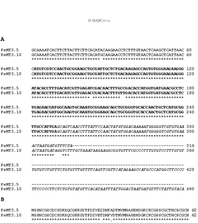
After the performed analyses, only two clones were proven to contain MT sequences. They were named pFeMT3.5 (312 bp) and pFeMT3.10 (479 bp) and submitted to the Blast Gene Bank with
acces-sion numbers DQ681063 and DQ681064. Both of the clones included a 189-bp-long coding region with STOP signal and a 61-bp one that belonged to the 5’-UTR. The clones shared identical 62-bp-long 3’-UTR with extension of 167 bp in clone pFeMT3.10 (Fig. 1A). This was evidence for the presence of at least two MT3 gene family members in the buck-wheat genome. In view of mind the importance of 3’-UTR sequences in messenger RNA stability and localization (H e s k e t h and V i l l e t t e , 2002),the detected differences could have an important impact on various functions of FeMT3 family members in buckwheat.
Both of the sequences encoded a 62-aa-long polypeptide which has the typical structure of plant MT-like proteins with two cysteine–rich termi-nal domains separated by a cysteine-free central domain. On the basis of the number and arrange-ments of cysteine residues in a specific and con-served pattern in two domains at the N- and C- ter-minals, clones pFeMT3.5 and pFeMT3.10 were con-firmed to belong to type 3 of plant MTs (C o b b e t t and G o l d s b r o u g h , 2002).Sequence comparison between the two identified clones showed 95% iden-tity on the protein level. Differences consisted of two amino acid changes in the spacer (central) region of the protein (Fig. 1B). One of them, Ile-Thr, could have influence on conformation and function of the spacer region, which is presumed to be involved in protein-protein and DNA-protein interactions (D o m e n e c h et al., 2006). Crystallographic as well as functional analyses of transgenics in those two MT protein variants should reveal potential biologi-cal relevance of these ‘’minor’’ changes.
FeMT3 expression analysis during flooding stress
FeMT3 transcripts have previously been detect-ed in green and senescent leaf, root, and developing seed. Influences of different stress conditions, such as oxidative and heavy metal stress, on the level of
The level of FeMT3 expression in control leaves was compared to that detected in leaves of plants completely submerged in water for different time periods. In contrast to mammalian MT gene tran-scripts, which increase in tissues under hypoxia during graft transplantation (L i et al., 2004), we observed a significant decrease in FeMT3 expression after 2 days of flooding stress. No significant change
was detected after 3 hours (Fig. 2). The same was observed after 2 hours and 1 day under the same stress conditions (data not shown).
The obtained results should be discussed in the light of complexity of the plant response to differ-ent stress conditions and that of the defined stress
per se. One of the most prominent consequences of
flooding is oxygen deprivation, since gas diffusion is extremely limited in water compared to air. Different plant species have developed different levels of tol-erance and different ways to overcome flooding, including biochemical as well as morphological changes. Transient increase in reactive oxygen spe-cies (ROS) concentration is detected shortly after submergence and has a role in hypoxia signaling pathways (Vo e s e n e k et al., 2006). Still, the overall oxidative status is not expected to be elevated, con-trary to mammalian hypoxic tissues experiencing oxidative stress due to nonspecific inflammation in graft transplantation (L i et al., 2004). Consequently, the need for MTs in plant vs. mammalian tissues during hypoxia is not the same. The most severe damage to the plant is incurred during oxidative stress upon its re-exposure to air. In some hypoxia-tolerant species, antioxidative enzymes like super-oxide dismutase (SOD) are upregulated gradually during submergence, preparing the plant for the postanoxic phase (M o n k et al., 1987.). However,
FeMT3 expression was not increased in the same manner, but rather decreased. Further investiga-tion will show if FeMT3 genes are upregulated after re-exposure to air, in the period of severe oxidative stress, thus when needed the most.
The existence of two or more highly homolo-gous genes belonging to the same gene family in one species is relevant both from the point of view
of molecular evolution and for studies of different mechanisms governing regulation of gene expres-sion. As mentioned above, various plant MT gene family members may have different expression pro-files, indicating their distinct but overlapping func-tions. Our future work will be focused on elucidation and comparison of particular roles of the two MT3 transcripts by Real Time RT-PCR methodology as the most sophisticated tool in expression analyses of sequence-similar genes.
Abbreviations used
MT) metallothionein; ROS) reactive oxygen species; SOD) superoxide dismutase; UTR) untranslated region. Acknowledgments — his work was supported by the Ministry of Science of the Republic of Serbia (Grant 143017).
REFERENCES
Brkljacic, J. M., Maksimovic, V. R., Radovic, S. R., and A. P. Savic (1999). Isolation of metallothionein-like cDNA clone from buckwheat. J. Plant Physiol.. 154, 802-804.
Brkljacic, J. M., Samardzic, J. T., Timotijevic, G. S., and V. R.
Maksimovic (2004). Expression analysis of buckwheat
(Fagopyrum esculentum Moench) metallothionein-like gene (MT3) under different stress and physiological conditions. J. Plant Physiol. 161 (6), 741-746.
Chabanon, H., Nury, D., Mickleburgh, I., Burtle, B., and H.
Hesketh (2004). Characterization of the cis-acting
ele-ment directing perinuclear localization of the metallo-thionein-1 mRNA. Biochem. Soc. Trans. 32, 702-704.
Chenchik, A., Diachenko, L., Moqadam, F., Tarabykin, V., Lukyanov, S., and P. D. Siebert (1996). Full-length cDNA cloning and determination of mRNA 5’ and 3’ ends by amplification of adaptor-ligated cDNA. Biotechniques 21 (3), 526-534.
Cobbett, C., and P. Goldsbrough (2002). Phytochelatins and
metallothioneins: roles in heavy metal detoxification and homeostasis. Annu. Rev. Plant Biol. 53, 159-182.
Coyle, P., Philcox, J. C., Carey, L. C., and A. M. Rofe (2002). Metallothionein: the multipurpose protein. Cell Mol. Life Sci. 59, 627-647.
Domenech, J., Mir, G., Huguet, G., Capdevila, M., Molinas, M., and S. Atrian (2006). Plant metallothionein domains: functional insight into physiological metal binding and protein folding. Biochimie 88, 583-593.
Fischer, E. H., and E. W. Davie (1998). Recent excitement
regarding metallothionein. Proc. Natl. Acad. Sci. USA 95, 3333-3334.
Guo, W. J., Bundithya, W., and P. B. Goldsbrough (2003).
Characterization of the Arabidopsis metallothionein gene family: tissue-specific expression and induction during senescence and in response to copper. New Phytol. 59, 369-381.
Hesketh, J. E. (1996). Sorting of mRNAs in the cytoplasm: mRNA localization and cytoskeleton. J. Exp. Cell Res. 225, 219–236.
Hesketh, J. E., and S. Villette (2002). Intracellular trafficking
of micronutrients: from gene regulation to nutrient requirements. Proc. Nutr. Soc. 61 (4), 405-414.
Kagi and Vallee (1960). Metallothionein: a cadmium- and zinc-containing protein from equine renal cortex. J. Biol.
Chem. 235, 3460-3465.
Li, X., Chen, H., and N. P. Epstein (2004). Metallothionein pro-Metallothionein pro-tects islets from hypoxia and extends islet graft survival by scavenging most kinds of reactive oxygen species. J.
Biol. Chem. 279 (1), 765-771.
Monk, S. L., Fagerstedt, V. K., and M. M. R. Crawford (1987).
Superoxide dismutase as an anaerobic polypeptide - a key factor in recovery from oxygen deprivation in Iris
pseudacorus? Plant Physiol. 85, 1016-1020.
Muller, P. Y., Janovjak, H., Miserez, A. R., and Z. Dobbie (2002). Processing of gene expression data generated by quanti-tative Real Time RT-PCR. Biotechniques 32, 1372-1374, 1376, 1378-1379.
Murphy, A., Zhou, J., Goldsbrough, P.B., and L. Taiz (1997).
Purification and immunological identification of metallothioneins 1 and 2 from Arabidopsis thaliana.
Plant Physiol. 113 (4), 1293-1301.
Palmiter, R. D. (1998). The elusive function of metallothioneins.
Proc. Natl. Acad. Sci. USA 95, 8428-8430.
Samardžić, J., Milisavljević, M., Brkljačić, J., Konstantinović, M., and V. Maksimović (2004) Characterization and evolu-tionary relationship of methionine-rich legumin-like protein from buchwheat. Plant Physiol. Biochem. 2 (42): 157-163.
Voesenek, L. A. C. J., Colmer, T. D., Pierik, R., Millenaar, F. F., and A. J. M. Peeters (2006). How plants cope with complete submergence. New Phytol.170, 213-226.
Wong, H. L., Sakamoto, T., Kawasaki, T., Umemura, K., and K.
Shimamoto (2004). Down-regulation of metallothionein, a reactive oxygen scavenger, by the small GTPase OsRac1 in rice. Plant Physiol. 135, 1447-1456.
АНАЛИЗА СТРУКТУРЕ ДВА ТРАНСКРИПТА ГЕНА ЗА МЕТАЛОТИОНЕИН ХЕЉДЕ И ИСПИТИВАЊЕ ЊИХОВЕ ЕКСПРЕСИЈЕ ТОКОМ ХИПОКСИЈЕ КОРИШЋЕЊЕМ
ТЕХНОЛОГИЈЕ REAL TIME RT-PCR
Драгана Б. Мајић, јелена Т. СаМарџић, Мира Ђ. МилиСављевић, а. M. КрСТић и веСна р. МаКСиМовић
Институт за молекуларну генетику и генетичко инжењерство, 11010 Београд, Србија
Металотионеини (МТ) припадају великој гру-пи протеина мале молекулске тежине богатих цистеином, иражене способности а веивање јона метала, укључених у процесе одржавања хомеостае металних јона и детоксификације од тешких метала. У раду је аналиирана структура