J of Evolution of Med and Dent Sci/ eISSN- 2278-4802, pISSN- 2278-4748/ Vol. 3/ Issue 50/Oct 06, 2014 Page 11798
RETROGRADE INTRAMEDULLARY INTERLOCKING NAIL: AN OPTIMAL
TOOL FOR THE TREATMENT OF SUPRACONDYLAR FEMORAL FRACTURES
S. B. Kamareddy1, Sanjeevi Bharadwaj2HOW TO CITE THIS ARTICLE:
S. B. Kamareddy, Sanjeevi Bharadwaj. Retrograde Intramedullary Interlocking Nail: An Optimal Tool for the Treatment of Supracondylar Femoral Fractures . Journal of Evolution of Medical and Dental Sciences 2014; Vol. 3, Issue 50, October 06; Page: 11798-11805, DOI: 10.14260/jemds/2014/3552
ABSTRACT: BACKGROUND: This prospective consecutive case series was done to evaluate indications, technical pearls and pitfalls and functional outcomes of distal femoral supracondylar fractures treated with retrograde intramedullary nailing. METHODS: The surgical outcome of 80 patients (59 males and 21 females) who were treated with retrograde intramedullary nailing for. Patients were followed-up both clinically and radiologically every 6 weeks for a mean duration of 20 (12 – 24) months. The patients were assessed based on Schatzker and Lambert criteria. RESULTS: The mechanism of injury was motor vehicle accident in 48 (60%) patients and fall in 32 (40%) patients. Sixty four (80%) fractures were closed and 16 (20%) fractures were compound. Average operation time was 2 hours and average blood loss was 300 ml. The mean union time was 5.6 months (5 – 7 months). Knee flexion of more than 120 degree was achieved in 60 patients, 110 degree in 16 patients and 100 degree in 2 patients. Six patients had anterior knee pain of transient nature which subsided within one year after rehabilitation, full weight bearing and self exercises. By the end of 1 year, overall outcome was excellent in 59 patients (74%), good in 12 patients (15%) and fair in 8 (10%) patients. CONCLUSION: Retrograde intramedullary femoral nailing is an optimal tool in the treatment of AO/ASIF type A and type C distal femur (supracondylar) fractures. It provides rigid fixation in a region of femur where, wide canal, thin cortices and frequently poor bone stock make fracture fixation difficult. It also provides excellent results in selected comminuted fractures of the distal femur with a low complication rate.
KEYWORDS: Retrograde supracondylar nailing, distal femoral fracture, union time, angular deformity, anterior knee pain.
INTRODUCTION: Supracondylar fractures of the femur account for 7% of all the femoral fractures. They are common both in elderly with osteoporotic bones and in young patients after motor vehicle accidents. These fractures may occur in young patients as a result of high energy trauma like motor vehicle accident or firearm injuries.(1,2, 3,4) These fractures require anatomical restoration to optimize
the function of the lower extremity, which necessitates surgical treatment.(1,2,5) Severely comminuted
distal femur fractures are especially difficult fractures to treat properly.
Obtaining adequate fixation may be technically challenging, especially when multiple fragments are present.(6) There is inadequate bony purchase of distal fragment of supracondylar
femoral fractures in cases treated with extra medullary implants like plate osteosynthesis,(2) condylar
blade plate,(7) and dynamic condylar screw (DCS).(8,9) Furthermore, open reduction and periosteal
stripping for placement of the implant may interfere with the healing process, resulting in high rate of non-union and infection.(10,11) Stress-shielding and re-fracture rate have been reported as a
J of Evolution of Med and Dent Sci/ eISSN- 2278-4802, pISSN- 2278-4748/ Vol. 3/ Issue 50/Oct 06, 2014 Page 11799 The less invasive stabilization system (LISS) developed over the last few years has now been used for fixation of distal femoral fractures, because the chances of usual complications associated with plating are decreased.(2,10,13,14,15,16,17) Flexible intramedullary nailing, modified ante grade nailing
and external fixation allows fracture fixation with minimal exposure of the fracture site, however, axial and rotational stability of these implants is inferior to the retrograde nailing, and early mobilization of the limb could result in loss of reduction.(18)
Wu et al. looked at the results of ante grade intramedullary nailing in extra-articular distal femoral fractures, and compared it to plating. They found a superior union rate (90% compared to79% for plating) and better range of knee movement.(19) However, Leung et al. concluded that in
the most distal fractures the standard ante grade nails would not be able to provide adequate control.(20)
Retrograde supracondylar nailing for distal femur fractures with use of a distal intercondylar intra-articular entry portal was introduced two decades back.(3,21) This method of treatment protects
the soft -tissue envelope due to its minimal invasive approach and closed reduction technique. In cases with severe metaphysical commination, retrograde supracondylar nailing offers a more biological method of fixation with good control of the distal fragment and less devitalisation of soft tissue.(22,23)
Recent studies show union and complication rates comparable to ante grade nails, with the exception of a higher incidence of knee pain in the retrograde technique.(24,25,26,27) However, there are
no reports of long term knee joint effects after retrograde nailing done transarticularly.(28) Currently
accepted indications for retrograde femoral nails are similar to those for ante grade nailing.
However, retrograde nailing is advantageous for the following indications: patients with multiple systemic injuries, multiple fractures, suspected or known spinal injuries, ipsilateral femoral neck and shaft fractures, peri-prosthetic fractures, floating knee, bilateral femur fractures, pregnancy, and morbid obesity.(24,25,26,29,30,31)
Advantages include minimal soft tissue exposure, closed reduction technique, shorter operating time, minimal blood loss and early recovery. There is need to put supracondylar nailing surgery into practice for distal femoral fractures because it is minimally invasive technique, better fixation and control of distal fragment, preservation of periosteal and fracture haematoma, negligible blood loss, less chance of post-operative infection, faster union and weight bearing associated with this procedure.(32) This prospective study aims to evaluate the surgical outcome of supracondylar
femoral fractures treated with retrograde supracondylar nailing.
MATERIALS AND METHODS: Between June 2011 to June 2014, a total of 80 patients with supracondylar femoral fractures were admitted to our hospital. The average age was 49 years (range 32 – 66 years). All the patients were operated upon by same orthopedic surgeon. Fractures were classified according to AO classification. Indications for retrograde intramedullary nail fixation included AO type A and type C supracondylar femur fractures.
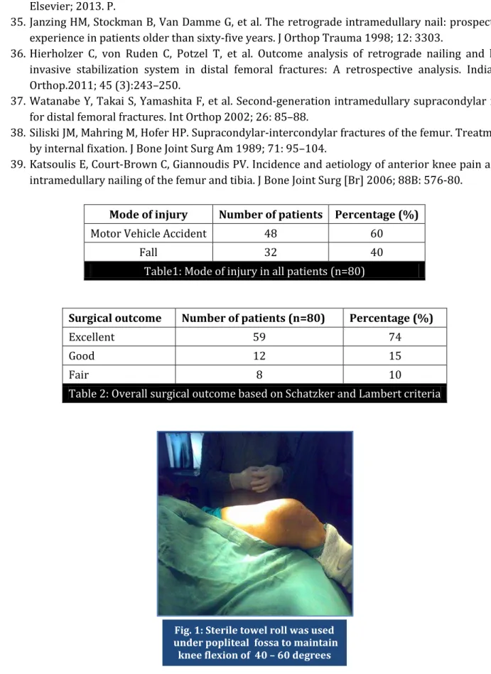
J of Evolution of Med and Dent Sci/ eISSN- 2278-4802, pISSN- 2278-4748/ Vol. 3/ Issue 50/Oct 06, 2014 Page 11800 SURGICAL PROCEDURE: Surgery was performed with the patient in supine position on a standard radiolucent operating table. C arm image intensifier was used in operating room for close reduction technique. Sterile towel roll was used under popliteal fossa to maintain knee flexion of 40 – 60 degrees (Fig. 1).
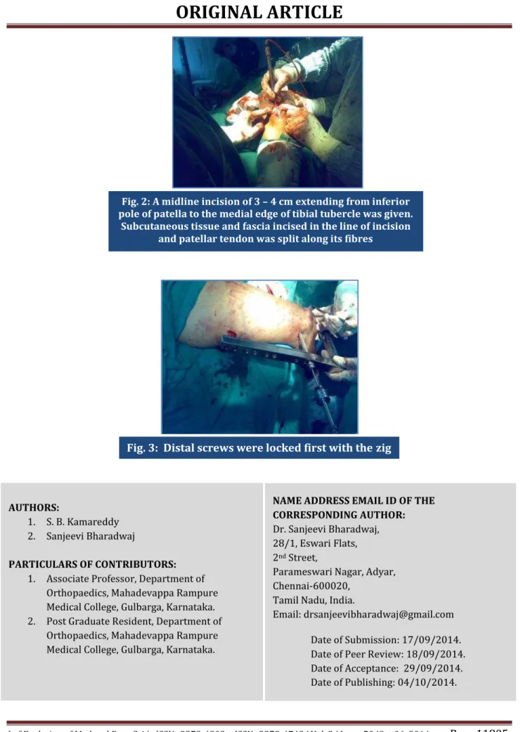
A midline incision of 3 – 4 cm extending from inferior pole of patella to the medial edge of tibial tubercle was given. Subcutaneous tissue and fascia incised in the line of incision and patellar tendon was split along its fibres (Fig. 2). The reference point of entry in intercondylar notch was anterior to Blumensaat`s line as seen on lateral image of knee on image intensifier.(2) We used shorter
version nails in all cases. The length of nails used was between 170 and 200 mm. The decision about nail diameter was taken preoperatively after reaming the femoral medullary canal. We used either 10 or 11 mm diameter nails.
The distal interlocking screws of the nail provided additional fixation to the condylar fragments. Distal screws were locked first with the zig (Fig. 3). Guided locking mode was used for only distal locking screws placement. Fixation of proximal locking screws was done with free hand technique. All patients started passive followed by active range of knee movements at 48 – 72 hours postoperatively. Partial weight bearing was allowed in initial 6 weeks. All patients were evaluated for duration of operation, average blood loss and postoperative complications.
All patients were followed up at 1st week, 6th week, 12th week and 24th week than every 3
months for 23 months (range 19 – 28 months). Follow-up X rays were performed in two planes and assessed for assessment of restoration of distal femoral articular surface, callus formation and varus-valgus and flexion extension deformity. Malalignment was defined as varus-varus-valgus greater than 5°, apex anterior-posterior greater than 10°, and rotational malalignment greater than 15°.
The criterion recommended by Schatzker and Lambert was used for overall outcome evaluation.(33) The data will be analyzed with SPSS 20. All quantitative variables was presented as
mean and standard deviation. All qualitative variables were presented as percentages and frequencies. P values < 0.05 are considered statistically significant.
RESULTS: The mode of injury was, motor vehicle accident in 48 (60%) and falls in 32 (40%) cases (Table 1). The mean duration of follow-up was 23 months (range 19 – 28 months). The mean operation time was 2 hours (range from 100 – 135 minutes). The average blood loss was 400 ml (range from 300 – 500 ml).
Postoperatively, knee joint mobilization without weight bearing was commenced on the first postoperative day and patients were with support of Zimmer frame non-weight bearing over operated leg. The average duration from the time of surgery to the time when the patient was ambulated with partial weight bearing was 3 weeks. Full weight bearing was allowed when the fracture was assessed as clinically and radio logically united. The mean time required to achieve union was 5.6 months (range: 5 – 7 months).
The assessment of limb length discrepancy and range of knee movements were based on the last recorded clinical evaluation. There were 5 cases (6%) of shortening of operated limb between 1 to 2 cm and remaining 75 cases (94%) had no limb length discrepancy. Knee flexion of more than 120 degrees was achieved in 60 patients, 110 degrees in 16 patients and 100 degrees in 2 patients.
J of Evolution of Med and Dent Sci/ eISSN- 2278-4802, pISSN- 2278-4748/ Vol. 3/ Issue 50/Oct 06, 2014 Page 11801 end of one year, overall outcome based on Schatzker and Lambert criteria was excellent in excellent in 59 patients (74%), good in 12 patients (15%) and fair in 8 (10%) patients (Table 2).
DISCUSSION: Open reduction and internal fixation using extra medullary implants has been the standard treatment for supracondylar fractures irrespective of age group. The drawbacks of this technique are the need for a large exposure with the possible risk of soft tissue damage, devascularisation of bone fragments and loss of the possible positive effect of the fracture hematoma. Moreover, early weight bearing is not advisable with these implants.(34)
Closed reduction technique and minimal exposure to facilitate the insertion of retrograde intramedullary nail results in little additional injury to the adjacent soft tissue, especially the periosteum, and fracture haematoma was preserved.
The load sharing mechanism of intramedullary nailing promotes secondary bone healing, and the cancellous bone from medullary reaming extravasated into the fracture site serves as a bone graft. All these factors likely contributed to the good union rate seen and the low incidence of soft tissue complications, especially infection.(34) Janzing et al. studied outcomes for 25 elderly patients
with distal femur fractures treated with supracondylar nailing.
Using the Neer scoring system criteria, they reported excellent or good results in 23 (92%) patients.(35) Canada LK et al. in their study on retrograde intramedullary nailing of comminuted
femoral diaphyseal fractures caused by low-velocity gunshots, observed 3 patients out of 73 had shortening >10 mm and 1 had angulation >10 degrees.
They concluded that there was low incidence of shortening, angular deformity, complication, and infection rates following treatment of femoral gunshot diaphyseal fractures with a retrograde nail, hence, retrograde nailing is an acceptable alternative for the treatment of femoral diaphyseal caused by low-velocity gunshot injuries.(4) Moment arm of varus/valgus bending force is significantly
reduced with intramedullary nailing compared with use of the lateral blade plate system and DCS.(34)
Four cortical fixations are also possible with 2 well-placed distal interlocking screws.(36)
Moreover, intramedullary positioning of the nail also provides 3-point fixation of the fracture to prevent flexion/extension displacement of the distal fragment. The combination of early union and stability of fixation seen with this approach effectively reduces the risks of angular malunion.
Angular malunion, in either the coronal or sagittal plane, may develop due to inadequate reduction or subsequent displacement during bone healing. Schatzker and Lambert classified fracture alignment of less than 100varus or valgus as good in their outcome evaluation. El-Kawy et al observed a high incidence of angular malalignment (39.2%) in their study conducted on elderly patients.13 Watanabe et al. noticed 12.5% incidence of angular malalignment.(37)
Siliski et al. reported that 15 of 52 patients in their series of distal femur fractures treated with plate fixation had limb shortening of 1 to 3 cm. Shortening was intentional in 11 patients to allow impaction for better bone healing.(38) In our series, we aimed to restore original bone length
and there were 5 cases of shortening of operated limb from 1 to 2 cm.
J of Evolution of Med and Dent Sci/ eISSN- 2278-4802, pISSN- 2278-4748/ Vol. 3/ Issue 50/Oct 06, 2014 Page 11802 Its aetiology is unclear, but a multi-factorial origin has been suggested like damage to the meniscus or cartilage of the tibial plateau, injury to the infra patellar branch of the saphenous nerve, splitting and repeated injury to the patellar tendon per operatively and nail protrusion at distal end.
However, the optimum point of entry and distal interlocking spares the articular surface and extra articular structures.(2,39) The mean union time of 5.6 months for fracture healing with no case of
non-union.
CONCLUSION: Retrograde intramedullary femoral nailing is an optimal tool in the treatment of AO/ASIF type A and type C distal femur (supracondylar) fractures. It provides a good bone purchase in the distal femur where fracture fixation is inherently difficult due to, wide canal, thin cortices and frequently poor bone stock. There is minimal soft tissue disruption and stable fracture fixation, which allows early ambulation and weight bearing with excellent results and low complication rates.
REFERENCES:
1. Handolin L, Pajarinen J, Lindahl Jan, et al. Retrograde intramedullary nailing in distal femoral fractures--results in a series of 46 consecutive operations. Injury 2004; 35 (5): 517-22.
2. Neubauer TH, Ritt er E, Potschka TH, et al. Retrograde Nailing of Femoral Fractures. Acta Chirurgiae Orthopaedicae 2008; 75: 158–166.
3. Reina R, Vilella FE, Ramirez N, et al. Knee pain and leg-length discrepancy after retrograde femoral nailing, Am J Orthop 2007; 36 (6): 325-328.
4. Cannada LK, Jones TR, Guerrero-Bejarano M, et al. Retrograde Intramedullary Nailing of Femoral Diaphyseal Fractures Caused by Low-velocity Gunshots. Orthopaedics 2009; 32 (3): 162.
5. Shahcheraghi GH, Doroodchi HR. Supracondylar fracture of the femur: closed or open reduction? J Trauma. Apr 1993; 34 (4): 499-502.
6. Crist BD, Della Rocca GJ, Murtha YM. Treatment of acute distal femur fractures. Orthopedics. Jul 2008; 31 (7): 681-90.
7. Johnson EE. Combined direct and indirect reduction of comminuted four-part intra articular T-type fractures of the distal femur. Clin Orthop. Jun 1988; (231): 154-62.
8. Helfet DL, Lorich DG. Retrograde Intramedullary Nailing of Supracondylar Femoral Fractures. Clinical Orthopaedics & Related Research. May 1998; (350): 80-84.
9. Placide RJ, Lonner JH. Fractures of the distal femur. Current Opinion in Orthopedics. February 1999; 10 (1): 2-9.
10.Gao K, Gao W, Huang J, et al. Retrograde Nailing versus Locked Plating of Extra-Articular Distal Femoral Fractures: Comparison of 36 Cases. Med Princ Pract 2013; 22: 161–166
11.Thomson AB, Driver R, Kregor PJ, et al. Long-term functional outcomes after intra-articular distal femur fractures: ORIF versus retrograde intramedullary nailing. Orthopedics 2008; 3: 748–750.
12.Bostman OM. Refracture after removal of a condylar plate from the distal third of the femur. J Bone Joint Surg Am 1990; 2: 1013–1018.
J of Evolution of Med and Dent Sci/ eISSN- 2278-4802, pISSN- 2278-4748/ Vol. 3/ Issue 50/Oct 06, 2014 Page 11803 14.Schutz M, Muller M, Kaab M. et al. Less invasive stabilization system (LISS) in the treatment of
distal femoral fractures. Acta Chir Orthop Traumatol Cech 2003; 70: 74–82.
15.Farouk O, Krett ek C, Miclau T, et al. Minimally invasive plate osteosynthesis: does percutaneous plating disrupt femoral blood supply less than the traditional technique? J Orthop Trauma 1999; 13: 401–406.
16.Kolb W, Guhlmann H, Windisch C, et al. Fixation of distal femoral fractures with the Less Invasive Stabilization System: a minimally invasive treatment with locked fixed-angle screws. J Trauma 2008; 65: 1425–1434.
17.Smith TO, Hedges C, MacNair R, et al. The clinical and radiological outcomes of the LISS plate for distal femoral fractures: a systematic review. Injury 2009; 40: 1049–1063.
18.Zehntner MK, Marchesi DG, Burch H, et al. Alignment of supracondylar/intercondylar fractures of the femur after internal fixation by AO/ASIF technique. J Orthop Trauma. 1992; 6 (3): 318-26. 19.Wu CC, Shih CH. Treatment of femoral supracondylar unstable communited fractures. Comparisons between plating and Grosse-Kempf interlocking nailing techniques. Arch Orthop Trauma Surg 1992; 111 (4): 232-6.
20.Leung KS, Shen WY, So WS, et. al. Interlocking intramedullary nailing for supracondylar and intercondylar fractures of the distal part of the femur. J Bone Joint Surg Am.1991; 73:332-340 21.Moed BR, Watson JT. Retrograde Nailing of the Femoral Shaft. J Am Acad Orthop Surg 1999; 7:
209-216.
22.Gurkan V, Orhun H, Doganay M, et al. Retrograde intramedullary interlocking nailing in fractures of the distal femur. Acta Orthop Traumatol Turc 2009; 43: 199–205.
23.Ostrum RF, Maurer JP. Distal third femur fractures treated with retrograde femoral nailing and blocking screws. J Orthop Trauma 2009; 23: 681–684.
24.Walcher F, Frank J, Marzi I. Retrograde nailing of distal femoral fracture – clear and potential indications. European Journal of Trauma. 2000; (4): 155-168.
25.Prayson M, Herbenick M, Siebuhr K, et al. An Alternative Direction for Proximal Locking in Retrograde Femoral Nails. Orthopedics August 2008; 31 (8): 757-60.
26.Ostrum RF, Agarwal A, Lakatos R, et al. Prospective comparison of retrograde and ante grade femoral intramedullary nailing. J Orthop Trauma. 2000; 14 (7): 496-501.
27.Ricci WM, Bellabarba C, Evanoff B, et al. Retrograde versus ante grade nailing of femoral shaft fractures. J Orthop Trauma. 2001; 15 (3): 161-169.
28.Yu CK, Singh VA, Mariapan S. et al. Ante grade Versus Retrograde Locked Intramedullary Nailing for Femoral Fractures: Which Is Better? Eur J Trauma Emerg Surg 2007; 33: 135–140.
29.Moed BR, Watson JT, Cramer KE, et al. Unreamed retrograde intramedullary nailing of fractures of the femoral shaft. J Orthop Trauma. 1998; 12 (5): 334-342.
30.Ostrum RF, Di Cicco J, Lakatos R, et al. Retrograde intramedullary nailing of femoral diaphyseal fractures. J Orthop Trauma. 1998; 12 (7): 464-468.
31.Riaz S. Retrograde Femoral Nailing: A Modified Technique for Unusual Femoral Shaft Fractures. Pak J Med Res 2006; 45: 1.
32.Yuvarajan P, Kaul R, Maini L. Review of concepts in distal femoral fractures management. Pb Journal of Orthopaedics 2009; 11 (1): 44–48.
J of Evolution of Med and Dent Sci/ eISSN- 2278-4802, pISSN- 2278-4748/ Vol. 3/ Issue 50/Oct 06, 2014 Page 11804 34.Canale S.T, Beaty J. H. Distal femur, In: (eds.) Campbell’s Operative Orthopaedics. 12th ed. USA:
Elsevier; 2013. P.
35.Janzing HM, Stockman B, Van Damme G, et al. The retrograde intramedullary nail: prospective experience in patients older than sixty-five years. J Orthop Trauma 1998; 12: 3303.
36.Hierholzer C, von Ruden C, Potzel T, et al. Outcome analysis of retrograde nailing and less invasive stabilization system in distal femoral fractures: A retrospective analysis. Indian J Orthop.2011; 45 (3):243–250.
37.Watanabe Y, Takai S, Yamashita F, et al. Second-generation intramedullary supracondylar nail for distal femoral fractures. Int Orthop 2002; 26: 85–88.
38.Siliski JM, Mahring M, Hofer HP. Supracondylar-intercondylar fractures of the femur. Treatment by internal fixation. J Bone Joint Surg Am 1989; 71: 95–104.
39.Katsoulis E, Court-Brown C, Giannoudis PV. Incidence and aetiology of anterior knee pain after intramedullary nailing of the femur and tibia. J Bone Joint Surg [Br] 2006; 88B: 576-80.
Mode of injury Number of patients Percentage (%)
Motor Vehicle Accident 48 60
Fall 32 40
Table1: Mode of injury in all patients (n=80)
Surgical outcome Number of patients (n=80) Percentage (%)
Excellent 59 74
Good 12 15
Fair 8 10
Table 2: Overall surgical outcome based on Schatzker and Lambert criteria
Fig. 1: Sterile towel roll was used under popliteal fossa to maintain
J of Evolution of Med and Dent Sci/ eISSN- 2278-4802, pISSN- 2278-4748/ Vol. 3/ Issue 50/Oct 06, 2014 Page 11805
AUTHORS:
1. S. B. Kamareddy 2. Sanjeevi Bharadwaj
PARTICULARS OF CONTRIBUTORS:
1. Associate Professor, Department of Orthopaedics, Mahadevappa Rampure Medical College, Gulbarga, Karnataka. 2. Post Graduate Resident, Department of
Orthopaedics, Mahadevappa Rampure Medical College, Gulbarga, Karnataka.
NAME ADDRESS EMAIL ID OF THE CORRESPONDING AUTHOR:
Dr. Sanjeevi Bharadwaj, 28/1, Eswari Flats, 2nd Street,
Parameswari Nagar, Adyar, Chennai-600020,
Tamil Nadu, India.
Email: drsanjeevibharadwaj@gmail.com
Date of Submission: 17/09/2014. Date of Peer Review: 18/09/2014. Date of Acceptance: 29/09/2014. Date of Publishing: 04/10/2014.
Fig. 3: Distal screws were locked first with thezig
Fig. 2: A midline incision of 3 – 4 cm extending from inferior
pole of patella to the medial edge of tibial tubercle was given. Subcutaneous tissue and fascia incised in the line of incision