4 artigo 635
ORIGINAL ARTICLE
1 – Physician; Head, Department of Pediatric Orthopedics, Hospital Universitário de Taubaté – Taubaté, SP, Brazil.
2 – Physician; Master’s degree; Doctor’s degree; Head, Orthopedics and Traumatology Clinic, Hospital Universitário de Taubaté – Taubaté, SP, Brazil. 3 – Physician; Resident in Orthopedics and Traumatology, Hospital Universitário de Taubaté – Taubaté, SP, Brazil.
Study conducted at the Orthopedics and Traumatology Clinic in Taubaté, SP.
Correspondence: Departamento de Ortopedia e Traumatologia do Hospital Universitário de Taubaté (UNITAU), Av. Granadeiro Guimarães, 270, Centro – 12020-130 – Taubaté, SP, Brazil. Email: [email protected]
Received for publication: 12/3/2011, accepted for publication: 4/27/2012.
suPracondylar fracTure of The humerus in
children: fixaTion WiTh TWo crossed
kirschner Wires
Roni Azevedo Carvalho1, Nelson Franco Filho2, Antonio Batalha Castello Neto3, Giulyano Dias Reis3,Marcos Pereira Dias3
ABSTRACT
Objective: To analyze and present the surgical results from unstable supracondylar fractures of the humerus in children, treated by means of reduction and percutaneous fixation using two crossed Kirschner wires. Methods: A cross-sectional study was conducted on 20 children, taking into consideration sex, age at the time of the fracture, age at the time of the assessment, side affected, type and mechanism of trauma, postoperative com-plications and radiographic and clinic variables. Results: Ten fractures were observed in the left arm and ten in the right arm. The age at the time of the fracture ranged from 2 to 13 years (mean: 5.9 ± 2.48 years). Three fractures were classified as type II and 17 as type III. The length of follow-up ranged from four
The authors declare that there was no conflict of interest in conducting this work
This article is available online in Portuguese and English at the websites: www.rbo.org.br and www.scielo.br/rbort
months to three years. Baumann’s angle ranged from 69 to 100 (mean: 78.3) and cubitus varus was observed in four patients (values ranging from 84 to 100). According to the modified Flynn’s criteria, 20 cases presented satisfactory outcomes: 17 excellent (85%), two good (10%) and one regular (5%). Two patients presented limited range of motion, two had paresthesia in the cubital region and one had transient neuropraxia of the ul-nar nerve for six weeks. Conclusion: Percutaneous fixation with two crossed Kirschner wires leads to good results when carried out under direct viewing and with isolation of the ulnar nerve.
Keywords – Child; Humerus Fractures; Bone Wires; Fracture Fixation
INTRODUCTION
Of the upper limbs in children, the elbow is the second most common site of occurrence of fractures, surpassed only by forearm fractures. Around 85% of elbow fractures in children occur in the distal humerus, and of these, 55 to 75% are supracondylar fractures, accounting for 3% of all fractures in children(1-4).
Supracondylar humerus fractures are caused by direct or indirect low-kinetic energy traumas, such as falls, which makes the occurrence of comminution, bone exposure or association with other fractures less frequent(1-4).
Due to the anatomical characteristics of this region, and the variations in fractured fragments, immediate complications (vascular-nerve injuries, compartment
syndrome) and late complications (vicious consolida-tion and angular deformities) can arise(5-8).
Despite the various types of treatment, high mor-bidity and complications related to this type of frac-ture make treatment urgent and essential(1).
Treatment with closed reduction, associated with plaster cast immobilization, has been gradually aban-doned, particularly due to the higher complication rate, such as loss of reduction, compartment syn-drome, and long hospitalization periods(9-11).
METHODS
Forty-six patients were identified, who had unsta-ble, closed (Gartland types II and III)(12) supracondylar
humerus fracture, and who underwent closed reduc-tion and percutaneous fixareduc-tion using two crossed
Kirschner wires, from April 2008 to August 2010.
The sample analyzed consisted of 20 patients;
chil-dren with any comorbidities that would interfere in the consolidation of the fracture were excluded, as well as children with neurovascular injury, association with other fractures, and exposed fractures (Table 1).
The following variables were taken into account: sex, age at the time of the fracture and of the assessment, date of surgery, side affected, mechanism and type of fracture (classification), postoperative complications and
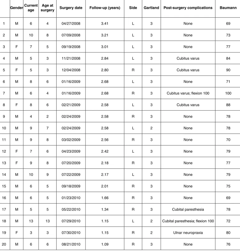
Table 1 – Database of patients assessed.
GenderCurrent age
Age at
surgery Surgery date Follow-up (years) Side Gartland Post-surgery complications Baumann
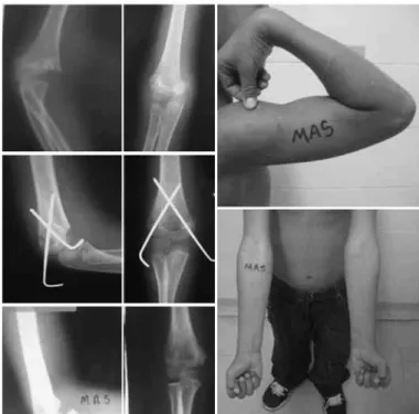
Figure 1 – Example of the results obtained.
radiographic variables (Baumann’s angle)(13).
With the consent of their respective legal guardians, and after revision of all medical data, the children were submitted to clinical and radiological analysis.
According to the protocol established at our clinic, all the patients initially underwent clinical evaluation, followed by analgesics and plaster cast immobilization, with no attempt at reduction. After preliminary evaluation, the patients were sent to the surgical center.
After general inhalational anesthetics with intubation, in the horizontal dorsal position, the reduction operation was then performed with the elbow extended and the forearm supinated, with longitudinal traction and contraction to recover the length, and clinical alignment of the load angle. Subsequently, the edges of both fragments were aligned, with hyperextension and supination as needed. At this point, maneuvers were performed in valgus or in varus to correct medial or lateral dislocations. When these two deviations were corrected, the angle of the distal fragment was corrected, flexing the elbow while pushing the distal fragment anteriorly.
After asepsis and antisepsis, the fracture was fixed with two smooth, crossed Kirschner wires, passing through the medial and lateral epicondyles. Before passing the wires, a small skin incision was made, of around 1 cm, to enable insertion of the wire without damaging the adjacent soft tissues due to torque, and to protect the ulnar nerve from injury. In this fixation, the wires were positioned at an angle of between 30 and 40°. The reduction was always checked through a radioscopy of the elbow, in the anteroposterior (with the elbow in maximum extension and supination) and lateral (with shoulder rotated externally) views. The Kirschner wires were cut and left outside the skin. The wound was dressed, and a full-arm cast was applied, with the elbow flexed at an angle of 90° and the forearm in neutral rotation. Active and passive finger exercises were prescribed from the time of arrival at the ward, and the upper limb that had been operated on was kept suspended during hospitalization. Patients were discharged within 24 to 48 hours, with an initial follow-up visit within seven to ten days, and a second within 14 to 17 days. The cast and the Kirschner wires were removed between the fourth and sixth week
after surgery, when mobilization was prescribed to recover the range of motion. Subsequent follow-up visits were held at every 30 days until outpatient discharge (Figure 1).
Patients were submitted to radiography analysis through measurement of the Baumann’s angle(13), and
clinical analysis according to the modified criteria proposed by Flynn et al(14) for fracture consolidation,
range of motion, and post-surgical neurovascular complications.
Elbows with Baumann’s angle under 64° were con-sidered in valgus(13), and those with Baumann’s angle
over 82° were considered cubitus varus(15-22).
RESULTS
Twenty children were observed, with ten fractures of the right elbow, and ten of the left elbow. The trau-ma mechanism was indirect, with extended limb in all cases. The children’s ages at the time of fracture ranged from two to 13 years (average of 5.9 ± 2.48; median 5.5 years). The children’s ages at the time of assessment ranged from three to 13 years (average of 7 ± 2.53; median 6.5 years). The Gartland classifica-tion(12) was used, resulting in three fractures being
classified as type II and 17 as type III. The follow-up period ranged from four months to three years.
observed in four patients (ranging from 84 to 100). According to the modified criteria established by Flynn et al(14), 20 cases were satisfactory, with no
unsatisfactory cases. Of the satisfactory results, 17 were excellent (85%), two were good (10%), and one fair (5%). The fractures of all patients were completely consolidated within the expected period, with no case of delay in consolidation or pseudarthrosis. Two patients had reduced range of motion, with 100° flexion in both cases. Two children had cubital paresthesia due to extrinsic compression, which was solved by replacing the cast. One child had transient neuropraxia of the ulnar nerve for six weeks, which disappeared when the wires were removed.
DISCUSSION
Supracondylar humerus fracture in children is an urgent condition, requiring rapid diagnosis and treat-ment, particularly due to the serious vascular and nerve complications associated with this injury(1,4,5-8,16,23-25).
The surgeon’s objective is to obtain anatomical reduc-tion and reestablish funcreduc-tion as soon as possible. The higher the level of instability, the higher the complica-tion rate(24). Thorough neurological examination and
palpation of the distal peripheral pulse are imperative during the initial analysis of all patients.
Percutaneous fixation is popular, and was de-scribed by Judet and Swenson as an osteosynthesis technique based on the work by Miller (1939) apud Fumo et al(26) which recommends this method for the
treatment of T-shaped injuries; its widespread use is due to a series of advantages, including low cost, safety, efficiency and minimal hospitalization times.
In our case series, we observed a distribution per sex (male-female) similar to that observed in the litera-ture (2.3:1), and there was no predominance in relation to affected side. The patient’s age at the time of fracture was also equally similar: 5.9 years(2,4,26-29).
The development of compartment syndrome is the most feared complication of this type of injury. Nerve injuries, such as neuropraxia, are relatively common, accounting for between 6 and 16% of all complications(2,3,26-29). Lesion of the ulnar nerve,
although rare (4.3%), is usually reversible, and is most commonly related to untimely treatment and edema in the region, which makes it difficult to feel and identify the anatomical accidents. One case (5%) of percutaneous fixation by extrinsic
compression of the wire was observed, which was resolved when the percutaneous wires were re-moved in the sixth week. Also, two cases (10%) had cubital paresthesia, which were solved after replacement of the plaster cast. Brauer et al(29) and
Woratanarat et al(27) stated that iatrogenic lesions of
the ulnar nerve are two to four times more frequent with cross-fixation, suggesting fixation with two parallel, lateral wires.
Authors who advocate the use of crossed wires de-fend the high rate of satisfactory results, as a conse-quence of the greater stability with restored anatomy, due to the strength applied medially through elastic de-formation of the wire, known as the “spring effect”(26).
A reduced arc of flexion-extension was observed in two patients (10%), who had 100° flexion at the time of assessment. It is believed that the short follow-up period of these patients (one year and six months, and two years and six months) is responsible for the high reduction rate of the observed flexion-extension, since mobility can be recovered partially or totally within a period of up to four years(14).
Cubitus varus is considered the most common late complication in this type of fracture, presenting an incidence of 20% in our series. Other authors who used closed reduction and percutaneous fixation re-ported lower rates of around 10%(1,3-6,14-18,22,25,27-29).
The initial deviation and ligamentotaxis are the main factors mechanically influencing the final result of the fracture. The lateral ligaments are firmer in pro-nation and more relaxed in supipro-nation(18,26). Closed
reduction associated with plaster cast is related to a higher incidence of varus deformity. Despite ana-tomical reduction, rotational deformity of the distal fragment may occur when the wires are not posi-tioned in the same plane, more commonly when one of the wires is located anterior to the other(1,2,4,26-29).
Despite the higher varus incidence in the present study, the results were considered satisfactory ac-cording to Flynn et al(14).
CONCLUSION
REFERENCES
1. Rouede RS, Giordano V, Amaral NP. Tratamento cirúrgico das fraturas su-pracondilianas desviadas do úmero na criança: análise dos resultados de 20 casos. Rev Bras Ortop. 2001;36(4):105-10.
2. Cheng JC, Shen WY. Limb fracture pattern in different pediatric age groups: a study of 3,350 children. J Orthop Trauma. 1993;7(1):15-22.
3. Canale ST. Fraturas e luxação em crianças. Canale ST. Cirurgia ortopédica de Campbell. 10a. São Paulo: Manole; 2006. In: p. 1437-53.
4. Amatuzzi MM, Zoppi Filho A, Montenegro NB. Fratura supracondiliana do úmero em crianças: estudo de 90 casos operados Rev Bras Ortop. 1997;32(6): 437-42.
5. Hungria Neto JS, Fregoneze M, Bezerra MJC, Mercadante MT, Teixeira AA, Yoneda T. Fraturas supracondilianas do úmero nas crianças: avaliação do resultado do tratamento cirúrgico. Rev Bras Ortop. 1996;31(7):575-83.
6. Lal GM, Bhan S. Delayed open reduction for supracondylar fractures of the humerus. Int Orthop. 1991;15(3):189-91.
7. Nacht JL, Ecker ML, Chung SM, Lotke PA, Das M. Supracondylar fractures of the humerus in children treated by closed reduction and percutaneous pinning. Clin Orthop Relat Res. 1983;(177):203-9.
8. Wilkins KE. Conduta operatória nas fraturas supracondilianas. Clin Ortop Am North. 1990;21:269-89.
9. Blount WP. Fractures in children. Baltimore: Williams e Wilkins; 1955.
10. Boyd DW, Aronson DD. Supracondylar fractures of the humerus: a prospective study of percutaneous pinning. J Pediatr Orthop. 1992;12(6):789-94.
11. Sutton WR, Greene WB, Georgopoulos G, Dameron TB Jr. Displaced supra-condylar humeral fractures in children. A comparison of results and costs in patients treated by skeletal traction versus percutaneous pinning. Clin Orthop Relat Res. 1992;(278):81-7.
12. Gartland JJ. Management of supracondylar fractures of the humerus in chil-dren. Surg Gynecol Obstet. 1959;109(2):145-54.
13. Baumann E. On the treatment of fractures of the elbow joint. Langen- beks Arch Klin Chir. 1960;295-300.
14. Flynn JC, Matthews JG, Benoit RL. Blind pinning of displaced supracondylar fractures of the humerus in children. Sixteen years’ experience with long-term follow-up. J Bone Joint Surg Am. 1974;56(2):263-72.
15. Terra DL, Santos MHB, Malheiros DS, Lima CLFA, Cunha FM. Nova aborda-gem no tratamento da fratura supracondilar do úmero, instável, em crianças e adolescentes. Rev Bras Ortop. 2005;40(1/2):42-51.
16. Keenan WN, Clegg J. Variation of Baumann’s angle with age, sex, and side: implications for its use in radiological monitoring of supracondylar fracture of the humerus in children. J Pediatr Orthop. 1996;16(1):97-8.
17. Köberle G. Prevenção de cúbito varo e cúbito valgo na fratura supracondiliana do úmero. Rev Bras Ortop. 2003;38(10):568-80.
18. D’Ambrosia RD. Supracondylar fractures of humerus--prevention of cubitus varus. J Bone Joint Surg Am. 1972;54(1):60-6.
19. O’Hara LJ, Barlow JW, Clarke NM. Displaced supracondylar fractures of the humerus in children. Audit changes practice. J Bone Joint Surg Br. 2000;82(2):204-10.
20. Mubarak SJ, Carroll NC. Volkmann’s contracture in children: aetiology and prevention. J Bone Joint Surg Br. 1979;61(3):285-93.
21. Royce RO, Dutkowsky JP, Kasser JR, Rand FR. Neurologic complications after K-wire fixation of supracondylar humerus fractures in children. J Pediatr Orthop. 1991;11(2):191-4.
22. Worlock P. Supracondylar fractures of the humerus. Assessment of cubitus varus by the Baumann angle. J Bone Joint Surg Br. 1986;68(5):755-7.
23. Jones ET, Louis DS. Median nerve injuries associated with supra-condylar fractures of the humerus in children. Clin Orthop Relat Res. 1980;(150):181-6.
24. Gruber MA, Hudson OC. Supracondylar fracture of the humerus in childhood. End-result study of open reduction. J Bone Joint Surg Am. 1964;46:1245-52.
25. Pirone AM, Graham HK, Krajbich JI. Management of displaced extension-type supracondylar fractures of the humerus in children. J Bone Joint Surg Am. 1988;70(5):641-50.
26. Fumo C, Bongiovanni RR, Dobashi ET, Pascarelli L, Righi LCS. Fraturas su-pracondilianas de úmero na infância. Arq Bras Cien Saúde. 2009;35(1):50-56
27. Woratanarat P, Angsanuntsukh C, Rattanasiri S, Attia J, Woratanarat T, Thak-kinstian A. Meta-analysis of pinning in supracondylar fracture of the humerus in children. J Orthop Trauma. 2012;26(1):48-53.
28. Slobogean BL, Jackman H, Tennant S, Slobogean GP, Mulpuri K. Iatrogenic ulnar nerve injury after the surgical treatment of displaced supracondylar frac-tures of the humerus: number needed to harm, a systematic review. J Pediatr Orthop. 2010;30(5):430-6.