A survey of East Palaearctic
Hersiliola
Thorell, 1870 (Araneae, Hersiliidae),
with a description of three new genera
Yuri M. Marusik1,2,†, Victor Fet3,‡
1 Institute for Biological Problems of the North, Magadan, Russia 2 Zoological Museum, University of Turku, Turku, Finland 3 Department of Biological Sciences, Marshall University, Huntington, West Virginia, USA
† urn:lsid:zoobank.org:author:F215BA2C-5072-4CBF-BA1A-5CCBE1626B08 ‡ urn:lsid:zoobank.org:author:C66A6241-5F83-4BDF-8F43-97E1A6CD06E8
Corresponding author: Yuri M. Marusik ([email protected])
Academic editor:Jason Dunlop | Received 1 March 2009 | Accepted 24 April 2009 | Published 29 July 2009
urn:lsid:zoobank.org:pub:D2CB2FAD-B1A2-4AA0-88A6-AB5BB6D735B8
Citation: Marusik YM, Fet V (2009) A survey of East Palaearctic Hersiliola h orell, 1870 (Araneae, Hersiliidae), with a description of three new genera. In: Stoev P, Dunlop J, Lazarov S (Eds) A life caught in a spider's web. Papers in arachno-logy in honour of Christo Deltshev. ZooKeys 16: 75-114. doi: 10.3897/zookeys.16.229
Abstract
h ree new genera and eight new species of Hersiliidae are described from the East Palaearctic (Afganistan, Iran, Kazakhstan, Kyrgyzstan, Pakistan, Tajikistan, Turkmenistan, and Uzbekistan). h e genus Hersiliola h orell, 1870 (Araneae: Hersiliidae) is revised, and four new species are described. h e genus includes nine species: H. afghanica Roewer, 1960 (Afghanistan); H. esyuninisp. n. (Uzbekistan); H. foordisp. n. (Iran); H. lindbergisp. n. (Afghanistan); H. macullulata (Dufour, 1831) (type species; from Spain and Algeria to Israel and Yemen); H. simoni (O.P.-Cambridge, 1872) (from Spain and Morocco to Israel); H. sternbergsisp. n. (Turkmenistan, Uzbekistan); H. versicolor (Blackwall, 1865) (Cape Verde); and H. xinjiangenis (Liang & Wang, 1989) (Xinjiang, China). A new genus Duninia gen. n. is described, with two new species, Duninia baehraesp. n. (type species; Turkmenistan) and D. rheimsaesp. n. (Iran). A new genus Deltshevia gen. n. is described, with two new species, Deltshevia danovisp. n. (type species; Turkmenistan, Kazakhstan) and D. gromovisp. n. (Uzbekistan, Kazakhstan). h e widely ranging Central Asian Hersiliola pallida Kroneberg, 1875 (Kazakhstan, Kyrgyzstan, Pakistan, Tajikistan, Turkmenistan, Uzbekistan) is transferred to a new monotypic genus, Ovtsharenkoia gen. n.
Keywords
Spider, Central Asia, China, new genus, new species, new combination, Hersiliola, Deltshevia gen. n., Duninia gen. n., Ovtsharenkoia gen. n.
www.pensoftonline.net/zookeys
Copyright Yuri M. Marusik, Victor Fet.This is an open access article distributed under the terms of the Creative Commons Attribution License, which permits unrestricted use, distribution, and reproduction in any medium, provided the original author and source are credited.
Introduction
Hersiliidae is a globally distributed entelegyne spider family that currently includes 159 species belonging to 12 genera (Platnick 2009; Marusik 2009). Most hersiliid spe-cies are found in tropical and subtropical regions. Ranges of only a few spespe-cies extend north of 40°N. Hersiliids are easily recognizable due to their very long posterior lateral spinnerets. All species of the family are ecribellate. Hersiliidae, together with Oecobii-dae, traditionally formed the superfamily Oecobioidea (Lehtinen 1967). Some arane-ologists also considered Eresidae as a family related to Oecobiidae and Hersiliidae, and placed these three families in Eresoidea (Coddington and Levi 1991).
During the last two decades, this family was subject to extensive studies, which re-sulted in exhaustive revisions of its Australian, Oriental, Neotropical, and Afrotropical faunas (Baehr and Baehr 1987, 1993, 1998; Rheims and Brescovit 2004; Rheims et al. 2004; Foord and Dippenaar-Schoeman 2005, 2006; Foord 2008), as well as a revision of fossil taxa (Penney 2006). Less attention has been devoted to the small Palaearctic genus Hersiliola h orell, 1870.
Some important changes af ecting Hersiliola composition have been introduced recently. South African species, formerly included in Hersiliola, have been transferred to a new genus Tyrotama by Foord and Dippenaar-Schoeman (2005). Fet (2008) transferred the dubious taxa Hersiliola brachyplura Strand, 1913 and H. b. demaculata
Strand, 1914 (both from Israel) to Oecobius (Oecobiidae). As a result, Platnick (2009) listed only i ve valid species of Hersiliola with the following geographic ranges: H. afghanica Roewer, 1960 (Afghanistan, Turkmenistan); H. macullulata (Dufour, 1831) (type species, Mediterranean to Turkmenistan, Burkina Faso); H. pallida Kroneberg, 1875 (Central Asia); H. simoni (O. P.-Cambridge, 1872) (Mediterranean, Nigeria, Cape Verde Islands), and H. versicolor (Blackwall, 1865) (Cape Verde Islands). In addi-tion, Marusik (2009) transferred one species to Hersiliola described from China (Xing-iang) as Hersilia xinjiangenis Liang & Wang, 1989.
Both Mediterranean species, Hersiliola macullulata and H. simoni, have been re-cently redescribed (Ribera et al. 1988, Levy 2003, Rheims et al. 2004, Foord and Dippenaar-Schoeman 2005). However, there was no study of H. macullulata across its presumed wide range in Central Asia. Also, no revision addressed two other Asian species, H. afghanica and H. pallida.
h e published range of the genus in Asia is quite large, and extends southwards to the Karakoram Mts (northeastern Pakistan; Caporiacco 1935; H. pallida) and east-wards to Xingiang (northwestern China; Marusik 2009; H. xinjiangenis). Hersiliola afghanica was reported as occurring widely across Afghanistan (Denis 1958, Roew-er 1960). From Turkey, H. macullulata was reported (Hatay Province; Yağmur et al. 2008), but the family was not included in the recent checklist (Bayram et al. 2008).
(Krivokhatsky and Fet 1982, Krivokhatsky 1987), and in pitfall traps (Kuznetsov and Fet 1984). Taxonomic placement of Central Asian species so far has been unclear; they were usually identii ed tentatively as either H. macullulata (often misspelled as
maculata; see below for discussion of correct spelling), H. pallida, or H. afghanica. No illustrations of male palps from Central Asian Hersiliola have been published.
Here, based on extensive new material, we reappraise all hersiliids found in Af-ghanistan, Kazakhstan, Kyrgyzstan, Tajikistan, Turkmenistan, and Uzbekistan, with a description of six new species and three new genera (Duninia gen. n., Deltshevia gen. n., and Ovtsharenkoia gen. n.). In addition, we describe two new species from Iran (the i rst record of Hersiliidae for this country).
Material and methods
h is paper is based on 238 specimens including 35 collected by the authors (28 specimens collected by VF during his resident work in Turkmenistan in 1975-1984, and 7 specimens collected by YM in Iran in 2000), as well as all available museum material (203 specimens, among them 31 collected by A.V. Gromov during a joint expedition with VF to Uzbekistan and Turkmenistan in 2002). Numerous new specimens from Central Asia reached us, due to the ef orts of Alexander Gromov and Dmitri Logunov, when this paper was already i nalized; therefore a few descriptions are based on older material (Ovtsharenkoia gen. n.) or on a subadult male with a well developed palp (Deltshevia gromovi sp. n.).
Specimens were photographed using an Olympus SZX12 stereomicroscope and Olympus Camedia C-5050 camera in the Zoological Museum, University of Turku, Finland. h e images were montaged using CombineZM image stacking software.
Whole specimens, palps and unmacerated epigynes were photographed in deep cups with a wax bottom. Depressions of dif erent shape and size were made in wax for i xing specimens in the correct position. Epigynes were macerated using either KOH or lactic acid. In some cases, to make weakly sclerotized structures within epigynes more visible, we used Amido Black 10B (Amidoschwartz, naphthol blue black) amido acid. All measurements are in mm.
Terminology.h ere are some contradictions in applying terminology for epigynal structures in Hersiliidae. Describing a hersiliid epigyne, Rheims and Brescovit (2004) and Foord and Dippenaar-Schoeman (2005) indicated both S (=spermatheca) and SR (=seminal receptacle). h e same two terms were used in their character matrix. In fact, these two terms, derived from either Greek (“spermatheca”) or Latin/English (“seminal receptacle”), refer to the same function: depository of sperm into the female epigyne. Ubick et al. (2005) treated these terms as synonyms. In our opinion, such confusion is caused by a specii c structure of epigyne, which in most hersiliids contains accessory globular or digitiform structures near the terminal part of fertilization duct. We call these structures “accessory glands” (Ag), although their function is unknown.
apo-physis; Bm, mesal part of basal apophysis; Dp, deep pocket; Eb, base of embolus; Em, embolus; Et, tip of embolus; Fd, fertilization duct; Fp, l attened part; Ha, horizontal arm of Te; Id, insemination duct; Lp, lateral pocket; Mp, median plate; Pp, pale (colour-less) part of epigyne; Sd, seminal duct; Se, septum; Sp, spermatheca (=receptaculum);
Te, tegular (median) apophysis; Ul, upper loop of Id; Va, vertical arm of Te; Wi, window.
Nomenclatural acts (in reference lists): D, described; T, transferred.
Depositories: CAS, California Academy of Sciences, San Francisco, California, USA; GNM, Göteborgs Naturhistoriska Museum, Göteborg, Sweden; HDO, Hope Department, Oxford University, Oxford, UK; MIZST, Museo ed Istituto di Zoologia Sistematica, Università di Torino,Turin, Italy; MNHN,Muséum national d’Histoire naturelle,Paris, France; MZUF, Museo zoologico “La Specola”, Università di Firenze, Florence, Italy; SMF, Senckenberg Museum, Frankfurt, Germany; ZMMU, Zoologi-cal Museum of Moscow State University, Moscow, Russia.
Taxonomy
Hersiliola h orell, 1870
Type species.Aranea macullulata Dufour, 1831, from Spain.
Diagnosis. Hersiliola can be easily distinguished from other Palaearctic hersiliid genera by short spinnerets (shorter than abdomen length) and the shape of copulatory organs: discoid tegulum; long, whip-like coiled embolus; a small tegular apophysis per-pendicular to the axis of the palp; insemination ducts coiled around fertilization ducts and uncoiled upper loop. h e other three Central Asian genera of Hersiliidae (described here) have either a globular tegulum or have more than one tegular apophysis and un-coiled insemination ducts. From habitually similar African genera (Tama Simon, 1882 and Tyrotama Foord & Dippenaar-Schoeman, 2005), Hersiliola can be distinguished by a digitate cymbium; presence of a hook-like median tegular apophysis; l attened bulbus of the male palp [=discoid tegulum]; a i liform, elongate, spirally coiled embolus; elon-gate, coiled copulatory ducts; small [relatively smaller] seminal receptacles (Foord and Schoeman 2005); and some somatic characters. See Foord and Dippenaar-Schoeman (2005) for a detailed redescription of the genus and the type species. See also below for the studied material of the type species and comments on its distribution.
Comments. For a long time this genus, as well as the Afrotropical Tama, was di-agnosed as a hersiliid with short spinnerets (shorter than abdomen length). All species with short spinnerets were placed in these two genera until Foord and Dippenaar-Schoeman (2005) recognized that species from southern Africa have copulatory organs and some somatic characters very dif erent from Hersiliola and Tama, and described a new genus Tyrotama.
Composition. Here, we assign nine species to Hersiliola, including four new spe-cies: H. afghanica Roewer, 1960 (♀, Afghanistan); H. esyunini sp. n. (♂♀, Uzbekistan);
Figure 1. Habitus of females of Hersiliola sternbergsi sp. n. (1), H. esyunini sp. n. (2), H. lindbergi sp. n. (3) and H. afghanica (4).
1
3 4
1 2
3
7 6
8
4 5
(Dufour, 1831) (♂♀, Spain, Algeria, Libya, Mali, Israel, Yemen); H. simoni (O.P.-Cambridge, 1872) (♂♀, from Morocco to Israel); H. sternbergsi sp. n. (♂♀, Turkmeni-stan, Uzbekistan); H. versicolor (Blackwall, 1865) (♀, Cape Verde); and H. xinjiangenis
(Liang & Wang, 1989) (♂♀, Xinjiang, China). Of these, two species, H. foordi sp. n. and H. lindbergi sp. n., known only from females, could in fact belong to other genera. It is very likely that actual diversity of Hersiliola is higher, and it seems that some junior synonyms could be revalidated. Hersiliola pallida Kroneberg, 1875 is transferred to a new genus, Ovtsharenkoia gen. n. (see below).
Distinguishing characters. Species of Hersiliola can be distinguished by the shape of the copulatory organs. h e most important diagnostic characters in the male palp are as follows: (1) position of the embolic base; (2) number of embolic coils; (3) shape and position of tegular apophysis; and (4) size of cymbial terminal part. In females, the most important characters are: (1) shape of median plate; (2) number of coils of insemination duct; (3) shape and size of spermathecae; and (4) position of insemina-tion duct attachment to the spermathecae.
Distribution. h e genus is found in Europe (Iberian Peninsula), Africa (Algeria, Burkina Faso, Cape Verde, Chad, Libya, Mali, Morocco, Niger), and Asia (from Mid-dle East to Xinjiang). h e northern limit of its distribution lies in Xinjiang (44°N). Two Mediterranean species have wide ranges within North Africa and the Middle East. Only one Central Asian species, H. sternbergsi sp. n., has a fairly large range. All other species are known either from a single locality or from a few closely located points.
Hersiliola macullulata (Dufour, 1831)
Figs 4.1, 5.1, 6.1
Aranea m. Dufour, 1831: 360, pl. 10, f. 2 (D♀).
Hersilia oraniensis Lucas, 1846: 129, pl. 4, f. 8 (D♂♀).
H. maculata: Ribera et al. 1988: 98, f. 1, 4, 7-8 (♂♀) (misspelling).
H. m.: Levy 2003: 28, f. 57-60 (♂♀).
H. simoni: Rheims and Brescovit 2004: 208, f. 12, 27, 39-42 (♂♀). Misidentii cation, at least of ♂. Geographical origin of illustrated specimens unknown, maybe Yemen.
H. simoni: Rheims et al. 2004: 344, f. 4-6, 24-32 (♂♀). Misidentii cation.
H. m.: Foord and Dippenaar-Schoeman 2005: 259, f. 2A-G (♂♀). ♂ appears to be misidentii ed.
For a full list of references, see Platnick (2009).
Diagnosis. Can be easily recognized by its long embolus, starting at position about 5 h and making 1.5 coils (Figs 4.1, 5.1, 6.1); and by the shape of tegular apophysis and epigyne.
Description. Redescribed in detail by Levy (2003) and Rheims et al. (2004, as
H. simoni).
Comments. h is species was described from Spain, and later reported from Algeria, Is-rael and Burkina Faso. Its types appear to be lost (Levy 2003). It is very likely that that spec-imens from Burkina Faso (males) illustrated by Foord and Dippenaar-Schoeman (2005) are not conspecii c with specimens from Algeria (females). Males from Burkina Faso have their embolus base in another position (at 04 hrs) compared to specimens from Spain or Is-rael (at 05 hrs). African males may belong to H. versicolor Blackwall, 1865, a species known from Cape Verde from females only. Another possibility is that specimen from Burkina Faso had a partially expanded bulbus, and its tegulum was slightly displaced (turned).
Figures of “H. simoni” in Rheims and Brescovit (2004) and Rheims et al. (2004) undoubtedly correspond to H. macullulata.
Distribution. H. macullulata is coni rmed here from Spain, Algeria, Israel (Levy 2003; Foord and Dippenaar-Schoeman 2005), Yemen (as H. simoni, Rheims et al. 2004), Libya (i rst record), and Mali (i rst record). Its records from Burkina Faso (Foord and Dippenaar-Schoeman 2005) and Turkey (Yağmur et al. 2008) could refer to another species. All reported Central Asian populations refer to H. sternbergsi sp. n. (Turkmenistan) or Deltshevia gromovi gen. n. sp. n. (Kazakhstan).
Note. h is name has been commonly listed as “H. maculata” although Dufour’s orig-inal name is Aranea macullulata. h e name was misspelled as “H. macululata” by Simon (1885: 28). Karsch (1881: 7) ‘corrected’ the name to “maculata” which was widely used. Bonnet (1957: 2180; footnote) suggested that macullulata is poor Latin, being derived from a non-existent word “macullula”(“a small spot”). h e consistent usage for over 100 years still does not validate Karsch’s emendation (ICZN Article 32b-c). In fact, Dufour’s original description (1831: 360) shows that his intention was to document a pattern of very small spots; and “macullula” is acceptable as an invented Latin word, parallel to the existing terms “medullula” or “ampullula” (H.D. Cameron, pers. comm. 1995).
Hersiliola simoni (O.P.-Cambridge, 1872)
Figs 4.2, 5.2, 6.2-3
Hersiliada simonii O. P.-Cambridge, 1872: 275, pl. 14, f. 9 (D♂♀).
Hersilidia lucasii O. P.-Cambridge, 1876: 562, pl. 58, f. 5 (D♂♀) (Egypt: Alexandria); synonymized by Levy (2003).
H. lucasi: Wiehle 1960: 470, f. 15 (♀) (specimen from Egypt).
H. s.: Ribera et al. 1988: 99, f. 2, 5, 9-10 (♂♀).
H. s.: Levy 2003: 25, f. 47-56 (♂♀, subad.).
H. s.: Rheims et al. 2004: 344, f. 4-6, 24-32 (♂♀). Misidentii cation; refers to H. macullulata.
H. s.: Foord and Dippenaar-Schoeman 2005: 261, f. 3A-G (♂♀). For a full li st of references, see Platnick (2009).
Material examined (28 specimens). Types of H. simoni: ISRAEL: Jerusalem, Jericho, holotype ♂, paratypes: 8♀, 6 ♂, 1 subad.♂, 9 subad.♀ (HDO, Bottle 400). Addi-tional material: LEBANON: 1♂, 1893, E. Festa coll., P. Pavesi det. (MIZST Ar. 336); 1♂, 1♀ “H. Simonii, Palästina, leg. 13.09.1931, Wiehle det.” (SMF 19407/2)”.
Diagnosis. Hersiliola simoni can be recognized by the position of the embolic base (ca. 10:30 hrs), a relatively short embolus forming only one coil, and the shape of epigyne. From H. sternbergsi sp. n.,it dif ers by a l atter palp with a smaller number of loops, a dif erent position of the embolic base, and a dif erent tegular apophysis.
Description. Redescribed in detail by Levy (2003).
Comments. Figures of H. simoni in Rheims and Brescovit (2004) and Rheims et al. (2004) undoubtedly correspond to H. macullulata.
Distribution.Hersiliola simoniwas reported widely from Spain, Morocco, Niger, Cape Verde, Tunisia, Egypt, Libya, Lebanon, Syria, and Israel, although some records are doubtful or incorrect. Records of this species from Cape Verde should, in our opin-ion, refer to H. versicolor,the only species described and properly recorded from these islands (cf. Rheims et al. 2004). h e record and illustrations of H. simoni from Yemen (Rheims et al. 2004) refer, in our opinion, to H. macullulata.
Hersiliola versicolor (Blackwall, 1865)
Fig. 7.1
Hersilia v. Blackwall, 1865: 81 (D♀).
H. v.: Simon 1893: 445.
H. v.: Foord and Dippenaar-Schoeman 2005: 261, f. 4A-D (♀).
Material examined: A slide of an epigyne (SMF, from the H. Wiehle collection) from “Kapeverde, Maio” [=Cape Verde Islands, Maio Island]. Identii ed as H. simoni.
Diagnosis.H. versicolor is similar to H. simoni, but dif ers from it by the shape of the median plate of the epigyne, which is much higher and has a wider septum.
Description. Redescribed by Foord and Dippenaar-Schoeman (2005).
Comments. h is species is known from females only. h e shape of the median plate illustrated by Foord and Dippenaar-Schoeman (2005: i g. 4A) appears to be misinterpreted, judging from the i gure of the vulva, and from the specimen (a slide of the epigyne) examined by us. Black dots on their i g. 4A and on our Fig. 7.1 seem to correspond to accessory glands. On the slide, glands seem to be displaced due to pressure from the cover glass.
Hersiliola sp.
Fig. 7.2
H. simoni: Benoit 1974: 997, f. 3, 6 (♀) (?)
Material examined. A slide of the epigyne (SMF, from the H. Wiehle collection) la-beled as “H. macullulata from Palestine.
Comments. Unlike other species from the Middle East (Israel), this specimen has
only one coil of the insemination duct, and it can be expected that its male should have a correspondingly shorter embolus. It could belong to H. lucasi (O.P.-Cambridge, 1876), which was described from Egypt and is currently synonymous with H. simoni. Confor-mation of the epigyne is similar to those illustrated by Benoit (1974) from Tunisia and identii ed as H. simoni. Benoit (1974) compared his specimen with those from Egypt.
Hersiliola afghanica Roewer, 1960
Figs 1.4, 7.3-4, 8.1
H. a. Roewer, 1960: 48, f. 16a-d (D♀).
Type material (examined). Holotype ♀, with label “Hersiliola afghanica n.sp. 1♀ Ty-pus, 1(♂inad.) A409” (GNM); inadult male is absent from the vial. According to the original publication (Roewer 1960), the holotype was collected in “AFGHANISTAN, Kabul, Mont Sher Dervazeh, under stones (K. Lindberg coll.)”.
Note. Roewer (1960) also listed 19 additional specimens (“paratypes”) across a wide range in Afghanistan (Mazari-Sharif, Kabul, Jalalabad, etc.), but all of them were indi-cated as juveniles. However, in the SMF we found a single adult female, clearly desig-nated as a paratype (allegedly of H. afghanica), with the following label: “1♀ Paratype, SMF 12996, No. 15, Afghanistan”. Holotype (GNM) and paratype (SMF) labels are written by the same hand, presumably by Roewer. Study of this SMF female demon-strated that it belongs to another species, which is described below as H. lindbergi sp. n.
Diagnosis.Hersiliola afghanica is most similar to H. sternbergsi sp. n., from which it can be distinguished by larger spermathecae, shape of the upper loop, and insemina-tion duct terminating on dorsal wall of the spermatheca.
Distribution. Afghanistan (Kabul Province).
Comments. Although this species was reported from a number of localities in Afghanistan, all records except for the holotype ♀ from Kabul are based on juveniles that could be misidentii ed since other hersiliids (H. lindbergi sp. n. or Ovtsharenkoia pallida) could occur in this area. Any of these species could also have been reported by Denis (1958) from Pirzada (Afghanistan) as H. simoni.
All records of H. afghanica from Turkmenistan are misidentii cations and refer either to Duninia baehrae gen. n. sp. n. or to Deltshevia danovi gen. n. sp. n.
Hersiliola lindbergi sp. n.
urn:lsid:zoobank.org:act:3609714D-77BE-4D7F-AA5F-34125B7693B9
Figs 1.3, 7.5-6, 8.3
Type material. Holotype ♀ with label “1♀ Paratyp, SMF 12996, No. 15, Afghani-stan”, in SMF.
Etymology. h e species name is a patronym honoring its collector, Dr. Knut Lind-berg (1892-1962) of Lund, Sweden, who conducted an important expedition to Af-ghanistan between 1957 and 1960.
Note. Roewer (1960) in his description of H. afghanica indicated only one adult specimen, the holotype female. All paratypes were juveniles. Labels of the holotype of
H. afghanica (GNM) and the SMF 12996 “paratype” were written by the same hand, probably by Roewer.
Description. Male unknown. Female: Total length 3.6. Carapace 1.5 long, 1.7 wide, femur I 2.5 long, femur I/carapace length ratio 1.66. General colouration light yellow-brown. Carapace without pattern but with dark margins. Abdomen with a wide heart spot and one transverse stripe; sides with brown bands. Legs without distinct annulations. Che-licerae and maxillae without strong setae. Coxae IV separated by less than one diameter.
Epigyne as in Figs 7.5-6, 8.3, with septum as wide as epigyne openings; median plate not separated from the epigynal plate; translucent spermathecae small and round, close to each other, their diameter approximately equals width of epigyne openings, windows absent; spermathecae separated from the opening by three diameters; in-semination ducts long, making six coils around fertilization duct; the upper loop well-developed; accessory glands small, digitiform.
Diagnosis. H. lindbergi sp. n. clearly dif ers from other species by the very long insemination ducts making six coils and an apical loop. It can be also distinguished by small round spermathecae, the shape of the septum and the epigyne openings.
Comments. h ere is a certain possibility that H. lindbergi sp. n. belongs to an-other genus. Unlike in H. maculullata, H. afganica, H. simoni, H. sternbergsi sp. n., H. versicolor, and Duninia baehrae sp. n., the median plate in this species is not separated from the epigynal plates and has no accessory sclerites or windows. h e position of H. lindbergi sp. n. cannot be coni rmed until a male is found.
Hersiliola foordi sp. n.
urn:lsid:zoobank.org:act:4915C8CC-EEF2-451F-A481-5BA06F4EF454
Figs 7.13-14, 8.5
Type material (3 specimens):Holotype 1♀, IRAN [01] Fars Province, 50 km NNE of Shiraz, Bamoo Reserve, 52°45’E 29°45’N, 18-28.05.2000, Yu.M. Marusik coll. (ZMMU). Paratypes: 1♀, 1 juv.♀, IRAN [09] Fars Province, Shiraz, 52.533°E 29.607°N, 18-26.05.2000, Yu.M. Marusik coll. (ZMMU).
Etymology. h e species name is a patronym honouring our colleague Stefan H. Foord (h ohoyandou, South Africa), for his contributions to in-depth modern studies of Hersiliidae.
Description. Male unknown. Female: Total length 5.01-5.75. Carapace 2.1-2.25 long, 2.2-2.45 wide, femur 3.1-3.75 long, femur I/carapace length ratio 1.47-1.67. Coxae IV separated only slightly. Abdominal pattern such as in holotype of H. afghanica
formed by short l at hairs of orange-light brown, dark brown, and white colour. Epigyne as in Figs 13-14, 8.5, with septum as wide as lateral arms of median plate; median plate not well separated from epigynal plate; translucent spermathecae oval, separated by their length (height) from the epigynal opening; insemination duct short, with only one loop; fertilization duct with a long appendix-like accessory gland extending along the duct.
Diagnosis.Hersiliola foordi sp. n.can be easily distinguished from all other con-geners by its very short and wide insemination ducts, unseparated median plate, and lack of windows.
Comments. h is species, like H. lindbergi sp. n., could belong to a separate genus. Its median plate is not separated from the rest of the epigynal plate. It also has no ac-cessory sclerites and no windows.
Distribution. h e species is known only from two nearby localities near Shiraz, southern Iran.
Hersiliola sternbergsi sp. n.
urn:lsid:zoobank.org:act:5D8AF1B1-75A1-4512-8E59-EFEAA36294D6
Figs 1.1, 2.1, 3.2, 4.3, 5.3, 6.4-5, 7.7-9, 8.2
H. maculata: Vlassov and Sytchevskaya 1937: 248 (Turkmenistan: Ashgabat); Kaplin 1978: 35 (Turkmenistan: Repetek); Krivokhatsky and Fet 1982: 69-70 (Repetek); Fet 1983: 837; Fet 1984: 259; Krivokhatsky 1987: 96 (Repetek). Misidentii cation and misspelling.
H. pallida: Spassky 1952: 196, 200 (in part); Sabirova 1975: 79 (Repetek); Ovt-sharenko and Fet 1980: 443 (Turkmenistan: Badghyz). Misidentii cation.
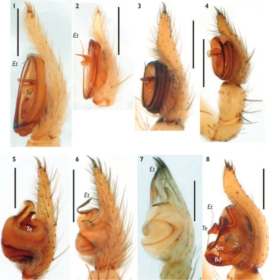
Figure 3. Retrolateral view of the male palp of Hersiliola esyunini sp. n. (1), H. sternbergsi sp. n. (2), Duninia baehrae sp. n. (3), Deltshevia danovi sp. n. (4), D. gromovi sp. n. (5), and Ovtsharenkoia pallida (6). Scale = 0.5 mm.
1
Et
Sd
Fp
Eb
Te
Te
Em Te
Sd
Et
Va Ha
Et Te
Bm
Bd
Bl Sd
4
2
5
3
Type material (49 specimens). Holotype: ♂ and paratypes 3♀, 4 juv. (ZMMU) with a label “TURKMENISTAN, Lebap Area, Karakum Desert, Repetek Reserve, NW environs of Repetek, 200 m, sands, 38°33’57-59”N 63°09’46”–10’13”E, 14.04.2002, A.V. Gromov coll.”
Paratypes.TURKMENISTAN: 1♂, 1♀, 2 subad.♀, 5 juv., Balkan Velayati (=Prov-ince), Turkmenbashi (=Krasnovodsk), 21.01.1978, K.G. Mikhailov coll. (ZMMU, Ta-3091); 3 juv., Balkan Province, Tuarkyr Mts., Kai gshem Plateau, 7.11.1982, V. Fet coll.; 1♀ (ZMMU), Lebap Province, East Karakum, Repetek Reserve, 170 m a.s.l., 05-06.1914, N.N. Plavilshchikov coll. (ZMMU); 1 subad.♀, attracted by light, 6.05.1972, V.I. Kuznetsov coll. (ZMMU); 2♀, 13.05.1981 (SMF); 1♂, 7.03.1982 (SMF); 1♂, 8.06.1982 (ZMMU), V.A. Krivokhatsky coll.; 1♂, 2♀ ZMMU), 18.04.1993, S.V.Ovtchinnikov coll.; 2♂, 2♀, Lebap Province, Karakum Desert, Repetek Reserve, Repetek, 170 m, 38°33’55”N 63°10’46”E, 17.04.2002, A.V. Gromov coll. (ZMMU); 1♂, Lebap Province, Amudarya Reserve, Kabakly, 4-14.05.1987, F. Zeleev coll. (ZMMU); 2♀ (ZMMU), Mary Province, Sultanbent, 05.1929, V.I. Sytchevskaya coll. (ZMMU); 1♀, 1juv., Mary Province, NE vicinities of Serhetabad (=Kushka), Kush-ka River left bank, 645 m a.s.l., 35°17’24”N 62°20’58”E, 12.04.2002, A.V. Gromov coll. (ZMMU). UZBEKISTAN: 1♂, 1♀, Navoiy (=Navoi) Viloyati (Province), Kyzyl-kum Desert, near Gazli, 21.05.1994 (S.V. Ovtchinnikov coll.) (ZMMU); 1♀, Buxoro (=Bukhara) Province, Kyzylkum Desert, near Gazli, 3.05.1998, S.V. Ovtchinnikov coll. (ZMMU); 6♀, 2 juv., Buxoro Province, Yagzakkum Sands, ca 7 km S of Nayumetan, 39°37’30”N 64°23’30”E, 20.04.2002, A.V. Gromov coll. (ZMMU).
Etymology. h e species name is a patronym honoring our late colleague and
friend Maris Šternbergs (1940-1996), of Riga, Latvia, the only Latvian araneologist of his generation, who also instructed VF in spider studies during his visit to Badghyz (Turkmenistan) in April 1977.
Diagnosis. Hersiliola sternbergsi sp. n. is most similar to H. simoni,from which it can be distinguished by a longer terminal portion of the cymbium, thicker seminal ducts, lower position of the tegular ridge, and position of the base of the embolus (at almost 12 hrs). Females of H. sternbergsi sp. n. dif er by a wider septum, thicker lateral arms, and only one coil of the insemination duct around the fertilization duct. h e epigyne of H. sternbergsi sp. n. is also similar to that of H. afghanica. h ese two spe-cies have an almost identical septum shape, but in H. sternbergsi sp. n. the epigyne is smaller in size, has smaller spermathecae, and fertilization ducts terminating on the ventral wall of the spermatheca (on the dorsal wall in H. afghanica).
Description. Male.Total length 4.6-5.4. Carapace 1.85 long, 2.0 wide, femur I 3.5, femur I/carapace length ratio 1.89. Colouration as in female. Palp as in Figs 3.2, 4.3, 5.3, 6.4-5, tip of cymbium as long as height of tegulum; seminal duct thick; em-bolus i liform, makes one coil (360°); emem-bolus base located at 11-11:30 hrs; tegular apophysis located almost at the center of the tegulum.
V-Figure 4. Retrolateral view of the male palp of Hersiliola macullulata (1), H. simoni (2), H. sternbergsi sp. n. (3), H. esyunini sp. n. (4), Duninia baehrae sp. n. (5), Deltshevia danovi sp. n. (6), D. gromovi sp. n. (7), and Ovtsharenkoia pallida (8). Scale = 0.5 mm.
1
5 Et
Et
Et
Te Te Te
Te
Et
Et
Bm Bd
2
6
3
7
4
8
shaped mark behind eyes. Abdomen with a heart spot and four pairs of transverse bands. h e upper transverse band originates from the heart spot. Sides of abdomen with dark spots. Epigyne as in Figs 7.7-9, 8.2, with a distinct median plate and win-dows, proportions of median plate and windows variable; insemination duct makes one coil around fertilization duct; insemination duct terminates on ventral side of the spermatheca.
men-Figure 5. Prolateral view of the male palp of Hersiliola macullulata (1), H. simoni (2), H. sternbergsi sp. n. (3), H. esyunini sp. n. (4), Duninia baehrae sp. n. (5), Deltshevia danovi sp. n. (6), D. gromovi sp. n. (7), and Ovtsharenkoia pallida (8). Scale = 0.5 mm.
1
5
2
6
3
7
4
8
tioned a wide distribution of “H. pallida” in the Turanian Zoogeographical Province, thus obviously assuming this name for the population from Ashgabat identii ed by Vlassov and Sytchevskaya (1937) as “H. maculata”. Our studyshows that this lowland desert Central Asian taxon is a new species, dif erent from H. macullulata (see above on H. macullulata).
1
4 5
6
2 3
8
9
7
Figure 6. Male palp of Hersiliola macullulata (1), H. simoni (2-3), H. sternbergsi sp. n. (4-5), H. esyunini sp. n. (6-7), Duninia baehrae sp. n. (8), and Ovtsharenkoia pallida (9). 1-2 ventral 3-4, 6 from below 5, 7, 9 from above 8 retrolateral. Scale = 0.5 mm.
Sd
Sd
Eb
Eb
Sd
Bd Te
Figure 7. Epigynes of Hersiliola versicolor (1), Hersiliola sp. (2), H. lindbergi sp. n. (3, 6), H. afghanica (4-5), H. sternbergsi sp. n. (7-9), H. esyunini sp. n. (10-12), and H. foordi sp. n. (13-14). 1-8, 10-11, 13-14 ventral 9, 12 dorsal. Scale = 0.5 mm.
1
4
7
10
2
5
11 12
13
14 8
3
6
9 Se
As Mp
Ag?
Ag
Ag
Wi
Wi Sp
Sp
Sp Sp
Mp
Mp Mp
As
Ul
Ul
Id
Id
Se
Se
sternbergsi sp. n. were recorded from November to May, and adult males, from Decem-ber to April, indicating a winter-spring mating period. Spiders were active in daytime.
Hersiliola esyunini sp. n.
urn:lsid:zoobank.org:act:406712E1-0BAC-4D22-816B-81D968015358
Figs 1.2, 2.2, 3.1, 4.4, 5.4, 6.6-7, 7.10-12, 8.4
H. xinjiangensis (in part): Marusik 2009: 153-156, f. 1-2, 4-13 (♂♀). Misidentii cation.
Type material. Holotype: ♂ together with paratypes 1♂ and 2♀ (ZMMU) from UZ-BEKISTAN: Namangan Province, Pap District, SE foothills of Kurama Mountains, ca. 5-5.5 km NW of Khanabad, ca. 850 m a.s.l., 40°54’05”N 70°45’44”E, 16.05.2002, A.V. Gromov coll.; paratype 1♂, Namangan Province, Pap District, SE foothills of Kurama Mountains, ca. 5.5 km NW of Khanabad, ca. 380 m a.s.l., under stones, 40°54’15”N 70°45’29”E, 15.05.2002, A.V. Gromov coll. (SMF).
Etymology. h e species name is a patronym honoring our friend and colleague
Sergei Esyunin of Perm, Russia.
Diagnosis. H. esyunini sp. n. is closely related to H. xinjiangenis, from which it can be distinguished by its smaller size, dif erently shaped tegular apophysis and the median plate of the epigyne. From all congeners H. esyunini sp. n. can be easily
distin-1
4 5
7 6
2 3
Ag FdAg
Lp
Lp Dp Dp
Ag
Sp
Sp
Sp
Ul
Ul
Ul
Id
guished by a relatively shorter tip of the cymbium, undivided tegular apophysis and a median plate of the epigyne with turned up lateral edges of the median plate.
Description. Male. Total length 3.4-3.65. Carapace 1.4-1.5 long, 1.5-1.6 wide,
fe-mur I 3.0-3.1 long, fefe-mur/carapace length ratio 2.1-2.14. Body yellow-light brown with pattern formed by brown hairs and spots. Carapace with brownish eye area and posterior cephalic part, and brown vertical stripe on clypeus (Figs 1.2, 2.2). Margins of carapace brown. Abdomen with indistinct pattern. Legs with broad, dark annulations, dark rings wider than light ones. Femur I almost entirely dark. Coxae IV separated by one diameter. Palp as in Figs 3.1, 4.4, 5.4, 6.6-7, with femur, patella+tibia and cymbium subequal in length, tibia in terminal part slightly wider than femur. Tegulum discoid, with long i liform embolus and tegular apophysis. Embolus starts at about 01 hrs, makes a loop of more than 270° and terminates around 10 hrs. Tegular apophysis perpendicular to tegu-lum. Apical part of the bulb slightly l attened (embolus straight, not rounded).
Female. Total length 3.75-4.0. Carapace 1.4-1.5 long, 1.5-1.6 wide, femur I 2.25-2.5 long, femur/carapace length ratio 1.6. Colouration as in female. Epigyne as in Figs 7.10-12, 8.4. Median plate anchor-like, with a pair of small accessory sclerites on the side that visually makes the basal part wider; spermathecae and seminal duct translu-cent through the integument. Accessory gland digitiform.
Comments. Earlier, Marusik (2009) confused this species with the related H. xin-jiangensis due to the similarity of their male palps, i.e. same shape of the apical part of the tegulum (character unknown in other Hersiliola species), same position of the em-bolic tip, and same shape of the tegular apophysis. Misidentii cation was made before we started this revision of East Palaearctic Hersiliola.
Distribution. h e species is known only from two nearby localities in eastern Uzbekistan.
Hersiliola xinjiangensis(Liang & Wang, 1989)
Figs 13.1-4
Hersilia Xinjiangensis Liang and Wang, 1989: 56, f. 1-4 (D♂♀).
Hersilia x.: Hu and Wu 1989: 78, f. 55.5-8 (♂♀, same i gs as in Liang and Wang 1989).
Hersilia x.: Song et al. 1999: 80, f. 32O-P, 33F-G (♂♀, same i gs as in Liang and Wang 1989).
H. x. (in part): Marusik 2009: f. 3a-d (T♂♀, same i gs as in Liang and Wang 1989).
Type material: 2♂, 2♀, CHINA: Xinjiang, Urumchi, not seen, apparently lost (see below).
Diagnosis. h is species can be easily distinguished from all congeners, except for
H. esyunini sp. n., by a relatively shorter tip of the cymbium, l attened apical part of the tegulum, and undivided tegular apophysis. From the sibling species H. esyunini sp. n., H. xinjiangensis can be distinguished by its larger size, dif erent shape of the tegular apophysis and median plate of the epigyne.
groove deep. h orax low and l at. Eye area brown, 8 eyes in 2 rows. Both AER, PER curved, AER curved stronger than PER. ALE white, others 6 eyes black, AME largest, located in the front of head; ALE small, located below in front of PLE. 4 eyes of PER near same size. Chelicera small, yellowish brown, with one promarginal tooth and no retromarginal. Pedipalps and legs yellowish brown, each segments of pedipalps and legs with blackish brown annuli in the median and the distal. Tarsus with 3 claws. Up-per claw with a single tooth. Leg formula 2143. Opisthosoma dorsally with yellowish brown scales and grayish brown spots. Heart spot black, with three pairs of muscular depressions. h e venter yellow, without marking. Anterior spinnerets robust, their distal segment small. Median spinnerets thin, nearly the same length as anterior ones. Rear spinnerets located on the sides of median ones. h e distal segment same length as opisthosoma, smaller than the basal one. Colulus present. Epigyne brown, septum inverse T-shaped.
Male. Total length 5.00. Habitus, colour and markings style as in female.
h is species inhabits crevices and holes of walls. It is a common species. Its coloura-tion is cryptic and therefore it is not easy to i nd specimens.”
Comments. Type specimens (2♂, 2♀) were supposedly deposited in the Depart-ment of Plant Protection, Xinjiang August 1st Agricultural College, Urumqi, Xinjiang,
China (Liang and Wang 1989). At our request, Shuqiang Li tried to i nd these types but failed. It seems that after the retirement of Tie Liang these specimens were lost or transferred. h e embolus base was not depicted by Liang and Wang (1989). Judging from the conformation of the epigyne in its sibling, H. esyunini sp. n., it is likely that median plate of the epigyne and vulva (endogyne) have been somewhat misinterpreted.
Distribution.H. xinjiangensis is known only from central Xinjiang (China). It is the northernmost species of the genus and of the entire family Hersiliidae. In Europe (Iberian Peninsula), the northernmost locality of Tama edwardsi (Lucas, 1846) (Ribera et al. 1988) is in northeastern Portugal ca. 41°N, while the type locality of H. xinjian-gensis lies north of 44°N.
Duninia gen. n.
urn:lsid:zoobank.org:act:64FD3C91-A822-42B1-9779-592D9D3EF7E2
Type species.Duninia baehrae sp. n.
Etymology. h e genus name is a patronym honoring our late colleague, friend, and a prominent araneologist Pyotr Dunin (1952-1998) who lived and worked in Baku (Azerbaijan) and Togliatti (Russia). Gender: feminine.
copulatory openings below the epigynal plate, but they are not heavily sclerotized as in
Duninia gen. n.; also, O. pallida does not have round spermathecae.
Description. Body length ca. 5 mm, carapace as long as wide from 1.75 to 2.25.
Pattern as in Hersiliola. Palp cymbium with a long tip, tegulum globular, seminal duct wide, embolus wide and not screw-shaped, tegular apophysis claw-like, with its claw part thinner than embolus. Epigynal plate with a septum-like structure, but without windows and openings, median plate without distinct margins. Copulatory opening is located below the epigynal plate in an epigastral fold; spermathecae round, coiled ducts absent.
Comments. Conformation of the epigyne in this genus is, in some respects, unique among entelegyne spiders. h e epigynal plate has no openings (furrows or fovea) but possesses two pairs of fovea on the vertical posterior wall located inside the epigastral fold. h e lateral pair of fovea is shallow and elongate. Another pair of fovea is closer to the median part and located deeper; they have a more heavily sclerotized wall and are very deep. It seems that these deep pockets have copulatory openings inside the ante-rior part. Lateral pockets seem to match the tegular apophysis of the male.
h ere are few taxa among entelegyne spiders that have a copulatory opening on the posterior vertical wall of the epigyne located in the epigastral fold. Besides some Erigoninae, we know of only one such genus, Paratus (Liocranidae) (cf. Marusik et al. 2008). Paratus has epigynal plate without furrows and fovea, and a small fovea hidden in the epigastral wall that leads to two closely separated copulatory openings.
h e male palp of Duninia gen. n. is unique among Hersiliidae because of its short, broad embolus and tegular apophysis extended along the cymbial axis.
Distinguishing characters. Two species of Duninia gen. n. can be easily distin-guished by the shape of the copulatory organs.
Composition and distribution. Duninia gen. n. includes two species: D. baehrae sp. n. (type species; Turkmenistan) and D. rheimsae sp. n. (northern Iran, Tehran Province).
Duninia baehrae sp. n.
urn:lsid:zoobank.org:act:1DD0B4E4-BFD2-4E35-B05C-BDC1EF3A9923
Figs 3.3, 4.5, 5.5, 6.8, 8.6, 11.1-4
Hersiliola pallida: Ovtsharenko and Fet 1980: 443 (Turkmenistan: Badghyz). Misiden-tii cation.
Hersiliola afghanica: Fet 1983: 837 (in part; Turkmenistan: Kopetdagh; see also Delt-shevia danovi sp. n.); Kuznetsov and Fet 1984: 52 (Turkmenistan: Central Kope-tdagh); Mikhailov and Fet 1994: 504. Misidentii cation.
Hersiliola afghana: Fet 1984: 259 (misspelling). Misidentii cation.
Central Kopetdagh, Kopetdagh State Reserve, Bolshie Katranki, 9-16.09.1978, G.T. Kuznetsov coll. (SMF); 1♀, Mary Province, Serhetabad (=Kushka) District, Morgu-novka, 29.06.1975, V. Fet coll. (SMF); 2♀, Balkan Province, Bolshoi Balkhan Mts., 6 km from Nebit-Dagh (now Balkanabat), 4.04.1993, S.V.Ovtchinnikov coll. (ZMMU).
Etymology. h e species name is a patronym honoring our colleague Barbara Baehr (Brisbane, Australia), for her contributions to in-depth modern studies of Hersiliidae.
Diagnosis. From its sibling D. rheimsae sp. n., females can be easily distinguished by a larger body and epigyne, diverging spermathecae, and deep pockets longer than the lateral ones.
Description. Male (holotype from Kurkulab, abdomen and most of legs are miss-ing): Carapace 2.12 long, 2.25 wide, femur I 5.0 long, leg I/carapace length ratio 2.36. Palp as in Figs 3.3, 4.5, 5.5, 6.8, bulbus globular, basal portion of seminal duct thick; embolus very massive, l at and short, twice shorter than the apical portion of the cymbium, opening large and clearly visible, tegular apophysis claw-like, large, located on the apical part of tegulum, extends parallel to cymbial axis, its terminal part much thinner than embolus.
Female: epigyne as in Figs 8.6, 11.1-4, lacks windows and distinct openings; medi-an plate twice as wide as high; trmedi-anslucent spermathecae round, separated from medimedi-an plate by more than one diameter; lateral sides of epigynal plates with “arches” clearly visible on dissected epigyne; vulva with two round spermathecae separated by one diameter and large lateral wings. Lateral arches lead to small pockets, and large wings correspond to large and deep pocket that seems to correspond to copulatory opening. It is not clear which structures of the vulva correspond to fertilization ducts, and also not clear whether the fertilization duct has accessory glands.
Comments. Kuznetsov and Fet (1984: 52) recorded this species (as Hersiliola af-ghanica) for several localities in Central Kopetdagh (Turkmenistan, bordering with Iran) within the territory of Kopetdagh State Reserve as well as its oi ce in Berzengi, suburb of Ashgabat. h ese records were based on six specimens, all captured in pitfall traps by Gennady Kuznetsov. In addition to Kurkulab, and B[olshie] Katranki localities (see labels above), Kuznetsov and Fet (1984) listed two specimens from Berzengi (05-06.1980, 1♀) and M[alyi] Dashtoi (22-29.07.1981, 1♂). h ree reported localities within the Kopetd-agh State Reserve lie at 900-2000 m a s.l (Kurkulab, 900-1000 m; Bolshie Katranki and Malyi Dashtoi, 1500-2000 m) (G. T. Kuznetsov. pers. comm. 2008). An additional, previously unpublished, specimen from Gaudan (ZMMU) was collected by an early lo-cal naturalist, Baron O. von Rosen, in 1895. h e species undoubtedly will be found in neighboring northeastern Iran (Khorassan Province) and northern Afghanistan. It is an element of the not yet fully explored mountainous fauna of the Turkmeno-Khorassan region, a transitional zoogeographic area between the western mountains of the Medi-terranean / Iran and eastern mountains of Central Asia / Himalayas (Fet 1994). h is area consistently yields interesting new spider taxa such as e.g. Paracedicus gennadii (Fet, 1993), P. ephthalitus (Fet, 1993) (Desidae), Synaphris orientalis Marusik & Lehtinen, 2003 (Synaphridae), or a hersiliid Deltshevia danovi gen. n. sp. n. (see below).
Duninia rheimsae sp. n.
urn:lsid:zoobank.org:act:1B60CF32-E7DF-483D-9575-9EF514922FDE
Figs 8.7, 11.5-7
Type material: Holotype subadult ♀ with a well-developed epigyne and paratypes 3 juvenile specimens: IRAN [25], Tehran Province, ca. 80 km E of Tehran, Damavand District, Aroo Village, 52°27’E 35°40’N, 15.06.2000, Yu.M. Marusik coll. (ZMMU).
Etymology. h e species name is a patronym honoring our colleague Cristina A.
Rheims (São Paulo, Brazil), for her contributions to in-depth modern studies of Hersiliidae.
Description. Male unknown. Female: Total length 5.0. Carapace 1.75 long, 1.85 wide, femur 2.15 long, femur I/carapace length ratio 1.23. Epigyne as in Figs 8.7, 11.5-7, with a sort of septum on the median plate, spermathecae converging and al-most touching each other, epigynal pockets subparallel, with hemispherical terminal part. Lateral pockets long, appear longer than their depth.
Diagnosis. D. rheimsae sp. n. can be distinguished from the sibling species D. baehrae sp. n. by its smaller body and epigyne size, converging spermathecae, and less slanting epigynal pockets bearing large, clearly visible hemispheres.
Comments. Judging from the conformation of the male palp and epigyne in its
sibling D. baehrae sp. n. and conformation of the epigyne in D. rheimsae sp. n., it is reasonable to suggest that male of this species should have a smaller embolus (matching a small, deep pocket of D. rheimsae sp. n.) and a larger tegular apophysis.
Distribution. Known only from type locality in northern Iran.
Deltshevia gen. n.
urn:lsid:zoobank.org:act:44D11CBC-5310-4DC0-A3C8-67F27C7A247D
Type species.Deltshevia danovi sp. n.
Etymology. h e genus name is a patronym honoring our colleague, friend, and a prominent araneologist, Christo Deltshev of Soi a, Bulgaria. We are especially glad to dedicate this genus to Christo on the occasion of his 70th birthday. Gender: feminine.
Diagnosis. Deltshevia gen. n. dif ers from other genera of Hersiliidae by a globular tegulum; thick and screw-shaped embolus with a tapering, sharp tip; large two-armed tegular apophysis extended upward; large epigyne openings; and thick copulatory ducts.
Description. Small hersiliids 5-7 mm long, with carapace 2.1-2.7 long and wide. Palp globular; embolus thick and screw-shaped; tegular apophysis large, with two arms: a massive vertical arm and a small horizontal arm. Epigyne with large copulatory openings; wide insemination ducts; septum and median plate distinct; spermathecae oval; fertilization duct short; accessory gland globular.
Distinguishing characters. h e two species of Deltshevia gen. n. can be easily distinguished by the shape of copulatory organs.
Composition and distribution. Deltshevia gen. n. includes two species: D. danovi
sp. n. (type species; Turkmenistan and Kazakhstan) and D. gromovi sp. n. (eastern Uzbekistan).
Deltshevia danovi sp. n.
urn:lsid:zoobank.org:act:47699594-B924-4F00-8435-D0200734C1C2
Figs 3.4, 4.6, 5.6, 9.1-5, 10.1-3
Hersiliola afghanica: Fet 1983: 837 (Turkmenistan: SW Kopetdagh). Misidentii ca-tion.
Hersiliola danovi: Fet 1985: 72 (nomen nudum; no description published).
Hersiliola sp. 1:Mikhailov and Fet 1994: 504.
Type material (31 specimens): Holotype: ♂ (ZMMU) and paratypes 3♀, 1 juv.♀
(ZMMU), 3♀ (SMF), TURKMENISTAN: Balkan Province, Southwest Ko-petdagh, Syunt-Khasardagh Reserve, N of Mt. Syunt, Damdam, 1000 m a.s.l., 8.07.1984, V. Fet coll.
Paratypes. TURKMENISTAN: Balkan Province, Southwest Kopetdagh, Gar-rygala (=Kara-Kala), Parkhai, 400 m a.s.l., 2 juv.♀, 08.1983; 1♀, 7.06.1984, V. Fet coll. (ZMMU); Balkan Province, Southwest Kopetdagh, Hodzhagala, 400 m a.s.l., under stones, 1 subad.♀, 05.1981, N. Yermakov coll. (ZMMU), 1♂, 11.05.1984, V. Fet coll. (SMF), 4 subad.♀, 11.05.1984, 2 subad.♂, 3 subad.♀, 4.06.1984, V. Fet coll. (ZMMU); Balkan Province, Gyzylarbat (=Kyzyl-Arvat), 12.05.1984, 2♀, 1 juv.♂, V. Fet coll. (ZMMU). KAZAKHSTAN: 3♂, 3♀, Atyrau (=Guryev) Prov-ince, Ustyurt Reserve, W of Baskorgan, 27.05.1989, Raikhanov and Ibrayev coll. (ZMMU).
Etymology. h e species name is a patronym honoring the late Rostislav Danov (1941-1993) of St. Petersburg, Russia, a naturalist, snake hunter, and artist, a friend of VF and his family, who spent many years working in Southwest Kopetdagh.
Diagnosis. Deltshevia danovi sp. n.is similar to D. gromovi sp. n.,from which it can be easily distinguished by the shape of the embolus, tegular apophysis and epigyne.
Figure 9. Somatic characters of Deltshevia danovi sp. n. (1-5),and D. gromovi sp. n. (6-7). 1, 7 female carapace, dorsal 2-3 abdomen of female, ventral and dorsal respectively 4-5 male carapace, frontal and lateral, respectively 6 female habitus.
1
4
6
5
7
Figure 10. Epigynes of Deltshevia danovi sp. n. (1-3),and D. gromovi sp. n. (4-5). 1, 3-4 ventral 2, 5 dorsal. Scale = 0.5 mm.
1
2 5
3
4
Wi Se
Ag Id
Fd Sp
danovi sp. n.has round openings (bell-shaped in D. gromovi sp. n.), a wider septum than in D. gromovi sp. n., a higher median plate, and larger spermathecae.
Female. Total length 6.0-7.0. Carapace 2.1-2.3 long, 2.4-2.5 wide (wider than long). Carapace pattern and eye arrangement as in Figs 9.4-5. Abdominal pattern as in Fig. 9.3, dorsal side with a dark heart band and four transverse stripes, sides of abdomen with dark spots, venter without pattern. Epigynal plate 1.00-1.14 wide, fovea 0.47-0.57 wide, plate/fovea ratio 2-2.1. Epigyne as in Figs 10.1-3, with distinct windows, well-separated septum, median plate anchor-like, epigynal opening distinct (with well-expressed borders), opening diameter larger than septum width and equal to median plate height; translucent spermathecae elongate and located aside of openings. Vulva simple, with large pockets (continuation of epigynal opening), with oval sper-mathecae separated by more than three times their widths; insemination duct short, accessory gland globular, poorly visible.
Distribution. Turkmenistan (southwest), Kazakhstan (southwest).
Deltshevia gromovi sp. n.
urn:lsid:zoobank.org:act:D7FA30A6-E286-4A5A-9C33-D799D815E437
Figs 3.5, 4.7, 5.7, 9.6-7, 10.4-5
Hersiliola macullulata: Zyuzin et al. 1994: 6 (Kazakhstan). Misidentii cation.
Note: Much new material from Central Asia reached us, due to the ef orts of Alexan-der Gromov and Dmitri Logunov, when this paper was already i nalized; therefore the following description and i gures of a male are based on a subadult with well-developed palp.
Type material (49 specimens): Holotype: ♀ and paratype subadult ♂ (with well-de-veloped palp)(ZMMU),UZBEKISTAN: Surkhandarya Province, Kattakum Sands, ca. 4 km NE of Uchkyzyl, ca. 330 m a.s.l., 37°22’33”N 67°16’38”E, 27.04.2002, A.V. Gromov coll.
2♂, 2♀, Buxoro (=Bukhara) Province, Gizhduvan District, SW foothills of Karaktau Mt. Range, ca 14.5 km N of Kanimekh, 40°24’51”N 65°08’57”E, 392 m, 5.06.2003, A.V. Gromov coll. (ZMMU); 1♂, 1♀, Navoi Province, Kanimekh District, Kyzyl-kum Desert, ca 65 km W of Chengel’dy, near Darvazatepa Mt., clay hills, 211 m, 40°57’26”N 64°07’51”E, 4.06.2003, A.V. Gromov coll. (ZMMU); 1♀, Kyzylkum Desert, Kuldzyktau Mt., Dzhengeldy, 22.05.1994, S.V. Ovtchinnikov coll. (ZMMU). KAZAKHSTAN: 2♂, 3♀, South Kazakhstan Province, Chimkent (now Shimkent) District, Kyzylkum Desert, Karaktau Mt. Range, Karamola Mt., 8.06.1989, A.A. Zy-uzin coll. (ZMMU); 4♀, Kazakhstan, South Kazakhstan Province, Kyzylkum Desert, Karaktau Mt. Range, Karamola Mt., 29.05.1993, A.A. Zyuzin coll. (ZMMU); 1♀, Kyzylkum Desert, Karamola Mt., 42°14’57.2”N 67°48’21.1”E, 228 m, 3.07.2006, A.V. Gromov coll. (ZMMU).
Etymology. h e species name is a patronym honoring its collector, our friend and
colleague Alexander V. Gromov of Almaty, Kazakhstan.
Diagnosis. Deltshevia gromovi sp. n.is similar to D. danovi sp. n.,from which it can be easily distinguished by the shape of the embolus, tegular apophysis and epigyne.
Deltshevia gromovi sp.n has a shorter and screw-shaped embolus, while D. danovi sp. n. has a longer, non-screw-shaped embolus. h e apical portion of the tegular apophysis in D. gromovi sp. n.has subparallel margins, while the apical portion of the tegular apophysis in D. danovi sp. n. is triangular, with slanting margins. Openings of the epigyne in D. gromovi sp. n. are bell-shaped but not round, copulatory ducts are much longer than in the sibling species, and accessory glands are much larger in comparison to the spermathecae.
Description. Male (subadult, but with well-developed palp). Total length 5.6. Carapace 2.0 long, 2.2 wide, femur I 2.75 long. Carapace with 4 pairs of dark marginal spots, 2 pairs of lateral spots in anterior half and unpaired median spot in thoracic part. Legs with distinct annulations. Palp as in Figs 3.5, 4.7, 5.7, with a long, non-screw-shaped embolus, which almost reaches the cymbium tip; tegular apophysis large, its apical part has almost parallel sides.
Female. Total length 5.0. Carapace 2.1 long, 2.35 wide, femur I 3.25 long, ep-igynal plate 0.71 wide, fovea 0.36 wide, ratio 0.5. Colouration as in male. Epigyne as in Figs 9.6-7, 10.4-5, with distinct large bell-shaped copulatory openings, thin taper-ing septum, and thin edge of median plate; opentaper-ings leads to voluminous copulatory ducts, their height is about three times longer than their diameter and two times longer than height of copulatory openings; spermathecae small, oval; accessory glands globu-lar, large (about half the size of a spermatheca)
Comments. It is possible that in mature males the embolus is screw-shaped, as in
D. danovi sp. n. h e single male that we studied was subadult, and its embolus was straightened being pressed between the cymbium and cuticle. It is possible that after the i nal molt the embolus would become partially screw-shaped.1
Distribution. Uzbekistan, Kazakhstan (south).
Figure 11. Epigynes of Duninia baehrae sp. n. (1-4),and D. rheimsae sp. n. (5-7). 1-2, 5 ventral 3 dorsal 4, 6-7 caudal. Scale = 0.5 mm.
1
3
5
2
4
7 6
Dp
Dp
Dp Dp Dp Lp
Lp
Lp
Lp Mp
Mp Mp Mp
Fd? Sp
Sp
Ovtsharenkoia gen. n.
urn:lsid:zoobank.org:act:22EE9CC4-BCC0-4E96-B57C-C84E920297E7
Type species. Hersiliola pallida Kroneberg, 1875.
Etymology. h e genus name is a patronym honoring our friend and colleague, and a prominent araneologist, Vladimir Ovtsharenko of New York, USA. Gender: feminine.
Diagnosis. Ovtsharenkoia gen. n. can be easily distinguished from other genera of Hersiliidae by its short spinnerets and the shape of the copulatory organs. h e male palp has a unique conformation for the family due to the presence of a complex out-growth in the basal part of the tegulum. All other genera similar to Hersiliola have only one apophysis (tegular). Females of Ovtsharenkoia gen. n. can be recognized by a small median plate of the epigyne, a transverse translucent fertilization duct, large pale areas next to the median plate, and absence of distinct spermathecae.
Comments. A discoidal tegulum and whip-like embolus in O. pallida indicate that
Ovtsharenkoia gen. n. is more closely related to Hersiliola than to two other Central Asian genera, Deltshevia gen. n. and Duninia gen. n., both of which have a globular tegulum and a thick, short embolus. A discoidal tegulum and whip-like embolus are also found in Hersilia. Embolus base modii ed, and dif ers from that in all other hersil-iid genera. h e epigyne of O. pallida has some similarity with those in Duninia gen. n. due to its pockets, but has an entirely dif erent vulva. h e type of embolus in O.pallida
is similar to those in some Neotama species (cf. i gs 45c, 46c in Baehr and Baehr 1993).
Composition and distribution. Type species only, Ovtsharenkoia pallida (Kro-neberg, 1875), found widely across the mountains and foothills from southern Turkmen-istan to UzbekTurkmen-istan, Kazakhstan, Kyrgyzstan, and TajikTurkmen-istan; south to northern PakTurkmen-istan.
Ovtsharenkoia pallida (Kroneberg, 1875)
Figs 2.3-8, 3.6, 4.8, 5.8, 6.9, 12.1-5
Hersiliola p.Kroneberg, 1875: 13-14, pl. 5, f. 41 (D♀).
Hersiliola p.:Kroneberg 1885: 545-547; Kroneberg 1888: 189-190; Simon 1893: 445; Reimoser 1919: 188; Charitonov 1932: 23; Caporiacco 1935: 142 (Pakistan); Spassky 1952: 196-200 (in part); Bonnet 1957: 2180; Andreeva and Tyshchenko 1969: 382; Andreeva 1976: 30-31 (Tajikistan).
Type material. Lectotype: ♀ (designated here): UZBEKISTAN: Samarkand, A.P. Fed-chenko coll. (ZMMU, Ta-1323); paralectotype ♀ (designated here): KYRGYZSTAN: Osh, A.P. Fedchenko coll. (ZMMU, Ta-1324). Vial with paralectotype female contains also adult female of Oecobius nadiae (Spassky, 1936). h is species and specimen were not men-tioned in Kroneberg’s text. It seems that he considered this specimen a juvenile Hersiliola.
Additional material examined (48 specimens). KAZAKHSTAN (i rst country
1
3 4
5 2
Dp? Dp?
Lp
Lp Mp
Pp Pp
Fd? Sp?
Id?
Figure 12. Epigyne of Ovtsharenkoia pallida.1-3 ventral 4 caudal 5 dorsal. Scale = 0.5 mm.
Figure 13. Hersiliola xinjiangenis, after Liang and Wang (1989). 1-2 epigyne, ventral and dorsal, respec-tively 3-4 male palp, retrolateral and ventral, respectively.
1
2 3 4
Fp Et
Diagnosis. Same as for the genus.
Description. Male (described here for the i rst time). Total length 4.8. Carapace 2.0 long, 2.25 wide, femur I 4.25 long, femur/carapace length ratio 2.13. Carapace light brown with dark margins, radial dark stripes and median dark band; cephalic part separated from the thoracic part by a dark V-shaped spot. Abdomen light brown with a brown heart spot, transverse stripes and dark sides. Dorsal pattern variable. Venter of abdomen without pat-tern. Palp as in Figs 3.6, 4.8, 5.8, 6.9, femur and cymbium equal in length, patella+tibia al-most as long as cymbium or femur; tegulum round, basal part extended; embolus whip-like, arched; embolus makes a half loop, base of embolus located on retrolateral side; tegulum with two apophyses: apical (?tegular) and basal. Basal apophysis (outgrowth) complicated, subdivided into three parts: lamellate, digitiform retrolateral, and mesal. Embolus passes below mesal part of basal apophysis. Apical apophysis perpendicular to the cymbial axis.
Female. Total length 4.2-6.0. Carapace 2.0-2.5 long, 2.25-2.75 wide, femur I 3.5, femur/carapace length ratio 1.75. Colouration as in male. Epigyne as in Figs 12.1-5, with strongly chitinized small median plate, septum well-developed, its length subequal to length of lateral arms; width of septum variable; windows absent, lateral sides of epigynal plate with small extensions, indicating pockets; aside of median plate epigyne is pale, size of pale part variable; upper part of epigynal plate with a pair of transverse stripes formed by translucent insemination (?) ducts. Distinct spermathecae absent, insemination ducts relatively thin, coils absent; fertilization ducts with globular accessory glands. Insemina-tion ducts have small globular extensions that possibly correspond to spermathecae proper. Size of epigyne variable, although size of median plate is not variable, as well as position of translucent fertilization ducts. Distance between epigastral fold and ducts is the same in large and small epigynes.
Comments. Details of epigynal structure remain uncertain. It is not clear whether openings on the median plates are real copulatory openings or just fovea, origins of copulatory (insemination) ducts as well as the position of the terminal part of the fer-tilization ducts. Most probably the insemination duct is weakly sclerotized and origi-nates in pockets lying below the epigynal plate.
h is species has been described from the important collections of the famous 1868−1871 expedition of Alexei P. Fedchenko (1844−1873) and his wife Olga A. Fed-chenko to Russian Turkestan. h e original type series includes two female syntypes; the lectotype from Samarkand and paralectotype from Osh are designated here. Andreeva and Tyshchenko (1969) and Andreeva (1976) reported juveniles of this species from Tajikistan (Bishkent Valley, Chiluchor-chashma, 4 juv., 8.05.1965, E. Martynova coll.; Khozratisho Range, 13 km from Muminabad along Obisurkh River valley, 1 juv., 9.06.1966, E. Andreeva coll.). Adults from Tajikistan are unknown.
Earlier records from lowland Turkmenistan were misidentii cations and refer to
Hersiliola sternbergsi sp. n. (Repetek, Badghyz) or Duninia baehrae sp. n. (Badghyz). However, new material collected by S. L. Zonstein in 1993 shows that this species is indeed found in Turkmenistan. It was collected in the very southwest of Badghyz, in the mountainous area bordering Iran (Zyulfagar Range in the watershed of Tejen, which forms an important zoogeographic boundary between the Turkmeno-Khurassan moun-tains to the west and all Central Asian/Himalayan mounmoun-tains to the east; see Fet 1994).
Distribution. Kazakhstan, Kyrgyzstan, Pakistan (Baltistan: Karakoram Mts.), Tajikis-tan, Turkmenistan (south), Uzbekistan. Unlike most species of Hersililola and other ha-bitually similar genera, this species is restricted exclusively to mountainous areas and seems to be the only species of hersiliids in the Palaearctic that penetrates human settlements and buildings (Bishkek). It could be a petrophilous species that inhabits clif s and walls.
Discussion
According to several authors (Baehr and Baehr 1987; Dippenaar-Schoeman and Joc-qué 1997; JocJoc-qué and Dippenaar-Schoeman 2006, etc.), Lehtinen (1967) was the i rst who indicated a close relationship between Hersiliidae and Oecobiidae. However, Lehtinen (1967: 305) clearly indicated that the priority of such a statement belonged
Map 1. Distribution of Hersiliidae in Central Asia.
Hersiliola afghanica; Hersiliola esyunini; Hersiliola foordi; Hersiliola lindbergi; Hersiliola sternbergsi; Hersiliola xinjiangensis; Duninia baehrae; Duninia rheimsae; Deltshevia danovi;
Deltshevia gromovi; Ovtsharenkoia pallida; ? identity uncertain afghanica
baehrae danovi esyunini foordi gromovi
to Bristowe (1938), who placed together Hersiliidae and Urocteidae in a special sec-tion among Clubionoidea. At the same time, Caporiacco (1938) suggested a separate superfamily Hersiliiformia that included Hersiliidae and Oecobiidae (but without the cribellate Urocteidae). Later, Mello-Leitão (1941) and Petrunkevitch (1958) proposed a superfamily Oecobioidea that included Oecobiidae, Urocteidae (now a subfamily of Oecobiidae), and Hersiliidae. h ese three families were united under this superfamily according to the structure of copulatory organs, spination, modii cation of carapace, eye pattern, and setation. In addition, Crome (1957) found some behavioral similari-ties between Hersiliidae and Oecobiidae (op. cit. after Lehtinen 1967: 305).
We have no doubt that Oecobiidae are related to Hersiliidae. In addition to clear so-matic characters indicated by earlier authors such as eye pattern, an almost round carapace (width equals length), etc., both families have elongated posterior lateral spinnerets and the same way of holding the palps (converging femora, and diverging tibia and terminal joint) in both sexes (Fig. 1.2, 2.1; i g. 71a in Jocqué and Dippenaar-Schoeman 2006). Similarities between Oecobiidae and Hersiliidae even led Strand (1913) to confusion when he described Oecobius (s.l.) brachyplura (Strand, 1913) in Hersiliola (see Fet 2008).
Coddington and Levi (1991) placed Oecobiidae and Hersiliidae in Eresoidea. Although Eresidae, like Oecobiidae and Hersiliidae, are very dif erent from the rest of the entelegyne spiders, we doubt that they should be placed in the same super-family due to entirely dif erent somatic morphology, morphology of copulatory or-gans, ecology, and behavior.
One of the additional diagnostic features of Hersiliidae, not indicated by earlier authors, is the presence of “accessory glands” in the epigyne. h ese structures appear to be present in the majority of hersiliid genera such as Hersilia, Hersiliola, Deltshevia
gen. n., Duninia gen. n., Yabisi Rheims & Brescovit, 2004, most of Tyrotama Foord & Dippenaar-Schoeman, 2005, Murricia Simon, 1882, Neotama Baehr & Baehr, 1993, and Tamopsis Baehr & Baehr, 1987. Some Tamopsis species appear to have two pairs of sack-like structures in addition to spermathecae (cf. i gs 25e, 28e in Baehr and Baehr 1987). Neotropical Iviraiva Rheims & Brescovit 2004 has acinoform spermathecae (not known in other entelegynes), and it is not clear whether they have accessory glands. Interestingly, structures that appear to be homologous to “accessory glands” have been observed in the related family, namely Uroctea limbata (C.L. Koch, 1843) and two uni-dentii ed species of Uroctea (Oecobiidae) (cf. i gs 10-16 in Baum 1972). Baum (1972) called these structures a “blind endender Anhang”. h ey were found in only half the studied species. Unlike in Hersiliidae, “acessory glands” in Uroctea are placed close to the spermathecae, but not close to the terminal part of the fertilization duct.