ESTUDO DAS MANIFESTAÇÕES NEUROPSIQUIÁTRICAS NOS
PACIENTES COM LÚPUS ERITEMATOSO SISTÊMICO DO
AMBULATÓRIO DE REUMATOLOGIA DO HOSPITAL DAS
CLÍNICAS DA UNIVERSIDADE FEDERAL DE MINAS GERAIS
UNIVERSIDADE FEDERAL DE MINAS GERAIS
PROGRAMA DE PÓS-GRADUAÇÃO EM CIÊNCIAS APLICADAS À
SAÚDE DO ADULTO
ESTUDO DAS MANIFESTAÇÕES NEUROPSIQUIÁTRICAS NOS
PACIENTES COM LÚPUS ERITEMATOSO SISTÊMICO DO
AMBULATÓRIO DE REUMATOLOGIA DO HOSPITAL DAS
CLÍNICAS DA UNIVERSIDADE FEDERAL DE MINAS GERAIS
Dissertação apresentada ao Programa
de
Pós-graduação
em
Ciências
Aplicadas à Saúde do Adulto da
Universidade Federal de Minas Gerais,
como requisito para obtenção do título
de Mestre em Ciências Aplicadas à
Saúde do Adulto.
Área
de
Concentração:Ciências
Clínicas
Orientador: Prof. Francisco Eduardo
Costa Cardoso
Co-orientadora: Profa. Gilda Aparecida
Ferreira
Ficha catalográfica elaborada pela Biblioteca J. Baeta Vianna – Campus Saúde UFMG
Maciel, Ricardo Oliveira Horta.
M152e Estudo das manifestações neuropsiquiátricas nos pacientes com Lúpus Eritematoso Sistêmico do ambulatório de Reumatologia do Hospital das Clinicas da Universidade Federal de Minas Gerais [manuscrito]. / Ricardo Oliveira Horta Maciel. - - Belo Horizonte: 2014.
102f.
Orientador: Francisco Eduardo Costa Cardoso.
Área de concentração: Ciências Clínicas Aplicadas à Saúde do Adulto. Dissertação (mestrado): Universidade Federal de Minas Gerais, Faculdade de Medicina.
1. Vasculite Associada ao Lúpus do Sistema Nervoso Central. 2. Transtorno Obsessivo-Compulsivo. 3. Transtorno do Déficit de Atenção com Hiperatividade. 4. Transtornos Cognitivos. 5. Dissertações Acadêmicas. I. Cardoso, Francisco Eduardo Costa. II. Universidade Federal de Minas Gerais, Faculdade de Medicina. III. Título
Prof. Jaime Arturo Ramires Vice- Reitora
Profa. Sandra Regina Goulart Almeida Pró-Reitor de Pós-Graduação
Prof. Ricardo Santiago Gomez Pró-Reitor de Pesquisa Prof. Renato de Lima Santos FACULDADE DE MEDICINA
Diretor
Prof. Tarcizo Afonso Nunes
Chefe do Departamento de Clínica Médica Prof. Ricardo de Menezes Macedo
PROGRAMA DE PÓS-GRADUAÇÃO EM CIÊNCIAS APLICADAS À SAÚDE DO ADULTO
Coordenadora
Profa. Teresa Cristina de Abreu Ferrari Sub-coordenador
Prof. Paulo Caramelli Colegiado
Prof. Marcus Vinícius Melo de Andrade Prof. Luiz Gonzaga Vaz Coelho Prof. Francisco Eduardo Costa Cardoso
Prof. Paulo Caramelli
Dedicatória
AGRADECIMENTOS
Ao meu orientador, prof. Francisco Cardoso, pelos ensinamentos e pela valiosa orientação na condução deste trabalho.
À minha co-orientadora profa. Gilda Ferreira, pela acolhida no serviço de Reumatologia e pela constante disponibilidade.
Aos colegas da Neurologia, pela amizade, apoio e auxílio na minha formação.
À toda equipe do Ambulatório de Reumatologia, pela acolhida e ajuda no encaminhamento de pacientes para o estudo.
Aos pacientes, pelo altruísmo na colaboração com este estudo.
À minha esposa Rita, pelo carinho, suporte e compreensão, sempre.
Aos meus pais, por todo o apoio, e pela compreensão nos momentos de ausência em virtude do trabalho
RESUMO
Introdução: O Lúpus Eritematoso Sistêmico (LES) pode afetar os núcleos da base, como por
exemplo, na coreia lúpica. A disfunção dos núcleos da base em outras doenças autoimunes
causa uma gama de sintomas não-motores neuropsiquiátricos, incluindo sintomas
obsessivo-compulsivos (SOC), disfunção executiva e o Transtorno de Déficit de Atenção e
Hiperatividade (TDAH).
Objetivo: Avaliar a frequência de manifestações neuropsiquiátricas, com ênfase na disfunção
executiva, e a presença de SOC e TDAH em pacientes com LES.
Métodos: Cinquenta e quatro pacientes com LES foram submetidos a uma bateria de testes
neuropsiquiátricos que incluíram o Mini-Exame do Estado Mental (MEEM), a Montreal
Cognitive Assessment(MoCA), a Bateria de Avaliação Frontal, os testes de fluência verbal FAS fonêmica e semântica (categoria animais), o Inventário de Obsessões e Compulsões –
Revisado, a Escala de Obsessões e Compulsões de Yale-Brown (YBOCS) e as Escalas de
Depressão e Ansiedade de Beck. O TDAH foi diagnosticado de acordo com os critérios da
DSM-IV.
Resultados: Seis (11,1%) pacientes apresentaram comprometimento cognitivo de acordo com
o MEEM e 33 (61,1%) quando avaliados pelo MoCA. Onze pacientes tiveram escores de
FAB anormais, e 5 (9,3%) tiveram fluência verbal semântica menor do que a esperada.
Quinze (27,8%) tinham SOC, dos quais 5 (9,3%) apresentavam sintomas moderados a graves,
de acordo com a YBOCS. Dezessete (31,5%) preencheram critérios diagnósticos para TDAH.
Disfunção executiva, depressão e SOC apresentaram correlação com atividade do LES.
Conclusão: Disfunção executiva, YDAH e SOC são comuns no LES. A importância de
ABSTRACT
Purpose of the Study: Systemic Lupus Erythematosus (SLE) can affect the basal ganglia, as is
the case of Lupus chorea. Basal ganglia dysfunction in other autoimmune diseases leads to
aarray of neuropsychiatric non-motor symptoms, including obsessive-compulsive symptoms
(OCS), executive dysfunction and attention deficit and hyperactivity disorder (ADHD).
Objectives: To evaluate the frequency of neuropsychiatric manifestations, with emphasis on
executive dysfunction, and the presence of OCD and ADHD in patients with SLE.
Methods: Fifty-four subjects with SLE were submitted to a battery of neuropsychiatric tests
that included the Mini Mental State Examination, the Montreal Cognitive Assessment, the
Frontal Assessment Battery, the FAS verbal and the categorical (animals) semantic fluency
tests, the Obsessive and Compulsive Inventory – Revised, the Yale-Brown Obsessive and
Compulsive Scale and Beck’s Anxiety and Depression Scales. ADHD was diagnosed
according to DSM-IV criteria.
Results: Six(11.1%) and 33 (61.1%) patients had cognitive impairment according the MMSE
and MoCA, respectively. 11 patients had abnormal FAB scores, and 5 (9.3%) had lower
semantic fluency scores than expected. 15 (27.8%) had OCS, of which 5 (9.3%) had moderate
to severe symptoms according to YBOCS. 17 (31.5%) met diagnostic criteria for ADHD.
Executive dysfunction, depression and OCS correlated with disease activity.
Discussion: Executive dysfunction, ADHD and OCS are common in SLE. The significance of
LISTA DE ABREVIATURAS
ACR American College of Rheumatology
CS Coreia de Sydenham
DH Doença de Huntington
DSM Diagnostic and Statistical Manual of Mental Disorders
FAB Frontal Assessment Battery
LES Lúpus Eritematoso Sistêmico
MEEM Mini-Exame do Estado Mental
MoCA Montreal Cognitive Assessment
OCI-R Obsessive-Compulsive Inventory – Revised
RM Ressonância Magnética
SAAF Síndrome de Anticorpos Antifosfolípides
mSLEDAI-2K ModifiedSystemic Lupus Erythematosus Disease Activity Index
2000
SLICC Systemic Lupus International Collaborative Clinics/American
College of Rheumatology Damage Index
TDAH Transtorno de Déficit de Atenção e Hiperatividade
TOC Transtorno Obsessivo-Compulsivo
SUMÁRIO
FOLHA DE ROSTRO ... i
FOLHA DA INSTITUIÇÃO ... ii
DEDICATÓRIA ... iii
AGRADECIMENTOS ... iv
RESUMO ... v
ABSTRACT ... vi
LISTA DE ABREVIATURAS ... vii
1. CONSIDERAÇÕES INICIAIS ... 8
2. OBJETIVOS ... 13
3. METODOLOGIA ...14
3.1 HIPÓTESES ... 14
3.1.1 HIPÓTESE NULA ... 14
3.1.2 HIPÓTESE ALTERNATIVA ...14
3.2 DESENHO DO ESTUDO ... 14
3.3 POPULAÇÃO DO ESTUDO ... 14
3.3.1 CRITÉRIOS DE INCLUSÃO ... 14
3.3.2 CRITÉRIOS DE EXCLUSÃO ... 14
3.4 VARIÁVEIS DO ESTUDO ... 16
3.4.1 VARIÁVEIS CATEGÓRICAS ... 16
3.4.2 VARIÁVEIS CONTÍNUAS ... 17
3.5 ETAPAS DO ESTUDO ... 18
3.5.1 PRIMEIRA ETAPA: COMITÊ DE ÉTICA ... 18
3.5.2 SEGUNDA ETAPA: COLETA DE DADOS ... 18
3.5.3 TERCEIRA ETAPA: ANÁLISE ESTATÍSTICA ... 18
4. REFERÊNCIAS BIBLIOGRÁFICAS... 19
5. RESULTADOS ... 24
5.1 ARTIGO ... 24
6. CONSIDERAÇÕES FINAIS ... 45
7. APÊNDICES ... 47
7.2 APÊNDICE 2: TERMO DE CONSENTIMENTO ... 60
7.3 APÊNDICE 3: INSTRUMENTO DE PESQUISA ... 62
7.4 APÊNDICE 4: MINI-EXAME DO ESTADO MENTAL …... 73
7.5 APÊNDICE 5: BATERIA DE AVALIAÇÃO FRONTAL ... 74
7.6 APÊNDICE 6: MONTREAL COGNITIVE ASSESSMENT ……... 77
7.7 APÊNDICE 7: TESTE DE FLUÊNCIA VERBAL F.A.S ... 78
7.8 APÊNDICE 8: CRITÉRIOS DIAGNÓSTICOS PARA TDAH... 79
7.9 APÊNDICE 9: INVENTÁRIO DE OBSESSÕES E COMPULSÕES REVISADO ..81
7.10 APÊNDICE 10: ESCALA DE SINTOMAS OBSESSIVOS E COMPULSIVOS DE YALE-BROWN………... 83
7.11 APÊNDICE 11: ESCALA DE ANSIEDADE DE BECK ... 88
7.12 APÊNDICE 12: ESCALA DE DEPRESSÃO DE BECK……….…….. 90
8.0 ANEXO ... 96
8.1 ANEXO 1: APROVAÇÃO DO COMITÊ DE ÉTICA ...96
8.2 ANEXO 2: DECLARAÇÃO DE APROVAÇÃO EM BANCA EXAMINADORA DE MESTRADO...97
1. CONSIDERAÇÕES INICIAIS
O Lúpus Eritematoso Sistêmico (LES) é uma doença autoimune multissistêmica e sintomas
neuropsiquiátricos formam, frequentemente, parte importante do quadro clínico da doença. O
American College of Rheumatology (ACR) organizou, em 1999, as diversas manifestações
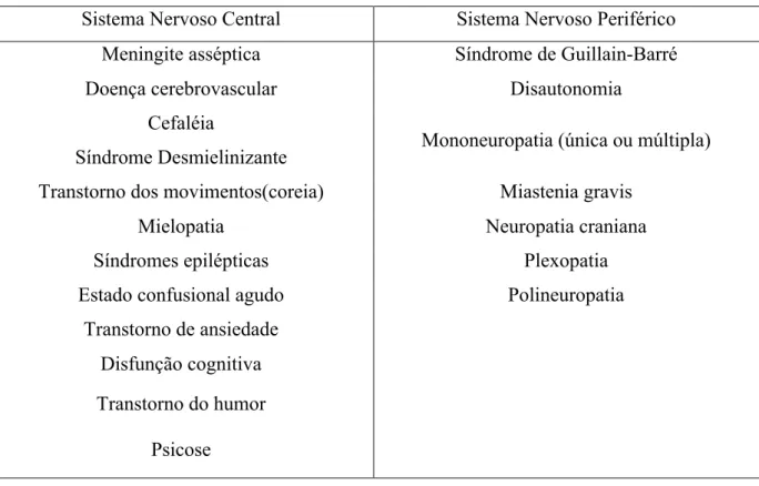
neuropsiquiátricas associadas ao LES em 19 síndromes bem definidas1 (tabela 1). O
acometimento neuropsiquiátrico no LES tende a ocorrer nos primeiros anos do diagnóstico,
ou mesmo ser o sintoma inaugural da doença, na ausência de outros sintomas sistêmicos que
possibilitem o diagnóstico definitivo de LES segundo os critérios clínicos recomendados pelo
ACR. As síndromes neuropsiquiátricas no LES variam, em um espectro de gravidade, desde
cefaleia e transtornos do humor, a coma. De fato, na ausência de marcadores laboratoriais ou
radiológicos bem definidos, a prevalência de acometimento neuropsiquiátrico no LES em
estudos epidemiológicos varia de 12 a 94%, a depender da metodologia utilizada e da
população estudada2. Soma-se a isto, a dificuldade de caracterizar determinadas
manifestações, comuns na população geral - como migrânea ou depressão, como realmente
provenientes da atividade autoimune da doença no cérebro3, 4.
Tabela 1 – Síndromes neuropsiquiátricas no LES
Sistema Nervoso Central Sistema Nervoso Periférico
Meningite asséptica Síndrome de Guillain-Barré
Doença cerebrovascular Disautonomia
Cefaléia
Síndrome Desmielinizante Mononeuropatia (única ou múltipla)
Transtorno dos movimentos(coreia) Miastenia gravis
Mielopatia Neuropatia craniana
Síndromes epilépticas Plexopatia
Estado confusional agudo Polineuropatia
Transtorno de ansiedade
Disfunção cognitiva
Transtorno do humor
A fisiopatologia envolvida no acometimento neuropsiquiátrico no LES não é completamente
conhecida. Múltiplos mecanismos etiológicos parecem estar envolvidos, e incluem,
principalmente, eventos trombóticos focais, como isquemia cerebral e trombose venosa
cerebral, e inflamação local com formação de auto-anticorpos. Alguns estudos mostram que a
presença de anticorpos antifosfolípides está associada a disfunção cognitiva, epilepsia, doença
cerebrovascular, cefaleia, neuropatia craniana, mielopatia e coreia5, 6. A presença de níveis
persistentemente elevados de anticardiolipina foi associada a pior performance cognitiva em
pacientes com LES, incluindo deterioração cognitiva discreta detectada em testes
neuropsicológicos prospectivamente7-9. A presença do anticoagulante lúpico está associada a
doença cerebrovascular em pacientes recém-diagnosticados com LES10 e a trombose venosa
cerebral11e declínio cognitivo em outras series12. O papel do anticorpo anti-P ribossomal no
LES neuropsiquiátrico (LES-NP) é mais controverso. Originalmente descrito em associação
com psicose13, enquanto alguns estudos apoiam esta associação10, 11, em outros a relação do
mesmo com acometimento neuropsiquiátrico foi questionado14, 15. No entanto, Matus et al.
demonstraram que o anti-P induz apoptose de neurônios no hipocampo e na amígdala em
ratos16, o que pode explicar o seu papel etiológico no acometimento de memória e de humor
em pacientes com LES. Outros fatores, como vasculopatia e aterosclerose acelerada estão
também associados17, 18. Infartos multifocais corticais, associada a dano microvascular, foi o
achado neuropatológico mais comum em uma série de casos de LES-NP levados à
necropsia19.
A ocorrência de déficit cognitivo em pacientes com LES foi melhor estudada a partir da
década de 80, com estudos iniciais mostrando alta prevalência de disfunção cognitiva
detectada através de testes neuropsicológicos adequados20. A prevalência de déficit cognitivo
leve chega a 80% em alguns estudos21, com uma média entre 14 a 75%, a depender da
metodologia empregada22, 23. A maior parte dos paciente têm um padrão flutuante de
acometimento, no entanto, alguns pacientes apresentam um declínio progressivo da função
cognitiva24. Não há um padrão único de acometimento, sendo provavelmente o acometimento
cognitivo no LES um conjunto de síndromes distintas, com etiologias variadas, podendo
mesmo ocorrer na ausência de outras manifestações neurológicas como doença
cerebrovascular ou epilepsia25. No entanto, são comuns o encontro de disfunção executiva,
problemas de atenção, memória e aprendizado. A patologia de substância branca e sua
tradução em disfunção cognitiva de padrão subcortical predominante parece ser importante no
Múltipla26, uma doença do sistema nervoso central que afeta predominantemente a substância branca.
Transtornos do humor e de ansiedade são comuns no LES, com frequência variando entre 1 a
75%, e 1 a 74%, respectivamente27. A presença de transtornos de humor ou de ansiedade não
se correlaciona com atividade da doença em pacientes com LES, de acordo com Japa E, et
al.28. Psicose, por outro lado, com uma prevalência entre 14 e 75% dos pacientes, ocorre na
maioria dos pacientes no primeiro ano do diagnóstico e na presença de outros marcadores de
atividade lúpica sistêmica29. A frequência de outros transtornos psiquiátricos não
contemplados na classificação do ACR de 1999 também parece ser maior no LES do que na
população geral. Bachen et al, mostraram uma frequência maior de fobia, transtorno do
pânico, transtorno obsessivo-compulsivo e transtorno bipolar I em mulheres lúpicas do que
em controles sadios30. De fato, a prevalência de transtorno obsessivo compulsivo em uma
coorte de LES foi 10 a 15 vezes maior do que a descrita na comunidade31. O transtorno de
déficit de atenção e hiperatividade raramente foi estudado no LES. A frequência de sintomas
de déficit de atenção e hiperatividade foi maior em pacientes com LES do que em controles
sadios em um estudo32.
Os distúrbios do movimento são manifestações neurológicas raras no LES, com prevalência
em torno de 0,5%2. O transtorno do movimento mais comum no LES é a coreia. A coreia no
LES afeta na sua maioria mulheres na terceira década de vida, e a presença de anticorpos
antifosfolípides é detectada em cerca de 60% dos casos33.
Pouco se entende sobre a fisiopatologia da coreia lúpica e sua fenomenologia. Como somente
uma parcela destes pacientes apresenta evidência de isquemia em exames de imagem,
levanta-se a hipótese que outros fatores, possivelmente imunológicos, contribuam para o
aparecimento de coreia neste subgrupo. Outros movimentos anormais descritos no LES
incluem o parkinsonismo34, a ataxia35 e a mioclonia36. Raros relatos também existem de
síndrome da pessoa rígida37, 38, blefaroespasmo, distonia cervical39 e tremor40 no LES, embora
a sua associação com a doença não esteja totalmente esclarecida. São poucos casos descritos
na literatura e não há dados suficientes para estimar a prevalência destes achados e fatores
associados à sua ocorrência. Algumas destas síndromes são de difícil reconhecimento, com
que eles possam ser sub-diagnosticados na prática do dia-a-dia, uma vez que a maioria destes
pacientes é acompanhada por clínicos sem treinamento formal em distúrbios do movimento.
Ademais a escassez, pelos fatos já mencionados, de estudos de séries consecutivas de
pacientes lúpicos com vistas à detecção de movimentos anormais, não há na literatura estudos
que investiguem a sua associação a transtornos comportamentais e cognitivos, frequentemente
vistos em pacientes com movimentos anormais de natureza semelhante, secundários ao
acometimento das mesmas vias e circuitos cerebrais (núcleos da base e suas conexões). A
Coreia de Sydenham (CS), por exemplo, é uma forma de coreia aguda em que há disfunção
dos núcleos da base após infecção estreptocócica. Pacientes com Coréia de Sydenham
apresentam maior frequência de Transtorno Obsessivo-Compulsivo (TOC) e de Transtorno de
Déficit de Atenção e Hiperatividade (TDAH) que controles saudáveis44, além de pior
desempenho em testes de função executiva, como na Torre de Londres, no teste de Stroop e
de fluência verbal45. Raramente, psicose46 pode fazer parte do quadro. Do mesmo modo, a
Doença de Huntington (DH), uma importante causa genética de coreia, é reconhecida, desde
sua descrição seminal por George Huntington, como uma doença com importante morbidade
psiquiátrica, sendo a doença marcada, em todo o seu curso, por expressivo acometimento das
esferas comportamentais e cognitivas47.
A maior frequência de TOC, TDAH e disfunção executiva em pacientes com CS e DH se
explica pela importância dos gânglios da base e suas conexões (especialmente com o córtex
pré-frontal, dorsolateral e com o cíngulo anterior) na cognição e no comportamento. De fato,
estudos de neuroimagem mostram alterações volumétricas no caudado48, córtex frontal
dorsolateral49, estriado50 e tálamo51 em pacientes com TOC. No TDAH, alguns estudos
evidenciam um menor volume do putâmen, globo pálido e caudado em compraração con
controles52, além de hipoativação dos gânglios da base durante testes de inibição (go/no-go)
na Ressonância Magnética funcional53.
Existem outras evidências, afora a coreia, do acometimento dos gânglios da base no LES
neuropsiquiátrico. Por exemplo, calcificações54 e hipersinal55 são comuns, respectivamente, à
Tomografia de crânio e à Ressonância Magnética (RM). Lim MK et al. encontraram uma
redução significativa da razão N-acetilaspartato-creatina na região dos gânglios da base em
pacientes com LES-NP em relação a pacientes sem manifestações neuropsiquiátricas pela
hipoperfusão unilateral ou bilateral nos gânglios da base em 22% dos pacientes com LES-NP
e em 9% daqueles sem nenhum acometimento neuropsiquiátrico57.
Considerando-se a evidência de que o LES pode causar lesões nos circuitos dos gânglios da
base, tanto clinicamente, em pacientes com coreia secundaria ao LES, quanto em estudos de
neuroimagem, e, traçando-se um paralelo com o conhecimento que nós temos sobre as
manifestações neuropsiquiátricas associadas a disfunção dos gânglios da base em outras
doenças autoimunes, como a CS, nossa hipótese é que o TOC, o TDAH e a disfunção
executiva são mais comuns em pacientes com LES do que na população em geral.
O objetivo deste estudo é, portanto, descrever em uma série de pacientes com diagnóstico de
LES, a frequênciadas síndromes neuropsiquiátricas conforme classificação do ACR, com
ênfase no estudo de movimentos anormais e sua associação com outras possíveis
manifestações clínicas de disfunção dos gânglios da base, como o TOC, o TDAH e a
2. OBJETIVOS
Objetivo Principal
Avaliar a frequência de manifestações neuropsiquiátricos em pacientes com LES do
ambulatório de Reumatologia do HC-UFMG, de acordo com a nomenclatura do Colégio
Americano de Reumatologia de 1999.
Objetivo secundário
Avaliar a presença de movimentos anormais, e sua associação com TDAH, TOC e disfunção
executiva em pacientes com LES do ambulatório de Reumatologia do HC-UFMG, e sua
3. METODOLOGIA
3.1 HIPOTÉSES
3.1.1 HIPÓTESE NULA
A frequência de Transtorno Obsessivo-Compulsivo, Transtorno de Déficit de Atenção e
Hiperatividade e disfunção cognitiva no LES são maiores do que as esperadas na população
geral.
3.1.2 HIPÓTESE ALTERNATIVA
A frequência de Transtorno Obsessivo-Compulsivo, Transtorno de Déficit de Atenção e
Hiperatividade e disfunção cognitiva no LES são iguais às esperadas na população geral.
3.2 DESENHO DO ESTUDO
Estudo observacional transversal.
3.3 POPULAÇÃO DO ESTUDO
Amostra de conveniência composta de 54 indivíduos com diagnóstico de LES do ambulatório
de Reumatologia do Hospital das Clínicas da Universidade Federal de Minas Gerais, em Belo
Horizonte, Brasil. Os pacientes foram submetidos, em única avaliação com tempo médio de
duas horas, a uma bateria de testes neuropsicológicos e ao exame clínico e neurológico por
avaliador único (ROH).
3.3.1 CRITÉRIOS DE INCLUSÃO
1) Diagnóstico de LES segundo os critérios do ACR58;
2) Idade entre 18 e 55 anos;
3) Mínimo de seis meses de acompanhamento ambulatorial e ao menos duas consultas
realizadas no ambulatório de Reumatologia;
4) Assinatura do termo de consentimento livre e esclarecido.
3.3.2 CRITÉRIOS DE EXCLUSÃO
1) Presença de outras doenças reumatológicas, exceto Síndrome de Anticorpos
2) Presença de outras doenças crônicas que impeçam ou dificultem a realização do protocolo
de pesquisa (ex.: cegueira), ou que interfiram na sua interpretação (ex.: doença psiquiátrica
grave prévia, não relacionada ao LES);
3.4. VARIÁVEIS DO ESTUDO
Foram coletadas, através de questionário, as seguintes variáveis categóricas e contínuas.
3.4.1 VARIÁVEIS CATEGÓRICAS
1) Sexo
2) Raça
3) Escolaridade
4) Medicações em uso (corticoides, imunossupressores, antimaláricos, antidepressivos,
ansiolíticos, antipsicóticos, benzodiazepínicos, antiepilépticos)
5) Manifestações iniciais do LES (Rash malar, rash discoide, fotossensibilidade, úlceras orais,
artrite, serosite, disfunção renal, disfunção hematológica, disfunção neurológica);
6) Critérios clínicos de classificação do LES de 199758;
7) Critérios clínicos de classificação do LES da Systemic Lupus International Collaborating
Clinics (SLICC) de 201259;
8) Presença de movimentos anormais como manifestação inicial
9) História prévia de aborto;
10) História prévia de trombose venosa profunda ou arterial;
11) Diagnóstico de SAAF60;
12) Presença de quaisquer das 19 síndromes neuropsiquiátricas associadas ao LES, segundo
classificação da ACR1, atuais e retrospectivas, através de revisão do prontuário;
13) Manifestações clínicas atuais do LES;
14) Presença de comprometimento cognitivo segundo o Mini-Exame do Estado Mental
(MEEM), traduzido para o português, com notas de corte adequadas para a escolaridade.
Foram considerados como anormais os seguintes valores: 1 a 3 anos de escolaridade – 20
pontos ou menos, 4 a 7 anos de escolaridade – 23 pontos ou menos, 8 anos ou mais de
escolaridade – 25 pontos ou menos61;
15) Presença de comprometimento cognitivo segundo a Montreal Cognitive Assessment
(MoCA), traduzida para o português. Foi utilizada a nota de corte de 25 pontos62;
16) Presença de comprometimento executivo através da Bateria de Avaliação Frontal (Frontal
Assessment Battery, FAB), traduzida para o português e com nota de corte adequada para escolaridade, segundo os critérios a seguir para disfunção executiva: menos do que 9 pontos
(1 a 3 anos de escolaridade), menos de 10 pontos (4 a 7 anos de escolaridade), menos de 12
17) Presença de diminuição da fluência verbal, aferida pelo teste de fluência verbal semântica
(categoria animais), com os seguintes valores de referência para resultado anormal: 1 a 8 anos
de escolaridade – 8 ou menos, 8 anos de escolaridade ou mais – 12 ou menos64;
18) Presença de Transtorno de Déficit de Atenção e Hiperatividade, avaliada através dos
critérios do Diagnostic and Statistical Manual of Mental Disorders (DSM), quarta edição,
excluindo-se a obrigatoriedade de presença de sintomas antes dos sete anos de idade;
19) Presença de sintomas obsessivos e compulsivos clinicamente significantes, avaliada
através do Inventário de Obsessões e Compulsões Revisado (Obsessive and Compulsive
Inventory – Revised, OCI-R), traduzido para o português, com o valor de corte de 21 para
sintomas clinicamente significativos65.
3.4.2 VARIÁVEIS CONTÍNUAS
Foram pesquisadas as seguintes variáveis contínuas:
1) Idade;
2) Anos de escolaridade formal;
3) Idade no momento do diagnóstico;
4) Dose de corticoide no momento da avaliação;
5) Pontuação no Índice de Atividade da Doença no Lúpus Eritematoso Sistêmico – 2000
Modificado (mSLEDAI-2K)66;
6) Pontuação no Escore do SLICC de Índice de Dano67;
7) Pontuação no MEEM68;
8) Pontuação na escala FAB69;
9) Pontuação na escala MoCA70;
10) Pontuação no teste de fluência verbal fonêmica com as letras F, A e S71;
11) Pontuação no Inventório OCI-R65;
12) Pontuação na Escala de Sintomas Obsessivos e Compulsivos de Yale e Brown
(Y-BOCS)72;
13) Pontuação na Escala de Ansiedade de Beck73;
3.5 ETAPAS DO ESTUDO
3.5.1 PRIMEIRA ETAPA: COMITÊ DE ÉTICA
O projeto foi aprovado pelo Comitê de Ética da Universidade Federal de Minas Gerais em 14
de dezembro de 2011 (projeto CAAE 0615.0.203.000-11).
3.5.2 SEGUNDA ETAPA: COLETA DE DADOS
Os dados epidemiológicos, clínicos e revisão de prontuário foram realizadas de janeiro de
2012 a novembro de 2013. Os pacientes foram submetidos a entrevista clínica, exame físico
por único examinador, e responderam aos questionários acima citados, na mesma ocasião.
Quando necessária, revisão de prontuários foi realizada a posteriori através de consulta ao
Serviço de Arquivo Médico e Estatístico do Hospital das Clínicas da UFMG.
3.5.3 TERCEIRA ETAPA: ANÁLISE ESTATÍSTICA
As análises estatísticas foram realizadas no pacote estatístico SPSS versão 20.0 (Statistical
Package for Social Sciences, IBM Corporation Software Group, USA)75. A análise descritiva
dos dados foi apresentada como média ± desvio padrão ou mediana e percentis 25 e 75, para
dados não-normais. Foi utilizado o teste de normalidade de Kolmogorov-Smirnov. Para
comparação entre variáveis categóricas e contínuas foram utilizados os testes de
Mann-Whitney, para variáveis não-normais e o teste t de Student para variáveis normais. Foram
utilizadas as correlações de Spearman e Pearson para variáveis contínuas não-normais e
normais, respectivamente. O teste qui-quadrado foi usado para comparar variáveis
4. REFERÊNCIAS BIBLIOGRÁFICAS
1. The American College of Rheumatology nomenclature and case definitions for neuropsychiatric lupus syndromes. Arthritis Rheum 1999;42:599-‐608.
2. Unterman A, Nolte JES, Boaz M, Abady M, Shoenfeld Y, Zandman-‐Goddard G. Neuropsychiatric Syndromes in Systemic Lupus Erythematosus: A Meta-‐Analysis. Semin Arthritis Rheum 2010;41:1-‐11.
3. Ainiala H, Hietaharju A, Loukkola J, et al. Validity of the new American College of Rheumatology criteria for neuropsychiatric lupus syndromes: a population-‐based evaluation. Arthritis Care Res 2001;45:419-‐423.
4. Hanly JG, McCurdy G, Fougere L, Douglas J-‐A, Thompson K. Neuropsychiatric events in systemic lupus erythematosus: attribution and clinical significance.J Rheumatol 2004;31:2156-‐2162.
5. Sanna G, Bertolaccini ML, Cuadrado MJ, et al. Neuropsychiatric manifestations in systemic lupus erythematosus: prevalence and association with antiphospholipid antibodies. J Rheumatol 2003;30:985-‐992.
6. Tomietto P, Annese V, D'Agostini S, et al. General and specific factors associated with severity of cognitive impairment in systemic lupus erythematosus. Arthritis Care Res 2007;57:1461-‐1472.
7. Menon S, Jameson-‐Shortall E, Newman SP, Hall-‐Craggs MR, Chinn R, Isenberg DA. A longitudinal study of anticardiolipin antibody levels and cognitive functioning in systemic lupus erythematosus. Arthritis Rheum 1999;42:735-‐741.
8. Hanly JG, Hong C, Smith S, Fisk JD. A prospective analysis of cognitive function and anticardiolipin antibodies in systemic lupus erythematosus. Arthritis Rheum 1999;42:728-‐734.
9. McLaurin EY, Holliday SL, Williams P, Brey RL. Predictors of cognitive dysfunction in patients with systemic lupus erythematosus. Neurology 2005;64:297-‐ 303.
10. Hanly JG, Urowitz MB, Siannis F, et al. Autoantibodies and neuropsychiatric events at the time of systemic lupus erythematosus diagnosis: Results from an international inception cohort study. Arthritis Rheum 2008;58:843-‐853.
11. Hanly JG, Urowitz MB, Su L, et al. Autoantibodies as biomarkers for the prediction of neuropsychiatric events in systemic lupus erythematosus. Ann Rheum Dis 2011;70:1726-‐1732.
12. Denburg SD, Carbotte RM, Ginsberg JS, Denburg JA. The relationship of antiphospholipid antibodies to cognitive function in patients with systemic lupus erythematosus. J Int Neuropsychol Soc1997;3:377-‐386.
13. Bonfa E, Golombek SJ, Kaufman LD, et al. Association between lupus psychosis and anti-‐ribosomal P protein antibodies. N Engl J Med 1987;317:265-‐271.
14. Gerli R, Caponi L, Tincani A, et al. Clinical and serological associations of ribosomal P autoantibodies in systemic lupus erythematosus: prospective evaluation in a large cohort of Italian patients. Rheumatology 2002;41:1357-‐1366.
15. van Dam A, Nossent H, de Jong J, et al. Diagnostic value of antibodies against ribosomal phosphoproteins. A cross sectional and longitudinal study. J Rheumatol 1991;18:1026-‐1034.
17. Mitsias P, Levine SR. Large cerebral vessel occlusive disease in systemic lupus erythematosus. Neurology 1994;44:385-‐393.
18. Ellison D, Gatter K, Heryet A, Esiri M. Intramural platelet deposition in cerebral vasculopathy of systemic lupus erythematosus. J Clin Pathol 1993;46:37-‐40.
19. Hanly JG, Walsh NM, Sangalang V. Brain pathology in systemic lupus erythematosus. J Rheumatol 1992;19:732-‐741.
20. Carbotte RM, Denburg SD, Denburg JA. Prevalence of cognitive impairment in systemic lupus erythematosus. J Nerv Ment Dis 1986;174:357-‐364.
21. Ainiala H, Loukkola J, Peltola J, Korpela M, Hietaharju A. The prevalence of neuropsychiatric syndromes in systemic lupus erythematosus. Neurology 2001;57:496-‐ 500.
22. West SG, Emlen W, Wener MH, Kotzin BL. Neuropsychiatric lupus erythematosus: A 10-‐year prospective study on the value of diagnostic tests. Am J Med 1995;99:153-‐ 163.
23. Sibley JT, Olszynski WP, Decoteau WE, Sundaram MB. The incidence and prognosis of central nervous system disease in systemic lupus erythematosus. J Rheumatol 1992;19:47-‐52.
24. Carlomagno S, Migliaresi S, Ambrosone L, Sannino M, Sanges G, Di Iorio G. Cognitive impairment in systemic lupus erythematosus: a follow-‐up study. J Neurol 2000;247:273-‐279.
25. Monastero R, Bettini P, Del Zotto E, et al. Prevalence and pattern of cognitive impairment in systemic lupus erythematosus patients with and without overt neuropsychiatric manifestations. J Neurol Sci 2001;184:33-‐39.
26. Benedict RH, Shucard JL, Zivadinov R, Shucard DW. Neuropsychological impairment in systemic lupus erythematosus: a comparison with multiple sclerosis. Neuropsychol Rev 2008;18:149-‐166.
27. Asano NM, Coriolano MD, Asano BJ, Lins OG. Psychiatric comorbidities in patients with systemic lupus erythematosus: a systematic review of the last 10 years. Rev Bras Reumatol 2013;53:431-‐437.
28. Jarpa E, Babul M, Calderon J, et al. Common mental disorders and psychological distress in systemic lupus erythematosus are not associated with disease activity. Lupus 2011;20:58-‐66.
29. Pego-‐Reigosa JM, Isenberg DA. Psychosis due to systemic lupus erythematosus: characteristics and long-‐term outcome of this rare manifestation of the disease. Rheumatology 2008;47:1498-‐1502.
30. Bachen EA, Chesney MA, Criswell LA. Prevalence of mood and anxiety disorders in women with systemic lupus erythematosus. Arthritis CareRes 2009;61:822-‐829. 31. Slattery MJ, Dubbert BK, Allen AJ, Leonard HL, Swedo SE, Gourley MF. Prevalence of obsessive-‐compulsive disorder in patients with systemic lupus erythematosus. J ClinPsychiatry 2004;65:301-‐306.
32. Garcia RJ, Francis L, Dawood M, Lai Z-‐W, Faraone SV, Perl A. Brief Report: Attention Deficit and Hyperactivity Disorder Scores Are Elevated and Respond to N-‐ Acetylcysteine Treatment in Patients With Systemic Lupus Erythematosus. Arthritis Rheum 2013;65:1313-‐1318.
34. García-‐Moreno JM, Chacón J. Juvenile parkinsonism as a manifestation of systemic lupus erythematosus: Case report and review of the literature. Mov Disord 2002;17:1329-‐1335.
35. Santos MJ, Reis P, da Silva JA, de Queiroz MV. Ischemic lesion of the CNS in patients with systemic lupus erythematosus. Acta MedPort 1994;7:201-‐206.
36. Joseph FG, Lammie GA, Scolding NJ. CNS lupus: A study of 41 patients. Neurology 2007;69:644-‐654.
37. Munhoz RP, Fameli H, Teive HA. Stiff person syndrome as the initial manifestation of systemic lupus erythematosus. Mov Disord 2010;25:516-‐517.
38. Goeb V, Dubreuil F, Cabre P, Jean-‐Baptiste G, Arfi S. Lupus revealing itself after a stiff-‐person syndrome. Lupus 2004;13:215.
39. Robert M, Sunitha R, Thulaseedharan NK. Neuropsychiatric manifestations systemic lupus erythematosus: a study from South India. Neurology India 2006;54:75-‐ 77.
40. Venegoni E, Biasioli R, Lamperti E, Rinaldi E, Salmaggi A, Novi C. Tremor as an early manifestation of systemic lupus erythematosus. Clin Exp Rheumatol 1994;12:199-‐ 201.
41. Newman EJ, Breen K, Patterson J, Hadley DM, Grosset KA, Grosset DG. Accuracy of Parkinson's disease diagnosis in 610 general practice patients in the West of Scotland. Mov Disord 2009;24:2379-‐2385.
42. Jain S, Lo SE, Louis ED. Common misdiagnosis of a common neurological disorder: how are we misdiagnosing essential tremor? Arch Neurol 2006;63:1100-‐1104. 43. Lalli S, Albanese A. The diagnostic challenge of primary dystonia: Evidence from misdiagnosis. Mov Disord 2010;25:1619-‐1626.
44. Maia DP, Teixeira AL, Quintão Cunningham MC, Cardoso F. Obsessive compulsive behavior, hyperactivity, and attention deficit disorder in Sydenham chorea. Neurology 2005;64:1799-‐1801.
45. Beato R, Maia DP, Teixeira AL, et al. Executive functioning in adult patients with Sydenham's chorea. Mov Disord 2010;25:853-‐857.
46. Teixeira AL, Maia DP, Cardoso F. Psychosis following acute Sydenham’s chorea. Eur Child Adolesc Psychiatry 2007;16:67-‐69.
47. Walker FO. Huntington's disease. Lancet 2007;369:218-‐228.
48. Baxter LR Jr, Schwartz JM, Mazziotta JC, et al. Cerebral glucose metabolic rates in nondepressed patients with obsessive-‐compulsive disorder. Am J Psychiatry 1988;145:1560-‐1563.
49. Lacerda AL, Nicoletti MA, Brambilla P, et al. Anatomical MRI study of basal ganglia in major depressive disorder. Psychiatry Res 2003;124:129-‐140.
50. Bartha R, Stein MB, Williamson PC, et al. A short echo H1 spectroscopy and volumetric MRI study of the corpus striatum in patients with obsessive-‐compulsive disorder and comparison subjects. A J Psychiatry 1998;155:1584-‐1591.
51. Perani D, Colombo C, Bressi S, et al. [18F]FDG PET study in obsessive-‐compulsive disorde. A clinical/metabolic correlation study after treatment. Br J Psychiatry 1995;166:244-‐250.
52. Nakao T, Radua J, Rubia K, Mataix-‐Cols D. Gray matter volume abnormalities in ADHD: voxel-‐based meta-‐analysis exploring the effects of age and stimulant medication. Am J Psychiatry 2001;168:1154-‐1163.
54. Miguel EC, Pereira RM, Pereira CA, et al. Psychiatric manifestations of systemic lupus erythematosus: clinical features, symptoms, and signs of central nervous system activity in 43 patients. Medicine (Baltimore) 1994;73:224-‐232.
55. Abreu MR, Jakosky A, Folgerini M, et al. Neuropsychiatric systemic lupus erythematosus: correlation of brain MR imaging, CT, and SPECT. J Clin Imaging 2005;29:215-‐221.
56. Lim MK, Suh CH, Kim HJ, et al. Systemic lupus erythematosus: brain MR imaging and single-‐voxel hydrogen 1 MR spectroscopy. Radiology 2000;217:43-‐49.
57. Shen YY, Kao CH, Ho YJ, Lee JK. Regional cerebral blood flow in patients with systemic lupus erythematosus. J Neuroimaging 1999;9:160-‐164.
58. Hochberg MC. Updating the American College of Rheumatology revised criteria for the classification of systemic lupus erythematosus. Arthritis Rheum 1997;40:1725. 59. Petri M, Orbai AM, Alarcón GS, et al. Derivation and validation of the Systemic Lupus International Collaborating Clinics classification criteria for systemic lupus erythematosus. Arthritis Rheum 2012;64:2677-‐2686.
60. Miyakis S, Lockshin MD, Atsumi T, et al. International consensus statement on an update of the classification criteria for definite antiphospholipid syndrome (APS). JThrom Haemost2006;4:295-‐306.
61. Brucki SM, Nitrini R, Caramelli P, Bertolucci PH, Okamoto IH. Suggestions for utilization of the mini-‐mental state examination in Brazil. Arq Neuropsiquiatr 2003;61:777-‐781.
62. Memoria CM, Yassuda MS, Nakano EY, Forlenza OV. Brief screening for mild cognitive impairment: validation of the Brazilian version of the Montreal cognitive assessment. Int J Geriatr Psychiatry; 2013;28:34-‐40.
63. Beato R, Amaral-‐Carvalho V, Guimarães HC, et al. Frontal assessment battery in a Brazilian sample of healthy controls: normative data. Arq Neuropsiquiatr 2012;70:278-‐ 280.
64. Brucki SMD, Malheiros SMF, Okamoto IH, Bertolucci PHF. Dados normativos para o teste de fluência verbal categoria animais em nosso meio. Arq Neuropsiquiatr 1997;55:56-‐61.
65. Foa EB, Huppert JD, Leiberg S, Langner R, et al. The Obsessive-‐Compulsive Inventory: development and validation of a short version. Psychol Assess 2002;14:485-‐ 496.
66. Uribe AG, Vila LM, McGwin G, Jr., Sanchez ML, Reveille JD, Alarcon GS. The Systemic Lupus Activity Measure-‐revised, the Mexican Systemic Lupus Erythematosus Disease Activity Index (SLEDAI), and a modified SLEDAI-‐2K are adequate instruments to measure disease activity in systemic lupus erythematosus. J Rheumatol 2004;31:1934-‐ 1940.
67. Gladman DD, Urowitz MB, Goldsmith CH, et al. The reliability of the Systemic Lupus International Collaborating Clinics/American College of Rheumatology Damage Index in patients with systemic lupus erythematosus. Arthritis Rheum 1997;40:809-‐813. 68. Folstein MF, Folstein SE, McHugh PR. "Mini-‐mental state". A practical method for grading the cognitive state of patients for the clinician. JPsychiatrRes 1975;12:189-‐198. 69. Dubois B, Slachevsky A, Litvan I, Pillon B. The FAB: a Frontal Assessment Battery at bedside. Neurology 2000;55:1621-‐1626.
71. Strauss E, Sherman E, Spreen O. Verbal fluency. A compendium of neuro-‐ psychological tests, Third Ed ed. New York: Oxford University Press, 2006: 499-‐526. 72. Goodman WK, Price LH, Rasmussen SA, et al. The Yale-‐Brown Obsessive Compulsive Scale. I. Development, use, and reliability. Arch Gen Psychiatry 1989;46:1006-‐1011.
73. Beck AT, Epstein N, Brown G, Steer RA. An inventory for measuring clinical anxiety: psychometric properties. J Consult Clin Psychol 1988;56:893-‐897.
74. Beck AT, Ward CH, Mendelson M, Mock J, Erbaugh J. An inventory for measuring depression. Arch Gen Psychiatry 1961;4:561-‐571.
75. IBM Corp. Released 2011. IBM SPSS Statistics for Windows, Version 20.0. Armonk, NY: IBM Corp.
5.0 RESULTADOS
5.1 ARTIGO
Os resultados serão apresentados no formato de artigo, a ser submetido a periódico
Original Article
Frequency of executive dysfunction, obsessive-compulsive symptoms, and attention deficit and hyperactivity disorder in Systemic Lupus Erythematosus: evidence for basal ganglia dysfunction?
Frequência de disfunção executiva, sintomas obsessivo-compulsivos, e déficit de atenção e
hiperatividade no Lúpus Eritematoso Sistêmico: evidência de disfunção dos núcleos da base?
Ricardo Oliveira Horta Maciel, MD1; Gilda Aparecida Ferreira, MD, PhD2; Bárbara Akemy;
Francisco Eduardo Costa Cardoso, MD, PhD1
1. Movement Disorders Clinic, Department of Neurology, Universidade Federal de Minas
Gerais
2. Rheumatology Clinic, Department of Locomotor System, Universidade Federal de Minas
Gerais
Corresponding Author: Francisco Cardoso MD PhD, Av Pasteur 89/1107, CEP 30150-290
Belo Horizonte, MG Brazil. Phone + 55 31 3231055 Fax +55 31 32134951. Email
cardosofe@terra.com.br
ABSTRACT
Purpose of the Study: Basal ganglia dysfunction in autoimmune diseases is associated with
obsessive-compulsive symptoms (OCS), executive dysfunction and attention deficit and
hyperactivity disorder (ADHD).
Objectives: To evaluate the frequency of executive dysfunction, OCD and ADHD in patients
with Systemic Lupus Erythematosus (SLE).
Methods: 54 patients underwent the Mini-Mental State Examination, the Montreal Cognitive
Assessment, the Frontal Assessment Battery, phonemic and semantic fluency tests, the
Obsessive and Compulsive Inventory – Revised, the Yale-Brown Obsessive and Compulsive
Scale and Beck’s Anxiety and Depression Scales. ADHD was diagnosed according to
DSM-IV criteria.
Results: 33 (61.1%) patients had cognitive impairment. 15 (27.8%) had OCS. 17 (31.5%) met
ADHD diagnostic criteria. Executive dysfunction, depression and OCS correlated with
disease activity.
Discussion: Executive dysfunction, ADHD and OCS are common in SLE. The significance of
basal ganglia pathology in SLE warrants further studies.
Keywords: Neuropsychiatric Systemic Lupus Erythematosus; obsessive-compulsive disorder;
INTRODUCTION
Cognitive impairment is common in patients with Systemic Lupus Erythematous (SLE).
Depending on the population or the methods used for assessment, from 14 up to 80% of
patients with SLE have some kind of cognitive dysfunction1. There is no predominant
cognitive domain affected, and no unique pattern of dysfunction has been recognized as
typical of SLE. Commonly, though, patients score lower than expected on tests of memory,
visuospatial abilities, attention and executive function. The pathophysiology of cognitive
impairment in SLE is not completely elucidated, but direct inflammatory damage to neuronal
circuits and ischemic injury is thought to play a significant role in most cases. Indeed, frontal
white matter lesions correlate with cognitive impairment in patients with SLE2.
The 1999 ACR classification of NPSLE recognizes three psychiatric syndromes related to
SLE activity in the Central Nervous System (CNS): psychosis, and mood and anxiety
disorders3. Although less studied, other psychiatric illnesses, such as obsessive-compulsive
disorder (OCD) and attention deficit and hyperactive disorder (ADHD), have been described
in patients with SLE4, 5. Indeed, OCD and ADHD also frequently occur in other autoimmune
diseases, such as Sydenham’s chorea (SC)6. In the latter, OCS, ADHD and chorea are related
to autoantibody-mediated basal ganglia dysfunction. In addition, OCD is more frequent in
patients with Rheumatic Fever than in controls, suggesting that anti-basal ganglia antibodies
can cause OCD irrespective of the presence of chorea7. The non-motor psychiatric symptoms
of SC are also similar, albeit less severe, to those seem in other diseases which affect the basal
ganglia, as for instance, Huntington’s Disease (HD) and Parkinson’s Disease (PD). In SLE,
basal ganglia dysfunction is associated with chorea and other more infrequent movement
disorders, such as parkinsonism, myoclonus and dystonia8. We hypothesize that OCS and
ADHD could be the non-motor manifestation of basal ganglia dysfunction (with or without
METHODS
We evaluated 54 consecutive patients with LES from the Rheumatology Clinic of the
Universidade Federal de Minas Gerais, a tertiary referral center in Belo Horizonte, Brazil,
between June 2012 and November 2013. Inclusion criteria were as follows: 1) Diagnosis of
LES according to the ACR criteria9, 2) age between 18 and 55 years, 3) minimum of 6
months follow-up and at least two office visits, 4) Informed consent for the participation in
the study. We excluded patients who had other rheumatologic diseases (other than
antiphospholipid syndrome) or other serious medical conditions that could impair completion
of the study protocol (e. g., blindness) or confound data interpretation (e. g., previous severe
psychiatric disease not related to SLE). This study was approved by the Ethics Committee of
the Universidade Federal de Minas Gerais (project number 0615.0.203.000-11).
We recorded patients’ demographics and reviewed charts to determine age of onset of the
disease, initial clinical manifestations, current and previous corticosteroids and
immunosuppressive treatments, and previous neuropsychological manifestations of SLE. The
presence of antiphospholipid antibodies was recorded. Patients underwent a battery of
cognitive tests, which included the Mini-mental State Examination (MMSE)10, the Montreal
Cognitive Assessment (MoCA)11, the Frontal Assessment Battery (FAB)12, the FAS verbal
fluency test and the category (animals) semantic fluency test13. The presence and severity of
obsessive and compulsive symptoms were assessed with the Revised Obsessive and
Compulsive Inventory (OCI-R)14 and the Yale-Brown Obsessive and Compulsive Scale
(YBOCS)15. A score equal to or greater than 21 was used as a cut-off for clinically significant
OCS14. The presence of attention deficit and hyperactivity symptoms was evaluated according
to the DSM-IV criteria with the exception of the criterion presence of symptoms before seven
years of age. Patients also completed Beck’s Depressive16 and Anxiety Inventories17. A score
equal or greater than 21 and 16 were considered as clinically meaningful, respectively.
Disease activity and severity were recorded according to the Modified Systemic Lupus
Erythematosus Disease Activity Index 200018(mSLEDAI-2K) and the Systemic Lupus
International Collaborative Clinics/American College of Rheumatology Damage Index
(SLICC)19. Clinical history and examination, with special attention to the presence of
all patients by a neurologist (RHM), and the presence of neuropsychological manifestations of
the disease was recorded, according to the 1999 ACR criteria. Confirmatory tests were
ordered when deemed necessary for diagnosis support.
Neuropsychiatric tests
Screening tests of global cognitive dysfunction: MMSE and MoCA
The MMSE is a widely used screening tool for global cognitive impairment. Different cut-off
scores were used according to degree of education: patients were considered as cognitively
impaired with a score below 21 if they had between 1 and 3 years of education, below 24 if
4-7 years of education or below 24 if 8 (or higher) years of education20. The MoCA is also a
screening instrument for global cognitive impairment; however, it is considered as an overall
more sensitive test than the MMSE in the detection of subtle cognitive deficits, particularly in
patients with higher education. MoCA more thoroughly evaluates global cognitive functions
not well represented in the MMSE (executive and visuospatial domains, specially) and is
more demanding to patients in tasks of language and memory21. Because of these features, it
has been found to be a valuable test in the detection of the subcortical cognitive impairment
characteristic of cerebrovascular disease and PD, for instance. Results below 24 were
considered abnormal22.
Tests of executive dysfunction: FAB and verbal fluency
The Frontal Battery Assessment is a brief screening tool that evaluates the presence of
executive dysfunction. Different cut offs were used according to years of education: 9 (for 1-3
years of education), 10 (for 4-7 years of education), 12 (for 8-11 years of education) and 13
(for more than 12 years of education)22, 23. The FAS verbal fluency and the category fluency
test assess phonemic and semantic fluency, respectively, by requesting the individual to
produce as many words (beginning with F, A or S in the FAS test, and in the category animals
in the semantic test) as possible in one minute. Both tests require the interaction of complex
cognitive processes, including operational memory, selective attention, mental set shifting,
which are all part of the executive function.
Continuous variables were tested for normality with the Kolmogorov-Smirnov test.
Descriptive analysis of data was done using SPSS 20.0 (Statistical Package for Social
Sciences, IBM Corporation Software Group, USA), and presented as mean ± standard
deviation when normal or median and quartiles when skewed. Pearson’s and Spearman’s
correlation coefficients were used respectively for normal or non-normal continuous
variables. To compare categorical and continuous variables the Mann-Whitney or Student’s t
-test were used, with a two-tailed p value <0.05 considered significant. The chi-square -test was
RESULTS
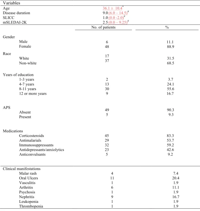
Of the 54 patients studied,17 (31.5%) were classified as white and37 (67.5%) as non-white.
Female to male ratio was 8:1. Mean age at assessment was 36.5 ± 10.4 years, and mean
disease duration was 11.1 ± 7.3 years. Median years of education were 10.0 (6.0 – 11.0)
years. Median mSLEDAI-2K was 2.5 (0 - 9.25) and median SLICC was 1.0 (0 - 2). The
majority of patients were using corticosteroids or immunosuppressants (83.3% and 59.2%,
respectively) and median corticosteroid dosage per day was 10.0 (5.0-15.0) mg. Five patients
(9.3%) had secondary antiphospholipid syndrome according to the 2006 Sydney criteria24.
Other demographical and clinical characteristics are summarized on Table 1.
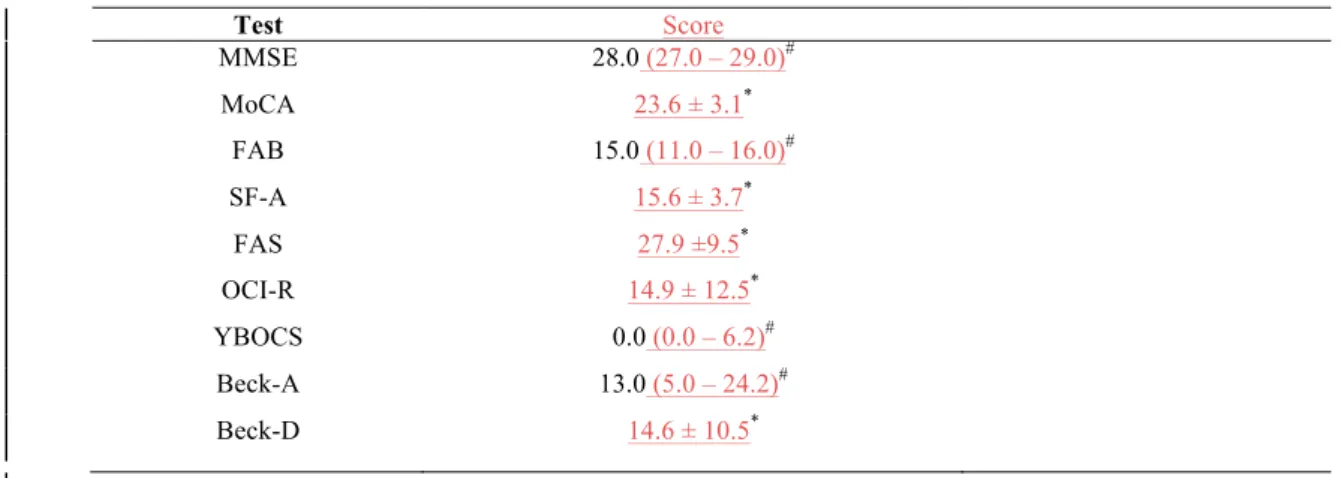
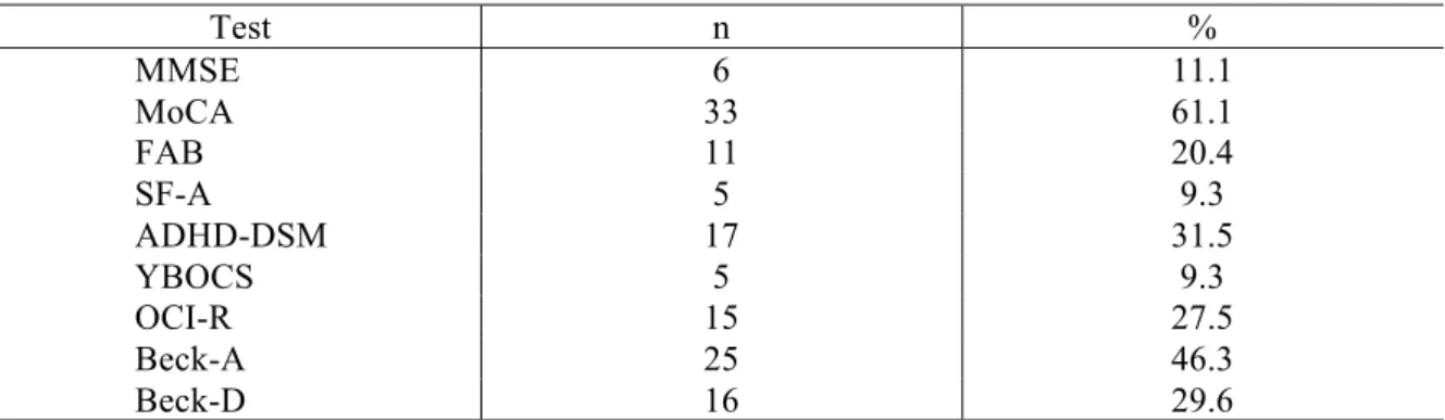
Cognitive impairment was detected in 6 (11.1%) patients according to the MMSE, but this
figure rose to 33 (61.1%) if the MoCA was used. Executive dysfunction was frequent in
patients, with 11 (20.4%) individuals scoring below normal on the FAB. FAB scores were
negatively correlated to SLICC scores (r = -0.302, p = 0.026). Five patients (9.3%) also
scored lower than expected according to education on the semantic fluency test. Mean scores
in the FAS verbal fluency were 27.9 ± 9.5. The overall frequency of cognitive dysfunction on
our sample, considering the presence of abnormal scores in either the MEEM, MoCA, FAB,
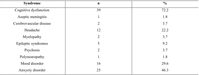
or verbal fluency scores, was 72.2 %(39 patients).
Obsessive-compulsive symptoms were frequent in our sample, 15 (27.8%) patients had
abnormal scores on the OCI-R. Five patients (9.3%) had moderate or severe symptoms, as
detected by the YBOCS. There was a strong positive correlation between OCI-R and YBOCS
scores (r = 0.653, p < 0.000). A diagnosis of ADHD was made in 17 patients (31.5%)
according to DSM-IV criteria. Of those, 12 (70.5%) were classified in the predominant
inattentive subtype, 3 (17.6%) were in the predominat hyperactive subtype and the remaining
2 (11.7%) were in the combined inattentive-hyperactive subtype. Executive dysfunction was
more common (p = 0.039) and mSLEDAI-2K scores were higher in patients with ADHD than
in patients without the latter -median score of 8 (2-14) vs 2.00 (0-4), p = 0.003. Presence of
clinically meaningful depressive and anxiety symptoms was found in 16 (29.6%) and 25
(46.3%) patients, respectively, and patients with more severe obsessive-compulsive
symptoms were more anxious (r = 0.544 p < 0.000). Mean and standard deviations of scores
OCI-R and YBOCS scores correlated with mSLEDAI-2K scores (r = 0.286, p = 0.036 and r =
0.369, p = 0.006, respectively). Patients with higher disease activity also were more depressed
(r = 0.286, p = 0.036) and scored lower on the FAS fonemic fluency test (r= -0.301, p =
0.027). Table 3 shows the frequency of abnormal results for each neuropsychiatric test.
We found a positive correlation between daily corticoid dosage and Beck’s Depressive
Inventory (r = 0.375, p = 0.006). Cognitive dysfunction also appears to be more common in
patients currently using corticosteroids, although statistical significance was not reached (p =
0.056). There was a negative correlation between FAB scores and Beck’s Depressive
Inventory (r = -0.289, p = 0.034). We found no correlation of depressive or anxiety symptoms
with MMSE, MoCA or verbal fluency tests.
Movement disorders was identified at the time of examination in one patient, who developed
parkinsonism associated with cognitive impairment, depression and systemic activity
characterized by nephritis, cutaneous vasculitis, hemolythic anemia, lymphopenia and low
complement. Other causes of parkinsonism as CNS infection and Wilson’s disease were ruled
out. She had a modest response to levodopaalthough there was poor compliance to treatment.
Another patient who had had chorea at age 22 but is currently in remissionwas identified
during chart review, retrospectively.
Table 4 shows other neuropsychiatric syndromes identified in our sample by clinical
examination and chart review. Overall frequency of neuropsychiatric SLE was 98.1%.
However, in a more conservative approach, in which we excluded the frequency of headache,
mood dysfunction and anxiety disorders, which are also frequent in the general population
and are difficult to directly attribute to SLE, yelds a frequency of 77.8% (42 patients). We
found no difference between patients with or without neuropsychiatric manifestations in
respect to age, disease duration, use of and daily dose of corticoid or immunosuppressive
DISCUSSION
Cognitive dysfunction is frequent in patients with SLE. Indeed, 39 (72.2%) patients had
cognitive impairment in our sample. This is in keeping with other studies, where up to 80% of
SLE patients had cognitive deficits25. Multiple cognitive domains can be affected in SLE,
including memory, attention, executive functions and visuospatial abilities26. Because it better
evaluates global cognitive function than the MMSE, we submit that MoCA is a more
adequate screening tool for the detection of cognitive impairment in SLE. In fact, MoCA has
been found to be superior to MMSE for the detection of cognitive dysfunction in diseases
with predominant subcortical deficits as expected in SLE, such as PD and cerebrovascular
disease27, 28. Adhikari et al. compared the MoCA with a computerized battery of
neuropsychiatric tests, called ANAM (Automated Neuropsychologic Assessment Metrics), a
well-validated tool for testing of cognitive impairment in SLE, which they considered a gold
standard. The MoCA identified cognitive dysfunction in 29.5% of patients, with a sensitivity
of 83% and a specificity of 73%29.
Executive dysfunction plays a major role in the production of cognitive impairment and
cognitive complaints in patients with SLE30To the best of our knowledge, this is the first
study to use the FAB in the evaluation of executive function in patients with SLE. Eleven
(20.4%) of cases in our series had abnormal FAB scores, whereas other studies have found
deficits in executive function in up to 52% of patients, when assessed with a battery of
neuropsychological test that included the FAS test, animal naming, Wisconsin Card Sorting
Test (WCST)and part B of the Trial Making Test.31. This difference in figures could be due to
the different methodologies used in evaluating executive dysfunction. In other subcortical
dementias such as Parkinson’s Disease Dementia (PDD) and HD, where executive function is
also prominentlyaffected, the FAB has been found to be a useful test in identifying cognitive
impairment in these diseases32, 33.
Executive dysfunction in patients with SLE correlates with white matter abnormalities, as
detected by MRI spectroscopy, even in patients without overt NPSLE2, 34. Concurrently, basal
ganglia dysfunction could play a part in executive impairment in SLE. Tao Ren et al.,
reported hypofunction of the globus palidus and thalamus of SLE patients,in comparison to
controls,in association withresponse inhibition and set-shiftingduring the WCST35. In SC,
demonstrating impaired verbal fluency, speed of processing of information, and planning,
even after remission of chorea36.
Patients with NPSLE have a more aggressive course of the disease than patients without CNS
manifestations of SLE37. In our study, we found a negative correlation between FAB scores
and SLICC ( r = -0.302, p = 0.026), meaning that patients with executive dysfunction had
higher rates of organ damage. Thus, in our sample, executive dysfunctionwas associated with
more aggressive forms of SLE. However, we found no differenceinmeasures of disease
activity or cumulative damage between patients with or without neuropsychiatric SLE.We
also found a correlation between FAB scores and the presence of depressive symptoms. Even
though the influence of psychiatric symptomsin performance on cognitive tests is well
know38-40, we found no association of depressive or anxiety symptoms in the other cognitive tests. We propose that the FAB is a potentially useful brief instrument for the detection of
executive dysfunction in patients with SLE.
The influence of corticoid therapy on cognitive dysfunction in SLE is not completely
elucidated. Data from the literature is conflicting, with some studies supporting an association
with corticosteroid therapy and decreased cognitive function41,42, while others have found no
such association30,38,43. In our sample, there was a trend towards higher corticoid dosage in
patients with cognitive dysfunction, however, statistical significance was not reached. The
possibility that higher corticoid dosage is a surrogate marker for disease activity should also
be considered. In relation to depressive symptoms, our finding of association of depressive
symptoms and corticoid use is in line with the know adverse effects of the medication44, and
it’s association with psychiatric symptoms in patients with SLE45, even though the attribution
of depressive symptoms to corticoid therapy in the face of NPSLE is complicated by the fact
that it could be a manifestation of the disease itself.
The frequency of OCS was very high in our study (27.8%) when compared to the prevalence
of 6.7 % found in a recent Brazilian community based survey46. To our knowledge, there is
only another study on the frequency of obsessive-compulsive symptoms in patients with SLE,
which found a similar frequency of OCD of 32%4. The pathophysiology of OCS in SLE is
unknown, but the orbito-frontal-striato-thalamo-cortical circuit is thought to play an important
role in thisneuropsychiatric disease. Basal ganglia lesions can produce OCS in patients after