Perilipin1 Deficiency in Whole Body or Bone
Marrow-Derived Cells Attenuates Lesions in
Atherosclerosis-Prone Mice
Xiaojing Zhao1, Mingming Gao2, Jinhan He3, Liangqiang Zou1, Ying Lyu1, Ling Zhang2, Bin Geng1*, George Liu2*, Guoheng Xu1,2
1Department of Physiology and Pathophysiology, School of Basic Medical Sciences, Peking University, Beijing, China,2The Key Laboratory of Molecular Cardiovascular Sciences, the Ministry of Education, Beijing, China,3Department of Pharmacy, State Key Laboratory of Biotherapy, West China Hospital of Sichuan University, Chengdu, Sichuan, China
*georgeliu@bjmu.edu(GL);bingeng@bjmu.edu.cn(BG)
Abstract
Aims
The objective of this study is to determine the role of perilipin 1 (Plin1) in whole body or bone marrow-derived cells on atherogenesis.
Methods and Results
Accumulated evidence have indicated the role of Plin1 in atherosclerosis, however, these findings are controversial. In this study, we showed that Plin1 was assembled and coloca-lized with CD68 in macrophages in atherosclerotic plaques of ApoE-/- mice. We further found 39% reduction of plaque size in the aortic roots of Plin1 and ApoE double knockout (Plin1-/-ApoE-/-) females compared with ApoE-/- female littermates. In order to verify wheth-er this reduction was macrophage-specific, the bone marrow cells from wild-type or Plin1 deficient mice (Plin1-/-) were transplanted into LDL receptor deficient mice (LDLR-/-). Mice receiving Plin1-/- bone marrow cells showed also 49% reduction in aortic atherosclerotic le-sions compared with LDLR-/- mice received wild-type bone marrow cells. In vitro experi-ments showed that Plin1-/- macrophages had decreased protein expression of CD36 translocase and an enhanced cholesterol ester hydrolysis upon aggregated-LDL loading, with unaltered expression of many other regulators of cholesterol metabolism, such as cel-lular lipases, and Plin2 and 3. Given the fundamental role of Plin1 in protecting LD lipids from lipase hydrolysis, it is reasonably speculated that the assembly of Plin1 in micro-phages might function to reduce lipolysis and hence increase lipid retention in ApoE-/- pla-ques, but this pro-atherosclerotic property would be abrogated on inactivation of Plin1.
Conclusion
Plin1 deficiency in bone marrow-derived cells may be responsible for reduced atheroscle-rotic lesions in the mice.
OPEN ACCESS
Citation:Zhao X, Gao M, He J, Zou L, Lyu Y, Zhang L, et al. (2015) Perilipin1 Deficiency in Whole Body or Bone Marrow-Derived Cells Attenuates Lesions in Atherosclerosis-Prone Mice. PLoS ONE 10(4): e0123738. doi:10.1371/journal.pone.0123738
Academic Editor:Christian Schulz, University of Munich, GERMANY
Received:April 25, 2014
Accepted:March 5, 2015
Published:April 9, 2015
Copyright:© 2015 Zhao et al. This is an open access article distributed under the terms of the Creative Commons Attribution License, which permits unrestricted use, distribution, and reproduction in any medium, provided the original author and source are credited.
Data Availability Statement:All data are included within the manuscript.
Funding:This work was supported by the Major National Basic Research Program of China (2012CB517806, 2012CB517505 and
2011CB503900) and the National Natural Science Foundation of China (91439119, 30821001 and 30930037). The funders had no role in study design, data collection and analysis, decision to publish, or preparation of the manuscript.
Introduction
All eukaryotic cells are able to store lipids in cytosolic lipid droplets (LD) [1,2]. The cellular lipid storage in non-adipose tissue is important with regard to the development of various ab-normalities, such as steatosis and atherosclerosis (As). In As, macrophages take up lipids in an uncontrolled manner making them transform into foam cells, the hallmark of As[3].
Plin1(perilipin)/Plin2(adipophilin/ADRP)/Plin3(Tip47)(PAT family) were found in macro-phages and foam cells [4–6]. In adipocytes, they were distributed in the surface of the LDs of different sizes. Plin1 was found in high abundance in adipocytes and steroidogenic cells [7,8], whereas, Plin2 (ADRP) is expressed ubiquitously [9]. Plin1 located on the surface of LDs to protect them from lipolysis. This protective effect disappears when Plin1 is phosphorylated by PKA and translocated to cytoplasm [10,11].
The role of Plin1 in atherogenesis had been implicated when Plin1 was found up-regulated in foam cells in human atherosclerotic plaques[12,13]. Expression of Plin1 is gradually in-creased in the differentiation of monocytes into macrophages[4]. Transfection of Plin1 in Chi-nese Hamster Ovary cells, human hepatoma cell line HepG2 cells and rat McArH7777 cells resulted in lipid droplet formation[14]. Moreover, overexpression of Plin1 in macrophage line THP-1 cell could increase lipid accumulation[4], transforming the cells to foam cell-like mac-rophages. By contrast, Langlois et al. reported that Plin1 deletion in male LDLR-/- mice showed accelerated As, with concurrent upregulation of ABCA1 and ABCG1[15]. Thus, the role of Plin1 in As and lipid metabolism is still uncertain. It is therefore necessary to investigate this issue further to clarify the role(s) of Plin1 in As and explore the potential mechanism involved in the process.
In our approach, we first demonstrated the presence of Plin1 in macrophages in atheroscle-rotic plaque of ApoE-/- mice by immunofluorescence staining. To investigate the role of Plin1 in As, we generated Plin1 and ApoE double knockout (Plin1-/-ApoE-/-) mice. Quantification of As in aortic roots indicated 39% decrease of As in Plin1-/-ApoE-/- mice compared with ApoE-/- littermates. To further clarify the nature of Plin1 in As, bone marrow cells (BMCs) with or without Plin1 were transplanted to irradiated LDLR-/- recipients. LDLR-/- mice that received BMCs without Plin1 showed attenuated As progression compared with that of BMCs with Plin1. When macrophages isolated from Plin1-/- mice were incubated with acetylated LDL or aggregated LDL in vitro, they showed reduced lipid accumulation, especially cholester-ol ester inside the cells.
Materials and Methods
Antibodies
Polyclonal antibodies against Plin1 or Plin2 [7,8,10,16] were gifts from the laboratory of C. Londos (US National Institutes of Health). This Plin1 antibody was used for immunostaining and another anti-Plin1 antiserum from Abcam (#ab3526) was used for immunoblotting. The sources of other antibodies used were listed as follows: rabbit anti-ATGL from Cayman Chemi-cal (Cat# 10006409), rabbit HSL from Cell Signaling Technology (Cat# 4107s), rat anti-CD36 from (R&D, MAB1955), mouse anti-ABCA1 (Abcam, ab18180), and mouse monoclonal anti-CD68 (Abcam, #ab955),
Animals and animal experiments
lidocaine infiltration. Plin1-/- mice on a 129/SvEv background were from the laboratory of C. Londos (US National Institutes of Health) [17,18]. This Plin1-/- mouse strain were crossed with the ApoE-/- mouse in C57BL/6J background to generate the F1 offspring (Plin1-/-ApoE-/-) with double ablation of the Plin1 and ApoE loci. The Plin1-/-ApoE-/- mice were con-tinuously backcrossed with ApoE-/- mice 6 times to obtain the F6 generation of Plin1-/-ApoE-/- mice on a pure C57BL/6 background. The Plin1-/-ApoE-/- littermates were used as control animals. All mice were housed and bred in a pathogen-free barrier facility.
At 8 weeks of age, 14 males and 20 females of Plin1-/-ApoE-/-mice, and 14 males and 22 fe-males of ApoE-/-mice were fed a high fat Western diet (21% fat, 0.15% cholesterol, 19.5% ca-sein, no sodium cholate; w/w) for 12 weeks. Atherosclerotic lesion was then analyzed in both genotypes of the mice. For bone marrow transplant study, LDLR-/-females with similar plasma cholesterol levels at 8 weeks old were distributed into 2 groups (n = 10 in each group) and sub-jected to a 9-Gy total body irradiation by using a cesium gamma source. Bone marrow cells used for repopulation were extracted from the femur and tibia of Plin1-/-or Plin1+/+ male mice at 6 weeks old. Six hours after irradiation, 5×10⁶bone marrow cells from either Plin1 -/-or Plin1+/+ male mice were suspended in 300μl of RPMI-1640 medium and injected in
irradi-ated mice via the tail vein. These mice were fed with similar Western diet for 12 weeks, and then atherosclerotic lesion development was analyzed.
Plasma levels of cholesterol (TC) and triglyceride (TG) were measured by using of the en-zyme-coupled assay kits (Applygen Technologies, Beijing)
Histology
Atherosclerotic lesion area in the aortic root was quantified on cross sections of the aorta as previously described [19]. In brief, mice were sacrificed and flushed with 20 ml phosphate-buffered saline, followed by perfusion of 4% paraformaldehyde through the left ventricle. Series sections of frozen heart tissue at 10μm thickness were obtained by cutting from the apex of the
heart towards the origin of the aorta. Sections were mounted from the point where all three aortic valve cusps became clearly visible. Every fifth section was used for oil red O (ORO) stain-ing and counterstained with hematoxylin, so that each ORO stained slide was separated in tis-sues with a 50-μm distance. Atherosclerotic lesion areas were measured using NIH image J
graphic Analysis System and were reported as the average ORO staining area per section in the first five such sections for each mouse. Lesion area of the aorta en face was quantified using NIH image J software and expressed as percent of the total aortic area or defined regions, such as the aortic arch, thoracic aorta, and abdominal aorta.
Immunofluorescence staining
The sections of aortic root were blocked with 10% goat non-immune serum, and incubated with the primary antibodies against Plin1 or CD68 for 30 min at room temperature. After washing, different secondary antibodies coupled with TRITC or FITC was added and incubat-ed for 30 min. Images were recordincubat-ed with using a fluorescence microscope (Leica DMI 3000B, German).
Preparation of aggregated LDL (aggLDL)
Human LDL (d =1.03 to 1.053 g/ml) were purchased from Peking Union-Biology Co.Ltd.
Experiments in cultured macrophages
Thioglycollate-elicited peritoneal macrophages were harvested as previously described [21]. The peritoneal macrophages were maintained in RPMI 1640 medium containing 25 mmol/L HEPES buffer and 10% fetal calf serum. Cells were seeded in 6-well plates at a density of 2×10⁶ cells/well. In some experiments, 50μg/ml aggLDL were added to the culture media. After 48
hours incubation with or without aggLDL, cells were washed 3 times with PBS, and then free cholesterol (FC), cholesterol ester (CE) and triglyceride (TG) were determined using commer-cial kits from Applygen Technologies (Beijing, China).
Cholesterol efflux assay
The harvested peritoneal macrophages were incubated for 24 h with 5μg /ml NBD-cholesterol
(Invitrogen), a fluorescent analogue of cholesterol, diluted in culture media. NBD-Cholesterol was first dissolved in absolute ethanol (ethanol concentration in cell culture media was under 0.2%). Then the Cells were washed with culture medium, sonicated with a microprobe for 15 seconds, and then centrifuged at 3000rmp/min for 5 min. These wells provided base-line (time 0) values for total NBD-cholesterol content. Other wells of cells were incubated without or with recombinant apoA1 at 10μg/ml for different time (from 1h to 6h). Cellular cholesterol
ef-flux was quantified by measuring the release of cellular NBD-cholesterol into the medium as a function of time. Two hundred microliter aliquots of the incubation medium were centrifuged for 2 min at 300g to pellet any floating cells. Fluorescence was read with Fluoroskan ascent FL (Labsystems), set at excitation 470 nm and emission 530 nm. The percentage of cholesterol ef-flux was calculated as a ratio of the fluorescence intensity in the medium against the total fluo-rescence within cells [22].
Oil red O and Nile red staining of macrophages
Cells were washed twice with PBS and were fixed with 1 ml 4% formaldehyde for 30 min. Thereafter cells were washed three times with 1 ml PBS and were incubated with 1 ml Oil Red O (0.3% in 60% isopropanol) for 30 min or Nile red (2.5μg/ml) for 10 min. Cells were again
washed twice with PBS. Then the cells were observed under a Nikon Eclipse 50i microscope [23].
Western blot
Aorta or peritoneal macrophages were isolated from mice and then lysed. The protein content was determined with BCA assay kit (Applygen Technologies, Beijing, China). Equal amounts of proteins were loaded and separated by SDS-PAGE. The proteins transferred on nitrocellu-lose membranes (Applygen Technologies, Beijing, China) were recognized by primary antibod-ies and then by secondary antibodantibod-ies conjugated to horseradish peroxidase. The blots were developed by use of the enhanced chemiluminescence detection reagents. If required, the anti-bodies bound to membranes were removed by a commercial stripping solution from Applygen Technologies Inc, and then the blots were reprobed with use of other antibodies and developed [24].
Quantitative real-time PCR
Statistical analysis
Data are presented as means ± SEM. Statistical comparison between the two groups was per-formed by Student’s t-test and among multiple groups by one-way ANOVA with use of Graph-Pad Prism 4.0. A value of P<0.05 was considered statistically significant.
Results
Plin1 expression in atherosclerotic plaques and its deficiency reduced
cholesterol accumulation in peritoneal macrophages
Immunoblotting showed that Plin1 was present in perivascular adipose tissue associated with the aorta (Fig 1A), but was absent in the aortic tissue without the perivascular adipose tissue, in wild-type mice (data not shown). Plin1b, a short variant of Plin1, seemed to express in a trace amount, in the aortic tissue of wild-type mice (Fig 1A). In contrast, the full length of Plin1 pro-tein was detected in peritoneal macrophages of mice and its expression was inducible by incuba-tion with modified LDL (Fig 1A). Immunostaining revealed that Plin1 was expressed in the region of the atherosclerotic plaque in ApoE-/- mice and colocalized with CD68, a marker of the macrophage (Fig 1B–1F). To assess the role of Plin1 in foam cell formation, the peritoneal mac-rophages isolated from Plin1-/-and Plin1+/+ mice were incubated with aggLDL (50μg/ml) for 48 h. Plin1 deficiency significantly reduced the accumulation of lipid droplets and cholesterol es-ters (CE) (Fig 2A–2C). Similarly, Plin1 deficiency also attenuated lipid droplet and CE accumu-lation in macrophages treated with acLDL (data not shown).
Reduced atherosclerotic lesions in ApoE-/-
Mice with Plin1
deficiency
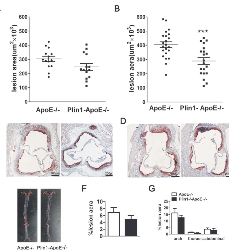
To assess the impact of Plin1 in atherogenesis in vivo, we crossed Plin1-/-mice with ApoE -/-mice to generate Plin1-/-ApoE-/-mice and using ApoE-/-littermates as controls. Despite the similar levels of plasma cholesterol and triglyceride between ApoE-/-and Plin1-/-ApoE -/-mice, Plin1-/-ApoE-/-males had reduced body weight compared with ApoE-/-male littermates (29.6 ± 0.8 g v.s. 34.2 ± 1.8 g; p = 0.035; n = 20) (Table 1). Quantification of Oil red-stained cross sections of aortic roots did not detect significant difference between male Plin1-/-ApoE -/-and ApoE-/-littermates (Fig 3A and 3C). Unlike males, the body weight of female Plin1 -/-ApoE-/-did not differ from female ApoE-/-littermates (Table 1). However, they showed re-duced plaque size in aortic roots when compared with female ApoE-/-mice (ApoE-/-females: 404 ± 21×103μm2n = 20; Plin1-/-ApoE-/-females: 290 ± 24×103μm2, n = 22) (Fig 3B and
3D). Histological analysis of aortic root lesions revealed no significant difference in macro-phage or smooth muscle cell content in atherosclerotic plaques of Plin1-/-ApoE-/-mice and ApoE-/-mice (data not shown). We also performed en face analysis of the aortic arch and de-scending aorta in these female mice but the results showed no statistically significant differ-ences in lesion area between the 2 groups of mice, although there was a trend toward decreased lesion area in Plin1-/-ApoE-/-mice (Fig 3E–3G). Taken together, Plin1 deficiency may have a protective role in As.
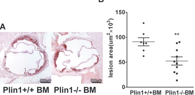
Role of Plin1-/- bone marrow cells in As
Fig 1. Plin1 expresses in atherosclerotic lesions.(A) Immunoblotting analysis of Plin1 expression in perivascular adipose tissue and in peritoneal macrophages isolated from Plin1-/- and wild-type mice. Incubation with modified LDL increases Plin1 expression in the macrophages. Anti-Plin1 antiserum from (Abcam, #ab3526) recognizes the C-terminal amino-acid residues of Plin1. (B) Unstained frozen section of arotic root of ApoE-/-mice. (C) The frozen section of arotic root in ApoE-/-mice was immunostained (Green) with the primary antibody against CD68, a macrophagemarker. (D) Immunostaining of Plin1 (Red). (E and F) Merged image shows colocalization of Plin1 and CD68 in atheroma. Boxed area in panel E (a-c) were magnified to show colocalization of Plin1 and CD68 (F, a-c) in macrophages in atherosclerotic lesion in ApoE-/- mice.
Fig 2. Lipid content is decreased in peritoneal macrophages and foam cells derived from Plin1-/- mice.Peritoneal macrophages from Plin1+/+ and Plin1-/-mice were incubated for 48 hours in the absence or presence of 50μg/ml aggLDL. (A and B) The contents of free cholesterol (FC), cholesterol ester (CE) and triglyceride (TG) were measured. n = 4,*P<0.05. (C) Nile red staining of LDs in peritoneal macrophages from Plin1+/+ and Plin1-/-mice with aggLDL (50μg/ml) (n = 6).
52 ± 8×103μm2) (Fig 4A and 4B). Therefore, absence of Plin1 in bone marrow-derived cells
alone is sufficient to reduce As in LDLR-/-mice.
Mechanisms of reduced lipid accumulation in macrophages lacking
Plin1
The lipid homeostasis in macrophages depends on the balance of influx and efflux of lipid in which receptors are critically involved. To explore the mechanism underlying the protective ef-fect of Plin1 deficiency, we examined the expression of several key factors in atherogenesis in macrophages. Immunoblotting results showed that the protein level of CD36 was significantly reduced in Plin1-/-macrophages but the expression of ABCA1 was not altered (Fig 5A and 5B). CE deposited in macrophages was hydrolyzed by neutral cholesterol ester hydrolase, mainly by hormone-sensitive lipase (HSL) and adipose triglyceride lipase (ATGL). However, immunoblotting showed that protein expression of HSL and ATGL was not altered between the two groups (Fig 5C and 5D).
AggLDL was incubated with macrophages for 48 hours to transform them into foam cells. The CE deposited in macrophages would be hydrolysed gradually. Although macrophages from Plin1+/+ mice had more CE than macrophages from Plin1-/-mice, the hydrolysis of CE was faster in Plin1-/-macrophages (Fig 5E). Neither HSL nor ATGL expression was altered by AggLDL treatment, in macrophages from Plin-/- and wild-type mice (Fig 5C and 5D). Because the free cholesterol hydrolyzed from CE is transported by ATP-binding cassette A1 (ABCA1), then we used NBD-cholesterol to determine the cholesterol efflux rate which reflects the activi-ty of ABCA1. We found cholesterol efflux between two groups was similar (Fig 5F). These re-sults demonstrated that Plin1 deficiency resulted in reduced CD36 expression and faster hydrolysis of CE in Plin1-/-macrophages, which then restricted the foam cell formation and retarded the development of As in ApoE-/-background.
After lipoproteins are taken up by macrophages, CEs are hydrolyzed by cellular lipases to re-lease free cholesterols that are effluxed to extracellular acceptors, or transported to the endo-plasmic reticulum and are re-esterified to CEs by acyl-CoA: cholesterol acyltransferase-1 (ACAT1) and stored in cytoplasmic LDs [25]. The process of intracellular cholesterol homeo-stasis is regulated by many other molecules, such as SR-A, CD36, ABCA1, ABCG1, SR-BI, Nie-man-Pick type C1 protein (NPC1), ACAT1, HSL and ATGL. There was no difference in the expression levels of any of these genes between the macrophages from Plin1+/+ mice and Plin1-/-mice (Data not shown). Also, quantitative PCR showed that there was no difference between the expressions of these genes in the aortic tissue of ApoE-/-and Plin1-/-ApoE -/-mice (Data not shown).
Table 1. Plasma cholesterol, triglyceride and body weight in ApoE-/- and Plin1-/-ApoE-/-mice after 12 weeks on western diet.
Genotype n Body Weight (g) Total Cholesterol (mg/dL) Triglyceride (mg/dL)
male
Plin1-/-ApoE-/- 15 29.59±0.8* 1464±133.0 107.1±5.7
ApoE-/- 15 34.19±1.8 1371±86.7 103.7±8.6
female
Plin1-/-ApoE-/- 20 22.78±0.7 1015±89.9 87.9±11.5
ApoE-/- 22 24.61±0.4 1101±99.7 102.1±10.9
*p<0.05 for Plin1-/-ApoE-/-vs. ApoE-/-.
Fig 3. Ablation of Plin1 restricts As in female ApoE-/- mice.(A and B) The mean lesion area (±SEM) in the aortic roots of male (A) and female (B) ApoE -/-and Plin1-/-ApoE-/-mice,***p<0.001. (C and D) Representative photographs of aortic roots from male (C) and female (D) ApoE-/-and Plin1-/-ApoE -/-mice stained with oil red O. (E) Representative photographs of the aorta en face from female ApoE-/-and Plin1-/-ApoE-/-mice. (F) Quantification of atheroma of total aortic lesion area. n = 15. (G) Regional analysis of lesion distribution in the aorta.
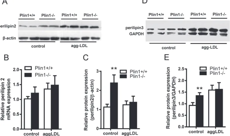
The expression of other genes of PAT family in Plin1-/-
mice
Plin2, another lipid droplet protein of Plin family, was associated with As [26]. Immunoblot-ting showed that Plin2 was up-regulated in Plin1-/-macrophages (Fig 6A); this phenomenon was consistent with the compensatory increase of Plin2 in adipocytes of Plin1-/-mice [17]. However, the upregulation of Plin2 ceased when Plin1-/-macrophages were incubated with modified LDL (Fig 6A and 6C). We examined the expression of Plin2 mRNA by quantitative PCR in these macrophages and the results showed no change in Plin2 mRNA expression (Fig 6B). Similarly, Plin3 was also expressed in the mouse peritoneal microphages and was not in-ducible by the modified LDL in Plin1-/- microphages (Fig 6D and 6E).
Discussion
Based on the relationship between Plin1 expression and foam cell formation we tested the hy-pothesis that Plin1 deficiency would possibly reduce As, by using different As-prone mouse models in absence of Plin1 gene. Although plasma levels of cholesterol and triglyceride re-mained unchanged between Plin1-/-ApoE-/-mice and ApoE-/-mice, the atherosclerotic le-sions at aortic root in Plin1-/-ApoE-/-mice reduced 39% in female and 15% in male, compared to that in ApoE-/-mice. This is likely due to the gender difference for the earlier development of atherosclerotic plaques in female ApoE-/-mice [27]. The presence of Plin1 in macrophages and its function in inhibiting TG hydrolysis in adipocytes would imply that Plin1 could in-crease foam cell formation via suppression of CE hydrolysis. In order to define the macrophage specific role in atherogenesis, we did bone marrow transplant using marrows from Plin1 +/+ and Plin1-/-mice as donors in another type of As-prone model, LDLR-/-mice. The mice receiving bone marrow cells from Plin1-/-mice had 49% reduction of lesion area, consistent
Fig 4. The bone marrow cells from male Plin1-/- mice transplanted to female LDLR-/- mice reduce atherosclerotic lesion.(A) Representative photographs of aortic roots from LDLR-/-mice received bone marrow cells from Plin1+/+ and Plin1-/-mice respectively. (B) The mean lesion area (±SEM) in the aortic roots in mice transplanted with bone marrow cells from Plin1+/+ or Plin1-/-mice.**p<0.01 for Plin-/- vs. Plin+/+.
Fig 5. The cholesterol metabolism in macrophages.(A) Immunoblotting of CD36 and ABCA1 in peritoneal macrophages and foam cells and (B) The density of the protein band was quantitated. (C) Immunoblotting of HSL and ATGL in peritoneal macrophages and foam cells and (D) the density of the protein bands was quantitated. (E) The rate of cholesterol ester hydrolysis in peritoneal macrophages from Plin1+/+ and Plin1-/-mice. (F) Time-course of cholesterol efflux to ApoA-I in peritoneal macrophages from Plin1+/+ and Plin1-/-mice.*p<0.05,**p<0.01 for Plin-/- vs. Plin+/+.
with Pln1-/-ApoE-/- mice. This finding implicated that the deletion of Plin1 in macrophages would be responsible for decreased atherosclerotic lesions in Plin1-/- mice.
In macrophages lipoprotein derived CEs are hydrolyzed and released FCs that are re-esteri-fied by ACAT1. The produced CEs are stored in cytoplasmic lipid droplets and are further hy-drolyzed by neutral cholesterol ester hydrolyase to release FC for efflux by ABCA1 or ABCG1. This is described as cholesterol ester cycle [25]. Modified LDL was taken up mainly by CD36 and SR-A [28]. We observed that the expression of CD36 protein but not its mRNA was de-creased significantly in Plin1-/-macrophages. It is known that CD36 deficiency markedly re-duces the lipid accumulation in macrophages [29]. Hence, down-regulation of CD36 in Plin1-/-macrophages would be accounted for one of factors affecting the cellular CE accumu-lation. However, it is unclear CD36 affect lipoprotein binding or uptake, or both. This will be clarified in the future. In adipocytes Plin1 coats the lipid droplet surface as a barrier to protect the hydrolysis of the droplet lipids by lipases, but Plin1 ablation leads to the loss of this protec-tive barrier and thus results in an elevated level of basal lipolysis [14,17]. Our in vitro experi-ments showed that CEs in Plin1-/-macrophages disappeared faster than wild-type
macrophages. The hydrolysis of CE was catalyzed by neutral cholesterol ester hydrolase (CEH) activity, which was the limiting step in cellular cholesterol efflux[30]. Based on the observations that 1) cAMP enhanced FC efflux from macrophages; 2) hormone sensitive lipase (HSL) was activated by cAMP and 3) CE was one of HSL preferred substrates, HSL was thought to be the
Fig 6. The expressions of Plin2 and Plin3 in Plin1+/+ and Plin1-/- macrophages and foam cells.(A) The expression of Plin2 protein and (B) Plin2 mRNA in the macrophages and foam cells of Plin1+/+ and Plin1-/-mice. (C) Protein band density of Plin2 shown in panel A. (D) Immunoblotting of Plin3 from the macrophages and foam cells of Plin1+/+ and Plin1-/-mice. (E) Protein band density of Plin3 shown in panel D.**p<0.01 for Plin-/- vs. Plin+/+.
likely candidate CEH for macrophage CE hydrolysis[31]. HSL expresses in murine macro-phages and indeed provides the main CEH activity in murine macromacro-phages [23]. Considering the role of HSL in lipolysis of adipocytes, we presume the similar mechanism in macrophages like in adipocytes, i.e., loss of Plin1 would expose accumulated CE to HSL hydrolysis. Although the expressions of mRNA and protein of HSL and ATGL were not altered, an increase in CE hydrolysis in Plin1-/-macrophages could be possible because the loss of Plin1 certainly abol-ishes the protective role of Plin1 on the droplet lipids, thus enhancing the enzymatic activity of these lipases.
Our results were in agreement with another study in which Paul found a protein related to Plin1 structurally and functionally, Plin2/ADRP, behaved similarly in its role to atherogenesis. Targeted deletion of Plin2 resulted in 40–50% reduction of aortic atherosclerotic lesions in ApoE null mice [26]. In further bone marrow transplant experiment these authors demonstrat-ed that the lack of Plin2 in macrophages was responsible for rdemonstrat-educdemonstrat-ed atherogenesis in Plin2 de-ficient mice. In their study they claimed inability to detect the signals of Plin1 in macrophages from both wild-type and Plin2-/-mice by Western blot. Therefore, there was no further experi-mental pursuit to the potential role of Plin1 in their study. We believe the discrepancy in detec-tion of Plin1 between their data and our results was due to the anti-mouse Plin1 antibodies chosen because we demonstrated clearly the presence of Plin1 atherosclerotic lesions and local-ized within plaque macrophages of ApoE-/-mice. During adipocyte differentiation Plin2 ap-peared early but then disapap-peared after the onset of Plin1, since Plin2 was replaced by Plin1 in fully differentiating adipocytes [32]. This phenomenon prompted us to think it could also hap-pen in transformation of macrophages to foam cells. Our immunostaining and immunoblot-ting data showed that both Plin1 and Plin2 proteins were expressed in the atherosclerotic plaques of ApoE-/- mice, and in some area Plin1 was colocalized with CD68, in the atheroscle-rotic microphages. Plin2 is a lipid-droplet marker ubiquitously expressed in all types of cells ex-cept for mature adipocytes [11]. Plin2, like Plin3 and Plin4, has been long assumed to prevent the LDs from the hydrolysis by cellular lipase, but this putative role remains not to be accessed. By contrast, the functional roles of Plin1 in both LD storage and degradation have been firmly established. Native Plin1 proteins barrier and protect the LDs from access of HSL and ATGL, thus prevent LD degradation and enhance LD formation [10,11]. On catecholamine stimula-tion, Plin1 is phosphorylated by protein kinase A and then induces the translocation of HSL from the cytosol to the LD surface and also indirectly activated ATGL [10,11]. In human ath-erosclerotic plaques, Plin1 protein is highly expressed in unstable athath-erosclerotic plaques and also expressed considerably in stable plaques [12]. Plin1 is mainly assembled in 3 subcellular locations of human atherosclerotic plaques, cells surrounding some cholesterol clefts, foam cells, and endothelial cells in newly formed vessels [12]. This observation is consistent with our present finding of Plin1 expression pattern in atherosclerotic plaques of ApoE-/- mice. In light of our immunostaining results, the circular fluorescence signals of Plin1 and CD68 indicated that Plin1 was associated with the LDs in CD68-labeled foam cells, but the irregular signal pat-terns implicated that Plin1 was also assembled in irregular vessel endothelium and in the cells sounded with cholesterol crystals. Given the fundamental role of Plin1 in protecting LD lipids from lipase hydrolysis, it is reasonably speculated that the assembly of Plin1 might function to reduce lipolysis and hence increase lipid retention in plaques. This could be a cellular basis by which Plin1 deficiency in bone marrow-derived cells resulted in reduced atherosclerotic lesions in mice.
we could not fully understand what caused the discrepancy between our and their results, we did find some disparate points: 1). We quantitated the As lesions both in the aortic root by multiple consecutive cryosections under microscope and in the en face entire aorta by Oil red staining,while they only did whole aorta en face lesion assessment by Sudan IV staining. 2).We chooses both males and females for the study and found significant difference in atherogenic lesions only in females after HFD feeding for 12 weeks. In their study they just had males and found increased As lesions after 20 weeks HFD feeding but not at 10 weeks interval. 3).We found consistently reduced As lesions in either ApoE null background when whole body Plin1 was deleted, or in LDLR null female mice when Plin1 in macrophages was depleted by bone marrow transplant approach. In contrast, they only had whole body Plin1 deficiency on LDLR null background. In explanation of the 7 fold up-regulation of SR-A expression as the cause for increased As lesions in Plin1 deficiency, they also showed a striking increase of mRNAs of ABCA1 and ABCG1 for nearly 20 and 5 folds, respectively. Though these changes were inter-preted as response to the up-regulated SR-A in Plin1 null mice,such robust increase of ABCA1 and ABCG1 would certainly diminish As lesions in theory via resulting super Cholesterol ef-flux. For instance, ABCA1 transgenic expression in mice resulted in 3 to 5 fold increase of ABCA1 in aorta or in macrophage and such increase in ABCA1 led to markedly reduction of aortic lesion areas in As-prone ApoE or LDLR null background [33]. Therefore, the inconsis-tent relationship in upregulated SR-A, ABCA1, ABCG1 and Plin1 deficiency with enhanced As lesions would require further careful examination.
Taken together, Plin1 deficiency in macrophages decreased lipid deposits by inhibition of foam cell formation and hence reduced As lesions in arterial walls. We postulated the major cause for reduced lipid accumulation in Plin1-/-macrophages was due to the disappearance of the barrier composed by Plin1, which facilitates the interactions of lipases with lipid droplets and results in faster hydrolysis of LD. In addition to this macrophage LD hydrolysis, down-reg-ulation of CD36 could lead less uptake of modified LDL, which may also to some extent con-tribute to reduced foam cell formation.
Author Contributions
Conceived and designed the experiments: GL GX BG. Performed the experiments: XZ MG JH L. Zhang L. Zou YL BG. Analyzed the data: XZ MG JH L. Zou YL. Contributed reagents/mate-rials/analysis tools: XZ MG L. Zhang BG GL GX. Wrote the paper: GL GX XZ MG BG.
References
1. Zweytick D, Athenstaedt K, Daum G. Intracellular lipid particles of eukaryotic cells. Biochim Biophys Acta. 2000; 1469: 101–120. PMID:10998572
2. Martin S, Parton RG. Lipid droplets: a unified view of a dynamic organelle. Nat Rev Mol Cell Biol. 2006; 7: 373–378. PMID:16550215
3. Ross R. Cell biology of atherosclerosis. Annu Rev Physiol. 1995; 57: 791–804. PMID:7778883
4. Larigauderie G, Bouhlel MA, Furman C, Jaye M, Fruchart JC, Rouis M. Perilipin, a potential substitute for adipophilin in triglyceride storage in human macrophages. Atherosclerosis. 2006; 189: 142–148.
PMID:16442115
5. Buechler C, Ritter M, Duong CQ, Orso E, Kapinsky M, Schmitz G. Adipophilin is a sensitive marker for lipid loading in human blood monocytes. Biochim Biophys Acta. 2001; 1532: 97–104. PMID:11420178
6. Buers I, Robenek H, Lorkowski S, Nitschke Y, Severs NJ, Hofnagel O. TIP47, a lipid cargo protein in-volved in macrophage triglyceride metabolism. Arterioscler Thromb Vasc Biol. 2009; 29: 767–773. doi:
10.1161/ATVBAHA.108.182675PMID:19286631
8. Servetnick DA, Brasaemle DL, Gruia-Gray J, Kimmel AR, Wolff J, Londos C. Perilipins are associated with cholesteryl ester droplets in steroidogenic adrenal cortical and Leydig cells. J Biol Chem. 1995; 270: 16970–16973. PMID:7622516
9. Jiang HP, Serrero G. Isolation and characterization of a full-length cDNA coding for an adipose differen-tiation-related protein. Proc Natl Acad Sci U S A. 1992; 89: 7856–7860. PMID:1518805
10. Sztalryd C, Xu G, Dorward H, Tansey JT, Contreras JA, Kimmel AR, et al. Perilipin A is essential for the translocation of hormone-sensitive lipase during lipolytic activation. J Cell Biol. 2003; 161: 1093–1103.
PMID:12810697
11. Brasaemle DL. The perilipin family of structural lipid droplet proteins: stabilization of lipid droplets and control of lipolysis. J Lipid Res. 2007; 48: 2547–2559. PMID:17878492
12. Faber BC, Cleutjens KB, Niessen RL, Aarts PL, Boon W, Greenberg AS, et al. Identification of genes potentially involved in rupture of human atherosclerotic plaques. Circ Res. 2001 89: 547–554. PMID:
11557743
13. Forcheron F, Legedz L, Chinetti G, Feugier P, Letexier D, Bricca G, et al. Genes of cholesterol metabo-lism in human atheroma: overexpression of perilipin and genes promoting cholesterol storage and re-pression of ABCA1 exre-pression. Arterioscler Thromb Vasc Biol. 2005; 25: 1711–1717. PMID:
15961705
14. Brasaemle DL, Rubin B, Harten IA, Gruia-Gray J, Kimmel AR, Londos C. Perilipin A increases triacyl-glycerol storage by decreasing the rate of triacyltriacyl-glycerol hydrolysis. J Biol Chem. 2000; 275: 38486–
38493. PMID:10948207
15. Beylot M, Neggazi S, Hamlat N, Langlois D, Forcheron F. Perilipin 1 ablation in mice enhances lipid oxi-dation during exercise and does not impair exercise performance. Metabolism. 2012; 61: 415–423.
doi:10.1016/j.metabol.2011.07.019PMID:21944269
16. Brasaemle DL, Barber T, Wolins NE, Serrero G, Blanchette-Mackie EJ, Londos C. Adipose differentia-tion-related protein is an ubiquitously expressed lipid storage droplet-associated protein. J Lipid Res. 1997; 38: 2249–2263. PMID:9392423
17. Tansey JT, Sztalryd C, Gruia-Gray J, Roush DL, Zee JV, Gavrilova O, et al. Perilipin ablation results in a lean mouse with aberrant adipocyte lipolysis, enhanced leptin production, and resistance to diet-in-duced obesity. Proc Natl Acad Sci USA. 2001; 98: 6494–6499. PMID:11371650
18. Zhai W, Xu C, Ling Y, Liu S, Deng J, Qi Y, et al. Increased lipolysis in adipose tissues is associated with elevation of systemic free fatty acids and insulin resistance in perilipin null mice. Horm Metab Res. 2010; 42: 247–253. doi:10.1055/s-0029-1243599PMID:20091459
19. Toh SY, Gong J, Du G, Li JZ, Yang S, Ye J, et al. Up-regulation of mitochondrial activity and acquire-ment of brown adipose tissue-like property in the white adipose tissue of fsp27 deficient mice. PLoS One. 2008; 3: e2890. doi:10.1371/journal.pone.0002890PMID:18682832
20. Griffin EE, Ullery JC, Cox BE, Jerome WG. Aggregated LDL and lipid dispersions induce lysosomal cholesteryl ester accumulation in macrophage foam cells. J Lipid Res. 2005; 46: 2052–2060. PMID:
16024919
21. Cramer T, Yamanishi Y, Clausen BE, Forster I, Pawlinski R, Mackman N, et al. HIF-1alpha is essential for myeloid cell-mediated inflammation. Cell. 2003; 112: 645–657. PMID:12628185
22. Lamant M, Smih F, Harmancey R, Philip-Couderc P, Pathak A, Roncalli J, et al. ApoO, a novel apolipo-protein, is an original glycoprotein up-regulated by diabetes in human heart. J Biol Chem. 2006; 281: 36289–36302. PMID:16956892
23. Buchebner M, Pfeifer T, Rathke N, Chandak PG, Lass A, Schreiber R, et al. Cholesteryl ester hydrolase activity is abolished in HSL-/- macrophages but unchanged in macrophages lacking KIAA1363. J Lipid Res. 2010; 51: 2896–2908. doi:10.1194/jlr.M004259PMID:20625037
24. Xu G, Sztalryd C, Lu X, Tansey JT, Gan J, Dorward H, et al. Post-translational regulation of adipose dif-ferentiation-related protein by the ubiquitin/proteasome pathway. J Biol Chem. 2005; 280: 42841–
42847. PMID:16115879
25. Brown MS, Ho YK, Goldstein JL. The cholesteryl ester cycle in macrophage foam cells. Continual hy-drolysis and re-esterification of cytoplasmic cholesteryl esters. J Biol Chem. 1980; 255: 9344–9352.
PMID:7410428
26. Paul A, Chang BH, Li L, Yechoor VK, Chan L. Deficiency of adipose differentiation-related protein im-pairs foam cell formation and protects against atherosclerosis. Circ Res. 2008; 102: 1492–1501. doi:
10.1161/CIRCRESAHA.107.168070PMID:18483409
28. van Berkel TJ, Out R, Hoekstra M, Kuiper J, Biessen E, van Eck M. Scavenger receptors: friend or foe in atherosclerosis? Curr Opin Lipidol. 2005; 16: 525–535. PMID:16148537
29. Febbraio M, Podrez EA, Smith JD, Hajjar DP, Hazen SL, Hoff HF, et al. Targeted disruption of the class B scavenger receptor CD36 protects against atherosclerotic lesion development in mice. J Clin Invest. 2000; 105: 1049–1056. PMID:10772649
30. Zhao B, Fisher BJ, St Clair RW, Rudel LL, Ghosh S. Redistribution of macrophage cholesteryl ester hy-drolase from cytoplasm to lipid droplets upon lipid loading. J Lipid Res. 2005; 46: 2114–2121. PMID:
16024911
31. Khoo JC, Reue K, Steinberg D, Schotz MC. Expression of hormone-sensitive lipase mRNA in macro-phages. J Lipid Res. 1993; 34: 1969–1974. PMID:8263420
32. Londos C, Sztalryd C, Tansey JT, Kimmel AR. Role of PAT proteins in lipid metabolism. Biochimie. 2005; 87: 45–49. PMID:15733736