Journal of Chemical and Pharmaceutical Research, 2017, 9(3):197
-
208
Research Article
ISSN : 0975
-
7384
CODEN(USA) : JCPRC5
197
PLGA Microspheres as New Strategy to Improve the Efficiency of Venom
Immunotherapy
Reginaldo Almeida da Trindade
1-3, Osvaldo Augusto Brazil Esteves
Sant’Anna
4,
Vanessa Cristina Rescia
2, Fernanda Miriani Bruni
5, Mônica Valdyrce dos Anjos
Lopes-Ferreira
5, Daiane Fernanda dos Santos
6, Roberto Nicolete
7, Lucia Helena Faccioli
6,
Pedro Soares de Araujo
8and Maria Helena Bueno da Costa
2*1
Faculty of Pharmacy, Federal University of Rio de Janeiro, Rio de Janeiro, RJ, Brazil 2
Laboratory of Microspheres and Liposome, Institute Butantan, São Paulo, SP, Brazil 3
Post-Graduation Program in Biotechnology, University of São Paulo, São Paulo, SP, Brazil 4
Laboratory of Immunochemistry, Institute Butantan, São Paulo, SP, Brazil 5
Center of Applied Toxinology, Institute Butantan, São Paulo, SP, Brazil 6
Faculty of Pharmaceutical Sciences of Ribeirão Preto, University of São Paulo, Ribeirão Preto, SP, Brazil. 7
Laboratory of Biotechnology applied to Health, FIOCRUZ, Rondônia, RO, Brazil 8
Institute of Chemistry, University of São Paulo, São Paulo, SP, Brazil
_____________________________________________________________________________
ABSTRACT
Background: Despite considerable knowledge of allergy and its physiopathology, the only treatment with long-lasting effects for bee venom (BV) allergic patients is venom immunotherapy (VIT). Its efficiency has been recognized worldwide for many years. However, some limitations still result in patient rejection and prevent its use, such as the long treatment period with a total of 30-80 injections administered over three to five years, the high cost of treatment, and the risk of anaphylaxis as a side effect. Objective: In order to overcome these inconveniences, a new formulation for VIT is proposed, consisting of BV encapsulated within microspheres composed of poly-lactide-co-glycolide (MS-PLGA) as a controlled delivery system. Methods: BV-MS-PLGA was prepared by a double emulsion/solvent evaporation method and its biological efficiency was tested by ex vivo macrophage uptake stimuli and in vivo antibody production in mice. Results: BV-MS-PLGA formulations prevented inflammatory reactions by impeding direct contact between BV and the organism/tissue. The increased phagocytosis rate of BV-MS-PLGAs by macrophages allowed for the delivery of antigens directly to the intracellular environment. Furthermore, with the use of BV-MS-PLGAs, antibody production was comparable to traditional VIT (free BV delivered in PBS) with the advantages of protecting the organism against the deleterious effects of BV toxins and reducing the number of injections needed to produce the same protective effect. Conclusion: MS-PLGA is a promising controlled delivery system to deliver BV for VIT.
Keywords: Allergy; Biodegradable microspheres; Protein delivery; PLGA microspheres.
_____________________________________________________________________________
INTRODUCTION
198
inflammatory mediators. Mast cell degranulation occurs since these cells are sensitized by specific IgE antibodies on the cell surface, which then cross-react with allergen molecules [9]. There are palliative and transient treatments involving the use of corticosteroids and antihistamines, which ameliorate and control the severity of such reactions, but they are not preventive against future accidents involving insect stings [6]. As a preventive therapeutic strategy for patients suffering from an allergy caused by bee venom (BV), allergen or venom immunotherapy (VIT) is the only treatment that has shown long-lasting effects. The efficiency of VIT has been recognized worldwide, and its application is approved by the World Allergy Organization (WAO), although the exact mechanism of action is not fully understood [7,8]. It is believed that a modulation of the immunological response from Th2 towards a Th1 profile is a key aspect of this treatment [8].
VIT is a well-established therapy consisting of subcutaneous injections of gradually increasing doses of allergen. This therapeutic strategy induces immunological tolerance in the organism by shifting the immunological response against the allergen from predominantly IgE towards IgG antibody production, which means a change from a Th2 to a Th1 response [8-10]. Despite its efficacy, VIT presents some inconveniences that have limited and prevented its wide application. For example, it is frequently rejected by patients because it entails a prolonged treatment period, commonly over three to five years, with a total of 30 to 80 injections. Additionally, it is a high-cost therapy not paid for or funded by public health programs [11,12]. Moreover, there is no safe dose for administering free BV (in aqueous solution) during treatment. Hence, each patient may respond in different ways, depending on their own reactivity threshold and sensitivity level. Therefore, the risk of side effects, however small, represents one of the greatest drawbacks preventing general and widespread application. In this context, the development of safer and more effective strategies for VIT is of great importance to improve the treatment of allergic diseases [13,14]. Taking into account all these described limitations, were propose in the current study that microspheres (MS) prepared using biodegradable polymers, such as poly-lactide-co-glycolide acid (PLGA), may represent an alternative controlled delivery system to encapsulate BV allergens for use in VIT.
MS-PLGA has been shown to be an efficient controlled delivery system capable of delivering antigens either continuously or by pulsatile release over prolonged periods of time for different therapeutic purposes [15-17]. MS-PLGA encapsulated antigens have been shown to be an alternative strategy to induce protective immunity even after a single subcutaneous injection, as the antigens can be released slowly over time, thus simulating the sequential injections of traditional VIT [18-20]. Moreover, the pharmaceutical strategy of encapsulating allergens within MS-PLGA helps to protect allergens from degradation by natural proteases present in the organism. This increases their bioavailability, which allows for prolonged stimulation of the immune system, and thereby represents the most promising approach in allergy treatment [3]. Furthermore, there is bi-directional protection, since microspheres also protect the organism from possible deleterious effects caused by direct contact with BV toxins. Finally, it is possible to direct these allergenic molecules to antigen-presenting cells such as macrophages and/or dendritic cells [21], where they can be adequately processed and provide a non-anaphylactic immunological stimulus upon the next contact with the same antigen, resulting in, for example, a Th1-polarized response.
Previous studies by our group have demonstrated the capacity of MS-PLGA to encapsulate BV proteins without losing their integrity, even after they have been submitted to the microencapsulation process [22]. Furthermore, characterization showed that they are adequate for VIT, by presenting a controlled-release profile of BV and by maintaining BV antigenicity for prolonged periods of time [23]. Therefore, the present study describes, to the best of our knowledge for the first time, the in vivo administration of whole BV encapsulated within MS-PLGA, which was prepared with six different polymers. Assessments were performed regarding tissue toxicity and inflammatory reactions, the ability to induce phagocytosis, and the ability to stimulate the production of specific neutralizing antibodies against BV proteins.
MATERIALS AND METHODS
Materials
Lyophilized BV was purchased from Sigma Chemical Co., St. Louis, MO, USA. PLGA polymers with different molecular weights with free carboxyl (12 kDa-H, 34 kDa-H and 63 kDa-H) and methylated terminal-carboxyl (12 kDa-Me, 34 kDa-Me and 63 kDa-Me) were purchase from Boehringer Ingelhein (Germany).
Dichloromethane was obtained from Aldrich Chemical® - Milwaukee – USA. Griess reagent mixtures were
purchased from Sigma Chemical Co. (St. Louis, MO, USA). Polyvinyl alcohol (PVA Mw 49000 - Mowiol® 40-88) were obtained from Sigma-Aldrich®. 3,3',5,5'-tetramethylbenzidine (TMB) was from Polysciences® and
Dimethylsulfoxide (DMSO) was from Merck®. Anti-mouse IgG1, IgG2a and IgE peroxidase conjugates were
199
Preparation of BV-loaded microspheresA water-in oil-in water (W1/O/W2) double-emulsion solvent evaporation [24] adapted method [25] was
employed to prepare microspheres containing bee venom proteins. 250 µl of BV (50 mg/mL) was mixed with 4
mL of dichloromethane containing PLGA, and emulsified (T 25 Basic IKA Labortechnik – Ultra Turrax®) at
24000 rpm for 2 min. The resultant first emulsion was added to 20 mL of PVA (2%), under agitation, and emulsification continued at 13500 rpm for 2 min. The emulsion was stirred continuously for 3 h at room temperature and atmospheric pressure till complete solvent evaporation. After the microspheres had formed, they were collected by centrifugation for 10 minutes at 2000 g, rinsed with water three times and then resuspended with 2 mL of 1 % PVA, freeze-dried for 24 hours and stored at -20 °C.
Encapsulation efficiency (EE)
EE was determined after MS alkaline digestion with some modifications from Sharif and O’Hagan’s method
(1995). Briefly, 20 mg of the freeze-dried MS was added to 5 ml of 0.1% SDS - 0.1 M NaOH solution [26]. The mixture was incubated under gentle stirring for 24 hours at 37 ± 1.0 °C. After centrifugation and filtration in 0.22 µm membranes, the clear supernatant liquid was assayed by the Lowry method. The amount of BV was determined using the standard curve equation (y = 1.00208X r=0.99931). The encapsulation efficiency (EE) can be calculated as follows:
EE = (Protein found in microspheres / Initial protein added) x 100%
Uptake of microspheres by J774 macrophages
Murine macrophages of the cell line J774 were used to study the phagocytic uptake of MS-PLGA. The cells were maintained in RPMI-1640 complete medium containing 20% FBS, antibiotics (10 µL/mL penicillin and 1
µL/mL gentamicin), and incubated at 37 °C under 5% CO2. The macrophages were washed, collected by
centrifugation and counted by Trypan blue exclusion; then, 1×10⁶cells/24-well were incubated for 4 h with
1mg/ml of non-loaded MS or BV-loaded MS [27]. After the incubation period, the medium was aspirated and non-ingested MS were washed off with additional medium. Cell suspensions were collected, cytocentrifuged and identified by panoptic staining [28]. MS uptake was assessed microscopically by counting the number of cells that had ingested at least one MS. The phagocytic index (PI) was calculated as:
PI = number of cells containing at least one MS / total number of cells X number of engulfed MS.
NO production by J774 macrophages
NO production by J774 cells were determined by the Griess reaction. Cell culture supernatants (0.1 ml) were incubated with an equal volume of Griess reagent (1% sulfanilamine, 0.1% N-(1-naphtyl)-ethylendiamine
dihydrochloride, 2.5% H3PO4) at room temperature for 10 min. The absorbance was measured in a microplate
reader at 540 nm and NO concentrations calculated from a sodium nitrite standard curve (y = 0.00525X
r=0.997). The data are presented as micromoles of NO2- (nitrite) (mean ± the SEM).
Intravital microscopy
Mice were injected with a muscle relaxant drug (0.4% Xilazin) and then anaesthetized with 2.5% chloral hydrate and the cremaster muscle was exposed for microscopic examination in situ [29-30]. Animals were maintained on a board thermostatically controlled at 37 °C, which included a transparent platform on which the tissue to be transilluminated was placed. After stabilization of the microcirculatory network, the number of rolling leukocytes was counted in post-capillary venules for a total period of 30 min (divided in periods of 5 minutes) before and after topical application of 0.3 µg BV (30 µ L of total volume) of free BV, BV-loaded MS or non-loaded MS in PBS. Values are the average of three independent experiments. The analysis of the microvascular system was performed with an optical microscope (Image.A1 Carl-Zeiss) coupled to a photographic camera (AxioCam ICc1) using an objective 40x/03.
VIT protocols
The VIT was performed with adult female Balb/C inbred mice, weighing 15-20 g, between 30-40 days old, all from the Central Animal Laboratory of the Institute Butantan. The animals were maintained at the Animal Care Service in the Biotechnology Center, kept in isolators with free access to water and food, with a cycle of light and temperature control according to the local guidelines. All experiments were conducted within ethical standards and the project was approved by the Ethics Committee on Animal Use of Institute Butantan (protocol n°. 336/06).
First protocol: adjuvant effects of MS-PLGA in VIT:
200
Sensitizations: all animals of each group were subcutaneously (SC) two times sensitized (30-day intervals) with
5 µg of BV adsorbed on Al(OH)3. Eight days after the sensitization, blood samples were collected from the
retro-orbital venous plexus.
Desensitizations (immunotherapy): 45 days after the first sensitization 0.3 µg of BV was applied (free in PBS
or encapsulated within the six different PLGAs) via SC. Injections were repeated every week during six weeks. Once immunotherapy has been finalized, other sera aliquots were collected from each group.
BV challenge: Ten days after this last bleeding the animals were SC challenged with 15 µg of native BV in
PBS. 24 hours after the challenge, a last bleeding was performed to analyze the production of antibody. All sera samples were stored at - 20 °C until analysis by ELISA.
Second protocol: effects of reduced injections through of BV within MS-PLGA:
Balb/C mice were divided only into four groups of six animals. Based on the results of the first protocol, in this
second assay, polymers of 12 kDa-COOH or 63 kDa-COOCH3 were studied.
Sensitizations: all animals of each group were two times sensitized, SC, (10-day intervals) with 5 µg of BV
adsorbed on Al(OH)3. Eight days after the sensitization, blood samples were collected from the retro-orbital
venous plexus.
Desensitizations (immunotherapy): were initiated 15 days after sensitizations. A control group (only
sensitized) received injections of PBS only. The group that represented the traditional VIT received six doses of 0.3 µg of native BV in PBS via SC at weekly intervals. The groups treated with reduced number of injections
received only two doses of 1.0 µg of encapsulated BV within MS (12 kDa-COOH or 63 kDa-COOCH3) with the
first injection in parallel with the first dose of BV-PBS and second injection in parallel with the fourth BV-PBS injections. Other blood collections were performed between the third and fourth doses, after seven and thirty days of desensitization.
BV challenge: was performed 40 days after the desensitizing treatment ended. 50 µg of native BV in PBS was
administered via SC. Two more bleedings were performed: two days before challenge and 24 hours just after challenge to assess changes in immunoglobulin production.
Quantification of antibody in serum
For detection of BV specific antibodies, microtiter 96-well plates were coated with 100 µL of 10 µg/mL BV in
buffered carbonate (pH 9.5) and incubated at 4 oC overnight. Plates were washed with PBS–0.05% Tween 80
(PBST) and blocked with 200 µL of 1.0% PBST containing dry milk (PBSTM) for 1 h. After washing, serial dilutions of individual sera in 100 µL PBST were incubated in the plates for 2 h. Subsequently, the plates were washed and incubated with anti-mouse IgG1 and IgG2a peroxidase conjugate in 100 µl PBSTM for 2 h. After this incubation, the plates were washed and the reaction was developed with 100 µL TMB used as substrate. After 15 minutes at room temperature (protected from light) the reaction was stopped by adding 50 µL of 1 M
H2SO4. The absorbances were read at 450 nm in a Multiskan reader Titerteck MCC/340.
For detection of specific IgE antibodies slight modifications were performed as follows: plates were coated with 100 µL of 20 µg/ml of BV in buffered carbonate (pH 9.5), and incubated overnight with serial dilutions of individual sera for better interactions between anti-BV IgE and BV on the plate. After washing, the plates were incubated with anti-mouse IgE peroxidase conjugate in 100 µl PBSTM for 4 h. All incubations were done at 37°C.
Assessment of hypersensitivity responses
The body temperature changes associated with anaphylaxy were monitored by measuring the rectal temperature before and 30 minutes after the challenge [31]. Symptoms of systemic anaphylaxis appeared within 15 to 30 minutes and reached a peak at 40 to 50 minutes after the first symptoms appeared. Symptoms were evaluated by using a scoring system from previous reports [32-34] and scored as follows: 0 = no symptoms; 1 = scratching and rubbing around the nose and head; 2 = puffiness around the eyes and mouth, pilar erecti, reduced activity, and/or decreased activity with increased respiratory rate; 3 = wheezing, labored respiration, and cyanosis around the mouth and the tail; 4 = no activity after prodding or tremor and convulsion; and 5 = death.
Statistical analysis
201
statistically significant differences were found, Tukey tests were performed. Statistical significance was set at p
≤ 0.05.
RESULTS
Efficiency of encapsulation (EE)
As previously reported [23] the efficiencies of BV encapsulations were 71.87 %; 74.76 %; 75.73 %; 55.14 %; 49.19 % and 70.10 % within the following MS-PLGAs 12 kDa-COOH; 34 kDa-COOH; 63 kDa-COOH; 12
kDa-COOCH3; 34 kDa-COOCH3; and 63 kDa-COOCH3 respectively. These values of EE were reproduced here
because they were used to calculate BV-MS-PLGA doses administrated in mice and other experiments.
Uptake of microspheres by J774 macrophages
Figure 1 (B and C) shows, respectively, the unloaded and BV-loaded MS uptake by macrophages at 37 ◦C, after
4 h of incubation.
Figure 1: Microscopy of macrophages in the presence of MS-PLGA. The cells were incubated for 4 hours in the presence of: A. RPMI medium; B. Non-loaded MS-PLGA; C. BV-Loaded MS-PLGA. Cells were identified after panoptic staining. Magnification:
1000x in oil immersion. The black arrows show the engulfed MS-PLGA
The assay revealed a greater number of cells that ingested at least one MS containing BV (54±4 %) when compared with those incubated with unloaded MS (26±7 %) (Figure 2A). J774 cells presented a greater phagocytic index when incubated with BV-loaded MS (63±12) than unloaded MS (11±6) (Figure 2 B).
Figure 2: Percentage of cells containing MS-PLGA in function of different formulations. The cells were incubated with different formulations: (A) Uptake of unloaded and BV-loaded MS-PLGA by J774 macrophages after 4 h of incubation. Data are expressed
in percentage of cells that engulfed at least one MS. (B) Phagocytic index. Results are presented as mean ± S.E.M. from three experiments (n = 3); (*) p<0.05, relative to values in the control group (unloaded MS-PLGA)
NO production by J774 macrophages
The nitrites levels produced by J774 macrophages were used as a prediction of NO generation meaning directly intracellular activations. There was low production of NO by J774 cells in the presence of non-loaded
MS-PLGA (2.39 ± 0.4 µM/5x105 cells) or BV-loaded MS-PLGA (2.56 ± 0.3 µM/5x105 cells) when compared to
medium (1.9 ± 0.2 µM/5x105 cells), indicating no stimuli to intracellular activation of these cells
202
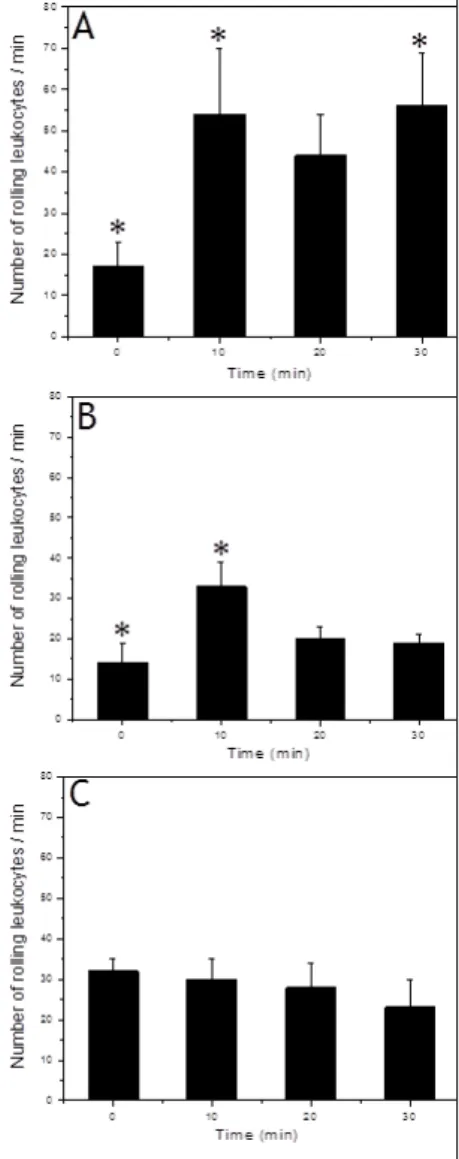
Intravital microscopyIt was observed that the administration of free BV-PBS increased markedly the leukocyte rolling flux in the microcirculatory tissue, about three times more circulating cells than normal (Figure 3 A). The empty PLGA MS (34 kDa-COOH) were nontoxic to mice (Figure 3 B) because maintained the leukocyte rolling flux close to the basal. When BV was administered encapsulated in MS-PLGA on the cremaster tissue, no change was observed in the number of circulating leukocytes (Figure 3 C), not even the initial increase in the number of circulating leukocytes, as observed with the empty MS. BV-MS-PLGA formulation was able to protect the organism from the myotoxic action of BV and inflammatory reaction (Figure 3 C). It was concluded that the MS protected the body against effects of direct contact with BV.
Figure 3: Rolling leukocytes in function of time. The (A): free BV; (B): MS-PLGA or (C): BV-loaded MS were added to mice cremaster muscle postcapillary venules (to observe leukocyte rolling as inflammatory marker). Results were mean ± S.E.M. from
three experiments (n = 3); (*) p<0.05, relative to values in the zero time (basal condition)
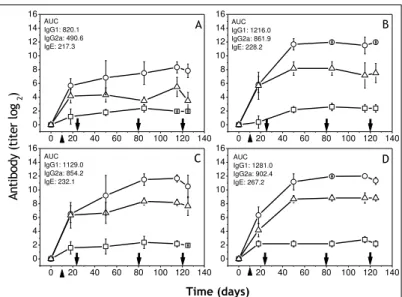
Adjuvant effects of BV loaded MS-PLGA in immunoglobulin productions
It is interesting to remind that the MS-PLGA VIT consisted of six injections with the different formulations. In
all treated mice with free BV or encapsulated within MS-PLGAs a higher production of IgG1 was observed,
showing that BV encapsulated retained their integrity and subsequently ability to trigger antibody production. However, the production rate was higher in the group that received BV within MS carboxy-terminal free
(angular coefficient: θ = 10) than the group treated BV within MS carboxy-terminal methylated (θ = 8) (Figure
4). This difference found in the velocity of antibody production was also observed for IgG2a. In this case, the
group treated BV within MS carboxy-terminal free (θ = 2) was much lower than the group immunized with BV
within MS carboxy-terminal methylated (θ = 6). The most interesting was the total absence of IgE, even after
203
Figure 4: Isotypes profiles of anti-BV serum antibodies in function of time and different formulations. The mice were injected with (A) BV-PBS; (B) BV-MS-12 kDa-COOH; (C) BV-MS-34 kDa-COOH; (D) BV-MS-63 kDa-COOH; (E) BV-MS-12 kDa-COOCH3;
(F) BV-MS-34 kDa-COOCH3 or (G) BV-MS-63 kDa-COOCH3. Frequently the blood was collected and (o) IgG1; (∆) IgG2a; (□) IgE
measured. Results are presented as mean ± S.E.M. from five animals (n = 5); Arrows (from left to right) means sensitization, treatment starting, treatment ending, and BV challenge, consecutively
Effect of reduced number of injections of BV loaded MS-PLGA in VIT
To study the influence of diminishing the number of injections in the VIT MS-PLGA mice were injected with two times (except the control with BV-PBS that received 6 injections) with the different formulations. All groups produced a homogeneous response of antibodies 20 days after sensitization. Along the treatment, the
magnitude of IgG1 and IgG2a production was quite similar in the groups receiving BV within the MS (Figure 5
C-D) and in the group which received BV-PBS (representing the control or the traditional VIT) (Figure 4 B).
After immunotherapy treatment with BV-MS, they achieved an antibody titer (log2) of 12 and 8 for IgG1 and
IgG2a, respectively, with the great advantage of smaller number of injections (2 in contrast with 6 for free
BV-PBS). This was reached without necessarily reducing the dose of antigen administered (Figure 5). Although, the control group, after sensitization, also produced antibody against BV, its titers were quite lower when compared with the BV-MS-PLGA (Figure 5). Another advantage of the treatment with BV-MS-PLGA was a reduced stimulus for IgE production (highly anaphylactic), when compared with BV-free, holding IgE production below
titer 2 (log2) (Figure 5).
Figure 5: Isotypes profiles of anti-BV serum antibodies in function of time. Mice were injected with (A) PBS; (B) PBS; (C) BV-MS-12 kDa-COOH; (D) BV-MS-63 kDa-COOCH3where (o) IgG1; (∆) IgG2a; (□) IgE. Results are presented as mean ± S.E.M. from
six animals (n = 6); Arrows (from left to right) means sensitization, treatment starting, treatment ending, and challenge, consecutively. AUC means area under curve and it is useful to compare antibody production among different treatments.
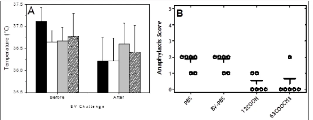
Hypersensitive reactions after BV challenge
In mice subjected to the second protocol of VIT, the changes in body temperature were observed after subcutaneous challenge with 50 µg BV / animal. The groups that received treatment with BV encapsulated
0 30 60 90
0 2 4 6 8 10 12 14 16
0 30 60 90
0 2 4 6 8 10 12 14 16
0 30 60 90
0 2 4 6 8 10 12 14 16
0 30 60 90
0 2 4 6 8 10 12 14 16
0 30 60 90
0 2 4 6 8 10 12 14 16
0 30 60 90
0 2 4 6 8 10 12 14 16
0 30 60 90
0 2 4 6 8 10 12 14 16 A n ti b o d y (t it e r Lo g2 ) A B E C Time (days) F D G
0 20 40 60 80 100 120 140 0 2 4 6 8 10 12 14 16
0 20 40 60 80 100 120 140 0 2 4 6 8 10 12 14 16
0 20 40 60 80 100 120 140 0 2 4 6 8 10 12 14 16
0 20 40 60 80 100 120 140 0 2 4 6 8 10 12 14 16 AUC IgG1: 820.1 IgG2a: 490.6 IgE: 217.3 A AUC IgG1: 1216.0 IgG2a: 861.9 IgE: 228.2 B AUC IgG1: 1129.0 IgG2a: 854.2 IgE: 232.1 C A n ti b o d y (t it e r lo
g 2
204
within MS prepared with polymers of 12-kDa-COOH or 63 kDa-COOCH3, were more resistant to the challenge.
This was verified by lower differences in the body temperature of these mice forty minutes after BV challenge (Figure 6 A). Systemic reactions after BV challenge were analyzed by clinical score of anaphylaxis (Li et al., 1999), which were less visible in the groups treated with BV-MS-PLGA as compared to control groups (only PBS) or free-BV in PBS (Figure 6 B).
Figure 6: Systemic reactions after BV challenge analysis through clinical score of anaphylaxis (A): Effect of BV challenge on body temperature of mice treated with different formulations: (black) PBS; (white) PBS; (gray) MS-12 kDa-COOH; (dense) BV-MS-63 kDa-COOCH3; (B) Anaphylaxis symptoms after 40 minutes of challenge: 0 = no symptoms; 1 = scratching and rubbing
around the nose and head; 2 = puffiness around the eyes and mouth, pilar erecti, reduced activity, and/or decreased activity with increased respiratory rate; 3 = wheezing, labored respiration, and cyanosis around the mouth and the tail; 4 = no activity after
prodding or tremor and convulsion; and 5 = death.
DISCUSSION
Microspheres or microparticles prepared with PLGA polymers (MS-PLGA) have been widely used in many pharmaceutical studies for a broad spectrum of applications in order to improve the efficacy of long-term treatments using drugs and other molecules, such as therapeutic proteins. Proteins are used for therapeutic purposes in many diseases, including hormonal and metabolic disturbances, for instance, for the treatment of growth hormone deficiency and diabetes with recombinant GH and insulin, respectively [37-38]. Additionally, the use of MS-PLGA for antigen delivery for immunological purposes has also been extensively studied. In this field, one of its main applications is as a carrier in vaccine development [39]. Regardless of the application, particulate systems are a promising tool for pharmaceutical uses because they can protect molecules from degradation in the body and promote their controlled delivery inside the organism. Based on these properties, we have investigated the use of MS-PLGA as a pharmaceutical system in order to replace the multiple injections that comprise traditional allergy or venom immunotherapy (VIT), also preventing direct contact between the allergen and the body.
Pharmacotechnical parameters, such as the efficiency of encapsulation, size, morphology and release profile, among others, are important factors contributing to the development of better systems for medical applications. The physicochemical properties of the BV-MS-PLGA formulations used in this study were described and fully discussed in a previous report by our group [23]. Therefore, these properties are not explored here again, since the focus of this study was on the biological application of BV-MS-PLGA formulations.
In the field of allergen vaccines, i.e. VIT, the strategy of encapsulating molecules can improve the safety and efficacy of such treatments because they release the antigen over prolonged periods of time [3, 17, 31, 35-36] and thereby reduce the need for frequent booster injections, as observed in the results of this study; this may also improve patient compliance. However, besides these beneficial effects of PLGA microspheres, it is also important to note the pharmacological rationale underlying therapeutic efficiency of VIT. It is known that allergy is a biological process that polarizes the immunological response towards Th2, frequently initiated by the sensitization of an organism to an allergen, and consequently inducing high production of IgE-class antibodies [9, 54-55]. IgE antibodies are specific molecules produced by B-lymphocytes when these cells are stimulated by mediators released by T-lymphocytes in the course of a Th2 immunological response, following contact with a protein-based allergen [9; 40, 55]. Once produced, IgE antibodies have the ability to bind to their receptor on mast cell surfaces, rendering them ready to respond specifically to allergen contact via cell degranulation and the rapid stimulation of inflammatory reactions [40]. The production of IgE antibodies as well
as their receptor (FcƐR) on mast cells is regulated by positive feedback; as new allergen molecules specifically
bind to their corresponding IgE on mast cells, more IgE and FcƐR are produced, which in turn exacerbate the
inflammatory response [41].
205
response toward Th1. One of these principal strategies is to prevent the binding of allergens to IgE on the mast cell surface, which in turn leads to the downregulation of IgE by negative feedback [41]. Another strategy is to allow allergens to be rapidly phagocytosed by tissue macrophages, which theoretically could direct the immunological response toward Th1 [40, 42]. This therapeutic rationale is the basis of VIT, i.e. repeated administration of very small quantities of allergen, aiming at rapid engulfment by phagocytic cells in order to prevent subsequent triggering of allergic reactions.
In the present study, it was demonstrated that BV proteins encapsulated within MS-PLGA can be delivered intracellularly due to rapid phagocytosis of these particles by J774 macrophages. It has been demonstrated by Bitencourt and colleagues [43] that even empty particles of PLGA are rapidly recognized by macrophages, which in turn stimulates their rapid phagocytosis. Nevertheless, in the present work, also reported by other authors, it was found that when particles or microspheres contain proteins, antigens or even other inflammatory molecules inside or bound to their surfaces, the stimulation of phagocytosis is stronger [42]. Similar results have been previously reported in the literature; for example, Nicolete and colleagues [28] have described that
microspheres of PLGA containing LTB4, an inflammatory lipid mediator, more robustly stimulate phagocytosis
compared to empty PLGA, with a phagocytic index of 13.3% and 3.3%, respectively. Dos Santos and colleagues [27] have also reported that the phagocytic index was much higher (208.37%) when macrophages were placed
in contact with PLGA containing cell-free antigens from Histoplasma capsulatum (CFAgs) plus LTB4
(LTB4/CFAgs-loaded MS) compared to unloaded PLGA (27.43%). The precise mechanisms by which
microspheres or microparticles interact with macrophage membranes and stimulate rapid phagocytosis are not fully explained yet. It is likely that the presence of molecules on the surface of microspheres stimulates the phagocytic process by binding to an unspecific membrane receptor. This is an important factor favoring the use of PLGA particles to delivery allergens in immunotherapy, since these systems can be rapid and efficiently engulfed by macrophages, removed from the local environment, and consequently directly presented to T-lymphocytes. Furthermore, the entrapped allergen would be less accessible for binding IgE on the surface of mast cells and basophils, which in turn may also protect the allergic patient from their effects [31].
In this work, the inflammatory stimulus was evaluated by assessing the release of NO from macrophages after they engulfed MS-PLGA or BV-MS-PLGA. Although a slight increase in NO production was observed following phagocytosis of MS-PLGA or BV-MS-PLGA compared to baseline levels produced by macrophages in RPMI medium, such differences were not significant, which shows that this delivery system would be innocuous to the organism. Here, it is worth emphasizing that some authors have stated that low levels of NO production by macrophages may promote Th1 differentiation [44], thus supporting the use of MS-PLGA in allergen immunotherapy. Still, regarding the levels of NO production observed in this study, Dos Santos and colleagues [45] have also reported that unloaded MS-PLGA can slightly stimulate NO production, but to a lesser extent production in response to loaded MS-PLGA. In terms of macrophage activation, some authors have
reported that even unloaded MS-PLGA can stimulate the production of inflammatory cytokines, such as TNF-α.
However, such stimuli are less intense compared to activation triggered by lipopolysaccharide (LPS), which is a potent inflammatory molecule derived from bacterial cell walls [43].
The phagocytic index, which represents the ability to stimulate phagocytosis, and consequently deliver molecules to the intracellular environment, is correlated with leukocyte influx to the site of injection; this is one of the initial cellular steps of inflammatory reaction [46, 47]. Our results show that when BV proteins are injected directly into the tissue and the microcirculation is exposed, this provided a strong stimulus to promote the influx of leukocytes to the site of injection (cell migration), whereas BV proteins encapsulated in MS-PLGA did not induce changes in the number of cells arriving at the site of injection. Although it was also observed that MS-PLGA produced slight changes in leukocyte influx when following contact with the microcirculation, these changes were transient rather than sustained. In fact, any substance that comes into contact with the tissues can initially trigger a slight perturbation in tissue homeostasis. Importantly, this phenomenon is not sustained and is rapidly resolved; thus, it does not induce a strong systemic reaction. Similar results have been found by Choe and colleagues [48], who demonstrated that empty MS-PLGA does not increase macrophage trafficking, but MS-PLGA loaded with LPS triggered robust and rapid influx of macrophages. However, it must be mentioned that LPS is a substance derived from bacterial cell walls and is a well-known inflammatory stimulus. A transient increase in the number of rolling leukocytes was also observed by Rücker and colleagues [49], who observed the same phenomenon after dorsal implantation of PLGA followed by studies on microhemodynamics. Summarizing this series of experiments, the tissue was protected from side effects, particularly from an exacerbated inflammatory stimulus caused by direct contact with an allergen (in this case BV proteins) since the allergen was encapsulated within MS-PLGA.
206
Here, we hypothesized that BV proteins encapsulated within MS-PLGA would provide the same stimulus in terms of antibody production and immunological response as free BV, but with no overreactions, which can be ultimately characterized by anaphylaxis. In mouse models, some classes of antibodies are more important in the
allergic response. For instance, high levels of IgG1a or IgE have been associated with the development of
allergic symptoms, and it is believed that they drive the immunological response with a Th2 profile; on the other
hand, IgG2a antibodies have been associated with the stimulation of an immunological response with a Th1
profile [51-52].
Although it was not possible to fully characterize or associate antibody production specifically to BV allergy in this study, we have shown that free BV or BV encapsulated within MS-PLGA (with different PLGAs) achieved sustained responses in terms of antibody production. In some cases, antibody production was not quantified in the first few days after sensitization, but in general, within thirty days of sensitization, all mice produced
moderate levels of specific antibodies. Except for BV-MS-PLGA 63 kDa-COOCH3, which produced only high
levels of IgG1, all other experimental groups had high levels of IgG1 and IgG2a. It is important to emphasize that
stimulation to IgG2a production indicates immunological maturity towards a Th1 response, probably via the
action of interferon-γ, whereas high levels of IgG1, although it plays a key role in the rapid neutralization of
antigens, still indicates a response polarized toward Th2. These results are in agreement with those of Schöll and colleagues [53] showing that antibody production after the immunization of mice with the major birch pollen
allergen Bet v1 encapsulated in particles of PLGA led to high levels of IgG1 and IgG2a production. Batanero and
colleagues [13] showed a complete shift from IgG1 to IgG2a production after the immunization of mice with
olive allergen encapsulated within PLG microparticles or adsorbed onto Al(OH)3. The adjuvant Al(OH)3 is a
well-known immunogenic substance that acts as a potent stimulus driving the development of a Th2 immune response with IgE production [53, 56].
Circulating antibodies, especially IgG1, are excellent antigen neutralizers and can rapidly protect the organism
from the deleterious effects triggered by direct contact with allergen proteins. This was confirmed by the fact that no anaphylactic reactions were observed when mice were challenged with a high dose of BV proteins. Therefore, we believe that the absence of BV toxicity was a direct consequence of BV neutralizing antibody production, along with the absence of IgE. These results demonstrate that BV encapsulated within MS-PLGA can provide the same stimulus to antibody production as free BV. Furthermore, the presence of high levels of specific antibodies reveals that BV proteins retain their integrity when they are released from MS-PLGA. Similar results were found when the number of injections was reduced for BV-MS-PLGA in the second VIT protocol compared to free BV. This strategy was employed to verify the sustained release of BV proteins from MS-PLGA compared to sequential injections of free BV. Briefly, we observed excellent performance of the treatment with fewer injections, which provided the same results as traditional VIT.
Since, in our VIT protocol, BV allergy was not well characterized or associated with IgE production, but only
led to specific IgG1 production (associated with allergic symptoms), we have also followed the development of
clinical reactions that resemble allergic symptoms. The most important clinical signs that can be used for that characterization of allergic reactions in mice are body temperature and clinical signs such as pilar erecti, reduced activity and prostration [32]. These events were monitored in mice challenged with a high dose of BV proteins; no signs related to allergic anaphylaxis were detected, indicating the benefits obtained from the encapsulation of BV within microspheres.
Another crucial aspect of this study is that the benefits obtained by the use of the BV-MS-PLGA formulations did not require the use of co-immunostimulatory molecules or any other adjuvant. Some authors have described allergic reactions using co-immunostimulatory molecules such as oligodeoxynucleotide (CpG) [31], which may lead the organism to be more prepared to respond against a specific antigen. In experimental studies using animals, this strategy represents a good alternative to fully characterize allergic reactions, but it is also well-known that the concomitant injection of different types of molecules can stimulate variable responses; obviously, in terms of clinical trials, this is not desirable.
To the best of our knowledge, the results presented in this study represent the first report using MS-PLGA as strategy to achieve sustained delivery of immunogenic BV proteins to the organism without the development of a strong inflammatory reaction. This formulation is especially intended to treat bee venom allergy. However, it is worth noting that, to better characterize the humoral immunological response, the kinetics of antibody production should be monitored after MS-PLGA injection to better describe this phenomenon. Furthermore, cytokines that promote a Th1 or Th2 profile should also be monitored during treatment. Additionally, detailed studies to clarify the specific interaction between MS-PLGA and phagocytes is of considerable interest, since there is conflicting information in the literature regarding the ability of MS-PLGA to trigger the activation of these cells and present engulfed antigens [43, 50].
207
Under our experimental conditions, it was possible to conclude that the encapsulation of BV proteins within MS-PLGA prevented systemic inflammatory reactions and promoted allergen delivery directly into the intracellular environment. Additionally, the injection of BV-MS-PLGA also prevented signs of anaphylaxis in vivo. Furthermore, these BV-MS-PLGA formulations can reduce the number of injections needed for venom immunotherapy with no concomitant reduction in antibody production, which also indicates the maintenance of BV protein integrity inside the particles. Therefore, a therapeutic strategy using MS-PLGA formulations may provide a promising and safe controlled delivery system to encapsulate BV for use in VIT.
Highlights
MS-PLGA represents an alternative allergen carrier for venom immunotherapy. BV-MS-PLGA is rapidly phagocytized by macrophages.
BV-MS-PLGA retains its integrity and ability to induce BV-specific antibody production.
ACKNOWLEDGEMENTS
We would like to thank to FAPESP (05/04514-2; 02/07293-9 and 00/10970-7) and Fundação Butantan for financial supports. M.H. Bueno da Costa has a CNPq Science Productivity fellowship (302047/2008-5) and R.A. Trindade was supported by a doctorate fellowship from CNPq (141052/2009-0 and 140974/2010-5).
Authors’ contribution: RAT and MHBC conceived the initial project; MHBC obtained financial support;
RAT, MHBC and OABES designed all immunological assays with animals (VIT protocols); RAT, OABES and VCR performed serological assays; RAT, FMB and MVALF performed intravital microscopy assays; RAT, DFS, RN and LHF performed macrophage assays; RAT, PSA and MHBC analyzed all data. RAT and MHBC wrote the paper. All authors read and approved the final manuscript.
Conflict of interest statement: The authors have declared no conflict of interest.
REFERENCES
[1] J Ring; B Eberlein-Koenig; H Behrendt. Ann Allergy Asthma Immunol, 2001, 87, 2-6.
[2] R Pawanker; GW Canonica; ST Holgate; RF Lockey. White Book on Allergy 2011-2012 Executive
Summary. World Health Organization, 2011.
[3] S Jilek; E Walter; HP Merkle; B Corthesy. J Allergy Clin Immunol, 2004, 114, 943-950.
[4] CJ Steen; CK Janniger; SE Schutzer; RA Schuwartz. Int J Dermat, 2005, 44, 91-94.
[5] EC Mingomataj; AH Bakiri; A Ibranji; GJ Sturm. Clin Rev Allergy Immunol, 2014, 47(1), 91-99.
[6] R Pawankar; CE Baena-Cagnani; B Bousquet; GW Canonica; AA Cruz; MA Kaliner; BQ Lanier. WAO
Journal 2008, S4-S17.
[7] UR Muller. Curr Opin Allergy Clin Immunol, 2003, 3, 299-303.
[8] D Antolín-Amérigo; CM Aguilar; A Vega; M Alvarez-Mon. Curr Allergy Asthma Rep, 2014, 14, 449.
[9] D Venarske; RD deShazo. South Med J, 2003, 96, 1049-1054.
[10] SJ Till; JN Francis; K Nouri-Aria; SR Durham. J Allergy Clin Immunol, 2004, 113, 1025-1034.
[11] SR Durham; SM Walker; EM Varga; MR Jacobson; FO Brien; W Noble; SJ Till; QA Hamid; QT
Nouri-Aria. N Engl J Med, 1999, 341, 468-475.
[12] RD Peseka; RF Lockey. Curr Opin Allergy Clin Immunol, 2014, 14, 340-346.
[13] E Batanero; P Barral; M Villalba; R Rodriguez. J Control Rel, 2003, 92, 395-398.
[14] J Stoevesandt; C Hosp; A Kerstan; A Trautmann. Dtsch Dermatol Ges, 2014, 12(3), 244-255.
[15] S Freiberg; XX Zhu. Int J Pham, 2004, 282, 1-18.
[16] W Jiang; RK Gupta; MC Deshpande; SP Schwendeman. Adv Drug Del Rev, 2005, 57, 391-410.
[17] V Saez; JR Hernández; C Peniche. Biotecnol Aplic, 2007, 24, 108-116.
[18] C Thomasin; G Corradin; Y Men; HP Merkle; B Gander. J Control Rel, 1996, 41, 131-145.
[19] JL Cleland. Res Immunol, 1998, 149, 45-47.
[20] M Manish; A Rahi; M Kaur; R Bhatnagar; S Singh. PLoS One, 2013, 29:8(4), e61885.
[21] Y Waeckerle-Men; M Groettrup. Adv Drug Del Rev, 2005, 57, 475-482.
[22] RA Trindade; PS Araujo; MH Bueno da Costa. Drug Deliv Lett, 2012, 2, 54-59.
[23] RA Trindade; PK Kiyohara; PS Araujo; MH Bueno da Costa. Int J Pharm, 2012, 423, 124-133.
[24] R Langer. Science, 1990, 249, 1527-1533.
[25] JAM Namur; ECM Cabral-Albuquerque; W Quintilio; MHA Santana; MJ Politi; PS De Araujo; AC
Lopes; MH Bueno da Costa. J Nanosci Nanotech, 2006, 6, 2403-2407.
208
[27] DF Dos Santos; R Nicolete; PRM De Souza; CS Bitencourt; RR Dos Santos Jr.; VLD Bonato; CL
Silva; LH Faccioli. Eur J Pharma Sci, 2009, 38, 548-555.
[28] R Nicolete; KM Lima; JMR Junior; MD Baruffia; AI Medeiros; MVLB Bentley; CL Silva; LH
Faccioli. Prostag Oth Lipid M, 2007, 83, 121-129.
[29] S Shigematsu; S Ishida; DC Gute; RJ Korthuis. Am J Physiol, 1999, 277, H161-H171.
[30] B Lomonte; JM Gutiérrez. Ver Biol Tropical, 1983, 31, 37-40.
[31] JM Martinez-Gómez; S Fischer; N Csaba; TM Kiindg; HP Merkle; B Gander; P Johansen. Pharm Res,
2007, 24, 1927-1935.
[32] OM Poulsen. Animal models for the study of allergy. In: Hau, J, editor. Handbook of Laboratory
Animal Science: Animal models. 1st edition Boca Raton: CRC Press, 1994, 2:29-41.
[33] XM Li; BH Schofield; CK Huang; GI Kleiner; HA Sampson. J Allergy Clin Immunol, 1999, 103,
206-214.
[34] L Navuluri; S Parvataneni; H Hassan; NP Birmingham; C Kelly; V Gangur. Int Arch Allergy Immunol,
2006, 140, 270-276.
[35] S Gómez; C Gamazo; B San Roman; A Grau; S Espuelas; M Ferrer; ML Sanz; JM Irache. J Immunol
Method, 2009, 348, 1-8.
[36] A Taluja; YS Youn; YH Bae. J Mater Chem, 2007, 17, 4002-4014.
[37] Y Capan; G Jiang; S Giovagnoli; KH Na; PP De Luca. AAPS Pharm Sci Techol, 2003, 4, 147.
[38] G Sharma; AR Sharma; J Nam; GPC Doss; SS Lee; C Chakraborty. J Nanobiotechnol, 2015, 13, 74.
[39] AL Silva; PC Soema; B Slütter; F Ossendorp; W Jiskoot. Hum Vaccin Immunother, 2016, 12(4),
1056-69.
[40] M Jones. Methods Mol Med, 2008, 138, 1-15.
[41] OT Burton; HC Oettgen. Immunol Rev, 2011, 242(1), 128-143.
[42] HR Nouri; A Varasteh; MR Jaafari; JM Davies; M Sankian. Immunol Res, 2015, 62, 280-291.
[43] CS Bitencourt; LB Silva; PAT Pereira, GM Gelfuso; LH Faccioli. Colloids Surf B Biointerfaces, 2015,
136, 678-686.
[44] W Niedbala; XQ Wei; D Piedrafita; D Xu; FY Liew. Eur J Immunol, 1999, 29, 2498-2505.
[45] DF Dos Santos; CS Bitencourt; GM Gelfuso; PAT Pereira; PRM De Souza; CA Sorgi; R Nicolete; LH
Faccioli. Eur J Pharm Sci, 2011, 44(5), 580-588.
[46] SR Zamuner; CFP Teixeira. Mediators Inflamm, 2002, 11, 351-357.
[47] K Newton; VM Dixit. Cold Spring Harb Perspect Biol, 2012, 4, a006049.
[48] SW Choe; AP Acharya; BG Keselowsky; BS Sorg. Acta Biomaterialia, 2010, 6(9), 3491-3498.
[49] M Rücker; MW Laschke; D Junker; C Carvalho; A Schramm; R Mülhaupt; NC Gellrich; MD Menger.
Biomaterials, 2006, 27(29), 5027-5038.
[50] A Luzardo-Alvarez; N Blarer; K Peter; JF Romero; C Reymond; G Corradin; B Gander. J Control Rel,
2005, 109(1-3), 62-76.
[51] A Oshiba; E Hamelmann; K Takeda; KL Bradley; JE Loader; GL Larsen; EW Gelfand. J Clin Invest,
1996, 97(6), 1398-1408.
[52] F Salari; AR Varasteh; F Vahedi; M Hashemi; M Sankian. Int Immunopharmacol, 2015, 29, 672-678.
[53] I Schöll; A Weissenbo; E Förster-Waldlz; E Untersmayr; F Walter; M Willheim; G Boltz-Nitulescu; O
Scheiner; F Gaborw; E Jensen-Jarolim. Clin Exp Allergy, 2004, 34, 315-321.
[54] S Romagnani. Ann Allergy Asthma Immunol, 2000, 85(1), 9-18.
[55] P Kids. Med Rev, 2003, 8(3), 223-246.