Document heading doi: 10.12980/JCLM.3.2015APJTB-2014-0364
©2015 by the Journal of Coastal Life Medicine. All rights reserved.
Isolation, purification and characterization of extracellular protease produced by marine-derived
endophytic fungus
Xylaria psidii
KT30
Bugi Ratno Budiarto
1,2, Apon Zaenal Mustopa
2, Kustiariyah Tarman
1* 1Department of Aquatic Products Technology, Faculty of Fisheries and Marine Sciences, Bogor Agricultural University, Bogor, Indonesia
2
Research Center for Biotechnology, Indonesian Institute of Sciences (LIPI), Indonesia
Journal of Coastal Life Medicine
*Corresponding author: Kustiariyah Tarman, Department of Aquatic Products Technology, Faculty of Fisheries and Marine Sciences, Bogor Agricultural University, Indonesia.
Tel: +62-251-8622915 E-mail: [email protected]
Funding Project: Supported by Ministry of Research and Technology, Republic of Indonesia (Grant No. RD-2013-0552).
1. Introduction
Fungal endophytes currently gain much attention in research in terms of their ability to produce diverse bioactive compounds that brings benefit in curing and preventing the emerging human diseases such as cancer and infectious diseases[1]. This group of microfungi is considered as one of factors that contribute in production and accumulation of many kinds of novel, unique and structurally broad spectrum of active metabolites in their hosts’ habitats. This natural phenomenon occurrs in fact that fungal endophytes tie a
symbiotic mutualism with host and their interaction mostly depends on virulence and defense equilibrium among fungi, host and pathogens[2].
Marine habitats are believed to be more rich in diversity compared to terrestrial environment that compose exclusively ecological interactions between microbiotas with their microbiome and create one of the best and plenty sources for fungal endophytes-derived novel bioactive compounds to be exploited[3]. Xylaria psidii (X. psidii) KT30 is one of marine-derived fungal endophytes with great potency as source for bioactive compounds. Ethyl acetate extract
PEER REVIEW ABSTRACT
K E Y W O R D S
Serine protease, Xylaria psidii KT30, Bioactive molecule
Objective: To isolate, purify and characterize extracellular protease produced by Xylaria psidii
(X. psidii) KT30.
Methods:In the present study, the extracellular protease secreted by X. psidii KT30 was isolated and purified by using three steps of protein purification, then the purified protease was characterized by applying qualitative and quantitative enzymatic assays.
Results:Extracellular protease with molecular mass 71 kDa has been purified successfully by applying diethylaminoethanol-Sepharose followed by sephadex SG75 with its final specific protease activity of 0.091 IU/mg. Protease was the most active at temperature 60 °C and pH 7. The activity of enzyme was abolished mostly by phenylmethanesulfonyl fluoride, showing it is family of serine protease.
Conclusions:Extracellular serine protease produced by X. psidii KT30 with good biochemical properties displayed some promising results for its further application in field of biotechnology or medicine.
Peer reviewer
Dr. Gabriella Caruso, Researcher at the Institute for Coastal Marine E nv i r o n m e n t , I t a l i a n N a t i o n a l Research Council (CNR-IAMC), Messina, Italy.
Tel: +39-090-6015423
E-mail: [email protected]
Comments
This paper is an interesting study focusing on the extraction and biochemical characterization of an extracellular protease produced by the marine fungus Xylaria psidii KT30. The biochemical properties displayed by this molecule suggest its promising application in the field of biotechnology or medicine, o b v i o u s l y f o l l o w i n g f u r t h e r experimentation.
Details on Page 61
Article history: Received 14 Aug 2014
Received in revised form 21 Aug 2014 Accepted 25 Sep 2014
of X. psidii KT30 broth culture showed antibacterial activity mostly against Gram-negative bacteria and some fish photogenic bacteria, and induced cytotoxicity against human bladder carcinoma cell line 5637[4]. Yet until now, the main active compound responsible for biological activity of X. psidii KT30 is still undergoing investigation. In this study, we try to reveal this active compound mainly focusing on proteases. The proteolytic enzymes have gained much attention to be explored due to their numerous biological activities such as antivirus[5], antibacterial[6], and anticancer activities[7].
Recently, almost all researches in novel bioactive compounds purified from fungal endophytes focused on non-protein secondary metabolites pathways-based new compounds isolations[8,9]; meanwhile, exploration of protein biomolecules as source for bioactive agents with high potency, especially from fungal endophytes, is still less attractive and this situation was sharp contrast to intensive research in investigating the new biomolecules with novel activity derived from macrofungi[10]. We do believe that biomolecules are more save compared to secondary metabolites, when applied as drugs or food preservatives in terms of their less toxicity effects and rapid metabolized by our body. In this study, we reported the isolation, purification and characterization of protease produced extracellulary by endophytic fungus X. psidii KT30.
2. Materials and methods
2.1. Materials
Culture of X. psidii KT30 was kindly obtained from Dr. Kustiariyah Tarman, Bogor Agricultural University, Indonesia. Diethylaminoethanol (DEAE)-cellulose was from Sigma Chemical Company, St. Louis, Missouri, USA. Sephadex SG-75 was from GE Heathcare Bio-Science, Uppsala, Sweden. Potato dextrose broth was from Himedia Laboratories Pvt. Ltd., India. Pierce bicinchoninic acid (BCA) protein assay kit and Pierce silver staining kit were from Thermo Scientific, USA. All other chemicals were purchased from Sigma Chemical Co.
2.2. Cultural condition
X. psidii KT30 was maintained on potato dextrose agar (PDA) at room temperature for 3-4 d at dark until the mycelia are mature enough to be recultured in liquid medium for extracellular proteases production. A mycelia, 0.5 mm×0.5 mm in length, was cultured in 300 mL of potato dextrose broth at 25 °Cwith 150 r/min for 10 d, then supernatant of the media was harvested for next experiment.
2.3. Purification of extracellular protease
Protein isolation was performed according to the protocol described by Wang et al. with slight modification[11]. Filtered-supernatant (crude extract) of 10-day X. psidii KT30 liquid culture was subjected to saturation using 90% solid ammonium sulfate, then resuspensed in 25 mmol/L Tris-HCl buffer (pH 7.4). Before applied on DEAE -Sepharose for first step purification, resuspended pellet of crude extract was firstly dialyzed in dialysis tubing (23 mm×15 mm, Sigma, molecular mass cut-off 14 kDa) using the same buffer for 24 h at 4 °C with gently agitation, and the buffer was replaced twice during dialysis process. The dialyzed crude extract was then applied onto DEAE-Sepharose column pre-equilibrated with 25 mmol/
L Tris-HCl buffer (pH 7.4). Crude protein-containing DEAE -Sepharose column was separated using gradient concentration of NaCl (0-0.8 mol/L) with flow rate fixed at 1 mL/min. Fractions showing protease activity were pooled and then applied onto a sephadex SG-75 column against 25 mmol/L Tris-HCl buffer (pH 7.4) at a flow rate of 1 mL/min. Fractions with protease activity was freeze-dried and resuspended in the same buffer up to 25% of the original volume. The presence of protein was determined by BCA method at the wavelength of 540 nm with mixing 10 µL of sample with working solution.
2.4. Determination of protein concentration
Protein contents in each samples were measured using Pierce BCA
protein assay kit as described in manual instruction. Bovine serum solution was used as standard proteins.
2.5. Directly extracellular protease assay on solid medium of
culture
Explants of X. psidii KT30 mycelium (0.5mm×0.5mm in size) was sliced aseptically from original stock, then placed on 0.1% gelatin-containing PDA. The solid culture was incubated for 14 d in the dark at room temperature. At Day 7 and 14, the clear zone formation around X. psidii KT30 was monitored.
2.6. Screening of protease using plate assay
Plate assay described by Hasan et al.[12] and More et al.[13] was adaptedwith minor modification. Protease activity in each purification steps was monitored through clear zone formation on gelatin-added agarose. About 30 mL of 0.5% agarose medium containing 0.2% of gelatin in 25 mmol/L Tris-HCl buffer (pH 7.4) was placed on a Petri disc. Sample (50 µL) was loaded into well then incubated for 24 h at 37 °C for enzymatic reaction. The development of clear zone around the wells was detected by applying Coomassie Blue (0.25% w/v) in methanol: acetic acid: water 5:1:4 (v/v/v) for 15 min at room temperature, followed by destaining step to remove staining solution using destain solution (66 mL methanol: 20 mL acetic acid: 114 mL H2O bidest) until the clear zone could be seen visually.
2.7. Protease activity assay
Activity of extracellular protease produced by X. psidii KT30 was measured using method developed by Cupp-Enyard with modification[14]. This method is based on the tyrosine formation during casein hydrolysis by protease and the tyrosine released was monitored at wavelength 540 nm. A total of 6 µL of test sample was firstly mixed with 6 µL phosphate buffer (pH 7.4, 25 mmol/L), then added with 6 µL of 1% (w/v) casein solution. The reaction mixture was incubated at 37 °C for 30 min. Following incubation, 12 µL of trichloroacetic acid was added to the mixture. After centrifugation at 12 000 r/min for 1 min, reaction mixture was added with 143 µL of reagent A [a mixture of Na2CO3 solution and CuSO4.5H2O solution
tyrosine released during enzymatic reaction per mL reaction mixture per minute under the experimental conditions.
2.8. Gel electrophoresis
Sodium dodecyl sulfate polyacrylamide gel electrophoresis ( SDS-PAGE) with 12% polyacrylamide gel was used to determine purity of protein as described by Laemmi[15]. Protein on gel was visualized using silver staining reagents according to the manual instruction. The band of protein was compared to the standard molecular mass of marker to determine its molecular mass.
2.9. Gelatin zymography
The method described by Raser et al.[16] and Kleiner and Stevenson[17] was adapted. As mentioned, this method is very powerful not only to detect protease activity but also to predict molecular mass of the target protein. Firstly, the sample was run on 0.2% gelatin-containing gel electrophoresis. After separation of protein, the protein was reactivated by incubating the gel in 2.5% Triton X-100 for 40 min at 37 °C. The third step is staining the gel in 0.05% Coomassie Blue solution for 2 h. The final step is removing the excess Coomassie Blue using destaining solution until clear band appeared on gel which indicates the protease activity.
2.10. Determination of molecular mass of target protein
Molecular mass of protease was predicted from the protein band appeared on gel electrophoresis by interpolation from a linear logarithmic plot of relative molecular mass against the Rf value
according to protocol explained by Martinez et al[18]. The Precise Plus Protein Dual-Xtra standards from BioRad Laboratories Inc. was used as reference of protein.
2.11. Biochemical characterization
The effect of temperature, pH, and protease inhibitor on protease activity and its enzyme kinetic was evaluated following method described by Zhang et al. with modification[19]. The optimum temperature was determined by carrying out the assay at different temperatures ranging from 20 °C to 90 °C with 10 °C interval. Meanwhile, the optimal pH for protease activity was determined within the pH range 4-10 using the following buffers: 0.1 mol/L citrate buffer (pH 4-6), 0.1 mol/L Tris-HCl (pH 7-9) and 0.1 mol/L glycine-NaOH (pH 10-11). For inhibition effect assay, sample was treated with either protease inhibitors phenyl methyl sulfonyl fluoride (PMSF) or ethylenediaminetetraacetic acid (EDTA). The residual enzyme activity was measured by performing the assay as described above respectively. The value of Km and Vmax was determined based
on the Lineweaver-Burk plot constructed by plotting the reciprocal of substrate concentration on the X-axis and reciprocal of the enzyme reaction velocity on the Y-axis by mixing sample with different concentration of casein ranging from 0.2%, 0.1%, 0.08%, 0.06%, 0.04%,and 0.02%.
3. Results
3.1. Assay of extracellular protease both directly on culture
and fraction
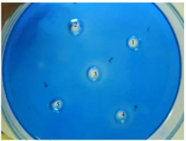
The activity of fungal extracellular protease was confirmed by directly observation on solid medium. Proteolytic activity was shown by clear zone formationsurrounding the active fungal growth as early as at Day 7 of incubation (Figure 1A). Plate assay showed protease activity particularly in resuspensed-pellet of crude extract after ammonium sulfate precipitation, and also both in undialyzed and dialyzed sample (Figure 2).
A B C
D
Figure 1.X. psidii KT30 on PDA medium.
A: At Day 7, clear zone formation of X. psidii KT30 on PDA medium containing 0.1% gelatin upon light exposure; B: At Day 14, clear zone formation of X. psidii KT30 on PDA medium containing 0.1% gelatin upon light exposure; C: At Day 7, X. psidii KT30 on PDA medium without gelatin upon light exposure; D: Culture of X. psidii KT30 with no direct exposure to light. Left is medium added with 0.1% gelatin and right is gelatin-free medium.
Figure 2. Results of plate assay.
1: Crude extract before ammonium sulfate precipitation; 2: Supernatant of crude extract after ammonium sulfate precipitation; 3: resuspensed-pellet of crude extract after ammonium sulfate precipitation and before dialysis treatment; 4: resuspensed-pellet of crude extract after dialysis treatment; 5: 25 mmol/L Tris-HCl with pH 7.
1 2
3
3.2. Purification of extracellular protease X. psidii KT30
Extracellular protease from X. psidii KT30 has been successfully purified from supernatant of 10-day medium culture by ammonium
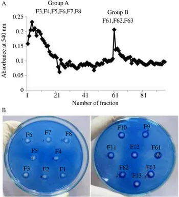
sulfate precipitation, DEAE-Sepharose column chromatography and sephadex SG75 column chromatography. Figure 3A exhibits two dominant peaks when anion exchanger column was eluted by gradient concentration of NaCl ranging from 0-0.8 mol/L and
designed as Group A and Group B. Plate assay was applied on both groups to confirm the activity of protease in each fraction. As shown in Figure 3B only fractions on Group A showed dominant clear zone formation. Base on this result, the Group A was chosen to be
applied in next step of purification. In the third step of purification by using sephadex-SG75, in total 40 fractions collected, only 7 fractions showed high peak when measured at wavelength 540 nm; and from those fractions only one fraction exhibited protease activity,
SG75-F14, (Figure 4B and Figure 5A), which was detected by using gelatin zymography with molecular mass 43 kDa. Yet, this molecular mass was bigger when purified protein was separated on SDS-PAGE,
showing single appearance, although the band appeared was very
thin (Figure 5B). This protein band resulted from the final steps of purification was also detected on SDS-PAGE sample of Group A DEAE -column result. On the other hand, proteins with molecular mass above 71 kDa that appeared in second steps of purification disappeared after
separated using sephadex-G75, indicating success of overall protein purification steps. Furthermore, the purity and protease specific activity of purified enzyme after all process of purification steps was 1.182 fold and 0.091 IU/mg respectively (Table 1).
Figure 3.Chromatography profile and protease activity screening.
A: DEAE-Sepharose chromatography profile of dialyzed-crude preparation; B: Protease activity screening on gelatin-containing agarose of samples resulted from anion exchange chromatography for purification.
0.25 0.2 0.15 0.1 0.05 0
Absorbance at 540 nm
A
B
F8
F9 F10
F11 F12 F61
F62 F63
F13 F7
F6
F5 F4
F3 F2 F1
1 21 41 61 81
Number of fraction Group A
F3,F4,F5,F6,F7,F8 Group B F61,F62,F63
Figure 4.Sephadex-G75 chromatography profile and gelatin zymogram
result.
A: Sephadex-G75 chromatography profile of Group A purified from DEAE-column chromatography; B: Gelatin zymogram result of protease activity in each fraction. M: Protein marker.
A
B
Number of Fraction
M SG75 SG75 SG75 SG75 SG75 SG75 E10 F12 F13 F25 F26 F328 0.030
0.025
0.020
0.015
0.010
0.005
0
Absorbance at 540 nm
E1 E3 E5 E7 E9 1 3 5 7 9 11 13 15 17 19 21 23 25 27 29
E10
F28 F25 F13
F14 F12
F26
Figure 5.Gelatin zymogram and SDS-PAGE result.
A: Gelatin zymogram of SG75-F14; B: SDS-PAGE result of SG75-F14, DEAE fractionation of Group A, dialyzed sample and unconcentrated crude extract. M: Protein marker.
SG75 SG75
M
A B
M DEAE
F2-F8
Dialysed sample
Crude extract
F14 F14
250 kDa 150 kDa 100 kDa 75 kDa
50 kDa
37 kDa
25 kDa
20 kDa
Table 1
Summary of purification of extracellular protease X. psidii KT30. Protease in different
purification steps
Volume (mL)
Total activity
(IU)
Protein (mg)
Specific activity (IU/mg)
Yield (%)
Purity (fold) Crude extract 300.00 14.400 186.200 0.077 100.000 1.000 Ammonium sulfate 2.00 0.205 5.697 0.036 1.424 0.467 DEAE-Sepharose 1.00 0.013 0.245 0.054 0.090 0.702 Sephadex-SG75-F14 0.25 0.006 0.066 0.091 0.042 1.182 Protease activity was assayed at basal condition with temperature at 37 °C and pH of 7.4. The yeild (%) of purified protein was calculated by dividing the total activity of protease in each fraction by total activity of protease in crude extract; meanwhile the purity was calculated by dividing specific protease activity in each fraction by specific protease activity in crude extract.
3.3. Characterization of extracellular protease of X. psidii
KT30
Relationship between temperature and casein hydrolysis of protease is shown in Figure 6. The proteolytic activity was very low
when enzyme was incubated at temperature ranging from 30 °C to
50 °C, but its activity rapidly increased and was maximum when
exposed to 60 °C, but gradually decreased over the temperature range
80-90 °C. Meanwhile, dependency of protease activity on different
pH was shown in Figure 7. Maximum value for protease activity was
achieved at pH 7, low activity was recorded at either pH 4 or 10.
Figure 6.Optimum temperature of extracellular protease X. psidii KT30.
100
80
60
40
20
0
Relati
v
e acti
vity (%)
20 30 50 60 80 90 Temperature (o
C)
Figure 7. Optimum pH of extracellular proteaseX. psidii KT30.
120
100
80
60
40
20
0
Relati
v
e acti
vity (%
)
0 2 4 6 8 10 12 pH
PMSF and EDTA were used to study the effect of protease inhibitors on protease activity. As pointed in Table 2, PMSF gave pronounced effect on enzyme activity compared to EDTA treatment. PMSF
treatment brought about more than 70% on protease activity loss;
meanwhile only 1 mmol/L EDTA treatment could bring more than
50% loss of protease activity. For kinetic parameters, as seen in
Figure 8 the values of Km and Vmax for protease tested are 0.183 mg/
mL and 7.01 µg/min, respectively.
Table 2
Effect of protease inhibitors on activity of extracellular protease X. psidii KT30.
Inhibitors Concentration (mmol/L) Protease remaining activity (%)
PMSF 1.00 13.05依0.38
0.04 22.54依0.29
EDTA 1.00 41.33依0.79
0.04 61.99依4.90
Protease activity was assayed at maximum condition with temperature at 60 °C and pH of 7. Results are expressed as mean依SD (n=3).
Figure 8.Determination of kinetic parameters of extracellular protease X.
psidii KT30 using Lineweaver-Burk plot.
1/V (30 min/Abs)
y=6.0027x+32.75 R2
= 0.9903 350
300 250 200 150 100 50 0
0 10 20 30 40 50 60 1/[S] (mL/g)
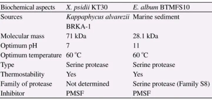
Biochemical characteristics result as explained above showed that our protease has much similarity in several biochemical aspects
with marine-fungal protease isolated from Engyodontium album
(E. album) BTMFS10 as reported by Chellappan et al., and the comparison was summarized in Table 3[20].
Table 3
Comparison of marine-derived extracellular protease from X. psidii KT30 and E. album BTMFS10.
Biochemical aspects X. psidii KT30 E. album BTMFS10 Sources Kappaphycus alvarezii
BRKA-1
Marine sediment
Molecular mass 71 kDa 28.1 kDa
Optimum pH 7 11
Optimum temperature 60 o
C 60 o
C
Type Serine protease Serine protease
Thermostability Yes Yes
Family of protease Not determined Serine protease (Family S8)
Inhibitor PMSF PMSF
4. Discussion
Extracellular protease has been successfully purified from supernatant of X. psidii KT30 at the 10th day. The 10-day culture was selected to isolate extracellular protease, in which the X. psidii
KT30 was on early stationary phase. This phase was considered as
the best stage to harvest the extracellular protease from some fungi
external factors such as starvation condition, nutritional status and
more importantly the availability of protein inducers could affect the
production of extracellular proteases[21,22]. In our experiment, the
protein inducers was absent in the medium for cultivating X. psidii
KT30, but X. psidii KT30 could still produce protease comparable to other fungi that produce extracellular proteases; and interestingly,
some particular fungal species producing extracellular proteases
is specific protein inducers[11,23-26]. If the proteases production in
X. psidii KT30 could also be modulated by protein inducers needs further clarification.
Extracellular protease of X. psidii KT30 had molecular mass of 43 kDa based on gelatin zymogram, but the molecular mass
was twice bigger, 71 kDa, after separation on SDS-PAGE gel. This discrepancy is attributed to the difference of fundamental principle
of protein separation between both methods. Reducing and boiling
of protein sample in SDS-PAGE is critical to gain similar shape and ionic charge of proteins sample that lead to protein separation on
the gel only based on their molecular mass. However, in zymogram,
the protein sample was diluted in non-reducing buffer and
boiling process was excluded prior to separation, so the proteins
migration along the gel will depend not only on sizes but also
on N-glycosylation and protein conformations which are almost
affected by presence of disulfide bonding that reflects the active
forms of protein. This phenomenon is also observed in other serine
protease such as proteinase Atl from Acremonium typhinum[27], fungal Ser protease proteinase K[28], bacterial serine protease
subtilisinand engineered subtilisin E from Bacillus subtilis[29,30]. Furthermore, oligomeric status of protein was proven by single clear
zone formation after renaturation in gelatin zymography, indicating
that extracellular proteaseof X. psidii KT30 has monomeric structure. Some other fungal serine protease such as extracellular
protease PoSland subtilisin like serine protease Eap also showed
similarity on structure, showing mature peptide at their monomeric
enzyme state[31,32].
Extracellular protease of X. psidii KT30 in its native state has molecular mass smaller than 205 kDa of serine protease isolated
from Acremonium typhinum[27]. However, molecular mass in its denatured state was in range of molecular mass of extracellular
serine protease purified from E. album (28.1 kDa)[20], Lecanicillium psalliotae (32 kDa)[26], Hirsutella rhossiliensisOWVT-1 (33 kDa)[11], Dactylella shizishanna (35 kDa)[20], Purpureocillium lilacinum (37 kDa)[33], Aspergillus fumigatus (88 kDa)[18], and
Paracoccidioides brasiliensis (66 kDa)[34]; all these exhibit numerous biological activities, or some are important in field of
biotechnological applications.
The factors such as temperature, pH and protease inhibitor influence the activity of enzymes. The optimum temperature for
activity of extracellular protease of X. psidii KT30 is 60 °C. The protease activity still remained above 50% when temperature raised
to 90 °C, indicating that it was moderately thermostabile protease.
Proteolytic activity was highest at pH 7 but low either at pH of 4 or
10. Surprisingly, the activity of enzyme remained above 50% when
pH was increased from 7 to 8. Furthermore, PMSF has tremendous effect on protease activity; meanwhile EDTA gave only mild effect. The above mentioned indicated that this enzyme was thermostabile
neutral serine protease type. The biochemical properties of protease
produced by X. psidii KT30 are nearly the same as what found in 88 kDa serine protease extracted from Aspergillus fumigatus in terms of temperature 60 °C, pH 7 and kinds of protease inhibitors (mostly
inhibited by PMSF but not by EDTA) for optimum enzymatic activity[19].
The kinetic parameters such as Km and Vmax are the essential
factors to predict behavior of enzymes in catalytic process of their
substrates. Using Lineweaver-Burk plot, we had predicted that the
value of Km and Vmax of extracellular protease X. psidii KT30 are
0.183 mg/mL and 7.01 µg/min (~233.67 µg/min/mL) and those values are lower than those Km (8.26 mg/mL) and Vmax (668 µg/min/
mL) of serine protease from Termitomyces albuminosus[35].
Protease enzymes particularly with thermostabile property have been used widely for their usefulness in biotechnological and
biomedical applications. This present research is the first study
reporting about proteases isolated from marine-derived endophytic
fungi and its characterization. Our further study will also focus
on the characterization of protease gene to gain more information
about the function of this fungal protease in X. psidii KT30 growth and development.
Conflict of interest statement
We declare that we have no conflict of interest.
Comments
Background
Isolation and characterization of bioactive compounds with potential applications in preventing human diseases is one of the
most attracting topics. Endophytic fungi are known to produce
several compounds with antibacterial, antitumoral and antiviral
properties. Therefore there is a growing interest in exploring new
sources of bioactive molecules.
Research frontiers
The extracellular protease secreted by X. psidii KT30 was isolated and purified using three steps of protein purification (ammonium
sulfate precipitation, DEAE-sepharose column chromatography, and sephadex SG75 column chromatography). Qualitative and
culture medium, screening on gelatin-agarose plates, protease
activity measurements at 540 nm) were used to characterize the
purified protease. Its molecular mass was also determined and the
optimum temperature, pH and the effects of protease inhibitors
(PMSF and EDTA) were assayed, showing that the molecule has maximum activity at 60°°C and pH 7 and belongs to serine-proteases group.
Related reports
Several examples of extracellular proteases produced from other fungal species are reported by other researchers. A
comparison between the biochemical properties of
marine-derived extracellular protease from X. psidii KT30 and E. album
BTMFS10 reported in other study is also performed in this present study.
Innovations and breakthroughs
The aim of this study is to isolate the main active molecule responsible of biological activity in X. psidii, since the ethyl acetate extract of X. psidii KT30 broth culture was known to have antivirus, antibacterial, and anticancer activity. Although
current literature refers on protease isolation from
marine-derived endophytic fungi, this is the first study reporting about its
isolation and characterization from X. psidii KT30.
Applications
The subject of the present study is attractive because most of current researches focus on secondary metabolites, while
exploring endophytic fungi as a source of active compounds
could be of greater interest since protein biomolecules are more
save compared to secondary metabolites when applied as drugs
or food preservatives in term of their less toxicity. Particularly the
thermostable properties of the isolated molecule are interesting
for its biomedical applications.
Peer review
This paper is an interesting study focusing on the extraction and biochemical characterization of an extracellular protease
produced by the marine fungus X. psidii KT30. The biochemical properties displayed by this molecule suggest its promising
application in the field of biotechnology or medicine, obviously
following further experimentation.
References
[1] Strobel GA. Endophytes as sources of bioactive products. Microbes Infect 2003; 5: 535-544.
[2] Kusari S, Hertweck C, Spiteller M. Chemical ecology of endophytic fungi: origin of secondary metabolites. Chem Biol 2012; 19:
792-798.
[3] Kim SK, Dewapriya P. Anticancer potentials of marine-derived fungal metabolites. In: Kim SW, editor. Marine microbiology: bioactive compounds and biotechnological applications. 1st ed.
Weinheim: Wiley-VCH; 2013, p. 237-243.
[4] Tarman K, Lindequist U, Wende K, Porzel A, Arnold N, Wessjohann LA. Isolation of a new natural product and cytotoxic and antimicrobial activities of extracts from fungi of Indonesian marine habitats. Mar Drugs 2011; 9: 249-306.
[5] Hu QX, Zhang GQ, Zhang RY, Hu DD, Wang HX, Ng TB. A novel aspartic protease with HIV-1 reverse transcriptase inhibitory activity from fresh fruiting bodies of the wild mushroom Xylaria hypoxylon. J Biomed Biotechnol 2012; doi: 10.1155/2012/728975.
[6] Park BT, Na KH, Jung EC, Park JW, Kim HH. Antifungal and anticancer activities of a protein from mushroom Cordyceps militaris. Korean J Physiol Pharmacol 2009; 13: 49-54.
[7] Sun J, Zhao Y, Chai H, Wang H, Ng TB. A novel alkaline protease with antiproliferative activity from fresh fruiting bodies of the toxic wild mushroom Amanita farinosa. Acta Biochim Pol 2011; 58: 567-572.
[8] Schulz B, Boyle C, Draeger S, Rommert AK, Krohn K. Endophytic fungi: a source of novel biologically active secondary metabolites. Mycol Res 2002; 106: 996-1004.
[9] Schumacher M, Kelkel M, Dicato M, Diederich M. Gold from the sea: marine compounds as inhibitors of the hallmarks of cancer. Biotechnol Adv 2011; 29: 531-547.
[10] Ng TB. Peptides and proteins from fungi. Peptides 2004; 25: 1055-1073.
[11] Wang B, Liu X, Wu W, Liu X, Li S. Purification, characterization, and gene cloning of an alkaline serine protease from a highly virulent strain of the nematode-endoparasitic fungus Hirsutella rhossiliensis. Microbiol Res 2009; 164: 665-673.
[12] Hassan MA, Haroun BM, Amara AA, Serour EA. Production and characterization of keratinolytic protease from new wool-degrading Bacillus species isolated from Egyptian ecosystem. Biomed Res Int
2013; doi: 10.1155/2013/175012.
[14] Cupp-Enyard C. Sigma’s non-specific protease activity assay casein
as a substrate. J Vis Exp 2008; doi: 10.3791/899.
[15] Laemmli UK. Cleavage of structural proteins during the assembly of the head of bacteriophage T4. Nature 1970; 227: 680-685.
[16] Raser KJ, Posner A, Wang KK. Casein zymography: a method to study mu-calpain, m-calpain, and their inhibitory agents. Arch Biochem Biophys 1995; 319(1): 211-216.
[17] Kleiner DE, Stetler-Stevenson WG. Quantitative zymography: detection of picogram quantities of gelatinases. Anal Biochem 1994; 218: 325-329.
[18] Hernández-Martínez R, Gutiérrez-Sánchez G, Bergmann CW, Loera-Corral O, Rojo-Dominguez A, Huerta-Ochoa S, et al. Purification and characterization of a thermodynamic stable serine protease from Aspergillus fumigatus. Process Biochem 2011; 46: 2001-2006.
[19] Zhang XQ, Liu QH, Zhang GQ, Wang HX, Ng TB. Purification and molecular cloning of a serine protease from mushroom Hypsizigus marmoreus. Process Biochem 2010; 45; 724-730.
[20] Chellappan S, Jasmin C, Basheer SM, Kishore A, Elyas KK, Bhat SG, et al. Characterization of an extracellular alkaline serine protease from marine Engyodontium album BTMFS10. J Ind Microbiol Biotechnol 2011; 38: 743-752.
[21] Valueva TA, Kudryavtseva NN, Gvozdeva EL, Sof’in AV, Il’ina NY,
Pobedinskaya MA, et al. Serine proteinases secreted by two isolates of the fungus Alternaria solani. J Basic Appl Sci 2013; 9: 105-115.
[22] Tunlid A, Rosen S, Ek B, Rask L. Purification and characterization of an extracellular serine protease from the nematode-trapping fungus Arthrobotrys oligospora. Microbiology 1994; 140: 1687-1695.
[23] Bonants PJ, Fitters PF, Thijs H, den Belder E, Waalwijk C, Henfling JW. A basic serine protease from Paecilomyces lilacinus with biological activity against Meloidogyne hapla eggs. Microbiology 1995; 141: 775-784.
[24] Segers R, Butt TM, Kerry BR, Peberdy JF. The nematophagous fungus Verticillium chlamydosporium produces a chymoelastase-like protease which hydrolyses host nematode proteins in situ. Microbiology 1994; 140: 2715-2723.
[25] Yang J, Huang X, Tian B, Wang M, Niu Q, Zhang K. Isolation and characterization of a serine protease from the nematophagous fungus,
Lecanicillium psalliotae, displaying nematicidal activity. Biotechnol
Lett 2005; 27: 1123-1128.
[26] Lindstrom JT, Belanger FC. Purification and characterization of an endophytic fungal proteinase that is abundantly expressed in the infected host grass. Plant Physiol 1994; 106: 7-16.
[27] Kolvenbach CG, Narhi LO, Lazenby K, Samal B, Arakawa T. Comparative study on proteinase R, T, and K from Tritirachiam album limber. Int J Pept Protein Res 1990; 36: 387-391.
[28] Narhi LO, Arakawa T. Sodium dodecyl sulfate polyacrylamide gel electrophoresis as a method for studying the stability of subtilisin. Biochim Biophys Acta 1989; 990: 144-149.
[29] Takagi H, Takahashi T, Momose H, Inouye M, Maeda Y, Matsuzawa H, et al. Enhancement of the thermostability of subtilisin E by introduction of a disulfide bond engineered on the basis of structural comparison with a thermophilic serine protease. J Biol Chem 1990; 265: 6874-6878.
[30] Faraco V, Palmieri G, Festa G, Monti M, Sannia G, Giardina P. A new subfamily of fungal subtilases: structural and functional analysis of a Pleurotus ostreatus member. Microbiology 2005; 151: 457-466.
[31] Jasmin C, Chellappan S, Sukumaran RK, Elyas KK, Bhat SG, Chandrasekaran M. Molecular cloning and homology modelling of a subtilisin-like serine protease from the marine fungus, Engyodontium album BTMFS10. World J Microbiol Biotechnol 2010; 26: 1269-1279.
[32] Simkovic M, Kurucova A, Hunova M, Varecka L. Induction of secretion of extracellular proteases from Trichoderma viride. Acta Chim Slovaca 2008; 1(1): 250-264.
[33] Cavello IA, Hours RA, Rojas NL, Cavalitto SF. Purification and characterization of a keratinolytic serine protease from Purpureocillium lilacinum LPS # 876. Process Biochem 2013; 48: 972-978.
[34] Parente JA, Salem-Izaccc SM, Santana JM, Pereira M, Borges CL, Bailao AM, et al. A secreted serine protease of Paracoccidioides brasiliensis and its interactions with fungal proteins. BMC Microbiol
2010; doi: 10.1186/1471-2180-10-292.