Regulation Between Proteases in the Human Protease
Web
Nikolaus Fortelny1,2,3, Jennifer H. Cox1,2¤a, Reinhild Kappelhoff2,3, Amanda E. Starr1,2¤b, Philipp F. Lange2,3, Paul Pavlidis4,5, Christopher M. Overall1,2,3*
1Department of Biochemistry and Molecular Biology, University of British Columbia, Vancouver, British Columbia, Canada,2Centre for Blood Research, University of British Columbia, Vancouver, British Columbia, Canada,3Department of Oral Biological and Medical Sciences, University of British Columbia, Vancouver, British Columbia, Canada,4Department of Psychiatry, University of British Columbia, Vancouver, British Columbia, Canada,5Centre for High Throughput Biology, University of British Columbia, Vancouver, British Columbia, Canada
Abstract
Proteolytic processing is an irreversible posttranslational modification affecting a large portion of the proteome. Protease-cleaved mediators frequently exhibit altered activity, and biological pathways are often regulated by proteolytic processing. Many of these mechanisms have not been appreciated as being protease-dependent, and the potential in unraveling a complex new dimension of biological control is increasingly recognized. Proteases are currently believed to act individually or in isolated cascades. However, conclusive but scattered biochemical evidence indicates broader regulation of proteases by protease and inhibitor interactions. Therefore, to systematically study such interactions, we assembled curated protease cleavage and inhibition data into a global, computational representation, termed the protease web. This revealed that proteases pervasively influence the activity of other proteases directly or by cleaving intermediate proteases or protease inhibitors. The protease web spans four classes of proteases and inhibitors and so links both recently and classically described protease groups and cascades, which can no longer be viewed as operating in isolationin vivo. We demonstrated that this observation, termed reachability, is robust to alterations in the data and will only increase in the future as additional data are added. We further show how subnetworks of the web are operational in 23 different tissues reflecting different phenotypes. We applied our network to develop novel insights into biologically relevant protease interactions using cell-specific proteases of the polymorphonuclear leukocyte as a system. Predictions from the protease web on the activity of matrix metalloproteinase 8 (MMP8) and neutrophil elastase being linked by an inactivating cleavage of serpinA1 by MMP8 were validated and explain perplexingMmp82/2versus wild-type polymorphonuclear chemokine cleavages in vivo. Our findings supply systematically derived and validated evidence for the existence of the protease web, a network that affects the activity of most proteases and thereby influences the functional state of the proteome and cell activity.
Citation:Fortelny N, Cox JH, Kappelhoff R, Starr AE, Lange PF, et al. (2014) Network Analyses Reveal Pervasive Functional Regulation Between Proteases in the Human Protease Web. PLoS Biol 12(5): e1001869. doi:10.1371/journal.pbio.1001869
Academic Editor:Andre Levchenko, Yale University, United States of America
ReceivedSeptember 10, 2013;AcceptedApril 16, 2014;PublishedMay 27, 2014
Copyright:ß2014 Fortelny et al. This is an open-access article distributed under the terms of the Creative Commons Attribution License, which permits unrestricted use, distribution, and reproduction in any medium, provided the original author and source are credited.
Funding:This work was supported by: Canadian Institutes of Health Research (CIHR MOP-37937, 11633, 111058 and 111055, http://www.cihr-irsc.gc.ca/) and Canadian Cancer Society Research (CCS 020483, http://www.cancer.ca/) grants (CMO); NIH (http://www.nih.gov/) grant GM076990 (PP); the Michael Smith Foundation for Health Research (http://www.msfhr.org/) (JHC, PFL), Natural Sciences and Engineering Research Council of Canada (http://www.nserc-crsng.gc.ca) (JHC), and CIHR Strategic Training Program STP-53877 (JHC), Michael Smith Foundation for Health Research (http://www.msfhr.org/) / Breast Cancer Society of Canada (http://www.bcsc.ca/) Research Trainee Award ST-PDF-03243 (11-1) BM (PFL), the Alexander von Humboldt Foundation (http://www.humboldt-foundation.de) and the German Federal Ministry of Education and Research (http://www.bmbf.de) (PFL). CMO is a Canada Research Chair in Metalloproteinase Proteomics and Systems Biology. The funders had no role in study design, data collection and analysis, decision to publish, or preparation of the manuscript.
Competing Interests:The authors have declared that no competing interests exist.
Abbreviations:a1-PI,a1-proteinase inhibitor; AUC, area under the curve; LIX, cytokine LIX (mCXCL5); MALDI, matrix-assisted laser desorption/ionization; MMP, matrix metalloproteinase; PMN, polymorphonuclear leukocyte; SDS-PAGE, sodium dodecyl sulfate polyacrylamide gel electrophoresis; TOF, time of flight.
* E-mail: [email protected]
¤a Current address: Inception Sciences Canada, Vancouver, British Columbia, Canada
¤b Current address: Department of Biochemistry, Microbiology and Immunology, University of Ottawa, Ottawa, Ontario, Canada
Introduction
Proteolysis, the hydrolysis of peptide and isopeptide bonds in protein substrates by proteases (also termed peptidases or proteinases [1]), affects every protein at some point during its lifetime. The outcomes of proteolysis are of two kinds: Protein degradation ablates protein function by breakdown to amino acids, whereas proteolytic processing is an irreversible posttrans-lational modification to precisely produce modified, stable protein
chains. The length of this cleavage product is defined by the substrate site specificity of the protease catalyzing the reaction, which can be exquisite. Processed proteins often have radically altered activity, protein interactions, structure, or cellular location and hence are implicated in many human diseases [2–4]. Recent research has focused on identifying the cleavage products of protease activity in cell culture and in vivo as a means of understanding their biological roles and hence guiding drug target identification and validation [5]. This need has led to the
development of genomics and proteomics approaches that have come to be termed degradomics [6,7] in which the specialized subfield known as terminomics that identifies N termini [8–10] and C termini [11,12] has seen recent rapid development. In one such terminomics analysis of murine skin in vivo, ,44% of identified N termini mapped to internal positions in proteins, revealing proteolytic cleavage after translation as part of protein maturation and function [13]. With,68% of identified N-termini being internal, human erythrocytes have been found to possess an even higher proportion of processed proteins [14]. These recent findings demonstrate that proteolytic processing is a widespread and functionally important posttranslational modification. There-by, proteolytic processing modifies the activity of many more proteins than currently appreciated from conventional shotgun proteomics analyses and biological studies.
As exemplified by N-terminal cleavage of chemokines [6], the activity of a protein often depends on the exact position and nature of its N and C termini [15]. Therefore, identifying the termini of proteins is essential for functional insight into protein bioactivity, annotation of proteins in the Human Proteome Project, and drug development [14]. However, deeper biological insight requires identifying the protease responsible for generation of neo-termini that distinguish cleavage products from the original protein termini. Whereas low- and high-throughput methods to identify the in vitro substrate repertoire of proteases, also known as the substrate degradome [7], are well established,in vivoidentification is problematic [16].In vitroexperiments can only indicate potential cleavagein vivobecause of difficulties assigning precise parameters governing cleavage in the actual biological system, such as protease and substrate colocalization spatially and temporally, presence of inhibitors, zymogen activation, pH, ion concentra-tions, interaction with nonprotein compounds [17], as well as O-glycosylation or phosphorylation of the protease or substrate [18]. Hence, posttranslational modifications of proteases, inhibitors, and their substrates add complexity to the dynamic nature of the
proteome and cell responses. Thus, an observed cleavagein vitro
might not occurin vivo—that is, ‘‘just because it can (in vitro) does not mean it does (in vivo)’’ [5].
In vivo studies, which rely on comparing samples of protease knockout or inhibition to controls, are hampered in particular because the underlying biological system reacts to the removal of a protease or inhibitor in complex and unpredictable ways. For example, a protease knockout can lead to alterations in gene expression profiles of proteases, inhibitors, and substrates [13,19], due to the biological consequences of altered substrate cleavagesin vivo, including cleavage of transcription factors [20]. Another factor is the activation of other proteases in the system through increasingly recognized activation cascades of protease zymogens by other proteases and the proteolytic regulation of protease inhibitor activity by nontarget proteases that cleave and inactivate the inhibitor. For example, serpins and cystatins inhibit serine and cysteine proteases, respectively, but when cleaved by a matrix metalloproteinase (MMP), the inhibitor is inactivated and the protease remains active [13,21–23]. Through activating and inactivating cleavages of other proteases and inhibitors, a protease thereby indirectly influences the activity of additional proteases. Such interactions can lead to knock-on effects that alter the cleavage of a range of additional protein substrates that are not direct substrates of the protease. Furthermore, titration of inhibitors upon covalent or tight interaction with one protease can reduce the availability of free inhibitors to regulate other proteases. Consequently, phenotyping protease and inhibitor genetic knockout mice is complicated, which also hampers biological understanding and drug target validation of proteases.
Protease biology is also complex due to the large protease numbers in humans (460) and mice (525), which form the second largest enzyme family after ubiquitin ligases in these organisms [24]. Moreover, an additional 93 and 103 are predicted to be inactive proteases in human and mouse, respectively, which often can function as dominant negative counterparts [24]. Protease numbers are almost equally distributed in the intracellular and extracellular environments, and other than some proteases that segue between these two compartments, this distribution partitions and limits their potential interactions with each other. In an effort to systematically comprehend this complex biology, proteases are grouped by the MEROPS database, which is assembled from biochemical experimental data curated from the literature, into seven classes, five of which are found in human and mouse, according to the active site residue catalyzing substrate cleavage, and into clans based on the structure of the active site [25]. Similarly, inhibitors are commonly grouped according to the class of proteases they inhibit, with several inhibitors exhibiting broad inhibitory activity against proteases from more than one class. Interactions between proteases of the same class are well established as part of classically described cascades of proteases such as the complement [26–28] and coagulation [29,30] systems, and newer recognized cascades such as kallikreins [31] and caspases in apoptosis [2,32–34]. However, wide-ranging additional protease interactions have also been proposed to extend more globally to link networks forming what was termed the protease web [35]. The protease web was defined as the universe of cleavage and inhibition interactions between proteases and their inhibitors. Stemming from examples in simple systems such asin vitro biochemical analyses and early in vitro and cell culture degradomics analyses of protease substrates [36–38], and mRNA transcript analyses in cancer upon administration of protease inhibitors or tissue inhibitor of metalloproteinase (TIMP) overex-pression and knockout studies [19], the protease web concept has been well supported. Extending terminomics analyses to in vivo
Author Summary
situations, for example skin inflammation in wild-type versus
Mmp2 knockout mice in vivo, has revealed hitherto biologically relevant and unsuspected critical connections of MMPs in regulating the complement and coagulation cascades and the plasma kallikrein system, which regulates vessel permeability through bradykinin excision and release from kininogen [13].
Such interactions between protease families were shown to create small networks in specific cases [13,19,39,40], but the full extent of the protease web, the fraction of proteases and inhibitors involved, and hence the regulatory potential of this network remain underexplored and underappreciated despite the poten-tially wide impact on the functional state of proteomes. Furthermore, the protease web is a black box with an unknown mechanism of regulation—it is unclear whether it follows a super structure of known cascades, where signals are amplified downstream, or forms more of a network, where signals can flow in multiple directions with multiple positive and negative feedback loops [35,40]. Similarly, it is unclear which are the main regulatory protein switches controlling subparts of the network. Descriptions of the protease web are difficult to assemble, as many proteases remain poorly studied and characterized. Likewise, many proteases have no described inhibitors and many predicted inhibitors have unknown protease targets and deorphanization examples are uncommon [41].
Here, we assessed the global extent and structure of protease interactions computationally. Graph models are used to describe multiple interactions between many elements and have been applied extensively in research on various biological networks. We represented existing biochemically validated data on protease cleavages and inhibition as annotated in the manually curated database TopFIND [42] as organism-specific networks. TopFIND stores established biochemical information on substrate cleavage and protease inhibition from MEROPS [25], the most complete collection of such data, most of it published, and combines it with published high-throughput terminomics and degradomics datasets as well as protein annotations from UniProt [43] for five different organisms. Our analyses revealed a large and pervasive network spanning all known cascades and four of the five protease classes present in human and mouse tissues. The network is highly connected in that via a few connections a protease can potentially influence many other proteases, with inhibitors often taking a special role as key connectors in the protease web. We demonstrate the utility of our analysis by applying the network to gain mechanisticin vivoinsights into protease web effects, which we then validatedin vitro, in cell culture, andin vivo.
Results
Protease Web Data
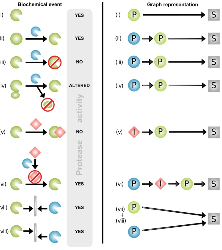
Functional protease interactions comprising cleavage and inhibition events influence the in vivo cleavage of substrates in many ways. Cleavage of a substrate by a protease is a direct event, and as shown in Figure 1, by cleaving other proteases and protease inhibitors, one protease can activate, inactivate, or alter the activity of a second protease, thereby indirectly influencing the cleavage of substrates of another protease. To assess the global extent of such effects, we represented protease interactions as a graph, connecting proteases and protease inhibitors to their established substrates and protease targets, respectively. The resulting graph contains nodes, which are proteins, and edges, which represent cleavages or inhibitions. Edges link proteases to their substrates and protease inhibitors to their target proteases. Therefore, edges are directed: an edge from protein X to protein Y signifies cleavage or inhibition of Y by X but does not contain
information about cleavage or inhibition of X by Y. In graph theory, the latter would require another edge with the opposite directionality. Figure 1 outlines functional protease interactions and how they are represented in small graph models, which were then aggregated to represent the full complexity of the protease web based on curated biochemical data as described below.
As input to our analysis of the protease network, we used the TopFIND v 2.0 knowledgebase [44] to retrieve validated cleavage and inhibition data mostly annotated from published experiments. TopFIND contained 4,774 cleavages forHomo sapiens, 3,679 for
Mus musculus, 426 for Escherichia coli, 190 for yeast, and 43 for
Arabidopsis thaliana. Due to the low number of cleavages annotated for other organisms, we focused our analysis on human and mouse. Only proteins performing an annotated cleavage or inhibition were added, and then these were connected via edges representing the biochemical reactions as explained in Figure 1. These networks extend the protease web, which contains only proteases and inhibitors, by also including all other substrates of proteases, and hence represent the annotated functional proteo-lytic interactions between the substrates in the proteome and the protease web. The human and murine networks (with 1,230 and 1,393 nodes, respectively) are shown in high resolution upon click-to-zoom in Figure S1 and available for download as a Cytoscape file, gml file, and R objects at www.chibi.ubc.ca/ProteaseWeb and http://clipserve.clip.ubc.ca/supplements/protease-web.
The human and murine proteolytic networks show that the majority of proteins are connected and only very few are in unconnected components. Thus, in both networks, the Largest Connected Component (i.e., the biggest group of nodes directly or indirectly connected) encompasses the vast majority of these proteins—1,183 of 1,230 (96%) in human and 1,377 of 1,393 (99%) in mouse (Table 1). This remarkable connectivity is particularly surprising given the incompleteness of annotation currently available in the databases. Indeed, Table 1 shows that of 460 human proteases, only 244 (53%) have one or more known and annotated substrates. In mouse this number is even lower, with only 88 of 525 (17%) proteases having a substrate annotated. Furthermore, even the data on these proteases are incomplete and biased, with most substrates assigned to few, well-studied proteases. Figure S2 shows the out-degree (i.e., the sum of cleavages catalyzed by a protease or the sum of inhibitions caused by a protease inhibitor) for proteases and inhibitors having any annotated cleavage or inhibition, respectively. Although few proteases have a large known substrate repertoire (higher out-degree), most proteases have very few known substrates. Although this could be due to high substrate specificity, it is more likely that these proteases simply received less attention in studies dedicated to discover substrate repertoires. This effect is especially pronounced for the mouse data, where 80% of total cleavages (2,938 of 3,679) are assigned to three proteases—cathepsin D (UniProt: P18242), cathepsin E (UniProt: P70269), and MMP2 (UniProt: P33434)—and are mostly derived from high-throughput proteomics screens. Accordingly, the annotations differ strongly between human and mouse. Although the networks have similar size (1,230 and 1,393 nodes, respectively), they overlap minimally, with only 126 of 3,852 connections in mouse (3.3%) reflected in 122 of 4,905 human connections (2.5%). However, we suggest that the small overlap is mostly due to differences in the state of data annotation between the networks rather than to actual differences in the evolution of these networks.
The human data are further biased in that proteases and inhibitors are largely overrepresented as substrates themselves (Figure S3). Strong representation of protease–protease cleavages is expected because many proteases are synthesized as zymogens
Figure 1. Biochemical protease interactions represented by graph theory.Proteases influence cleavage of substrates both directly and also indirectly through cleavage of other proteases and inhibitors. Protease interactions as represented biochemically (left) and by graph theory (right). Proteases are green or blue, inhibitors are red, and other substrates are grey. Examples of protease interactions (cleavage and inhibition events) are outlined on the left: (i) In the simplest case, a protease directly cleaves a substrate, as indicated by the presence of proteolytic activity, with no further interactions. A protease can also indirectly influence cleavages by cleaving another protease for (ii) zymogen activation [76], (iii) catalytic domain removal, or (iv) exosite domain removal [77]. This will increase (ii), decrease (iii), or alter (iv) [78] the activity of the affected protease and thereby influence the cleavage of its substrates. (v) If a protease inhibitor is present, the protease does not cleave substrates. (vi) An inhibitor can be cleaved and inactivated by another protease [13,79], which leads to increased cleavage of substrates by its cognate protease. Proteases also compensate for loss of function of other proteases or complement their activity by (vii) cleaving the same substrate at the same site or (viii) substrate cleavage by one protease can depend on prior cleavage by another protease at a different site. By graph theory of protease interactions (right), all proteins are nodes. Proteases (P) are represented as green or blue circles, inhibitors (I) as red diamond shapes, and substrates (S) are grey squares or rectangles. An edge from protein A to protein B signifies a direct regulatory influence from A on B. Such a regulatory effect could either be a cleavage or inhibition, resulting in higher, lower, altered, or unchanged activity of the target.
requiring proteolytic cleavage for activation by other proteases. Indeed, this strong enrichment is found in the human TopFIND/ MEROPS data, but less so in mouse. We compared these values to a terminomics data set of cleavages in mouse skin [13], which more accurately reflects reality because terminomics analyzes N termini in an unbiased fashion. However, in thisin vivodata set, inhibitors, and not proteases, were overrepresented as processed proteins, indicating that the overrepresentation of proteases as cleavage substrates in the human in vitro database is likely exaggerated.
The observed data biases likely resulted from the nature of biochemical studies, where many substrates were identified for some ‘‘interesting’’ proteases (target bias) and ‘‘interesting’’ proteins are more likely to be tested as substrates (substrate bias). Substrate bias is especially found for proteases themselves, which are preferably tested as substrates in zymogen activation studies. With the advent of degradomics utilizing proteomics methods dedicated to substrate discovery, we anticipate both an increase in target bias in the future with many substrates identified for a few proteases, and a decrease in substrate bias where any protein can be identified as a substrate without prior selection of interesting candidates. Therefore, the cleavages annotated represent a biased fraction of the biochemically possible cleavages in the organism compared with an unknown number of as yet uncharacterized cleavages. On these grounds, the high connectivity in both the mouse and human networks is even more noteworthy because future information can only further increase connectivity. The observed, extensive interactions between proteases and inhibitors are further characterized as described in the following.
Protease Web Structure
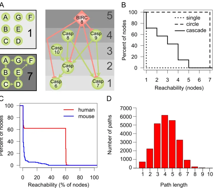
In the interactions between proteases in proteolytic signaling pathways, there are major upstream regulators or initiation factors, whose proteolytic activity leads to the cleavage of downstream proteases, which in turn activate even further downstream factors that finally cleave and activate the effector molecules at the end of the pathway. A special case of proteolytic pathways are activation cascades, where signal amplification occurs to generate large quantities of the end protein products in seconds as classically described for coagulation [29,30]. To investigate whether the connections in the overarching protease web follow such a pathway or cascade (hierarchical) structure, we used a graph measure termed reachability. Reachability of node X denotes the number of other nodes Y where there is a path from X to Y in the network. A path is a sequence of directed edges connecting X and Y, following the directionality of edges in the network. The path from X to Y can therefore be different from the path from Y to X (and the existence of one does not guarantee the existence of the other). In the protease web, reachability corresponds to the number of proteins that can be influenced by one protease or inhibitor. Figure 2A outlines reachability values of nodes in three theoretical examples: (i) an unconnected (single), (ii) a strongly connected (circle), and (iii) a cascade-like network
(cascade). Figure 2B shows the respective distribution of reach-ability values of these three theoretical examples.
We next compared the theoretical reachability distributions with the distributions observed in our human and mouse protease networks. In order to specifically describe the selective connectivity between proteases and inhibitors, which form the protease web, we excluded from further analysis other simple substrates (nonprotease and noninhibitor proteins), whose reachability in the network is 1 by definition. Table 2 summarizes the resulting protease web networks for human (340) and mouse (220) proteins that have annotated cleavages or inhibitions. In analyzing the human and mouse protease webs, we further identified one dominant ‘‘largest connected component’’ comprised of 255 proteins for human and 187 proteins for mouse. Figure 2C compares the distribution of reachability scores in the largest connected component in mouse (blue curve) and human (red curve). In mouse, reachability indicates a cascade-like, hierarchical network, where most nodes have a very low reachability and fewer nodes have gradually higher reachability. In contrast, the reach-ability distribution of the human network follows a strongly bimodal distribution: 158 (62%) nodes reach 153 (60%) or more nodes. This is very high reachability that is most similar to the circle graph in Figure 2B, where any node can reach any other node. For a biological system, this implies that 158 proteases or inhibitors have the potential to regulate the activity of 153 or more other proteases and inhibitors in the network. In other words, there are one or more directed paths between 24,166 pairs of proteases in the human protease web, which are 37% of all 64,770 possible directed connections between pairs of 255 proteins. This number of connections between pairs rises to 141,523 paths when substrates are added (network with 1,230 nodes). This highlights the high degree of connectivity between proteases and inhibitors. Reachability between nodes does not take the path length between nodes into account and so might be the result of very long and hence biologically irrelevant paths in the network. However, this possibility can be excluded as most paths have a length of just four (Figure 2D). The lack of connectivity in the mouse network is not surprising given the small overlap between the two networks. We assume that this difference is due to data biases rather than a real biological difference, and accordingly we focused on characteriz-ing the extensive and more complete human network.
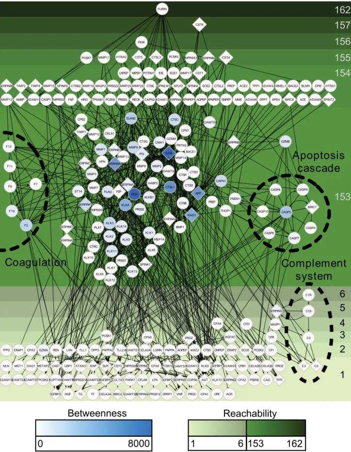
High connectivity in the human protease web is due to a strongly connected component (87 nodes), a subgroup of nodes within the largest connected component, that can directly or indirectly reach each other and hence have the same reachability value of 153 (Table 3). We visualized this effect in Figure 3, where nodes of the human protease web are shown separated by their reachability. Upstream of the strongly connected component are 71 nodes with reachability higher than 153; these nodes can reach the strongly connected component, but cannot be reached from it. Downstream (with reachability smaller than 7) are 97 nodes, which cannot reach the strongly connected component. The nodes in Figure 3 are also colored according to their centrality in the
Table 1.Human and mouse proteolytic networks created from all annotated proteases, inhibitors, and substrates.
Organism Total Nodes Largest Connected Component Proteases with Substratesa Inhibitors with Target Proteasesa Edges
Human 1,230 1,183 244 41 4,905
Mouse 1,393 1,377 88 47 3,852
aProtease and inhibitor numbers refer to those with at least one annotated cleavage or inhibition, respectively, in MEROPS and TopFIND. doi:10.1371/journal.pbio.1001869.t001
network, as measured by node betweenness [45]. Betweenness is calculated by first finding the shortest paths (as explained above) between all 64,770 pairs of nodes in the network and then counting the number of times a node appears in these paths. Notably, all nodes with high betweenness are found in the strongly connected component; these nodes tether the network together. Nodes with high betweenness or reachability are listed in Table S1.
Figure 3 shows that our network data from MEROPS/ TopFIND contain all the known proteolytic pathways (e.g.,
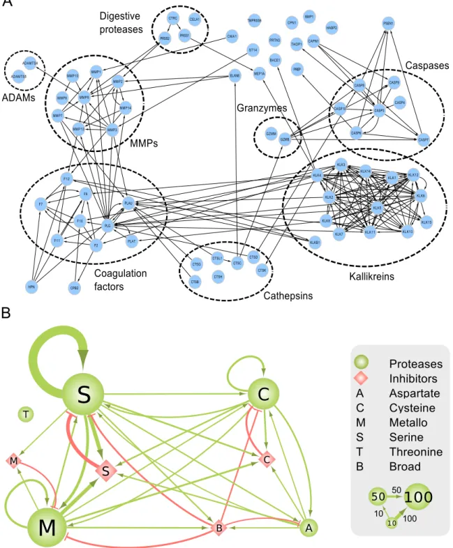
coagulation, complement system, apoptosis, and kallikreins) as they were discovered, published, and annotated previously in MEROPS (detailed in Figure S4). In addition, these proteolytic pathways are extended by connections linking known pathways with other pathways and additional proteases. Details of these connections can be found in Figure 4A, which shows separated protease groups in the strongly connected component after removing inhibitors. Figures 3, 4A, and S4 show that the observed connectivity in the protease web is caused by the concerted action
Casp
8
Casp
9
Casp
10
Casp
3
Casp
7
Casp
6
BIRC
8
C
B
A
5
4
3
2
1
D
A
B
C
F
G
E
D
1
Reachability (nodes)
Reachability (% of nodes)
Percent of nodes
Percent of nodes
A
B
C
F
G
E
D
7
0
20
40
60
80
100
0
20
40
60
80
100
human
mouse
Number of paths
Path length
1
2
3
4
5
6
7
0
20
40
60
80
100
single
circle
casc.
single
circle
cascade
0
1 2 3 4 5 6 7 8 9 10
1000
2000
3000
4000
5000
6000
7000
Figure 2. Reachability in network examples and the human and murine protease webs.Connectivity in the protease networks as measured by the reachability distribution of nodes in the network. (A) Reachability in three theoretical model networks: In an unconnected network without edges, each node has a reachability of 1. In a strongly connected network, where each node can reach each other node, the reachability of each node is the sum of nodes. In a hierarchical, cascade-like network (apoptosis cascade taken from KEGG [80]), reachability values are high for upstream regulators and decrease as one descends the cascade towards the downstream effector proteins. For each protein, the corresponding reachability values are shown on the right. Proteases are represented as green circles and inhibitors as red diamonds. Edges are cleavages (green, with arrow head) and inhibitions (red, with ‘‘T’’ head). Although these two types of edges have biologically distinct interpretations, the implication for the graph model and reachability is identical. (B) Reachability values of nodes in a theoretical hierarchical cascade (cascade), unconnected (single), or strongly connected (circle) networks shown in (A). Reachability is plotted as an inverse cumulative function of the percentage of nodes, which can reach a given minimum number of nodes in the corresponding network. (C) Inverse cumulative function of reachability values of the largest connected components of the human protease web (255 nodes, red line) and the mouse protease web (187 nodes, blue line). Reachability is plotted as the inverse cumulative function of the percentage of nodes that can reach a given minimum percentage of nodes in the corresponding network. (D) Histogram of the path length of all shortest paths in the human network comprised of a total of 24,166 paths.
of defined protease cascades and key protease inhibitors: alpha-2-macroglobulin (A2M, UniProt: P01023), amyloid precursor protein (APP, UniProt: P05067), kininogen 1 (KNG1, UniProt: P01042), and alpha-1-antitrypsin (also known as serpin A1) (SERPINA1, UniProt: P01009). Whereas intragroup connections are pervasive as expected, intergroup connections are also considerable, in particular between coagulation factors and kallikreins or MMPs, but also including cathepsins and caspases. These findings are confirmed in Figure 4B, which shows that connections among four of the five classes of proteases and protease inhibitors in human are extensive. Importantly, Figure 4B also shows proteases frequently cleaving inhibitors of other protease classes, an important regulatory aspect of protease activity. Only threonine proteases, which are found exclusively in large specialized cell organelles termed the proteasome and immunoproteasome, remain isolated from connections with other proteases and inhibitors according to current data. Note added in proof: However, a recent publication shows that the threonine proteasomal proteases are cleaved by intracellular MMP-12. Thus, all five classes of proteases in human and mouse are intercon-nected [81].
Theoretical Network Analysis of the Protease Web
From a biological standpoint, the highly interconnected (reachable) nature of the protease web was surprising and underappreciated in the literature. To explore the degree to which this result is statistically surprising given the properties of the proteins making up the network, we investigated theoretical network models as well as randomized versions of the network. We first compared the protease web to two commonly used generative network models, the Erdo˝s-Re´nyi model (ER) and the Barabasi-Albert model (BA), with parameters chosen to mimic the properties of the real network’s member proteins (see Materials and Methods). We found that neither model (each 500 networks) adequately explains the data, yielding networks that have either much higher (ER) or lower (BA) reachability on average (Figure S5A–C). These experiments therefore leave open the statistical nature of the process that generates the network, which we stress currently involves both biological components and experimenter biases, the latter being due to the incomplete nature of the underlying biochemical analyses (many potential edges have not been tested). We next generated two types of edge-shuffled networks, one maintaining in- and out-degree of each node (‘‘Shuffled’’) and a second preserving overall in- and out-degree distributions of the network, but not for each node (‘‘Shuffled2’’). The mean reachability was lower in the real network (72.09) than in 353 Shuffled networks (70.6% of all 500; average reachability was 73.96 across all 500 networks; see Figure S5D) but higher than all 500 Shuffled2 networks (average 34.8; Figure S5C). Taken together, these results indicate that high reachability emerges quite readily in a network composed of proteins with the measured
in- and out-degrees found in a real biological network, such as the protease web described here. In fact, a network without such high reachability—as it is often assumed in biochemistry and cell biology—would be surprising from these results. Importantly, this further suggests that the current biochemical description of cascades and individual proteases working in isolation is unlikely.
High Connectivity in the Protease Web Is Robust to Possible Annotation Errors
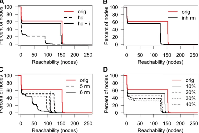
To assess reliability of high connectivity in the protease web, which we observed assuming that all cleavage and inhibition data are trustworthy, we addressed the possibility of erroneous data passing through database annotations into our network. A possibility of validating our findings is to compare the network to another second network derived from an orthologous data source. However, MEROPS being the only database of similar coverage, we instead tested whether the same connectivity can be observed by removing nodes in anticipation that some interactions are wrongly annotated. Protease specificity is mostly influenced by three factors: substrate sequence, substrate folding, and the encounter of protease and substrate [46]. In MEROPS/ TopFIND, annotations are mostly derived from in vitro experi-ments where a protease is incubated with a substrate. Although some proteases are specific for given substrate sequences, others will cleave a wider range of sequences, but in both cases, possible cleavage sites can be masked in the protein structure of the substrate. Hence, experimental parameters of protease cleavage assays are designed to preserve protein folding and activity of both the protease and substrate in order to prevent unspecific cleavage of denatured substrates. Colocalization of proteases and substrates
in vivois an important factor but not unambiguously determinable, with unexpected localization recently revealed [20,37,47–49,81]. In addition, most experiments are only performed if it can be assumed that the protease and substrate will colocalize in vivo. Assuming that most annotations are correct but individual assignments can be wrong, we randomly and selectively removed edges from the protease web (focusing on the regulatory core, the largest connected component with 255 nodes) to test how reachability is maintained or influenced by such modifications. We utilized the term ‘‘physiological relevance,’’ as annotated in MEROPS and TopFIND, to first create a high-confidence network (abbreviated as ‘‘hc’’ in Figure 5A) by removing all edges that were annotated with physiological relevance other than ‘‘yes.’’ As a consequence, the reachability of the resulting network was markedly decreased (Figure 5A), with the area under the curve (AUC) reduced to 22% of the original network. This was mostly due to the removal of all inhibitors (abbreviated as ‘‘i’’ below) as all 131 human inhibitions in TopFIND have a physiological relevance annotation of ‘‘unknown’’; that is, their physiological relevance is not annotated in MEROPS from which TopFIND
Table 2.Proteins comprising the human and mouse protease webs.
Organism
Proteins with
MEROPS ID Protease Weba Largest Connected Component
Nodes Edges Nodes Proteasesb Inhibitorsb Edges
Human 755 340 1,264 255 215 40 1,238
Mouse 696 220 415 187 141 46 404
aOnly nodes having a MEROPS ID and are part of a cleavage or inhibition are in the protease web. b
Proteases and inhibitors are assigned based on the MEROPS IDs of the proteins. doi:10.1371/journal.pbio.1001869.t002
data are largely derived. Upon adding back the inhibitors to the high confidence network (‘‘hc+i’’), but still removing all ‘‘low confidence’’ nonphysiological cleavages, high reachability was largely recovered as indicated by an AUC of 88% of the original network. The observation that limiting the cleavages to high-confidence cleavages only barely reduces network connectivity strengthens the result that the protease web is not due to incorrect annotations. Moreover, removing inhibitions from the network severely impacted reachability and thus connectivity, highlighting the essential role of inhibitors in connecting the protease web.
Given the observed importance of inhibitors, we assessed the possibility of incorrect annotation of cleavages of inhibitors. The molecular mechanism of cysteine or serine protease inhibition by serpins involves cleavage of the serpin at its flexible reactive loop, which displays ‘‘bait’’ amino acids. Following cleavage, an induced conformational change leads to entrapment and inactivation of the protease [50,51]. Because the trap occurs after formation of the acyl intermediate during catalysis, the inhibited serine proteases, but also some cysteine proteases, remain covalently bound to the inhibitor. In contrast, metallo-proteinase and aspartic protease cleavage of serpins in the reactive loop does not result in their inhibition, as the nucleophile of these proteases classes is a water molecule. Thus, these proteases are not trapped and therefore escape inhibition, but the serpin is now inactivated. Mechanisms of trapping upon cleavage have also been observed for some metalloprotease inhibitors [52] and for A2M or pregnancy zone protein (PZP, UniProt: P20742), which use a physical trapping mechanism to inhibit all classes of proteases, except exopeptidases [53,54]. Therefore, annotated cleavages of a protease inhibitor comprise cleavages that reflect either a regulatory inhibition of the protease or a regulatory inactivation cleavage of the inhibitor. To date, this distinction is not annotated in the databases, but is one that we suggest implementing. As a conservative estimate, we removed all cleavages of serpins by serine or cysteine proteases and from any protease to A2M or PZP (‘‘inh rm’’ in Figure 5B). Therefore 144 edges were deleted from the original 1,238 edges of the largest connected component of the protease web (‘‘orig’’ in Figure 5B). Notably, this removal only moderately reduced reachability (AUC 74% of original) and preserved a bimodal distribution. Thus, the high connectivity is not a result of unspecific inhibitors. Hence, the observed connectivity in the network is not an artifact attributable to ambiguous annotation of inhibitor cleavage and so further supports the importance of inhibitors in connecting the protease web.
We next assessed the dependence of reachability on individual nodes of the network. By removing each node individually, we found that reachability in the protease web is not dependent on any one single node (Figure S6). Indeed, by iteratively removing all nodes with the highest betweenness from the network, we
identified the six most important nodes: plasminogen (PLG; UniProt: P00747), alpha-1-antitrypsin, A2M, cathepsin L1 (CTSL1; UniProt: P07711), alpha-1-antichymotrypsin (also known as serpin A3) (SERPINA3; UniProt: P01011), and kallikrein-4 (KLK4; UniProt: Q9Y5K2) (Figure 5C). Removing all six nodes simultaneously removes 227 edges whereupon this significantly breaks down the bimodal distribution of reachability values, an effect not observed when removing any combination of five out of the six connectors. Thus, high connectivity in the protease web is robust in that it depends not on a single protein, but rather on six important connectors. Furthermore, even after removal of those six nodes the reachability for many proteins remains high with many long paths in the network. Notably, none of these six important nodes are digestive tract proteases, such as trypsin or chymotrypsin, which are broad-acting proteases and ones that might have been expected to form many connections. However, we predict that the identity and number of these key connector proteins will change as more information on the protease web is uploaded to the databases with further experimentation.
Finally, we addressed the possibility of incorrect annotations by removing a fixed percentage of edges, thereby simulating a situation where these edges are incorrect cleavage or inhibition annotations and therefore would have to be removed from the network (Figure 5D). We randomly removed 10%, 20%, 30%, and 40% of all edges (cleavages and inhibitions) 200 times and then plotted the worst case for each experiment. The AUC was reduced to 78%, 65%, 47%, and 52%, respectively, but nonetheless even removal of 40% of edges still preserved the bimodality of the reachability values. Therefore, again the protease web shows a strong resistance to removal of elements, which further increases confidence in the description of a highly connected protease web with inherent robustness to change. This also leads to biological resilience and shows the importance of proteases that can nonetheless be resiliently maintained in genetic deficiencies or pathological perturbations of the system.
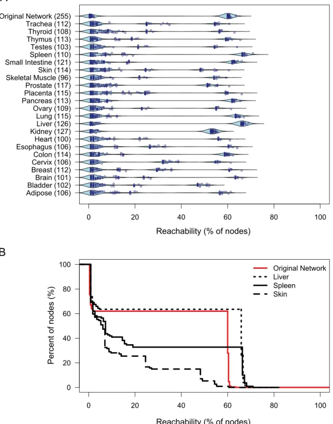
Human Tissue-Specific Protease Webs
Our analyses suggested that the protease web represents a robust regulatory system of high complexity and flexibility enabling complex patterns of regulation of proteins at the posttranslational level. We next assessed how this system is implemented in vivo where only a fraction of proteases and inhibitors is expressed or active at the same time in the same cell, compartment, or tissue. We constructed tissue-specific networks based on protease and inhibitor gene expression levels in 23 different human tissues quantified by CLIP-CHIP microarray (Kappelhoff et al., unpublished data available at http://clipserve. clip.ubc.ca/supplements/protease-web). We used negative con-trol spots on this microarray to define a threshold of expression at detectable levels and then limited networks to those proteins expressed above this threshold. We next plotted the reachability of the nodes in the largest connected component of the resulting networks for all 23 tissue-specific protease webs (Figure 6A). Figure 6B shows liver, spleen, and skin results in more detail. Although most tissue-specific networks (e.g., skin) show low reachability values, some preserve the strong connectivity of the original network totally (e.g., kidney and liver) or partially (e.g., spleen, small intestine, pancreas, lung, colon). Notably, the tissue-specific networks also show that reachability is highly dependent on expression of the same six network connectors shown in Figure 5C (Figure S7).
Table 3.Reachability values of nodes in the human protease web.
Number of Nodes Reachability
97 ,7
87 153
71 .153
Figure 3. The largest connected component of the human protease web.The structure of the core of the human protease web is comprised of 255 connected proteases and inhibitors that form the largest connected component. Proteins are designated by their UniProt gene names. Proteases are circles and inhibitors are diamonds. Nodes are color-shaded according to their betweenness. All nodes are positioned from top to bottom by decreasing reachability, which is indicated by the depth of shade of the green background. Edges are cleavages (with arrow head) or inhibitions (‘‘T’’ head). Nodes of known protease cascades are labeled and marked by dashed circles. The figure is rendered in high resolution for click to zoom.
doi:10.1371/journal.pbio.1001869.g003
Figure 4. Interactions between protease groups in the human protease web.(A) Click-to-zoom figure of detailed connections between pathways and protease groups in the strongly connected component of the network. The network presented is limited to proteases (no inhibitors) with a reachability of 153 from Figure 3. Nodes are proteases and edges are cleavages. Proteases are designated by their UniProt gene names. (B) Interactions between classes of proteases and their inhibitors. Nodes are classes of proteins: classes of proteases are green circles; classes of protease inhibitors are red diamonds. The size of the nodes represents the number of proteins in each class as exemplified with groups of 10, 50, and 100 nodes in the legend. Protein classification: ‘‘M’’ are metallo, ‘‘S’’ are serine, ‘‘C’’ are cysteine, ‘‘A’’ are aspartate, and ‘‘T’’ are threonine proteases (as classified in MEROPS) or the corresponding inhibitors (as annotated in neXtProt). ‘‘B’’ are broad-spectrum inhibitors that are annotated to inhibit more than one class of protease and include A2M, serpin B4, serpin B9, PZP, histidine-rich glycoprotein, ovostatin homolog 1, and reversion-inducing cysteine-rich protein with Kazal motifs. Edges are cleavages (green, with arrow head) or inhibitions (red, with ‘‘T’’ head). Thickness of edges corresponds to the number of cleavages or inhibitions between the classes as exemplified with edges corresponding to 10, 50, or 100 interactions in the legend.
Evidence for the Protease Web in Other Data
In agreement with our findings based on biochemical interactions, general biological literature also shows that proteases and their inhibitors can be involved in multiple biological processes (Figure 7A). It is easy to imagine that this multi-functionality is partly due to the interplay in the protease web. Indeed most of the proteins in Figure 7A are found in the strongly connected component of our protease web, indicating that they serve in connecting different biological processes. One example is TIMP1 (UniProt: P01033). Protein expression levels of TIMP1, an MMP inhibitor mainly involved in extracellular matrix remodeling and organization, were found associated with hemostasis [55]. This finding, which is derived from orthogonal data to the protease web, primed us to search for connections linking TIMP1 to coagulation factors, which we could indeed identify (Figure 7B). Together, these provide a plausible mechanism of action of TIMP1 and hence MMPs on coagulation and could explain the association observed. Hence, the protease web can be used to explain multifunctionality of proteases, which in turn strengthens our conclusion of a large interplay between proteases.
Using the Protease Web to Decipherin VivoNetwork Effects
We were able to test the utility of our graph representation of the protease web by deciphering a previously inexplicable resultin vivo. We analyzed the MMP8-dependent cleavage of the murine chemokine C-X-C motif chemokine 5 (CXCL5, UniProt: P50228), also known as lipopolysaccharide (LPS)-induced C-X-C chemokine LIX (LIX). LIX is a potent chemoattractant chemokine for polymorphonuclear (PMN) leukocytes, and MMP8 (UniProt: O70138) is PMN specific. It was previously demonstrated in anin vivoairpouch model that MMP8 knockout mice showed reduced PMN migration in response to LPS [56]. This was attributed to MMP8 processing and activation of LIX at position Ser4QVal5, with a second cleavage at Lys79QArg80of the 92-residue protein. Indeed the MMP8-truncated activated form of LIX (5–79) showed equal cell migration in wild-type and knockout mice, validating LIX as a physiological MMP8-dependent mechanism for promoting neutrophil infiltrationin vivo. However, a neoepitope antibody specific to the MMP8-generated neo-N terminus failed to detect truncations at Ser4QVal5 in the airpouch model. Thus, cleavage of LIX is a MMP8-dependentbut
Figure 5. Reachability in the human protease web after various perturbations.Reachability of the largest connected component of the protease web (shown in Figures 2C and 3) after various perturbations. Reachability is plotted as the inverse cumulative function of the percentage of nodes that can reach a given minimum number of nodes in the corresponding network. (A) Reachability in the high confidence network comprised of nodes annotated as having physiological relevance. The reachability distribution of the original network (‘‘orig,’’ red solid line as also shown in Figure 2C) is compared to networks where edges were removed to create a high confidence network (‘‘hc,’’ black dashed line) and the high confidence network plus inhibitors (‘‘hc+i,’’ black solid line). (B) Reachability before (‘‘orig,’’ red line) and after (‘‘inh rm,’’ black line) removing edges, reflecting cleavages of inhibitors. Cleavage edges were removed if (i) the inhibitor is annotated to be a serine protease inhibitor and the protease is a serine or cysteine protease or (ii) the inhibitor is A2M or PZP. (C) Reachability after removal of six nodes from the original network (PLG, alpha-1-antitrypsin, A2M, CTSL1, alpha-1-antichymotrypsin, and KLK4). The reachability after removing these six nodes (‘‘6 rm,’’ black solid line) is compared to the reachability distribution of the original network (‘‘orig,’’ red line) and to six networks representing each possible combination of keeping one of the six nodes and removing the other five (‘‘5 rm,’’ black dotted lines), each showing much smaller reduction in reachability. (D) Reachability after removal of random edges. The reachability in the original network (‘‘orig,’’ red line) compared to networks where 10%, 20%, 30%, or 40% of edges were removed at random. In each case, random edge deletion was carried out 200 times and the worst AUC value was selected for plotting. doi:10.1371/journal.pbio.1001869.g005
Figure 6. Reachability in tissue-specific protease webs.(A) Beanplot of reachability distributions in the largest connected components of 23 human tissue-specific networks based on gene expression in the corresponding tissues and the original protease web reachability distributions. Overlaid is a scatterplot of the precise values of each node. Numbers in parentheses refer to the size of the network. (B) Inverse cumulative distribution plot of reachability values for skin (dashed black line), spleen (black solid line), liver (dotted black line), and original network (red solid line). Reachability is plotted as an inverse cumulative function of the percentage of nodes that can reach a given minimum percentage of nodes in the corresponding network.
MMP8-indirecteventin vivothat could not be explained, prompting a further analysis of alternate MMP8-dependent proteolytic pathways predicted using our representation of the protease web. To examine the importance of neutrophil-derived MMP8 in LIX processing and activation, we isolated bone marrow neutrophils from wild-type and MMP8 knockout mice. Neutro-phils were stimulated with phorbol myristate acetate (PMA) followed by incubation of the activated neutrophils with chemokine for up to 3 h. Truncations of LIX generating the bioactive products LIX (9–92) and LIX (9–78), as determined by MALDI-TOF mass spectrometry from the still inactive form LIX (1–78), were readily apparent, even after only 1 h of incubation (Figure 8A). However, both the MMP8 knockout and wild-type neutrophils showed identical cleavage sites (Ala8QThr9 and Ala78QLys79) and cleavage kinetics. Because these sites differ from the MMP8 cleavage sites (Figures S8, S9, and 8B), MMP8 is not the dominant neutrophil protease cleaving LIX in the cellular
context. Investigating protease web effects that may account for this, we found that LIX cleavage by neutrophils was inhibited by the serine protease inhibitor 2-aminoethyl benzenesulfonyl fluo-ride hydrochlofluo-ride (Figure 8C). This showed that one or more of the four serine proteases in neutrophils—neutrophil elastase (UniProt: Q3UP87), cathepsin G (UniProt: P28293) [57], proteinase-3 (UniProt: Q61096), or the recently described neutrophil serine proteinase 4 (UniProt: Q14B24) [58]—were responsible for LIX cleavage. Using low concentrations of the endogenous serine proteinase inhibitors a1-proteinase inhibitor (a1-PI, UniProt: P07758) [21] and secreted leukocyte proteinase inhibitor (SLPI, UniProt: P97430) (Figure 8C), we excluded proteinase-3 and neutral serine proteinase 4 as candidates, as SLPI does not inhibit these proteinases [58,59]. Moreover, neutral serine proteinase 4 has a stringent substrate specificity that does not fit our observed cleavage sites. Cathepsin G did not cut after Ala8 and required high enzyme concentrations (.100 nM) in generating the Figure 7. Proteases and their inhibitors involved in multiple, discreet biological processes.(A) A matrix showing the annotation of proteases and inhibitors with selected, protease-specific biological processes based on Gene Ontology [61]. Proteins annotated with more than one term are displayed. (B) A subnetwork of the protease web connecting TIMP1 to coagulation: TIMP1 (UniProt: P01033) inhibits MMP10 (UniProt: P09238) and MMP1 (UniProt: P03956), which both cleave and activate MMP9 (UniProt: P14780), which cleaves PLG (UniProt: P00747). Similarly, MMP1 and MMP9 cleave and inactivate serpin A1 (UniProt: P01009), which is an inhibitor of PLG.
doi:10.1371/journal.pbio.1001869.g007
C-terminal cleavage (Figure S9) as it was inefficient with a kcat/KM
60 M21s21. Thus, neutrophil elastase was the strongest candidate, and indeed 1 nM elastase efficiently cleaved LIX with a kcat/KM
1,200 M21s21at Ala8QThr9and Ala78QLys79(Figures 8D, S8, and S9). Because MMP8 cleaves N-terminal to the Ala8QThr9
elastase site and C-terminal to the Ala78QLys79 elastase site, truncations by elastase will remove evidence of any MMP8 cleavage. Furthermore, MMP8 is less efficient (kcat/KM 600 M21
s21) than elastase in cleaving LIX. Thus, elastase is the dominant protease for LIX cleavage by neutrophilsin vivo.
To explain the paradoxical result that in theMmp82/2mouse LIX is not cleaved in vivo despite the presence of neutrophil elastase, we employed path finding in the protease web to identify potential regulatory effects from MMP8 on neutrophil elastase. Although no path was found in the murine network, the more extensive human network contains a path that had potential to explain this perplexing result (Figure 8E). Human MMP8 is known to cleave and inactivate humana1-PI [21], the potent inhibitor of neutrophil elastase, but SLPI is resistant to MMP8 cleavage [60]. We verifieda1-PI cleavage by MMP8 using mouse proteins for the first time at various enzyme-to-substrate ratios and in time course experiments (Figure 8F) from which we found that murine MMP8 efficiently cleaves and inactivates murinea1-PIin vitrowith a kcat/
KM7.76103M21s21.
We next validated the in vitro results in vivo. In murine bronchioalveolar lavage collected following 24 h of treatment with LPS, both the full-length and high molecular weight forms of
a1-PI, which were present as inhibitor-serine protease complexes, were greatly enhanced inMmp82/2 mice compared to wild type (Figure 8G). Together, these in vitro and in vivo data show that efficient cleavage ofa1-PI occurs by MMP8in vivoand indicates the importance of MMP8 in modulating the balance of functional
a1-PI protein and activityin vivoand hence elastase activity. This result further shows that MMP9, which also cleaves alpha1-PI in vitro, does not functionally compensate for MMP8 in vivo. This is despite MMP9 being in the same cytosolic granules as MMP8 and being present at elevated concentrations in the neutrophils from the MMP8 knock out mouse. Finally, we confirmed neutrophil elastase-dependent LIX cleavagein vivousing a specific neutrophil elastase chemical inhibitor (GW311616). Specific elastase inhibi-tion reduced the relative numbers of neutrophils in wild-type mouse bronchioalveolar lavage similar to the decrease in cell migration in the MMP8 knockout versus the wild-type mouse bronchioalveolar lavage (Figure 8H). We conclude that MMP8 cleaves and inactivatesa1-PIin vivoacting as the ‘‘metallo-serpin’’ switch leading to increased neutrophil elastase activity and LIX activation, which thereby promotes neutrophil infiltrationin vivo. Evidence of LIX cleavage by MMP8 is lost following elastase cleavage in vivo, which is also catalytically more efficient than MMP8. Thus, the protease web enabled deconvolution of a complex biologically relevant proteolytic event and in turn formulation of a testable hypothesis that was confirmed in vitro
andin vivo.
Discussion
To our knowledge, this is the first systematic bioinformatics analysis of the extent and structure of the protease web. We assembledin siliconetworks comprising all biochemically annotat-ed interactions between proteases and their inhibitors, which therefore represent the potential of regulation among proteases based on current biochemical data. By representing the human protease web as a graph, we show the depth of how proteases and inhibitors regulate each other across families and even catalytic
classes. Thus, known cascades and proteases do not act in isolation, as often assumed, but crosstalk extensively. The structure of the human protease web is not cascade-like and hierarchical but multidirectional with connections between top and bottom proteins of known cascades with six proteases and inhibitors identified as key connectors in this network. Although other connectors might be identified in future versions of the network, this shows how regulatory switches, especially inhibitors, tether subnetworks of the overall network. Notably, the observed potential for regulatory crosstalk between proteases and inhibitors is not an artifact of data annotation as it persists robustly despite various perturbations we tested (Figure 5). On the contrary, the extent of such crosstalk is an underestimation because current data on protease cleavage and inhibition are largely incomplete.
As high-throughput terminomics analyses continue to massively add new information, more connections will undoubtedly be found, thereby further increasing the observed connectivity. In fact, a decrease in connectivity can only occur if current annotations are proven wrong and are corrected by removing edges from the network. However, we demonstrated that connectivity in the protease web is highly robust against such modifications, further validating the existence of a pervasive network of proteases and inhibitors embedded in different proteomes. Investigating tissue-specific implementations of the protease web, we found that gene expression shapes the protease web specifically in various tissues. Thus, subnetworks of the entire network are active at any place and time in different tissues. Some human tissues exhibit a protease web with connectivity close to the global network, further validating the existence of such a network
in vivo. Mouse annotations are currently focused on few proteases and can therefore not yet display large-scale network features. Despite this and the current lower connectivity in the murine network (Figure 2C), we expect that with further annotations the murine network will morph to form more of a multidirectional, highly connected structure similar to the described human network. The utility of the protease network as a concept and as a tool was demonstrated in successfully deciphering a paradoxicalin vivo
result involving cleavage of the murine chemokine LIX by neutrophils, an important inflammatory cell in innate immunity, which had been previously shown to be a substrate of the neutrophil-specific MMP8 [56]. Our analyses showed that even though MMP8 cleaves LIXin vitroand in theMmp82/2mouse LIX cleavage is also reduced, it was not cut by MMP8in vivo. Rather, we identified neutrophil elastase as the relevant proteasein vivo. Path finding in the protease web enabled us to then prove that MMP8 potently but indirectly facilitated LIX cleavage through direct MMP8 cleavage and inactivation of the elastase inhibitor
Principles of Regulation in the Protease Web
Critical control of protease activity is exerted at the protein level. Proteases from one class (e.g., metalloproteases) frequently cleave proteases from other classes (e.g., serine proteases) or their
cognate inhibitors (serpins), and subnetworks can thereby be activated or inactivated. In this process, we found that protease inhibitors take an important connecting role in the web—they are highly enriched as substrates of all classes of proteases and removal Figure 8. Protease web affects validationin vivo.(A) Tris-Tricine 15% SDS-PAGE and MALDI-TOF mass spectrometry analyses of LIX cleavage following incubation with wild-type (WT) or MMP8-deficient (KO) murine polymorphonuclear leukocytes (PMNs) for up to 3 h after PMA stimulation to release PMN proteases to the culture medium. (B) Sequence of the N- and C-terminal regions of LIX with cleavage sites by PMN MMP8 and the unknown protease (?) annotated. (C) Tris-Tricine SDS-PAGE analysis of LIX cleavage by PMNs after addition of protease inhibitors: AEBSF, 2-aminoethyl benzenesulfonyl fluoride hydrochloride;a1-PI,a1-proteinase inhibitor; SLPI, secreted leukocyte proteinase inhibitor. (D) LIX cleavage by murine (m) MMP8 and murine neutrophil elastase (mNE) analyzed by 15% Tris-Tricine SDS-PAGE analysis and MALDI-ToF mass spectrometry. E:S, enzyme-to-substrate ratio; ‘‘Marker,’’ molecular weight markers as indicated. (E) Network effects on LIX cleavage. Proteases are green, inhibitors red, and other substrate proteins are grey. Edges are cleavages (green, with arrow head) or inhibitions (red, with ‘‘T’’ head). (F) MMP8 cleavage ofa 1-proteinase inhibitor (a1-PI). The serine protease inhibitora1-PI was incubated with MMP8 for 16 h at 37uC in 50 mM Tris, 200 mM NaCl, 5 mM CaCl2,
pH 7.4 containing 1 mM APMA. The enzyme-to-substrate (E:S) ratio ranged from 1:5 to 1:5,000 (w:w). Reactions were visualized on a 10% SDS-PAGE (silver stained). Below, a time course of MMP8 cleavage ofa1-PI at 1:50 (w:w) E:S ratio. (G) Bronchioalveolar lavage of mice stimulated with LPS. WT, wild-type mouse; KO, MMP8 knockout mouse. LPS (2mg) was instilled in the lungs of female mice, and after 48 h, the mice were sacrificed and the
lungs lavaged with PBS. Cell-free bronchioalveolar lavage from three mice was pooled and concentrated by acetone precipitation.a1-PI detection was with Alexa-conjugated antibodies (Molecular Probes) on the LiCOR Odyssey. (H) Numbers of PMNs in the bronchioalveolar lavage after LPS stimulation with (n= 3) and without (n= 3) instillation of GW311616 (GW), a specific neutrophil elastase inhibitor.
doi:10.1371/journal.pbio.1001869.g008
of inhibition strongly decreases reachability of all nodes in the network. Protease inhibitors often lack specificity and inhibit families of proteases rather than just individual enzymes. Thus, inhibitors function as key on/off switches of entire subnetworks within the protease web, enabling rapid and efficient activation of proteolytic processes upon their cleavage. We provided a new example of a metallo-serpin switch controlling chemokine activation. As an important biological consequence of this, removal of inhibition is therefore recognized to be as important as zymogen activation in cascades in controlling proteolysis. Indeed this was recently demonstrated in skin inflammationin vivo, where MMP2 was found to cleave and inactivate serpin G1, also known as complement C1 inhibitor [13]. Dynamically regulating the activity levels of serpin G1 inhibition allowed complement activation to cascade, which otherwise was greatly reduced in the
Mmp22/2mouse, where excess amounts of intact functional serpin
G1 were proteomically quantified by TAILS terminomics. The central role of this metallo-serpin inhibitor switch in the protease web was further shown in the regulation of another subnetwork involving plasma kallikrein cleavage of kininogen to release the vasoactive peptide bradykinin. The network representation of the protease web emphasizes that proteases of one family and class can markedly regulate the activity of proteases from different families and classes.
Applicability of the Protease Web
Understanding a complex biological network, such as the protease web, can only be achieved via systematic storing and sharing of biochemical information in order to enable network-based predictions to generate testable hypotheses. Applying this strategy, we gainedin silico insights into in vivoprocesses and validated these biochemically, in culture and in vivo. We forecast that through further identification and biochemical characterization of cleavage and inhibition events, the repre-sentation of protease interactions can be improved to strengthen its predictive power. The resulting network could then be used to simulate the effects of protease and inhibitor knockouts and protease drug targeting in disease, which will enhance confidence of targeting the correct protease and thereby increase the success rate of clinical trials by reducing unexpected side effects.
In conclusion, our analysis of the protease web reveals a multidirectional rather than a hierarchical structure, as has been proposed [40], with deep connections in regulation of the proteome by specific proteolytic processing in addition to degradation. As the structure of the human protease web is multidirectional rather than cascade-like and hierarchical, it has high connectivity that is robust to change. Biologically this implies that regulation by proteolysis is a consistent and pervasive force in all tissues. In comparison to phosphorylation, which is limited to intracellular proteins and pathways, proteolysis affects all proteins and pathways inside and outside the cell, and it is irreversible and pervasive and needs to be considered in functional analyses of the proteome.
Materials and Methods
Protease Web Data
Tables containing proteases and their substrates (cleavages) and protease inhibitors and their target proteases (inhibitions) as well as tables mapping UniProt IDs to MEROPS IDs and gene names were collected from the TopFIND MySQL database (http://clipserve.clip.ubc.ca/topfind/; downloaded January 15, 2012).
Classifying Proteases and Inhibitors
Proteases were classified based on their MEROPS IDs in TopFIND. Determining the inhibitor class specificity of human protease inhibitors was performed by downloading lists of UniProt ACs for Gene Ontology [61] annotations cysteine-type (GO:0004869, n= 49 proteins), metallo- (GO:0008191, n= 11 proteins), or serine-type (GO:0004867,n= 95 proteins) endopep-tidase inhibitor from neXtProt [62] on May 24, 2012. A term ‘‘aspartic-type endopeptidase inhibitor’’ (GO:0019828) exists, but no proteins are annotated with this term. Inhibitors were labeled ‘‘broad’’ if they are annotated to inhibit more than one class of protease based on (i) their GO terms from neXtProt or (ii) their annotated inhibitions from TopFIND.
Network Construction and Analysis
The network representation of cleavages and inhibitions was obtained via R [63] scripts, heavily relying on the use of theigraph
library [64]. Proteins are represented as nodes. Cleavages are represented as directed edges from the proteases node to the substrate node. Accordingly, inhibitions were represented as directed edges from the inhibitor to the inhibited protease. Reachability of a node was calculated by counting all proteins where a shortest path can be found using theshortest.pathfunction ofigraph. Betweenness of nodes was calculated using thebetweenness
function of theigraphpackage. By recalculating betweenness after removing each node, the iterative identification of nodes with the highest betweenness was performed. Paths from MMP8 to neutrophil elastase were identified in the network using the
get.all.shortest.paths function of the igraph package. Erdo˝s-Re´nyi networks with the same number of nodes and edges as the original graph were generated using the erdos.renyi.game function of the
igraphpackage, and Barabasi-Albert networks were generated with thebarabasi.gamefunction, forcing the same out-degree distribution as the protease web. Edge-shuffled random graphs were generated using the degree.sequence.game function once keeping out- and degree distributions the same so that each node has the same in-and out-degree as in the original network (Shuffled) in-and once shuffling those distributions before passing them to the method (Shuffled2). Inverse empirical cumulative distribution functions were calculated and plotted using an inverted version of the empirical cumulative function ‘‘ecdf’’ in R. The AUC was calculated by calling theintegratefunction in R on the cumulative function.
Mapping Mouse to Human Proteins
Mouse and human networks were compared by identifying connections, which occur between homologous proteins. The homology mapping between UniProt ACs of the two species was performed by mapping UniProt ACs to Ensembl protein IDs via the Ensembl database of thebiomaRt package [65] in R obtained from Bioconductor [66]. The homology mapping between Ensembl protein IDs was performed using the InParanoid [67] database via thehom.Hs.inp.db[68] package in R/Bioconductor.
Network Figures
Network figures were plotted using Cytoscape 2.8.3 [69].
Involvement of Proteases and Inhibitors in Biological Processes