RAQUEL SOARES MAIA GODOY
MORFOFISIOLOGIA DO INTESTINO MÉDIO DE ADULTOS DO
MOSQUITO NÃO HEMATÓFAGO Toxorhynchites (Lynchiella)
theobaldi (DIPTERA, CULICIDAE)
VIÇOSA
MINAS GERAIS - BRASIL 2015
ii SUMÁRIO
RESUMO...iii
ABSTRACT ...iv
1. INTRODUÇÃO ... 1
1.1. CULICIDAE ... 1
1.2. TOXORHYNCHITINI ... 3
1.3. SISTEMA DIGESTIVO DE INSETOS ... 5
1.4. INTESTINO MÉDIO DE MOSQUITOS ... 9
2. REFERÊNCIAS BIBLIOGRÁFICAS ... 15
3. OBJETIVOS ... 23
4. ARTIGO... 25
iii RESUMO
GODOY, Raquel Soares Maia, M.Sc., Universidade Federal de Viçosa, julho de 2015. Morfofisiologia do intestino médio de adultos do mosquito não hematófago Toxorhynchites (Lynchiella) theobaldi (Diptera, Culicidae). Orientador: Gustavo Ferreira Martins. Coorientador: Kenner Morais Fernandes.
iv
ABSTRACT
GODOY, Raquel Soares Maia, M.Sc., Universidade Federal de Viçosa, July, 2015.Midgut of the non-hematophagous mosquito Toxorhynchites theobaldi (Diptera, Culicidae). Adviser: Gustavo Ferreira Martins. Co-adviser: Kenner Morais Fernandes.
In most mosquito species, the females require a blood-feeding for complete egg
development. However, in Toxorhynchites mosquitoes, the eggs develop without
blood-feeding, and both females and males exclusively feed on sugary diets. The midgut is a
well-understood organ in blood-feeding mosquitoes, but little is known about it in
non-blood-feeding ones. In the present study, the detailed morphology of the midgut of
Toxorhynchites theobaldi was investigated using histochemical and ultrastructural
methods. The midgut of female and male T. theobaldi adults consists of a long, slender
anterior midgut (AMG), and a short, dilated posterior midgut (PMG). The AMG is
subdivided into AMG1 (short, with folds) and AMG2 (long, without folds). Nerve
branches and enteroendocrine cells are present in AMG and PMG, respectively.
Compared with the PMG of blood-feeding female mosquitoes, the PMG of T. theobaldi
is smaller; however, in both mosquitoes, PMG seems be the main region of food
digestion and absorption, and protein secretion. The epithelial folds present in the AMG
of T. theobaldi have not been reported in other mosquitoes; however, the midgut muscle
organization and endocrine control of the digestion process are conserved in both T.
1
1. INTRODUÇÃO
O gênero Toxorhynchites pertence à família Culicidae, subfamília Culicinae, tribo Toxorhynchitini. Apesar de compartilhar várias características simplesiomórficas com os demais membros da família, Toxorhynchites possui hábito alimentar peculiar e modificações adaptativas que o difere dos outros gêneros de culicídeos. No entanto, sua biologia é pouco estudada, o que dificulta o entendimento de suas distinções e o esclarecimento de sua história evolutiva.
Um dos aspectos mais intrigantes da biologia de Toxorhynchites é seu comportamento alimentar cujas fêmeas, diferentemente dos culicídeos em geral, não são hematófagas. Como o tipo de alimentação está intimamente ligado às características morfofuncionais do trato digestivo, o conhecimento desse trato nas espécies do gênero traz informações-chave para o entendimento de sua biologia, auxiliando também na compreensão da morfofisiologia geral dos mosquitos.
1.1. CULICIDAE
O táxon Culicidae é monofilético e engloba todas as espécies de mosquitos. Seus representantes, quando no estágio adulto, são delgados, possuem peças bucais alongadas e longas pernas, além de escamas em grande parte do corpo, tornando-os facilmente reconhecíveis (Harbach e Kitching, 1998). Devido à falta de ferramentas de estudo mais conclusivas, a classificação atual dos membros do táxon Culicidae não reflete inteiramente sua história evolutiva. Portanto, apesar de estar sujeita a modificações, a família Culicidae é atualmente composta por duas subfamílias, Anophelinae e Culicinae (Harbach e Howard, 2007; Reidenbach et al, 2009; WRBU, 2014). Culicinae é a maior subfamília do táxon Culicidae, abrangendo 3059 espécies (Harbach, 2014). Seus
representantes são denominados comumente como “mosquitos verdadeiros” e seus
membros são considerados mais derivados que os da subfamília Anophelinae (Pawlowski et al, 1996; Besansky e Fahey, 1997; Miller et al, 1997; Harbach e Kitching, 1998; WRBU, 2014).
2
esses animais um enorme sucesso evolutivo (Grimaldi e Engel, 2005). Eles habitam regiões tropicais e temperadas, sendo algumas espécies encontradas até bem além do Círculo Polar Ártico (Foley et al, 2007). No entanto, seu número e diversidade são muito maiores em ambientes de floresta tropical (Harbach e Howard, 2007).
Os mosquitos são normalmente encontrados em ambiente cuja umidade é alta. Algumas espécies vivem a poucos metros do solo, enquanto outras, principalmente as silvestres, vivem no dossel de florestas. O tempo de vôo e a duração da atividade alimentar são geralmente característicos de cada espécie. Em relação ao período de atividade, há culicídeos noturnos, crepusculares, ou ativos durante o dia (Harbach e Howard, 2007).
Culicídeos são insetos holometábolos (de metamorfose completa), pois possuem distintos estágios de desenvolvimento, com seu ciclo de vida incluindo as fases de ovo, larva, pupa e adulto. O estágio de pupa é uma fase quiescente, mas os estágios larvais precisam se alimentar para dar continuidade ao seu desenvolvimento (Wiegmann et al, 2009). As fases imaturas dos mosquitos são encontradas em um amplo espectro de ambientes aquáticos, ocupando principalmente corpos temporários e permanentes de água subterrânea (Harbach e Howard, 2007).
Fêmeas e machos adultos de Culicidae são em geral morfologicamente distintos, principalmente quanto às antenas, peças bucais e genitália. As diferenças são mais visíveis em relação à morfologia das antenas e do aparelho bucal. Fêmeas possuem antenas pilosas, estruturas de perfuração no aparelho bucal e são geralmente mais robustas que os machos, que, por sua vez, têm antenas plumosas e não apresentam probóscide com peças perfurantes (Forattini, 2002).
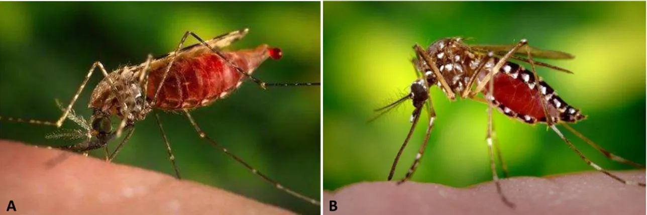
Todos os mosquitos machos se alimentam exclusivamente de líquidos de plantas, como néctar, mel, sumos de frutas e exudados. No entanto, com exceção dos gêneros Toxorhynchites, Malaya e Topomyia, as fêmeas de mosquitos se alimentam também de sangue de animais vivos, o que faz delas excelentes veículos para transmissão de patógenos, exibindo uma enorme importância médica (Fig. 1) (Harbach e Howard, 2007). Dentre os três gêneros de mosquitos não-hematófagos, Toxorhynchites possui um número muito maior de espécies e ampla distribuição geográfica, tornando-o mais representativo.
3
(autogenia). Fêmeas anautógenas não produzem ovos sem o repasto sanguíneo. Nas fêmeas autógenas, um lote de ovo é desenvolvido após a emergência do estágio adulto, mas alimentações de sangue são necessárias para a produção dos lotes posteriores (Clements, 1992).
Figura 1: Fêmeas hematófagas adultas de mosquitos realizando repasto sanguíneo. A: Fêmea da espécie Anopheles quadriannulatus, representante da subfamília Anophelinae. B: Fêmea da espécie Aedes aegypti, representante da família Culicinae. Fonte: https://www.vectorbase.org/.
A hematofagia surgiu independentemente várias vezes no curso da evolução dos artrópodes e muitas adaptações comportamentais, anatômicas e fisiológicas acompanharam essa tendência evolutiva (Justice et al, 2003). Dentre essas adaptações, as mandíbulas dos mosquitos foram modificadas para facilitar o acesso ao sangue; as glândulas salivares passaram a produzir moléculas capazes de bloquear a hemostasia; e o intestino médio foi modificado para neutralizar reações imunológicas mediadas pelo sangue e otimizar a digestão dos componentes sanguíneos (Ribeiro, 1996; Stark e James, 1996). No entanto, o grupo que engloba os mosquitos do gênero Toxorhynchites sofreu novas modificações adaptativas, perdendo a habilidade de se alimentar de sangue (Marquardt, 2005).
1.2. TOXORHYNCHITINI
A subfamília Culicinae é composta por 11 tribos, sendo a Toxorhynchitini uma delas (WRBU, 2014). Toxorhynchitini possui apenas um gênero, Toxorhynchites, que por sua vez engloba 4 subgêneros, Toxorhynchites, Afrorhynchus, Ankylorhynchus e Lynchiella. O subgênero Toxorhynchites ocorre apenas no Velho Mundo; Afrorhynchus é encontrado na África; e Ankylorhynchus e Lynchiella (a qual inclui a espécie em
4
estudo) são confinados ao Novo Mundo. Ao todo, são reconhecidas 90 espécies do gênero atualmente. Toxorhynchites foi anteriormente encaixado em uma subfamília exclusiva (Toxorhynchitinae) dentro do táxon Culicidae. Hoje, por apresentar maiores semelhanças morfológicas com culicíneos do que com anofelinos, e por se acreditar que a maioria das características distintivas deste gênero são características adaptativas secundárias relacionadas ao grande tamanho e aos hábitos de alimentação das larvas e adultos, Toxorhynchites foi incluído na subfamília Culicinae (WRBU, 2014).
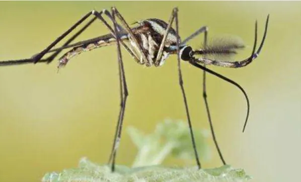
O gênero Toxorhynchites inclui os maiores mosquitos já identificados. Os adultos são facilmente reconhecidos pelo grande tamanho e pela peculiar probóscide dobrada (Fig. 2). O corpo é coberto por escamas iridescentes de cores vivas e os segmentos abdominais posteriores possuem tufos de escamas laterais. As larvas também são grandes e apresentam cores que variam entre o rosa, vermelho e roxo, o que também as torna de fácil identificação (WRBU, 2014).
Figura 2: Macho de Toxorhynchites rutilus, representante da tribo Toxorhynchitini (subfamília Culicinae). Fonte: http://bugguide.net/node/view/721549/bgpage
Toxorhynchites é encontrado na maioria das regiões tropicais e também em algumas regiões subtropicais e temperadas do mundo, e os principais tipos de vegetação que eles ocupam são as florestas (Muspratt, 1951). Suas larvas geralmente habitam buracos de árvores e bambus, mas algumas são vistas nas axilas de folhas, em plantas carnívoras, fendas de rochas e recipientes artificiais (Schreiber, 2007).
5
As larvas de Toxorhynchites são predadoras e, por isso, possuem partes bucais modificadas, compostas por 10 ou menos filamentos espessos achatados utilizados para agarrar as presas (WRBU, 2014). Elas se alimentam de invertebrados aquáticos, incluindo larvas de outros mosquitos, sendo úteis em tentativas de controle de culicídeos de importância epidemiológica, como Aedes aegypti, Aedes albopictus e Culex quinquefasciatus (Regis, 1995; Collins e Blackwell, 2000; Shaalan e Canyon, 2009; Nyamah et al, 2011). Algumas dessas tentativas falharam, o que pode ter ocorrido, pois, na elaboração de estratégias de controle biológico, é indispensável que a biologia de ambos espécie-alvo e agente potencial de controle sejam entendidas completamente, o que não é o caso do gênero Toxorhynchites. Além disso, a utilização desses predadores no controle de outras espécies esbarra na dificuldade de criação em laboratório para obtenção de mosquitos em grande quantidade (Steffan e Evenhuis, 1981; Collins e Blackwell, 2000; WRBU, 2014).
Como o gênero não está incluído na lista de “espécies-praga”, e não é vetor de doenças por não apresentar comportamento de hematofagia, a biologia geral e a taxonomia de Toxorhynchites têm sido amplamente negligenciadas. Exceções foram descrições da ecologia de algumas espécies, somado a um pequeno número de estudos taxonômicos (Collins e Blackwell, 2000).
Por fim, o peculiar comportamento alimentar das fêmeas de Toxorhynchites somado à falta de estudos sobre sua biologia revela a importância da investigação da morfofisiologia do órgão digestivo de espécies do gênero, a fim de revelar que modificações relacionadas com a dieta podem ter ocorrido durante sua evolução, além de aperfeiçoar o conhecimento geral sobre o sistema digestivo de Culicidae.
1.3. SISTEMA DIGESTIVO DE INSETOS
6
regiões principais: intestino anterior, intestino médio e intestino posterior (House, 1974; Chapman, 1985; Dow, 1986).
Invaginações do ectoderma denominadas estomodeu e proctodeu originam os intestinos anterior e posterior, respectivamente. Por serem derivadas de células ectodérmicas, essas porções do intestino são revestidas internamente por cutícula, característica que oferece limitações para sua capacidade de absorção e digestão (Snodgrass, 1935). A cobertura cuticular é secretada pelas próprias células epiteliais dessas regiões e é contínua com a cutícula do revestimento corporal (House, 1974). O intestino médio é a única estrutura do inseto que deriva da endoderme, e, por isso, não possui revestimento cuticular. Os túbulos de Malpighi se inserem entre o intestino médio e o intestino posterior como evaginações ectodérmicas desse último (Chapman, 1985; Lehane e Billingsley, 1996).
A primeira região do intestino anterior é a faringe, que é seguida pelo esôfago. Esse último consiste geralmente em um tubo simples que desemboca no intestino médio, embora em alguns insetos possa estar modificado em um “estômago” ou papo dilatável utilizado para estocar alimento (Klowden, 2007). Em insetos adultos das
ordens Diptera e Lepidoptera, o “estômago” existe como um divertículo separado do
resto do esôfago por um ducto (Winteringham, 1965; Kathuria, 1972). O alimento composto de açúcares é primeiro estocado no “estômago” e então passa vagarosamente para o intestino médio, enquanto proteínas são enviadas diretamente para o intestino médio (Clements, 1963; Klowden, 2007). A cobertura cuticular do “estômago” limita sua capacidade absortiva, fazendo com que este funcione basicamente como um reservatório alimentar, apesar de permitir a absorção de alguns lipídios. Embora a absorção seja limitada, a digestão pode ocorrer nessa região devido à ação continuada de enzimas das glândulas salivares presentes no bolo alimentar. A parte posterior do intestino anterior é modificada em um proventrículo muscular (cárdia) que funciona como um esfíncter, regulando a passagem do alimento para o intestino médio (House, 1974; Klowden, 2007).
7
superfície absortiva e secretora. Elas contêm várias redes de retículo endoplasmático necessárias para a produção de enzimas digestivas e absorvem a maioria dos nutrientes do lúmen intestinal (Chapman, 1985, Lehane e Billingsley, 1996). Essas células estão em constante renovação a partir das células-tronco regenerativas, que por sua vez estão localizadas na região do intestino médio adjacente à hemolinfa. As células regenerativas se dividem assimetricamente e tem a capacidade de se diferenciar tanto em células colunares quanto endócrinas (Chapman, 1985). Essas últimas, por fim, estão em contato com o lúmen e/ou com a hemolinfa, contêm muitos grânulos secretórios citoplasmáticos eletron-densos e são encontradas dispersas no epitélio intestinal geralmente como células simples, embora algumas vezes possam estar organizadas em pequenos grupos (Andriès e Tramu, 1985, Klowden, 2007).
A região anterior do intestino médio pode conter divertículos denominados cecos gástricos, que aumentam a área de superfície do órgão para a absorção e secreção de substâncias e criam um fluxo contracorrente dentro do intestino, resultante de sua absorção diferencial de água (Terra, 1990; Billingsley e Lehane, 1996). Esta é secretada no lúmen pelo intestino posterior, e se movimenta para frente para ser absorvida na região cecal, permitindo que sólidos não digeridos se movimentem de volta para serem finalmente digeridos e absorvidos no intestino médio (Billingsley e Lehane, 1996; Klowden, 2007).
Devido à ausência de cutícula, as células do intestino médio estão constantemente sujeitas a abrasão pelo alimento. Na grande maioria dos insetos, células do intestino médio produzem uma matriz extracelular denominada matriz peritrófica (MP), que consiste em uma estrutura saculiforme composta por microfibrilas de quitina, proteínas e glicoproteínas em uma matriz de proteoglicano (Moskalik et al., 1996; Lehane, 1997). As várias proteínas que suprem a MP são denominadas peritrofinas e possuem domínio de ligação a moléculas de quitina (Dinglasan et al., 2009). Proteínas e precursores de quitina são secretados por células digestivas do intestino médio, e a MP se organiza por intercalação de fibrilas de quitina e peritrofinas (Hegedus et al, 2009; Rose et al, 2014).
Existem dois tipos de MP com base em seus sítios de síntese (Peters, 1992;
8
alimento ao chegar no intestino médio, como ocorre tipicamente em insetos hematófagos. A MP do tipo II é formada sempre continuamente por um anel de células localizado na cárdia (ou válvula estomodeal), que produz um tubo em movimento retrógrado com função de englobar o bolo alimentar. Esse tubo então se estende revestindo o interior dos intestinos médio e posterior (Jacobs-Lorena e Oo, 1996; Lehane, 1997).
Distintos estágios de vida de um mesmo inseto podem produzir MPs de diferentes tipos (Tellam et al, 1999). Todos os tipos de MP contém poros e apresentam permeabilidade a algumas enzimas digestivas e certos produtos da digestão. Essa permeabilidade seletiva cria dois compartimentos; o espaço endoperitrófico, circundado pela MP; e o espaço ectoperitrófico, localizado entre a parede intestinal e a MP. A compartimentalização das enzimas digestivas e seus produtos resultante da presença da MP leva a uma maior eficiência na digestão por manter as enzimas e seus substratos específicos concentrados em um mesmo compartimento (Lehane, 1977; Hegedus et al, 2009).
O intestino médio também age como uma barreira contra patógenos, cuja principal importância está ligada à transmissão de doenças pelos vetores hematófagos (Arias-Goeta et al, 2013). As enzimas digestivas e a MP secretadas nessa porção do intestino oferecem resistência ao desenvolvimento dos parasitas. Além disso, a permeabilidade seletiva da MP pode proteger o inseto contra possíveis toxinas presentes no alimento, impedindo-as de atravessarem a MP e afetarem diretamente as células intestinais (Abraham and Jacobs-Lorena, 2004; Arias-Goeta et al, 2013; Agrawal et al, 2014).
9
Após a digestão do alimento no intestino médio, o material não digerido segue em direção ao intestino posterior, que é composto por piloro, íleo e reto. O piloro é a válvula de onde os túbulos de Malpighi surgem. O íleo consiste em um tubo estreito seguido pelo reto, que é mais alargado (Klowden, 2007). O intestino posterior, cuja principal função é osmoregulação, é revestido por uma cutícula mais permeável que a do intestino anterior e contém a abertura dos túbulos de Malpighi, que por sua vez funcionam produzindo uma “pré-urina” isosmótica a partir da hemolinfa contendo íons, aminoácidos e metabólitos (Hanrahan et al, 1984). O intestino posterior reabsorve seletivamente água, aminoácidos e alguns íons, produzindo uma urina hiper ou hiposmótica, que é excretada juntamente com o bolo fecal. Esse bolo é formado por produtos não digeridos e materiais descartáveis que passam pelo intestino posterior e perdem água (Phillips et al, 1987, Klowden, 2007).
1.4. INTESTINO MÉDIO DE MOSQUITOS
Somente os mosquitos que se alimentam de sangue estão envolvidos na transmissão de patógenos. Por esse motivo, as pesquisas são mais direcionadas ao seu estudo, sendo grande parte da revisão bibliográfica voltada ao conhecimento sobre sistema digestivo de fêmeas hematófagas.
Alguns estudos com glândulas salivares de mosquitos mostraram que tais estruturas são sexualmente dimórficas nas espécies em que as fêmeas são hematófagas. As glândulas salivares dos machos são menores e não têm, ou possuem pouca atividade relacionada com a ingestão de sangue, como a produção de substâncias antiplaquetárias, anticoagulantes e vasodilatadoras (Ribeiro et al, 1984; Rossignol e Lueders, 1986; Marinotti et al, 1990; Moreira-Ferro et al, 1999). Porém, experimentos utilizando Toxorhynchites revelaram que suas glândulas salivares são sexualmente monomórficas, refletindo o tipo idêntico de dieta do macho e da fêmea (Jariyapan et al, 2004). Tal achado oferece uma pista de que modificações evolutivas importantes ocorreram no trato digestivo desses insetos.
10
propícia a modificações adaptativas relacionadas ao tipo específico de dieta, seu estudo torna-se de maior interesse.
O intestino médio dos mosquitos compreende duas regiões distintas que se diferem morfológica e funcionalmente: intestino médio anterior (IMA) e intestino médio posterior (IMP). O IMA configura-se como um tubo simples relacionado com a digestão/absorção de açúcares (Billingsley, 1990). O IMP está mais envolvido com a digestão de sangue e compreende um saco expansível cujas células são multifuncionais para desempenhar pelo menos três papéis: regulação hídrica pós-repasto sanguíneo; síntese e secreção de enzimas digestivas e componentes da MP; e absorção de nutrientes (Billingsley, 1990).
Figura 3: Visão geral do trato digestivo de mosquitos, com destaque para o intestino médio. Modificado
de Rebecca et al, 2014.
Quando o alimento é uma solução açucarada, como néctar, ele é estocado primeiramente no “crop” ou “estômago”, e vai passando aos poucos para o intestino médio. Se o alimento é sangue, ele vai diretamente para o intestino médio, sendo armazenado no IMP. O principal fator que determina o destino do alimento é sua composição química (Gooding, 1972), apontando para a existência no mosquito de receptores para glicose e/ou para alguma substância presente no sangue. Além disso, sabe-se que o néctar possui inibidores de proteinases, e isso pode ser um dos motivos pelo qual ocorre essa separação espacial do tipo de alimento (Gooding, 1973), essencial no caso das fêmeas, por possuírem ambos os tipos de dietas.
Nas espécies de mosquitos cujas fêmeas são hematófagas, o intestino médio de
fêmeas e machos difere quanto ao tamanho e morfologia. As fêmeas possuem região
anterior reduzida e região posterior mais desenvolvida que os machos (Christophers,
11
O epitélio do IMA é responsável pela digestão e absorção de carboidratos
(Billingsley e Hecker, 1991; Billingsley, 1990). Nas fêmeas hematófagas, a presença de um plugue de material da matriz peritrófica (MP) no início do IMP impede o envolvimento direto do IMA na digestão de sangue (Perrone e Spielman, 1988; Billingsley e Rudin, 1992). Além disso, glicosidases secretadas pelas células do IMA ajudam a suportar um papel dessa porção do intestino na digestão de carboidratos (Billingsley, 1989). Esta porção é bem suprida por terminações nervosas, ao contrário do IMP, o que colabora com a idéia de que essas duas regiões funcionam independentemente (Hecker, 1977; Billingsley, 1990).
Tanto as células epiteliais do IMA quanto as do IMP possuem características ultraestruturais que apontam para as funções de estocagem, síntese, secreção e absorção de substâncias (Bertram e Bird, 1961; Rudin e Hecker, 1976).
As células que compõem o IMA normalmente possuem uma lâmina basal contínua em contato com a hemolinfa, microvilos densamente empacotados e um labirinto basal bem formado (Hecker et al, 1974; Hecker, 1977). As membranas plasmáticas altamente dobradas dos microvilos (região apical) e labirinto (região basal) são típicas do epitélio absortivo. O retículo endoplasmático rugoso (RER) das células do IMA ocorre como pequenas cisternas simples em Aedes aegypti, e cisternas mais longas e pobremente organizadas em Anopheles gambiae, Anopheles stephensi, e Culex pipiens fatigans. O retículo endoplasmático liso (REL) é encontrado denso e altamente organizado nessa região (Hecker, 1977).
O IMP possui células secretoras apresentando pilhas e espirais de retículo endoplasmático rugoso (RER); aparelho de Golgi numeroso e pobremente organizado; e vesículas secretoras. Células com função de estocagem também foram encontradas nessa região, contendo depósitos de lipídio e glicogênio (Bertram e Bird, 1961; Reinhardt e Hecker, 1973). O glicocálice das células do IMP de Culex tarsalis possuem glicosaminoglicanos e/ou glicoproteínas ácidas, enquanto que as membranas plasmáticas laterais, o labirinto basal e a lâmina basal são altamente aniônicos, consistente com seu papel regulatório (Houk et al, 1986a; Houk et al, 1986b).
12
presença de junções intercelulares em suas membranas laterais. Em espécies de Anopheles, as células do IMP possuem apenas desmossomos septados como junções intercelulares (Hecker, 1977) Em culicíneos, são encontradas junções do tipo zonula continua próximas ao ápice celular, e desmossomos nas regiões basais (Reinhardt e Hecker, 1973; Hecker, 1977; Houk e Hardy, 1979).
As células epiteliais do intestino médio de fêmeas adultas de mosquitos produzem MP do tipo I em resposta à ingestão de sangue. Vários papéis foram propostos para essa estrutura, dentre eles o de proteção contra patógenos e proteção física do epitélio; filtração semipermeável de proteínas e enzimas digestivas; barreira de retenção para inibidores de proteinases; e uma camada para prevenir o entupimento dos microvilos por material do lúmen (Marquardt, 2005).
A MP impede que o alimento ingerido fique em contato direto com o epitélio. A formação dessa matriz ocorre diferentemente em culicíneos e anofelinos. Nesses últimos, o material precursor da matriz já está presente em grânulos, que são liberados após a ingestão de sangue (Hecker, 1977). Em culicíneos, a MP é formada “de novo” pelas células do IMP (Rudin e Hecker, 1976).
Para a formação da MP em anofelinos, grânulos de secreção apicais das células digestivas são liberados no lumen do IMP, exclusivamente em resposta ao estresse mecânico durante a alimentação. Esses grânulos são repostos enquanto ocorre a digestão subsequente (Berner et al, 1983; Billingsley e Rudin, 1992). Os seus conteúdos coalescem e então condensam para formar a MP (Berner et al, 1983).
Em culicíneos, algum tempo após a refeição sanguínea, forma-se uma MP discernível mostrando uma estrutura densamente laminada próxima aos microvilos, que por sua vez tornam-se progressivamente menos estruturados e, finalmente, a MP atinge sua estrutura altamente organizada, com numerosas camadas (Perrone e Spielman, 1988).
13
(Clements, 1992). As células endócrinas podem ser distinguidas das demais células do sistema digestivo devido à sua estrutura refinada. Em A. aegypti, pelo menos 500 células endócrinas estão presentes por epitélio do IMP, sendo concentradas posteriormente e constituindo o maior órgão endócrino do mosquito (Brown et al., 1985). Pelo menos dois tipos de células endócrinas estão presentes, as células claras e as escuras, e cerca de 25% dessas células são encontradas adjacentes às células regenerativas do epitélio. Dentre esses dois tipos, são encontradas células que atingem e que não atingem a superfície luminal, essas primeiras contendo microvilosidades e em nenhuma delas havendo labirinto basal. Vesículas secretoras, produzidas pelo complexo de Golgi, são geralmente concentradas no terço basal do citoplasma dessas células, e várias formas de corpos lamelares estão também presentes. O conteúdo das vesículas é liberado diretamente na hemolinfa por exocitose (Brown et al., 1985; et al., 1987; Billingsley e Lehane, 1996).
Dois hormônios peptídicos, o neuropeptídeo semelhante ao FMRFamida de moluscos e o hormônio semelhante ao polipeptídio pancreático de vertebrados (PP), foram localizados nas células endócrinas intestinais de algumas espécies de mosquitos. Ambos os hormônios são encontrados em vesículas tanto das células endócrinas claras quanto das escuras, mas nem todas as células apresentam imunoreatividade (Brown et al, 1986;Glanttli et al, 1987). A marcação das células enteroendócrinas com anticorpos anti-FMRFamida é a mais utilizada para auxiliar na sua distinção entre os tipos celulares do epitélio.
Em relação ao processo digestivo enzimático, quando o mosquito ingere sangue, a sua presença no intestino médio estimula a secreção de enzimas digestivas no lúmen intestinal, cujas principais são as proteolíticas (Billingsley, 1991; Terra, 1996). Enzimas proteolíticas (ou proteases) são divididas em endo e exo-peptidades. Endopeptidases são moléculas relativamente pequenas (~25-30 kDa) que clivam grandes complexos proteicos e são capazes de passar através dos poros e espaços da MP. Exopeptidades são enzimas maiores (>100 kDa) geralmente ligadas à membrana plasmática do epitélio intestinal que hidrolisam as regiões terminais de pequenas proteínas e peptídeos (Borges-Veloso et al., 2012; Weidlich et al., 2012).
14
de proteases podem aumentar 20 vezes. A síntese da tripsina inicial é aparentemente parte de um sistema único de transdução de sinais e sua síntese geralmente é induzida pela presença de aminoácidos livres encontrados no bolo sanguíneo (Noriega et al, 1999; Marquardt, 2005). Acredita-se que sua função pode ser determinar se há proteína suficiente para suportar o ciclo gonadotrófico. Quando isso ocorre, vias de transdução de sinais ativam a transcrição da tripsina tardia, e de genes que codificam outros tipos de enzimas digestivas para digerir o sangue. Dentre esses outros tipos, as principais são as amino e carboxipeptidades (Marquardt, 2005;Weidlich et al, 2012).
Imediatamente após a emergência das fêmeas adultas de mosquito, o intestino médio ainda não está completamente diferenciado, o que as impede de se alimentarem de sangue (Hecker, 1971). As células do IMP de A. aegypti nesse momento são extremamente irregulares na forma, possuem poucos microvilos e um aparato sintético-secretório precariamente desenvolvido. Dentro de um dia após a emergência, desmossomos se desenvolvem ao longo da margem celular apical, os microvilos formam uma “borda em escova” completa, e o retículo endoplasmático rugoso se desenvolve em pilhas e espirais bem organizadas. Três dias após a emergência, as células do IMP estão maduras formando um epitélio colunar regular e os mosquitos estão prontos para realizar o repasto sanguíneo (Hecker, 1971; Hecker et al, 1974) . Em Anopheles spp., processo de maturação semelhante ocorre, mas não envolve desenvolvimento de desmossomos e inclui síntese de vesículas apicais contendo precursores da MP (Berner et al, 1983; Hecker, 1977). Se o mosquito não realiza o repasto sanguíneo, as células do IMP exibem sinais de processos degenerativos, como aumento das frações de lisossomos e mitocôndrias, e perda drástica da quantidade de retículo endoplasmático (Bauer et al, 1977).
15
2. REFERÊNCIAS BIBLIOGRÁFICAS
Abraham, E.G. and Jacobs-Lorena, M. Mosquito midgut barriers to malaria parasite development. Insect Biochemistry and Molecular Biology 34, 667-671 (2004).
Agrawal, S.; Kelkenberg, M.; Begum, K.; Steinfeld, L.; Williams, C.E.; Kramer, K.J.; Beeman, R.W.; Park, Y.; Muthukrishnan, S. and Merzendorfer, H. Two essential peritrophic matrix proteins mediate matrix barrier functions in the insect midgut. Insect Biochemistry and Molecular Biology 49, 24-34 (2014).
Andriès, J.C. and Tramu, G. Ultrastructural and immunohistochemical study of endocrine cells in the midgut of the cockroach Blaberus craniifer (Insecta, Dictyoptera). Cell and Tissue Research 240, 323-332 (1985).
Arias-Goeta, C.; Mousson, L.; Rougeon, F. and Failloux, A.B. Dissemination and Transmission of the E1-226V Variant of Chikungunya Virus in Aedes albopictus are Controlled at the Midgut Barrier Level.Plos One 8, e57548 (2013).
Bauer, P.G.; Rudin, W. and Hecker, H. Ultrastructural changes in midgut cells of female Aedes aegypti L. (Insecta, Diptera) after starvation or sugar diet. Cell Tissue Research 177, 15-19 (1977).
Berner, R.; Rudin, W. and Hecker, H. Peritrophic membranes and protease activity in the midgut of the malaria mosquito, Anopheles stephensi (Liston) (Insecta: Diptera) under normal and experimental conditions. Journal of Ultrastructure Research 83, 195-204 (1983).
Bernick, E.P.; Moffett, S.B. and Moffett, D.F. Ultrastructure and morphology of midgut visceral muscle in early pupal Aedes aegypti mosquitoes. Tissue and Cell 40, 127-141 (2008).
16
Besansky, N.J. and Fahey, G.T. Utility of the white gene in estimating phylogenetic relationships among mosquitoes (Diptera: Culicidae). Molecular Biology and Evolution 14, 442-454 (1997).
Billingsley, P.F. Blood Digestion in the Mosquito, Anopheles stephensi Liston (Diptera:
Culicidae): Activity and Distribution of Trypsin, Aminopeptidase, and α-Glucosidase in
the Midgut (1991).
Billingsley, P.F. The midgut Ultrastructure of hematophagous insects. Annual Review of Entomology 35, 219-48 (1990).
Billingsley, P.F. and Hecker, H. Blood Digestion in the Mosquito, Anopheles stephensi Liston (Diptera: Culicidae): Activity and Distribution of Trypsin, Aminopeptidase, and
α-Glucosidase in the Midgut.Journal of Medical Entomology 28, 865-871 (1991).
Billingsley, P.F. and Lehane, M.J. Structure and ultrastructure of the insect midgut.
Biology of the Insect Midgut, 3-30 (1996).
Billingsley, P. F. and Rudin, W. The role of the mosquito peritrophic membrane in
digestion and malaria infectivity. Journal of Parasitology 78, 430-40 (1992).
Borges-Veloso, A., Saboia-Vahia, L., Cuervo, P., Pires, R. C., Britto, C., Fernandes, N.,
d’Avila-Levy, C. M. and De Jesus, .J. B. Proteolytic profiling and comparative analyses of active trypsin-like serine peptidases in preimaginal stages of Culex quinquefasciatus. Parasites and Vectors 5, 113-123 (2012).
Brown, M. R., Raikhel, A. S. and Lea, A. O. Ultraestructure of midgut endocrine cells in the adult mosquito, Aedes aegypti (Diptera). Tissue and Cell 17, 709-721 (1985). Brown, M.R.; Crim, J.W. and Lea, A.O. FMRFamide- and pancreatic polypeptide-like immunoreactivity of endocrine cells in the midgut of a mosquito. Tissue and Cell 18, 419-428 (1986).
Chapman, R.F. Structure of the digestive system. Comprehensive insect physiology, biochemistry, and pharmacology 4, 165–211 (1985).
17
Clements, A.N. The Biology of Mosquitoes. Development, Nutrition, and Reproduction. Chapman & Hall, New York and London (1992).
Consoli, R. A. G. B. and Lourenço-de-Oliveira, R. Principais mosquitos de importância sanitária no Brasil. Rio de Janeiro, Editora Fiocruz, 225p (1994).
Collins, L.E. and Blackwell, A. The biology of Toxorhynchites mosquitoes and their potential as biocontrol agents. Biocontrol News and Information 21, 105-116 (2000). Dinglasan, R.R., Devenport, M., Florens, L, Johnson, J.R., McHugh, C.A., Donnelly-Doman, M., Carucci, D.J., Yates, J.R. and Jacobs-Lorena, M. The Anopheles gambiae adult midgut peritrophic matrix proteome. Insect Biochemistry and Molecular Biology 39, 125-134 (2009).
Dow, J.A.T. Insect midgut function. Advances in insect physiology 19, 87–328 (1986). Eliason, P. A. Feeding adult mosquitoes on solid sugars. Nature 200 289 (1963).
Foley, D.H.; Rueda, L.M. and Wikerson RC. Insight into global mosquito biogeography from country species records. Journal of Medical Entomology 44, 554-567 (2007). Forattini, O.P. Culicidologia Médica. EDUSP, São Paulo, 864p. (2002).
Glattli, E.; Rudin , W. and Hecker, H. Immunoelectron microscopic demonstration of pancreatic polypeptide in midgut epithelium of hematophagous dipterans. Journal of Histochemistry and Cytochemistry 35, 891-896 (1987).
Gooding, R.H. Digestive processes of haematophagous insects. I. A review literature. Quaestiones entomologicae 8, 5-60 (1972).
Gooding, R.H., Cheung, A.C. and Rolseth, B.M. The digestive processes of haematophagous insects. III. Inhibition of trypsin by honey and the possible functions of the oesophageal diverticula of mosquitoes (Diptera). Canadian Entomologist 105, 433-436 (1973).
18
Hanrahan, J. W., Meredith, J., Phillips, J. E. and Brandys D. Methods for the Study of Transport and Control in Insect Hindgut. Measurement of Ion Transport and Metabolic Rate in Insects in Experimental Entomology 19-67 (1984).
Harbach, R.E. Mosquito Taxonomic Inventory, http://mosquito-taxonomic-inventory.info/, (2014).
Harbach, R.E. and Kitching, I. J., Phylogeny and classification of the Culicidae (Diptera). Systematic Entomology 23, 327–370 (1998).
Harbach, R.E. and Howard, T.M. Index of currently recognized mosquito species (Diptera: Culicidae). European Mosquito Bulletin 23, 1-66 (2007).
Hecker, H. Structure and function of midgut epithelial cells in Culicidae mosquitoes (Insect, Diptera). Cell Tissue Research 84, 321-4 (1977).
Hecker, H.; Brun, R.; Reinhardt, C. and Burri, P.H. Morphometric analysis of the midgut of female Aedes aegypti (L.) (Insect, Diptera) under various physiological conditions. Cell Tissue Research 152, 1-49 (1974).
Hecker, H.; Freyvogel, T.A.; Briegel, H. and Steiger, R. Ultrastructural differentiation of the midgut epithelium in female Aedes aegypti (L.) (Insecta, Diptera) imagines. Acta Tropica 28, 80-104 (1971).
Hegedus, D., Erlandson, M., Gillott, C. and Toprak, U. New insights into peritrophic matrix synthesis, architecture, and function. Annual Review of Entomology 54, 285-302 (2009).
Houk, E.J. and Hardy, J.L. In vivo negative staining of the midgut continuous junction in the mosquito, Culex tarsalis (Diptera : Culicidae). Acta Tropica 36, 267-75 (1979). Houk, E.J.; Chiles, R.E. and Hardy, J.L. Unique midgut basal lamina in the mosquito, Aedes dorsalis (Meigen) (Insecta, Diptera). International Journal of Insect Morphology and Embryology 9,161-164 (1980).
19
Houk, E.J., Hardy, J.L. and Chiles, R.E. Histochemical staining of the complex carbohydrates of the midgut of the mosquito, Culex tarsalis Coquillett. Insect Biochemistry 16, 667-675 (1986a).
Houk, E.J.; Hardy, J.L. and Chiles, R.E. Mesenteronal epithelial cell surface charge of the mosquito, Culex tarsalis Coquillett. Binding of colloidal iron hydroxide, native ferritin and cationized ferritin. Journal of Submicroscopic Cytology and Pathology 18, 385-96 (1986b).
House, H.L.. Digestion. In The Physiology of Insecta, Academic Press, New York, 5, 63-117 (1974).
Jacobs-Lorena, M. and Oo, M-M. The peritrophic matrix of insects. In: Beaty BJ, Marquardt WC, editors. The Biology of Disease Vectors. Niwot, Colorado: University Press of Colorado, 318-332 (1996).
Jariyapan, N.; Choochote, W.; Jitpakdi, A. and Bates, P.A. Salivary gland of Toxorhynchites splendens Wiedemann (Diptera: Culicidae): ultrastructural morphology and electrophoretic protein profiles. Journal of Medical Entomology 41, 569-574 (2004).
Kathuria, O. P. Metamorphic changes in posterior region of foregut of Papilio aristolochae F.( Lepidoptera: Papilioidae). International Journal of Insect Morphology and Embryology 1, 163-168 (1972).
Klowden, M.J. Physiological Systems in Insects. Second Edition (2007).
Lehane, M.J. Peritrophic matrix structure and function. Annual Review of Entomology 42, 525-550 (1997).
Lehane, M.J., Billingsley, P.F. The biology of the insect midgut. Chapman & Hall, London (1996).
Marquardt, W.C. Biology of Disease Vectors, 2nd ed. Elsevier Academic Press, Burlington, Massachusetts. Second Edition, 785 p. (2005).
20
Miller, B.R.; Crabtree, M.B. and Savage, H.M. Phylogenetic relationships of the Culicomorpha inferred from 18S and 5.8S ribosomal DNA sequences (Diptera: Nematocera). Insect Molecular Biology 6, 105-114 (1997).
Moreira-Ferro, C.K.; Marinotti, O. and Bijovsky, A.T. Morphological and biochemical analyses of the salivary glands of the malaria vector, Anopheles darlingi. Tissue and Cell 31, 264-273 (1999).
Moskalyk, L.A. Oo, M.M. and Jacobs-Lorena, M. Peritrophic matrix proteins of Anopheles gambiae and Aedes aegypti. Insect Molecular Biology 5, 261-268 (1996). Muspratt, J. The bionomics of an African Megarhinus (Diptera, Culicidae) and its possible use in biological control. Bulletin of Entomological Research 42, 355-370 (1951).
Noriega, F.G., Colonna, A.E. and Wells, M.A. Increase in the size of the amino acid poll is sufficient to activate translation on early trypsin mRNA in Aedes aegypti midgut. Insect Biochemistry and Molecular Biology 29, 243-247 (1999).
Nyamah, M.A.; Sulaiman, S. and Omar B. Field observation on the efficacy of Toxorhynchites splendens (Wiedemann) as a biocontrol agent against Aedes albopictus (Skuse) larvae in a cemetery. Tropical Biomedicine 28, 312-319 (2011).
Pawlowski, J.; Szadziewski, R.; Kmieciak, D.; Fahmi, J. and Bittar, G. Phylogeny of the infraorder Culicomorpha (Diptera: Nematocera) based on 28S RNA gene sequences. Systematic Entomology 21, 167-178 (1996).
Perrone, J. B. and Spielman, A. Time and site of assembly of the peritrophic membrane of the mosquito Aedes aegypti. Cell and Tissue Research 252, 473-78 (1988).
Peters, W.. Peritrophic membranes. In: Bradshaw D, Burggren W, Heller HC, Ishii S, Langer H, Neuweiler G, Randall DJ, editors. Zoophysiology, 130, Berlin (1992).
21
Reidenbach, K.R., Cook, S., Bertone M.A., Harbach, R.E., Wiegmann, B.M. and Besansky, N.J. Phylogenetic analysis and temporal diversification of mosquitoes (Diptera: Culicidae) based on nuclear genes and morphology. BMC Evolutionary Biology 9, 298 (2009).
Reinhardt, C. and Hecker, H. Structure and function of the basal lamina and cell junctions in the midgut epithelium (stomach) of female Aedes aegypti L. (Insecta, Diptera). Acta Tropica 30, 13-36 (1973).
Reynolds E.S. The use of lead citrate at high pH as an electron-opaque stain in electron microcopy. Journal of Cell Biology 17, 208-212 (1963).
Ribeiro, J.M.C.; Sarkis, J.J.F.; Rossignol, P.A. and Spielman, A. Salivary apyrase of Aedes aegypti: Characterization and secretory fate. Comparative Biochemistry and Physiology, 79, 81-86 (1984).
Rose, C.; Belmonte, R.; Armstrong. S.D.; Molyneux, G.; Haines, L.R.; Lehane, M.J.; Wastling, J. and Acosta-Serrano, A. An investigation into the protein composition of the teneral Glossina morsitans morsitans peritrophic matrix. Plos Neglected Tropical Diseases 8, e2691 (2014).
Rossignol, P.A.; Lueders, A.M. Bacteriolytic factor in the salivary glands of Aedes aegypti. Comparative Biochemistry and Physiology 83, 819-822 (1986).
Rudin ,W. and Hecker, H. Morphometric comparision of the midgut epithelial cells in male and female Aedes aegypti L. (Insecta, Diptera).Tissue and cell 8, 459-470 (1976). Schreiber, E.T. Toxorhynchites. Journal of the American Mosquito Control Association 23, 129-132 (2007).
Shaalan, E.A.S. and Canyon, D.V. Aquatic insect predators and mosquito control. Tropical Biomedicine 26, 223-261 (2009).
Silva, C.P.; Lemos, F.J.A. and Silva, J.R. Tópicos avançados em Entomologia Molecular. Instituto Nacional de Ciência e Tecnologia em Entomologia Molecular (2012).
22
Springer-Verlag.Preuss, U.; Landsberg, G. and Scheidtmann, K.H. Novel mitosis-specific phosphorylation of histone H3 at Thr11mediated by Dlk/ZIP kinase. Nucleic Acids Research 31, 878-85 (2003).
Steffan, W.A.; Evenhuis, N.L. Biology of Toxorhynchites. Annual Review of Entomology, 26, 159-181 (1981).
Tellam, R.L.; Wijffels, G. and Willadsen, P. Peritrophic matrix proteins. Insect Biochemistry and Molecular Biology 29, 87-101 (1999).
Tellam, R.L. and Eisemann, C. Chitin is only a minor component of the peritrophic matrix from larvae of Lucilia cuprina. Insect Biochemistry and Molecular Biology 30, 1189-2001 (2000).
Terra, W.R. Evolution and function of insect peritrophic membrane. Ciência e Cultura
48, 317324 (1996).
Terra, W.R. Evolution of digestive systems of insects. Annual Review of Entomology
35, 181-200 (1990).
Thompson, M.T. Alimentary canal of the mosquito. Proceedings of the Boston Society of Natural History 32, 145-202 (1905).
Weidlich, S.; Huster, J.;, Hoffmann, H. and Woodring, J. Environmental control of trypsin secretion in the midgut of the two-spotted field cricket, Gryllus bimaculatus. Journal of Insect Physiology 58, 1477-1484 (2012).
Wiegmann, B.M.; Trautwein, M.D.; Kim, J.W.; Cassel, B.K.; Bertone, M.A.; Winterton S.L. and Yeates, D.K. Single-copy nuclear genes resolve the phylogeny of the holometabolous insects. BMC Biology 7, 34 (2009).
Winteringham, F.P. Some distinctive features of insect metabolism. Biochemical Society Symposium 25, 29-37 (1965).
23
3. OBJETIVOS
3.1. OBJETIVOS GERAIS
Analisar e caracterizar a estrutura do intestino médio de mosquitos adultos de Toxorhynchites theobaldi.
3.2. OBJETIVOS ESPECÍFICOS
a) Descrever a morfologia e caracterizar os tipos celulares do intestino médio; b) Verificar se há diferenças morfológicas entre os epitélios do intestino médio anterior e posterior.
24
4. ARTIGO
Midgut of the non-hematophagous mosquito Toxorhynchites theobaldi (Diptera,
Culicidae)
Raquel S. M. Godoy, Kenner M. Fernandes, Gustavo F. Martins*
Departamento de Biologia Geral - Universidade Federal de Viçosa, 36570-900 Viçosa,
Minas Gerais, Brasil
Email list:
Raquel S. M. Godoy: raquel.godoy@ufv.br
Kenner M. Fernandes: kennerbio@yahoo.com.br
Gustavo F. Martins: gmartins@ufv.br
* Corresponding author: Professor Gustavo Ferreira Martins
Departamento de Biologia Geral - Universidade Federal de Viçosa, 36570-900 Viçosa,
Minas Gerais, Brazil
Tel# +553138993492
25
Abstract
In most mosquito species, the females require a blood-feeding for complete egg
development. However, in Toxorhynchites mosquitoes, the eggs develop without
blood-feeding, and both females and males exclusively feed on sugary diets. The midgut is a
well-understood organ in blood-feeding mosquitoes, but little is known about it in
non-blood-feeding ones. In the present study, the detailed morphology of the midgut of
Toxorhynchites theobaldi were investigated using histochemical and ultrastructural
methods. The midgut of female and male T. theobaldi adults consists of a long, slender
anterior midgut (AMG), and a short, dilated posterior midgut (PMG). The AMG is
subdivided into AMG1 (short, with folds) and AMG2 (long, without folds). Nerve
branches and enteroendocrine cells are present in AMG and PMG, respectively.
Compared with the PMG of blood-feeding female mosquitoes, the PMG of T. theobaldi
is smaller; however, in both mosquitoes, PMG seems be the main region of food
digestion and absorption, and protein secretion. The epithelial folds present in the AMG
of T. theobaldi have not been reported in other mosquitoes; however, the midgut muscle
organization and endocrine control of the digestion process are conserved in both T.
theobaldi and blood-feeding mosquitoes.
26
Introduction
The family Culicidae (Diptera) is monophyletic and consists of all mosquito
species 1, including species of the tribe Toxorhynchitini 2. This tribe includes a single
genus, Toxorhynchites, comprising approximately 93 species 3. Unlike most
mosquitoes, in Toxorhynchites, females are not hematophagous 4,5,6. Hence, egg
development does not depend on blood supply and, as adults, both males and females
feed exclusively on nectar, honey, and other sugary substances 3,4,7. Lack of
hematophagy is not an exclusive characteristic of Toxorhynchites and is shared with
other genera (e.g., Malaya and Topomyia) in the family Culicidae. Among the
non-hematophagous mosquitoes, Toxorhynchites has a greater number of species and wider
geographic distribution 8, making this genus more representative.
The midgut is the portion of the digestive tract responsible for digestion of
food in mosquitoes 9,10. In adult mosquitoes, the midgut has two portions, which differ
morphologically and functionally: the anterior midgut (AMG) is mainly associated with
sugar digestion and absorption 11,12; and the posterior midgut (PMG), which is an
expandable sac whose cells are involved in blood digestion (females exclusively), water
regulation, digestive enzyme and peritrophic matrix (PM) component synthesis and
secretion, and nutrient absorption 9,13,14.
Unlike the PMG, the AMG of adult mosquitoes is well supplied by nerve
endings 13. However, both AMG and PMG are enclosed externally by circular and
longitudinal muscles, which assist in food movement and provide structural integrity
10,15
. The midgut epithelium is adjacent to the muscle fibers, and is predominantly made
up of digestive cells. These cells actively participate in nutrients digestion and
absorption, with two typical types of cell membrane specializations: microvilli and
27 cells, related to the control of digestive processes through the release of hormones and
neuropeptides; and regenerative cells, responsible for the renewal of midgut epithelium
10,13,16 .
The midgut in blood-feeding female mosquitoes is the site of blood digestion
and the gateway for establishment of various human pathogen, including viruses,
protozoa, and nematodes 17-19. This explains why the midgut is one of the most
understood organs in mosquitoes. However, there has been little research on the midgut
of non-hematophagous mosquitoes, such as Toxorhynchites. Therefore, in the present
study, the midgut morphological and functional characteristics of female and male
Toxorhynchites theobaldi were investigated, and the differences between this species
and blood-feeding mosquito species were discussed. Additionally, this study will also
help in understanding the overall morphophysiology of the Culicidae midgut.
Results
General morphology and histology
The midguts of both female and male T. theobaldi consist of a long, slender
AMG, and a smaller, dilated PMG. In both females and males, the AMG is divided into
two distinct parts: AMG1, with folds on the surface and located in the thorax; and
AMG2, without folds and located in abdomen (Fig. 1a and Sup. Fig. a). The total length
of the midgut was 6.1 mm in females and 4.5 mm in males, however, length and width
of the different regions of the midgut were proportional between females and males.
The length of the AMG corresponded to ~84% of the total midgut length. The length of
AMG1 corresponded to a quarter of the total length of the AMG. The width of PMG
28 In the three regions of the T. theobaldi midgut (AMG1, AMG2, and PMG)
there was a single cell layer epithelium with cells displaying brush borders (Figs. 2a, 2g,
and 3a). The AMG1 epithelium was continuous with the cardia epithelium
(proventriculus or the transition between the foregut and midgut) and had many
wrinkles or folds (Figs. 2a and 2d). In AMG2 and PMG, no folds were seen, but
undulations occurred in the basal region of the epithelium, where the circular muscles
are inserted (Figs. 2g and 3a). In AMG1, digestive cells were approximately of the same
height (Fig. 2a), unlike AMG2, where cells exhibited different heights, forming a
narrow lumen with an “X” shape when cross-sectioned (Fig. 2f). In the PMG, digestive
cells were aligned with the nucleus positioned at the same height (Fig. 3a). The height
of the epithelium and the thickness of brush border in each of the three-midgut regions
were similar between females and males (p > 0.05). Digestive cells in PMG were higher
(43.34 µm) and had thicker brush borders (11.31 µm) than AMG1 and AMG2, which
had similar measurements (22.78 µm epithelial height and 3.59 µm brush border) (Fig.
1c).
In the three regions of the midgut, digestive cells had elongated or rounded
nuclei, and their supranuclear portion was predominantly acidic (Figs. 2c and 3a). The
subapical cell region, immediately below the brush border, was negative for the PAS
reaction, but rich in proteins. This region was thicker or more evident in PMG (Figs. 2d,
3d-3e). The brush border on all regions of the midgut was positive for the PAS reaction
(Figs. 2a, 2f and 3d). Besides digestive cells, other cells were seen in the basal region of
the midgut epithelium (Fig. 2c and 3a). These basal cells could correspond to
regenerative or enteroendocrine cells. In whole mounts, they had small nuclei with no
defined shape, unlike the digestive cells with large, elongated, and regular nuclei (Figs.
29 The lumen of AMG1 and AMG2 was narrow, sometimes with opposite brush
borders very close to each other (Figs. 2a and 2f), while the PMG lumen was large (Fig.
3c). In AMG1 and PMG, structures protruding from the apical side of the digestive cells
towards midgut lumen were visualized, characterizing the process of aprocrine
secretion. These structures were positive for proteins (Figs. 2d and 3b), and negative for
PAS reaction (Supplementary Figs. d-e). In AMG1, these structures were basic, while in
PMG, they were acidic (Figs. 2c and 3a). This type of cell process was not seen in
AMG2.
In midguts where the food bolus is being transferred to the hindgut, digestive
cells of AMG and PMG exhibited intense staining for the PAS reaction. This staining
was observed in the apical and basal regions, or only in the basal region of cells (Figs.
2e and 3d).
A PAS-positive material, corresponding to the PM, was also seen in the lumen
of midgut, including inside epithelial folds of AMG1 (Figs. 2b, 2g and 3d). This PM
was thin and loosely organized (Figs. 2b and 3d). The presence of PM in the lumen of
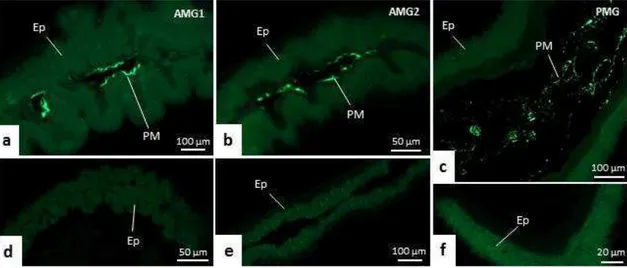
all midgut regions of T. theobaldi were confirmed by WGA-FITC staining (Figs. 4a–f).
In the AMG1 (Fig. 4a) and AMG2 (Fig. 4b), the labelling was seen just above the brush
border. Differently, the labelling in PMG were more diffused or located in the center of
the lumen (Figs. 4c).
By phalloidin-FITC labeling (actin marker), muscle bundles were seen in the
midgut of T. theobaldi, forming a network covering the outer wall of the organ. This
labeling revealed the arrangement of circular and longitudinal bundles in the three
regions of the midgut (Fig. 5a–c). The longitudinal bundles had similar width in the
30 thicker in AMG1, becoming narrower in the passage between AMG1 and AMG2 (Figs.
5d–f).
The circular bundles were organized orthogonally to the longitudinal ones. Each
circular bundle was interconnected to neighbor circular bundles. The interconnection of
the circular muscle bundles always occurred at the same position along the midgut
length (Figs. 5d-e and 5g).
The longitudinal bundles were parallel and with few branches, seen only in
some muscle bundles of AMG1 and PMG (Figs. 5d and 5g). The longitudinal bundles
were not all continuous from the beginning to the end of the midgut, with some of them
terminating early in the PMG, while others originated from the rear end of the PMG,
extending the transition of AMG2 to PMG (Fig. 5f).
Transmission electron microscopy
AMG1
The AMG1 digestive cells had densely-packed microvilli in the apical region
and invaginations in the basal region, forming an extensive and sparse labyrinth, which
occupied nearly half the cell (Figs. 6a and 6f). The microvilli were thin, tall, numerous,
and contained extracellular material with granular aspect on its ends, corresponding to
the PM (Fig. 6b).
The digestive cells of AMG1 had many mitochondria and lamellar bodies in the
apex (Fig. 6c). Golgi apparatus, small autophagic vacuoles, lamellar, and multilamellar
bodies were also seen (Figs. 6d, 6e, and 6g). The basal lamina was compact and
continuous, and had undulations and depressions (Figs. 6f and 6h). Muscle cells were
31 Regenerative cells were seen in the basal region of the epithelium of AMG1,
AMG2, and PMG (Figs. 6h and 7d). These small cells had few organelles and extensive
lateral expansions, which establish a connection with the neighboring regenerative cells
and the basal lamina. Differentiating regenerative cells had emerging microvilli and
basal labyrinths (Fig. 6h).
AMG2
AMG digestive cells had long and slender microvilli (Figs. 7a, inset and 7b). As
well as in AMG1, microvesicle-like structures were seen close to the PM, with single or
double layers (Fig. 7c). The digestive cell cytoplasm had autophagic vacuoles (Fig. 7b,
inset), electrondense lysosome-like structures, and many mitochondria and lamellar
bodies concentrated in the apex (Fig. 7b). The basal labyrinth was extensive, but less
developed than in AMG1, and the basal lamina was compact and continuous (Fig. 7d).
Enteroendocrine cells were seen in AMG2, and in PMG. In both regions, these
cells had electron-lucent nuclei, few mitochondria, and many small electrondense
granules. These cells established contact with the basal lamina through extensive
cytoplasmic processes (Figs. 7d, 8h, and 8i).
PMG
The microvilli of digestive cells of PMG were thin, numerous, and higher than
that of AMG and were also associated to microvesicle-like structures (Fig. 8a, inset).
Autophagic vacuoles of various sizes, multilamellar bodies, and Golgi apparatus were
also found here (Figs. 8b–f). Digestive cells in the PMG were rich in rough endoplasmic
reticulum with their concentric lamellae accumulated in the supranuclear region (Figs.
8f-g). Large vesicles, or inclusion bodies, containing eletrondense structures or a
32 labyrinth in PMG was less expressive and the basal lamina was thick and compact in
some intervals in comparison to AMG (Figs. 8h-i).
Scanning electron microscopy
The midgut topography was similar in T. theobaldi females and males. AMG1
was continuous with the cardia, a dilated structure that connected the esophagus to
AMG (Figs. 9a and 9b). As seen in the histological sections, the AMG1 epithelium had
folds (Figs. 9a and 9d) that were not seen in AMG2 (Fig. 9e).
Ganglia were located just above the cardia and nerve fibers extended along
AMG. Nerves ramified and connected to the longitudinal muscle bundles (Figs. 9b and
9c). Tracheoles were seen on the entire surface of the midgut and were most commonly
found in AMG2 and PMG (Figs. 9e and 9f).
In AMG1 and AMG2, only the longitudinal muscle bundles were seen (Figs.
9c–e) under SEM. In AMG1, longitudinal bundles were more widely spaced, but the
circular bundles still could not be seen, as they were hidden in the furrows formed
between the epithelial folds (Fig. 9c). In AMG2, there were many tracheoles and the
longitudinal bundles were very close to each other, hiding the circular bundles (Fig. 9e).
In the PMG, the longitudinal bundles were widely spaced, allowing the visualization of
circular muscle bundles (Figs. 9f-g).
Cell proliferation
Cell proliferation was not detected in any of the three regions in the midgut of 5-
to 10-day-old adult mosquitoes under experiment conditions. However, in the positive
control, corresponding to the midguts of A. aegypti (4th larval stage), labeled nuclei
33
FMRFamide-like positive cells
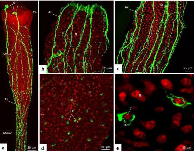
The anti-FMRFamide antibodies labeled neurons and endocrine cells in T.
theobaldi midgut. The pattern of this labeling was similar in female and male T.
theobaldi adults. FMRFamide-like positive ganglions were seen above the cardia and
their ramifications were seen overlying more than half of the AMG (Figs. 10a–c).
Enteroendocrine cells (i.e., FMRFamide-like positive cells) were abundant and
scattered among the digestive cells of the extreme end of AMG2 (close to PMG) and
throughout PMG (Figs. 10d and 10e). The number of enteroendocrine cells was similar
in males and females (p = 0.842), with approximately 99 cells per midgut.
Discussion
The general morphology of the midgut in female and male T. theobaldi
resembled that of the midgut of male mosquitoes whose females are hematophagous. In
this regard, similar to these males, the AMG of T. theobaldi was thin and long, while
the PMG was enlarged and reduced in size. Different of this, in blood-feeding female
mosquitoes, the AMG is short and the PMG is expanded (Sup. Figs. b and c) 13,20-23.
The AMG of T. theobaldi was subdivided into two morphologically distinct
regions: AMG1 and AMG2. In other Culicidae (both females and males) this
subdivision is not evident (or absent), and the AMG is slender and without folds, similar
to AMG2 of T. theobaldi. By being wider than AMG2 and containing folds, AMG1
seems to function as a first site of food digestion. The presence of folds increase the
contact surface between the food and the digestive epithelium, and probably reduce the
speed in which nutrients pass through the lumen, facilitating the digestion and
34 The epithelium characteristics of the three midgut regions of T. theobaldi were
compatible with the secretory, digestive, absorptive, and nutrient transport functions as
reported elsewhere 13,25. Both AMG1 and PMG seem to be more involved in enzyme
secretion and nutrient absorption compared with AMG2. These two regions presented
apocrine secretion of proteins and intense labeling for carbohydrates, especially in the
basal portion of the digestive cells. However, the acidic character of apocrine secretion
in AMG1 versus the basic character of this secretion in PMG indicates that the secreted
proteins are probably different in the two regions.
The AMG1 and PMG digestive cells of T. theobaldi showed greater
carbohydrates accumulation when food was being transferred to the hindgut. The
carbohydrates accumulation, such as glycogen, is common in insect digestive cells
during absorption activity 10,26, and in the PMG digestive cells of larval and adult
mosquitoes fed with sugar or blood 27-29. These carbohydrate deposits seen in PMG
seem to accumulate because of the digestion process, functioning as energy reserves, or
facilitating the subsequent absorption of more carbohydrates 30.
Apocrine secretions are typically released during the digestive process of
insects, and it is speculated that this is also related to regions that perform nutrients
absorption 31. Corroborating this, AMG1 and PMG are apparently more involved with
the carbohydrate absorption, and are the regions where apocrine secretion occurred.
Another possibly secretory mechanism present in the midgut of T. theobaldi is
the microapocrine secretion. The small single and double membrane structures seen
across the midgut lumen of T. theobaldi resemble microapocrine secreted vesicles found
in the midgut lumen of various insects 31. Enzymes, such as amylase, and various
35 existence of this type of secretory mechanism is something that needed to be clarified in
adults of non-hematophagous mosquitoes.
The abundant rough endoplasmic reticulum (RER) lamellae in the PMG are
also found in the PMG of hematophagous females when a blood meal is acquired. The
marked presence of these organelles occurs in cells that are specialized in protein
secretion 9,13,29. Accordingly, it is possible to infer that the bloodmeal in blood-feeding
female mosquitoes, and the sugar meal in T. theobaldi stimulate intense activity of
protein secretion in PMG digestive cells. In addition to the PMG, the AMG1 also had
many RER lamellae in the digestive cells, which is probably related to the apocrine
secretion of proteins as demonstrated by histochemistry with bromophenol blue.
Autophagic vacuoles were seen in the digestive cells of all regions of T.
theobaldi midgut, being larger in size and quantity in PMG. These vacuoles are related
to the recycling of membranes that occurs due to endo- and exocytosis during digestion
29
. Large inclusion bodies were also seen in the digestive cells of T. theobaldi, similar to
those observed in the PMG of blood-feeding mosquitoes post bloodmeal 16,29. The
function of these inclusion bodies is unknown, but it has been proposed for recycling
membranes, along with the autophagic vacuoles 16. By containing a large amount of
autophagic vacuoles and inclusion bodies compared with AMG, PMG digestive cells
may be more involved in vesicular transport than AMG digestive cells.
Lamellar bodies were also abundantly found in digestive cells throughout the
T. theobaldi midgut. These structures are composed primarily of lipids and proteins, and
their biogenesis involves endocytic and/or autophagic pathways 33. In some vertebrate
digestive epithelia, lamellar bodies are secreted to protect cell membranes from
digestive enzymes and the abrasion of food flow 34. However, the function of these